PhD school in
“Innovation in immuno-mediated and haematological disorders” Curriculum: “Immunology and immunopathology”
XXXII cycle
PhD thesis
“Play it again, SAMHD1”: the hard life of an antiviral restriction factor during the intrinsic immune response against human cytomegalovirus infection.
PhD school coordinator
Prof. Angela Santoni
Supervisor PhD student Prof. Cristina Cerboni Simone De Meo
1
Contents
Summary ... 3 1. Introduction ... 4 1. HCMV ... 4 a. General characteristics ... 4 b. Virion structure ... 5c. Genome structure and variability ... 7
d. Virus entry, replication and latency ... 9
e. HCMV and cell cycle regulation ... 12
f. Immune responses ... 13
g. HCMV and immune evasion ... 16
2. Restriction factors ... 20
a. HCMV and restriction factors ... 21
3. SAMHD1 ... 23
a. Expression ... 23
b. Structure ... 24
c. Metabolic activity and functions ... 26
d. Mechanisms of regulation ... 29
e. Mutations ... 32
4. SAMHD1 and viral restriction ... 33
a. HIV-1 restriction ... 33
b. SAMHD1 restriction of RNA and DNA viruses ... 34
2. Aim of the study ... 38
3. Materials and methods ... 40
1. Cells and culture conditions ... 40
2. HCMV preparation and infection ... 40
3. Real-time PCR ... 41
2
5. Immunoblot analysis ... 42
6. Immunofluorescence and FACS analysis... 42
7. Chemical compounds ... 43
8. Small interfering RNA ... 43
9. VLP generation ... 43
10. Retroviral vectors production and infection ... 44
11. Confocal microscopy analysis ... 44
12. Nuclear/cytoplasmic fractionation ... 45
13. Cryo-immunoelectron microscopy ... 45
14. Immunoprecipitation and mass spectrometry ... 45
15. Statistical analysis ... 46
4. Results ... 47
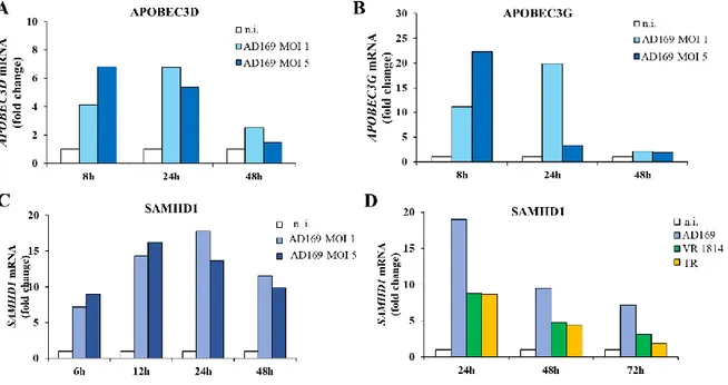
1. SAMHD1 and APOBEC3s expression are up-regulated following HCMV infection ... 47
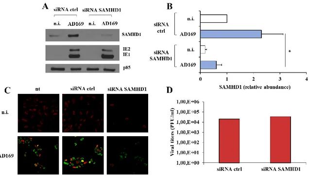
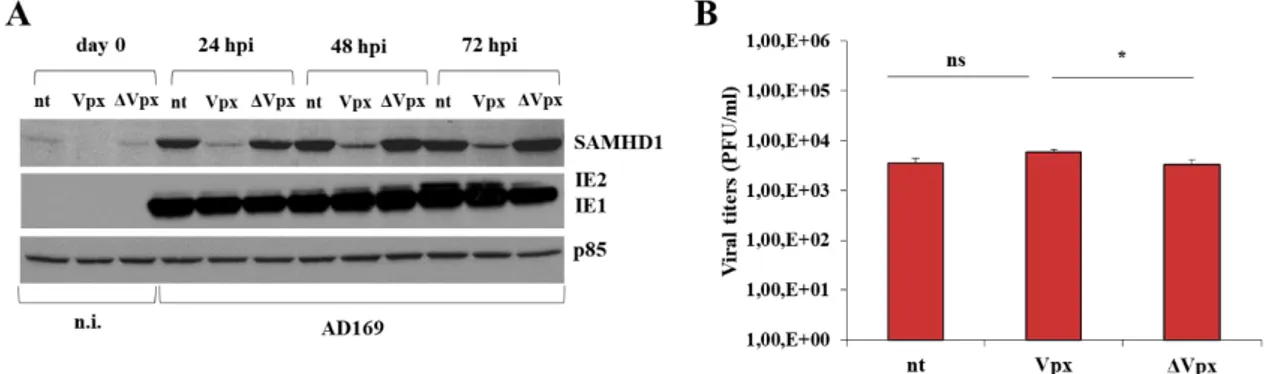
2. SAMHD1 silencing and Vpx-mediated knocking-down marginally influence HCMV replication ... 49
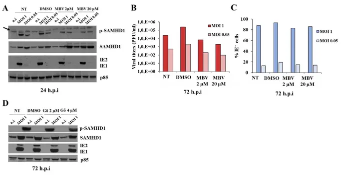
3. SAMHD1 is phosphorylated at Threonine 592 in HCMV infected cells ... 52
4. The viral pUL97 kinase inhibitor Maribavir reduces HCMV replication but has only a slight effect on SAMHD1 phosphorylation ... 53
5. The cellular Cdk1 inhibitor CGP74514A reduces HCMV replication and abrogates SAMHD1 T592 phosphorylation ... 55
6. HFFs overexpressing wild type or T592 SAMHD1 mutants are equally permissive to HCMV replication ... 56
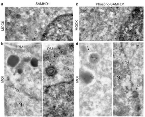
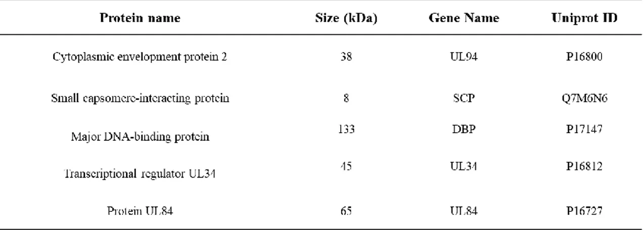
7. Phospho-T592-SAMHD1 preferentially localizes in the cytoplasm after HCMV infection 58 8. SAMHD1 association with HCMV infectious particles, non-infectious dense bodies and viral proteins ... 60
9. Discussion ... 62
3
Summary
Sterile α-motif and histidine-aspartate domain-containing protein 1 (SAMHD1) is a cellular deoxynucleotide triphosphates (dNTPs) triphosphohydrolase that hydrolyses dNTPs into deoxynucleosides and inorganic triphosphates. This activity is associated with its antiviral restriction function and it inhibits replication of many RNA and DNA viruses. The antiviral activity is negatively regulated by Thr-592 phosphorylation, that destabilizes the active tetrameric form of the protein. Human cytomegalovirus (HCMV) is an opportunistic pathogen in immunocompromised hosts, while it is asymptomatic in the general population. Infection is life-long, and latency is a pivotal feature of the virus, which interferes with host anti-viral responses, including intracellular restriction factors. During my PhD project, we investigated the role of SAMHD1 in HCMV replication and potential viral evasion mechanisms, which could contribute to the unsuccessful clearance of the virus. After infection of different cell types with different HCMV strains, we observed an increase in SAMHD1 mRNA and protein levels. This was associated with the induction of phosphorylation at the regulatory residue Thr-592, likely involving the cellular kinase Cdk1, but not the viral kinase pUL97. Both SAMHD1 knock-down and, on the other hand, overexpression of wild-type and Thr-592 mutants showed negligible influence on HCMV viral production, suggesting that SAMHD1 activity could be overcome by HCMV during lytic infection, independently from its phosphorylation status. We also observed, by various experimental approaches, phospho-SAMHD1 localization in the cytoplasm of infected fibroblasts and its association with viral particles, suggesting the idea of a mechanism of de-localization and evasion from its nuclear-associated antiviral activity. Despite recent observations of SAMHD1 restriction during early steps of HCMV replication, our work suggests that SAMHD1 is unable to limit viral lytic infection, probably due to re-localization in the cytoplasm, setting a cellular environment permissive for a productive HCMV replication.
4
1. Introduction
1. HCMV
a. General characteristics
Human cytomegalovirus (HCMV) is a ubiquitous herpesvirus that latently infects most of the world’s population (Mocarski et al., 2013). It is a double strand DNA (dsDNA) virus, belonging to the β-herpesvirinae subfamily and it has the largest genome of all known human viruses, with a length of 235 kbp. In vitro and in vivo infections result in a characteristic cytopathology of enlarged (cytomegalic) cells, that gives to this virus its name (Britt, 2011). Like other human herpesviruses, HCMV clearance is never complete and it remains latent during the entire life of the host. Primary infection does not cause severe illness in the general immunocompetent population, except for an occasional febrile illness as well as infectious mononucleosis, but it is an important opportunistic pathogen in immunocompromised hosts (i.e. AIDS patients or transplant recipients). Susceptibility to HCMV infection is particularly related with defects in CD4+ and CD8+ T cell functions in
immunocompromised or not fully competent hosts (Mocarski et al., 2013). For all these reasons, the virus remains clinically important despite antiviral therapies aimed at reducing disease burden. The clinical spectrum of HCMV infection of congenitally infected children, for example, involves numerous organs and tissues and comprises sensorineural hearing loss, microcephaly, splenomegaly, pneumonitis and even death (Britt, 2011). At cellular level, HCMV infects various cell types: monocytes, macrophages, neuronal, retinal, epithelial, endothelial and dendritic cells (DCs) (Revello and Gerna, 2010). Viral latency occurs in bone marrow-derived hematopoietic cells, where viral DNA is present at a very low copy number and it is associated with low viral transcription (Bego and St. Jeor, 2006).
As a result of long-standing infections, many genetically different strains of HCMV evolved and spread in the general population (Bradley et al., 2008), through contact with body secretions (e.g. urine, milk, saliva and uterine secretions). Breastfeeding is the most common way of mother-to-child transmission, but the transplacental one is the most
5 clinically important, and it occurs in women who are already infected before conception, as well as in women who experience primary infection during pregnancy (Britt, 2011).
b. Virion structure
The HCMV virion consists of three different regions: the envelope, the tegument and the capsid containing the viral genome (Figure 1). The envelope is composed of a variety of proteins that are largely still unknown in structure and function. Analysis of viral genome sequences indicated that almost 50 viral open reading frames (ORFs) can be predicted as membrane glycoproteins, and despite the exact number of envelope glycoproteins is still unknown, at least 12 different glycoproteins were identified (Britt and Boppana, 2004). Many of them (gB, gH, gL, gM and gN) have homolog function and structure in other herpesviruses, and they can exist as complexes hold together by disulfide bonds located in the portion of the protein inside the virion (gM/gN, gH/gL/gO and gH/gL/UL128/UL130/UL31A) (Britt and Boppana, 2004). gB is encoded by viral UL55 ORF and is expressed as a trimer, named glycoprotein complex I (gcI), and it mediates attachment to the plasma membrane, fusion and entry. The gcI trimer is important for virus spread and cell-to-cell membrane fusion, leading to formation of cellular syncytia (Mocarski et al., 2013). Several putative gB receptors were described: cell surface integrins (Feire et al., 2010), the epidermal growth factor receptor (EGFR) (Wang et al., 2003) and platelet-derived growth factor receptor alpha (PDGFRα) (Soroceanu et al., 2008). Nevertheless, the question of receptor requirement for virus entry is still under debate, as it has been reported that gB does not need cellular receptors during the membrane fusion (Wille et al., 2013). gM and gN glycoproteins are linked by disulfide bonds and form a heterodimeric complex named gcII. These proteins are encoded by UL100 and UL73 ORFs respectively, and they are the most abundant envelope glycoproteins (Nguyen and Kamil, 2018). The cytoplasmic tail of gM is essential during the stage of virion assembly (Krzyzaniak et al., 2007), while gN cytoplasmic tail is essential at the envelopment stage (Mach et al., 2007). Notably, the UL73 gene (gN) has one of the most variable sequences among different HCMV strains (Pignatelli et al., 2003). Moreover, gcII complex is a target of adaptive immune responses (Shimamura et al., 2006).
6 Figure 1: HCMV virion structure
HCMV structure consists of an outer bilayer envelope, composed of many glycoprotein complexes. Inside the envelope, there is the tegument, composed by proteinaceous matrix, which cover the icosahedral capsid containing the double stranded linear DNA genome.
gH, gL and gO form the heterotrimeric complex gcIII, and each protein is encoded by UL75, UL115 and UL74 ORFs, respectively (Nguyen and Kamil, 2018). PDGFRα acts as a receptor for the trimer and its role in viral entry is independent from its tyrosine kinase activity (Wu et al., 2018). The HCMV pentameric complex is composed by gH and gL heterodimer bound with UL128, UL130 and UL131 gene products (Ryckman et al., 2008). The pentamer has been described as necessary for infection of many cell types, including leukocytes, epithelial and endothelial cells (Adler, 2006). A putative cellular receptor binding the HCMV envelope pentamer is neuropilin-2, essential for attachment and entry in endothelial and epithelial cells (Martinez-Martin et al., 2018).
The tegument is the most complex and heterogeneous structure of HCMV virion. The tegument is composed by many viral proteins and RNAs and it is generally assumed as an amorphous layer between the envelope and the capsid (Chen et al., 1999). HCMV can include cellular proteins in its tegument and this strategy can be exploited by the virus to seize host anti-viral proteins, such as those mediating the intrinsic innate immune response (Dell’Oste et al., 2014). Tegument proteins are usually phosphorylated and many of them have a regulatory function in HCMV replication. In addition, they can regulate various
7 pathways in an infected cell: block of intrinsic cellular responses, stimulation of cell cycle progression, enhancement of immediate-early (IE) transcription and help in viral DNA replication (Baldick et al., 1997; Hayashi et al., 2000). One of the most abundant viral protein in the tegument is pp65, encoded by UL83 ORF, which is highly immunogenic: indeed, a percentage ranging from 2 to 5% of CD8+ T lymphocytes in the peripheral blood
of healthy seropositive individuals are specific for this protein (Khatamzas et al., 2002). The capsid is the inner structure of the virion and consists of 162 capsomere subunits, divided in 150 hexons and 12 pentons built together in an icosahedral symmetry (Chen et al., 1999). The capsid is partially assembled in the cytoplasm, guided by viral UL80a ORF, that acts as a scaffold for the generation of individual capsomeres (Gibson, 2008). Capsids containing viral dsDNA exit the nucleus and are enveloped in the cytoplasm.
c. Genome structure and variability
The HCMV genome is one of the largest of all human viruses and is composed of two main coding domains, the unique long (UL) and unique short (US) domain. They are flanked in the outer and inner side of the genome by the long and short terminal repeated (TRL, TRS) and internal repeated (IRL, IRS) sequences (Bankier et al., 1991) (Figure 2).
Figure 2: HCMV laboratory and clinical strain genomes
Organization of AD169 (a), Merlin (b), TR (c) and VR-1814 (d) genome. (a) AD169 genome comprise two unique blocks of ORFs (UL and US), each one flanked by repeated blocks of ORFs (TRL, IRL; IRS, TRS) (b) In Merlin genome, TRL and IRL blocks of ORFs are missing, the UL segment is larger and contain RL1-14 and UL1-151. (b)(c) TR and VR-18RL1-14 clinical isolates, after the unique domain RL1-13 and UL1-150, contain a block of genes (UL133–UL151) separated from the UL segment, and the US1-36 region is inverted in VR-1814 genome (adapted from Murphy et al., 2003)
8 The genome is tightly packaged inside the nucleocapsid and it circularizes during the replication phase of the virus, in a mechanism called “rolling circle amplification”, that can generate multiple copies of the genome, linked in tandem (Gibson, 2008).
An extensive analysis of protein encoded by the well-known AD169 laboratory strain genome was conducted for the first time in 1990. The authors reported that AD169 HCMV laboratory strain contains 208 encoding ORFs and 14 of them are duplicated in TRL and IRL regions (Chee et al., 1990). Because of AD169 adaptation to in vitro cell culture conditions, AD169 completely misses genes from UL133 to UL151, encoding immune evasion proteins (Prichard et al., 2001).
Merlin strain has been extensively studied and its genome has been used as a reference wild-type genome (Martí-Carreras and Maes, 2019). Merlin has been also designated from the World Health Organization (WHO) as the international standard model for HCMV (Wilkinson et al., 2015). In 2004, Merlin strain was predicted to code for 165 protein (Dolan et al., 2004), but later research identified additional transcripts and more coding region could thus exist (Gatherer et al., 2011). In the same study, AD169 has been used as a model of high-passaged laboratory strain, and VR-1814 and TR strains were used as low-passaged clinical strain models. Compared to AD169, VR-1814 and TR are characterized by less DNA mutations and by the presence of the UL133-UL151 region (Murphy et al., 2003). At the present time, more than 300 full-length complete viral low and high passaged strain genomes have been published and it is estimated that HCMV contains ̴ 751 translated ORFs (Martí-Carreras and Maes, 2019; Stern-Ginossar et al., 2012).
The severity of clinical outcomes caused by HCMV could be related to viral genetic variability, that has been shown to be remarkably high inside and between hosts (intra- and interhosts variability) (Renzette et al., 2015). As a consequence of HCMV ability to infect many different cell types and organs, viral populations sequenced from different tissue samples in the same individual can be as different as viral populations sequenced from distinct individuals (Renzette et al., 2013). It has been proposed that immune responses can trigger a selective pressure on HCMV during infection, favouring the selection of viral populations containing mutations advantageous for the successful spreading in the hosts (Renzette et al., 2011). Regarding interhost variability, it has been recently reported that viral strains freshly isolated from congenitally infected children are genetically and
9 phenotypically different, and characterized by different aggressiveness in models of in vitro infection (Galitska et al., 2018).
d. Virus entry, replication and latency
HCMV entry is very promiscuous and it can bind, penetrate and start replication in almost all cell types. This characteristic was originally explained with the hypothesis that the receptor for HCMV is distributed on the surface of a great variety of cells (Nowlin et al., 1991). In 1993, Compton and colleagues reported that virus entry requires the presence of the polysaccharide heparan sulfate on the cell surface, which is expressed in almost all cells (Compton et al., 1993). However, from a biochemical point of view, Boyle and Compton reported that HCMV can engage other molecules than heparan sulfate (Boyle and Compton, 1998). One of them was identified in 2003 to be the epidermal growth factor receptor (EGFR) (Wang et al., 2003). In their paper, the authors reported the co-immunoprecipitation of gB and EGFR after chemical crosslinking, but they did not resolve the exact mechanism of interaction between these two proteins. After this important finding, in the following years other HCMV receptors were reported: αvβ3 integrin (Wang et al., 2005), the platelet-derived growth factor receptor (PDGFR) (Soroceanu et al., 2008), and CD147 for the entry into epithelial and endothelial cells (Vanarsdall et al., 2018). OR14I1 and neuropilin-2 have been identified as receptors for the viral pentameric complex and responsible for the epithelial/endothelial tropism (Martinez-Martin et al., 2018; Xiaofei et al., 2019). After plasma membrane binding, regardless the receptor, the virus can entry the cell in a pH-independent manner through macropinocytosis, like in fibroblasts (Li et al., 2015a), or through endocytosis in a low pH-dependent manner, like in epithelial and endothelial cells (Ryckman et al., 2006). Then, the envelope/cell membrane fusion results in activation of several cellular signaling pathways and start of the viral lytic cycle (Figure 3).
10 Figure 3: Overview of the HCMV lytic cycle
HCMV enters the cell through interaction with EGFR and other cellular receptors. Capsid is delivered into the cytoplasm and travels to the nucleus, where the genome is transcribed and replicated. The expression of viral genes follows three temporal moments: expression of immediate early (IE), early (E) and late genes (L). In the nucleus, late genes expression set up capsid assembly. Capsid translocates to the cytoplasm, associates with tegument components and is trafficked to the viral assembly complex (VAC). Subsequently, the capsids acquire viral envelope by budding into intracellular vesicles. After envelopment, viral infectious progeny is then released outside the cell.
HCMV lytic cycle can be divided in three distinct phases, characterized by the expression of different viral proteins, named immediate early (IE), early (E) and late (L) proteins, in relation to different stages of expression. After entry, the capsid is transported into the nucleus and the IE1 and IE2 genes are expressed without any active viral protein synthesis. Some viral proteins could be already present inside the virion, and host transcription factors are probably involved in the early induction of viral protein synthesis (Britt and Boppana, 2004). IE1 and IE2 genes result from transcription of the same viral genome region, the major immediate-early (MIE) gene and the resulting proteins derive from alternative splicing of the primary polyadenylated transcript. IE1 is a 72 kDa phosphoprotein detectable at very early times post-infection, while IE2 is an 86 kDa trans-activating factor of viral early and late genes. Together, IE1 and IE2 autoregulate their
11 expression, activate cellular genes, establish nuclear sites of lytic viral DNA synthesis and contribute to the latency/reactivation switching (Mocarski et al., 2013).
The MIE gene is located inside the so-called major immediate-early promoter (MIEP), in turn controlled by MIEP enhancer, that requires, for its regulation, the activity of tegument proteins pp71 and ppUL69. After MIE protein expression, the virus expresses early genes (E), that are important for alteration of cell cycle progression and apoptosis, for viral DNA transcription and production of structural virion proteins. Early genes comprise almost 65 proteins and a set of miRNAs that accumulate and act through 18 to 24 hours post-infection (h.p.i), at the time of viral DNA synthesis. During this time, the virus expresses late genes (L), that encode virion structural proteins and are required for the assembly of infectious particles (Mocarski et al., 2013).
DNA synthesis occurs inside the nucleus of infected cells starting at 14 h.p.i and reaching more than 10,000 copies from 24 to 48 h.p.i. The mechanism of synthesis starts from the oriLyt site and viral DNA replication occurs in a rolling circle dynamic of replication, leading to the formation of a long linear concatemer of multiple copies of HCMV genome that are packaged into pre-formed capsids inside the nucleus (McVoy and Adler, 1994). The replication start is dependent on viral replisome formation, composed by the viral UL54 DNA polymerase core subunit, UL44 DNA polymerase processivity subunit, UL57 DNA single strand-binding protein and the viral helicase-primase heterotrimeric complex (Mocarski et al., 2013). DNA polymerase UL54 core subunit is the target of the antiviral drugs ganciclovir and foscarnet, and mutations in UL54 gene sequence have been described to contribute to antiviral drug resistance (Chou et al., 2003). Capsid localization, packaging and cleavage of viral DNA are positively regulated by phosphorylation from pUL97 kinase and cellular cyclin-dependent kinases (Cdks). The exit of the capsid from the nucleus is mediated by a herpesvirus conserved nuclear egression complex (NEC) localized in the nuclear inner membrane. NEC acts as a mechanism of packaging quality control of DNA-containing capsids. During this phase, the nucleocapsid temporary envelopes from the nuclear inner membrane that is subsequently lost in the cytoplasm (Britt, 2011). After this stage, the nucleocapsid reaches a perinuclear area known as virus assembly complex (VAC), where the virus acquires the tegument and it is enveloped from endosomal/exosomal membranes before the final egress from the cell (Mocarski et al., 2013) (Figure 3).
12 Latency is a characteristic of all herpesviruses, and HCMV is not an exception. Latent infection has been shown in cells of the myeloid lineage, such as CD34+ progenitors, macrophages and CD14+ monocytes (Hahn et al., 1998). HCMV can reactivate upon differentiation of these cell types, and mature circulating DCs and macrophages were reported to be sites of reactivation (Poole et al., 2015; Reeves and Sinclair, 2013). Despite the mechanisms that favor the establishment of latency are still largely unknown, a hallmark of this process is the suppression of MIEP activity and, subsequently, one of the first events of reactivation is the production of IE proteins. Given that, MIEP activity can be regulated by transcription factors and epigenetic/chromatin structure modifications (Sinclair, 2009; Stinski and Isomura, 2008). In particular, the latent genome has a MIEP chromatin structure associated with transcriptional repression, including trimethylation of Lysine-27 and Lysine-9 sites inside H3 histone. In addition, the activity of histone deacetylase (HDAC) maintains viral chromatin in a repressed conformation (Elder and Sinclair, 2019). Indeed, treatment of latently infected cells with HDAC inhibitors is associated with reactivation of IE gene transcription (Krishna et al., 2018).
e. HCMV and cell cycle regulation
HCMV can subvert and modify host cell cycle to favor its own replication and the establishment of a lytic or latent infection. The cell cycle is tightly regulated by the activity of cyclins and cyclin-dependent kinases (Cdks), that form active heterodimers and phosphorylate serine and threonine residues of target proteins, to obtain their activation or inactivation. Human cells express about 20 Cdks and 13 cyclins to regulate transition through the cell cycle, but also to control genetic transcription, epigenetic modifications, metabolic activity and DNA repair. Moreover, the activity of Cdks and cyclins are intertwined with cell cycle checkpoints, that are essential to ensure a correct progression during the multiple phases of the cycle (Roskoski, 2019).
One of the main characteristics of HCMV-infected cells is the arrest of cell cycle before mitosis, and it is particularly interesting the fact that they express G1, S and
M-phase-associated proteins but are blocked in DNA synthesis (Spector, 2015). Indeed, HCMV influence on cell cycle mechanisms is very deep: it influences expression, translation, post-translation modifications, stability and localization of cellular proteins. For example, the
13 viral product pUL97, a serine/threonine kinase, can hyper-phosphorylate Rb to release E2F and allow active gene transcription (Jault et al., 1995). IE1 and IE2 proteins are able to modify cell cycle progression in different ways: IE1 can stimulate entry into the S phase removing the p107 repression of E2F promoters, and IE2 can support p53 expression and accumulation in the nucleus and entry into S-phase (Castillo et al., 2000). Moreover, the virus targets the anaphase promoting complex (APC), to modulate its activity and avoid degradation of thymidine kinase (TK) and ribonucleotide reductase (RNR), to ensure the production of enough nucleotide triphosphates for viral genome replication (Fehr and Yu, 2013).
Thus, cell cycle manipulation is an important strategy carried out by the virus to stimulate its own DNA replication and disadvantage synthesis of host DNA, starting already at early times post-infection.
f. Immune responses
In hosts with an intact and fully competent immune system, innate and adaptive immunity can limit HCMV spread in the organism and prevent the arise of serious illness. Studies in experimental murine models helped to delineate the role of these two arms of the immune system in controlling virus replication, and the key importance of DCs, natural killer (NK) cells, CD4+ T helper cells and CD8+ cytotoxic T lymphocytes (CTL), interferon responses and virus-specific antibodies (Figure 4). In immunocompromised hosts, defects of innate and adaptive immune responses predispose to HCMV infection, resulting in an increasing probability of morbidity and mortality (Britt, 2011).
The main characteristic of the immune responses in immunocompetent individuals is a robust CTL and neutralizing antibody response to the infection. Despite that, the control of the virus is not complete, and clearance is impossible. This is largely due to the expression of viral gene products that modulate and disrupt host innate and adaptive immune responses, establishing a life-long balance between immune-evasion mechanisms of the virus and the immune system that tries to eradicate the pathogen.
14 Figure 4: HCMV immune responses
Schematic representation of immune cells and their effector mechanisms against HCMV-infected cells. Intracellular restriction factors (RFs) interfere with many steps of HCMV replication. pDCs release IFN-α, IL-6, IL-10 and TNF-α to mediate humoral and cell-mediated immune responses. NK cells limit HCMV producing IFN-γ and killing infected cells by secretion of cytotoxic granules. Neutralizing antibodies target viral epitopes preventing cell attachment, penetration and HCMV dissemination. CD8+ T cells release
cytotoxic granules to kill HCMV-infected cells. CD27 and CD28 double positive CD4+ T cells produce
IFN-γ and TNF-α, support CD8+ T cell persistence and maintain viral-specific antibody production.
The very first line of defence against viral pathogens is grouped in the innate arm of the immune system, known as “intrinsic immunity”, that involves various proteins that act as defence mechanisms at the cellular level. The most characterized and studied intrinsic immune responses are accomplished by cellular “restriction factors” (RFs), constitutively expressed intracellular proteins able to interfere with many steps of viral replication (Bieniasz, 2004). Because RF response to HCMV infection is the focus of this thesis, it will be discussed in a separate paragraph later in the text (see page 20).
NK cells are key components of innate immune responses. Individuals with NK cell defects are susceptible to herpesvirus infections, including HCMV (Biron et al., 1989; Gazit et al., 2004). In general, one of the main features of NK cell responses to infected cells is the release of proteins with cytotoxic activity, leading to a rapid killing of infected cells (Santoli et al., 1978). NK cells can also produce cytokines and chemokines, to modulate inflammatory responses and to recruit various effector cells to the site of infection (Fehniger et al., 1999; Loza et al., 2002). NK cell activation, proliferation and
15 effector functions depend on a balance between positive signals provided by many activating receptors (i.e. NKG2D, NKG2C, NKp46) and negative signals promoted by inhibitory receptors (i.e. KIRs, NKG2A) interacting with major histocompatibility complex (MHC) class I molecules (Lanier, 2005). Moreover, HCMV infection triggers an enrichment of a particular NK cell subset: adult hematopoietic stem cell recipients experimenting HCMV reactivation from latency, as well as infected children, show an expansion of a mature CD56dim CD16− NK cell subset expressing NKG2C, probably
involved in the control of HCMV reactivation (Goodier et al., 2018; Gumá et al., 2004). Furthermore, NK cells can limit HCMV transmission in epithelial, endothelial cells and fibroblasts, directly producing IFN-γ and inducing IFN-β production in these infected cells (Iversen et al., 2005; Wu et al., 2015).
At the same time, DCs are also important players in the innate immune responses against pathogens. They are crucial for the efficacy of antiviral responses, by stimulating chemokine and cytokine production to limit viral dissemination, and by triggering the activation of adaptive immune responses. During HCMV infection, plasmacytoid DCs (pDCs) are particularly important, because they are non-permissive to viral HCMV replication, but remain functional to facilitate the mounting of an effective immune response (Varani et al., 2007). For example, experiments conducted on pDC isolated from human peripheral blood mononuclear cells, showed that HCMV infection triggers IFN-α, IL-6, IL-10 and TNF-α production by pDCs, critical for humoral and cell-mediated immune responses (Varani et al., 2007).
Immunocompetent individuals infected with HCMV, show a high proportion of CD4+ T cells that recognize pp65 and IE-derived immunodominant epitopes (Jackson et al., 2011). During primary infection of kidney transplant recipient, CD4+ T cells specific for HCMV epitopes appear approximately 7 days after detection of viral DNA in peripheral blood (Rentenaar et al., 2000). Moreover, during the acute phase of infection, these cells are CD27 and CD28 double positive and produce IFN-γ and TNF-α cytokines (Rentenaar et al., 2000), while in the latent phase, these cells are prevalently CD27 and CD28 double negative and acquire a cytolytic function, characterized by the expression of perforin and granzyme B (van Leeuwen et al., 2006).
Together with CD4+ T cell responses, CTL responses emerge and persist also during the latent phase of infection, and are crucial for HCMV control of reactivation (Kuijpers et al.,
16 2003). In bone marrow transplantation recipients, regain of CTL populations protects from HCMV-associated disease (Avetisyan et al., 2007). During viral persistence, impairment of CD8+ T cell maturation and effector functions contributes to the failure of the immune system in controlling viral replication and disease (Gamadia et al., 2003). Over the entire life of infected hosts, a high number of memory CD8+ T cells specific for HCMV expand
with aging and persist in the circulation. This peculiarity is also named memory inflation, characterized by memory CD8+ T cells expressing 7 receptor and thus responding to
IL-7 mediated homeostatic proliferation (Waller et al., 2008). Moreover, during memory inflation, CD4+ T cells contribute to a lesser extent in controlling HCMV reactivation,
compared to CD8+ T cells, sharing the characteristic of IL-7 receptor-dependent expansion
and persistence at aging of infected individuals (Gamadia et al., 2004; Libri et al., 2011). During HCMV infection, many specific neutralizing antibodies are produced, preventing cell attachment, entry and dissemination in the organism. Confirming the importance of HCMV envelope glycoprotein complexes for viral entry in host cells, gB and gH (components of the gcI, gcIII and pentameric complexes) are two of the main immunogenic proteins encoded by HCMV targeted by neutralizing antibodies (Mocarski et al., 2013), together with gM/gN, components of gcII complex (Shimamura et al., 2006). Moreover, specific antibodies can also target the tegumental protein pp150 as well as the DNA polymerase processivity factor subunit pp52 (encoded by UL44 gene) (Beqaj et al., 2008; Greijer et al., 1999). The role of anti-HCMV antibodies can be deducted from studies in B-cell deficient mouse models and from correlation of these studies in humans, recapitulating the importance of antibodies in limiting the severity of HCMV-related disease but not to prevent infection and viral dissemination. However, to date, a clear evidence demonstrating the effectiveness of anti-HCMV neutralizing antibodies in protecting humans from viral reactivation and spread still lacks (Krmpotić et al., 2019).
g. HCMV and immune evasion
HCMV can efficiently establish a latent infection in the host and this reflects its ability to evade immune responses. In fact, the genome encodes various immune evasion proteins that can target different molecular pathways and cells of the immune system (Table 1).
17 Among innate responses, HCMV can influence NK cell recognition of target cells in different ways. The UL18 viral glycoprotein, an HLA-I homolog, can bind to the inhibitory receptor leukocyte immunoglobulin-like receptor-1 (LIR-1) (Cosman et al., 1997) and suppress LIR-1+ NK cell activation (Prod’homme et al., 2007). In a similar way, a peptide encoded by the viral UL40 protein can upregulate HLA-E expression on target cells, which is recognized by the inhibitory complex CD94/NKG2A on NK cells and thus inhibit NK cell-mediated lysis (Cerboni et al., 2001; Ulbrecht et al., 2000). Other HCMV-encoded NK cell modulators comprise RL11, UL119-118, UL148, all implicated in the reduction of NK cell-mediated antibody-dependent cellular cytotoxicity (ADCC), and the NK cell activating ligand modulators UL16, UL148A, US9, US18, US20, UL141 and UL142, that downregulate the ligands of the activating receptors NKG2D and DNAM-1 (Patel et al., 2018).
HCMV can also compromise DC function. Studies of HCMV interaction with DCs focused mainly on monocyte-derived dendritic cells (MDDCs), useful for their characteristic to be easily stimulated in vitro. HCMV can productively infect MDDCs, by the binding of its envelope protein gB to the membrane protein DC-SIGN (Halary et al., 2002). Infected MDDCs were reported to have an impaired production of IL-12 and TNF-α in response to LPS stimulation (Moutaftsi et al., 2002), and they can suppress T lymphocyte priming and proliferation and induce their CD95L- and TRAIL-mediated apoptosis (Haspot et al., 2012; Raftery et al., 2001). HCMV can also impair MDDC chemotaxis to lymph nodes (Gredmark-Russ and Söderberg-Nauclér, 2012). The impaired migratory ability has been associated with a reduction of the C-C chemokine receptors 1 and 5 (CCR1 and CCR5) expression by MDDCs (Varani et al., 2005).
The US2, US3, US6, US10 and US11 viral products can modulate and interfere with surface expression of cellular MHC class I and II and antigen presentation in different ways, resulting in evasion from CD8+ and CD4+ T cell recognition and activation,
respectively, and causing a severe impairment of adaptive immune responses (Jackson et al., 2011; Noriega et al., 2012). In particular, US3 acts during the immediate early phase of infection, retaining MHC class I molecules in the endoplasmic reticulum (ER) (Jones et al., 1996). During the early phase, US2 and US11 can induce ubiquitination and degradation of MHC class I or retain the heavy chains in the ER, blocking MHC-peptide complex formation and expression on cellular surface (Furman et al., 2002; Shamu et al., 2001;
18 Tomazin et al., 1999). During late phases, US6 can inhibit TAP activity and translocation of processed peptides to MHC class I complexes in the ER (Ahn et al., 1997). Beside the influence on MHC class I expression, US2 and US3 have been reported to downregulate the expression of MHC class II molecules as well (Tomazin et al., 1999). In addition, the truncated UL111A transcript, expressed during latency, can downregulate MHC class II molecules on the surface of infected myeloid progenitors, reducing their ability to present antigens to CD4+ T lymphocytes (Cheung et al., 2009). At the same time, HCMV can
interfere with the IFN-γ-mediated upregulation of MHC class II molecules through disruption of the Jak/STAT pathway and subsequent antigen presentation to CD4+ T cells,
limiting the activation and function of these cells (Miller et al., 1998).
HCMV is also able to counteract the specific antibodies produced against its immunogenic epitopes. To the best of our knowledge, the virus encodes four distinct glycoproteins which are homologues of the receptors for the Fc fragment of IgG (vFcγR): gp68, encoded by the UL119-UL118 gene, gp34, encoded by the TRL11/IRL11 gene (Atalay et al., 2002; Lilley et al., 2001), gpRL13 and gpRL12, encoded by RL11 gene family members (Cortese et al., 2012). The precise biological meaning of this strategy of immune evasion is unclear, but it has been proposed that these four glycoproteins, expressed on the surface of HCMV-infected cells, can bind and mask the Fc domain of host IgG antibodies, blocking complement fixation and activation, and preventing ADCC (Atalay et al., 2002).
Beside viral proteins, HCMV also encodes microRNAs (miRNAs) to prevent infected-cell recognition and clearing by the immune system (Ng et al., 2015). For example, viral miR-UL112 was the first to be bioinformatically predicted and then experimentally validated to target the NK cell-activating ligand MICB mRNA and to reduce its translation (Stern-Ginossar et al., 2007). Interestingly, miR-US25-2-3p targets tissue inhibitor of metalloprotease 3 (TIMP3), leading to activation of extracellular metalloproteases. Subsequently, active metalloproteases increase MICA and MICB shedding, reducing NK cell-mediated recognition and killing (Esteso et al., 2014). Viral miRNAs can also influence CTL responses, transcribing miR-US4-1 and miR-UL112-5p, that affect ER aminopeptidase 1 (ERAP1)-mediated antigen processing (Kim et al., 2011; Romania et al., 2017); miR-UL112-1, that modulates IL-32-mediated inflammatory responses (Huang et al., 2013) and miR-UL112-3p, that influences TLR-2 activity (Landais et al., 2015).
19 Table 1: HCMV immunomodulatory molecules (adapted from Patro, 2019)
HCMV products Mechanism of evasion and effect on host immune
system
Reference
UL18 HLA-I homolog; inhibition of NK cells Cosman et al., 1997;
Prod’homme et al., 2007; Cerboni et al., 2006
UL40 Up-regulation of HLA-E; inhibition of NK cells Ulbrecht et al., 2000; Cerboni
et al., 2001 RL11,
UL119-118, UL40
Reduction of NK cell-mediated ADCC Reviewed in Patel et al., 2018
UL16, UL148A, US9, US18, US20, UL141, UL142
Downregulation of ligands of the NK cell-activating receptors NKG2D and DNAM-1
Reviewed in Patel et al., 2018
US3, US2, US11, US6, UL111A
Downregulation of HLA-I and HLA-II expression; inhibition of CD8+ and CD4+ T cell recognition
Reviewed in Jackson et al., 2011
gp68, gp34, gpRL13-gpRL12
Viral FcR homologs Lilley et al., 2001; Atalay et
al., 2002; Cortese et al., 2002
miR-UL112 Reduction of MICB translation Stern-Ginossar et al., 2007
miR-US25-2-3p Reduction of TIMP3 expression; increase of MICA and MICB shedding
Esteso et al., 2014
miR-US4-1, miR-UL112-5p
Reduction of ERAP1 expression; negative regulation of antigen processing
Kim et al., 2011; Romania et al., 2017
miR-UL112-1 Negative regulation of IL-32 mediated inflammatory responses Huang et al., 2013
miR-UL112-3p Negative regulation of TLR-2 activity Landais et al., 2015
HCMV can also influence the mechanisms of intrinsic immunity, comprising cellular RFs. Despite the many RFs known to inhibit HCMV replication, the virus can effectively replicate in host cells, suggesting an evolution of evasion strategy also from restriction activity. RF responses to HCMV infection are the focus of this thesis work and will be extensively described in the next chapter.
20
2. Restriction factors
Antiviral immune responses comprise also those ones mediated by the intrinsic innate immune system. This is characterized by restriction factors (RFs), host proteins that limit viral infection and replication at the cellular level and target different steps of viral life cycle, from entry to replication and spreading in the organism. The cellular activities grouped as intrinsic immunity were first reviewed in 2004 by Paul Bieniasz and at that time, they comprised Friend virus susceptibility protein-1 (Fv1), Fv-like proteins (e.g. the TRIM family) and apolipoprotein B editing catalytic subunit-like (APOBEC) gene families (Bieniasz, 2004). Historically, research on intrinsic immunity pathways started in 1960s, focused on cellular responses to retroviral infections, and only later this field comprised a broader set of viral pathogens, including HCMV. From the early 1970s, when the cellular Fv1 protein was described to protect mice from lethal doses of murine leukaemia virus (MLV), many research works aimed to describe the role of RFs during viral infections and, at the present time, many of them have been described. RFs are very different proteins that share some common characteristics: most are encoded from interferon stimulated genes (ISGs), are germline encoded and can be found in almost every cell type (Kluge et al., 2015). Notably, many RFs are constitutively expressed and ready to act during very early steps of infection (i.e. viral entry in the cell) and most interestingly, they can affect the virus specificity for target cells.
From a molecular and structural point of view, RFs are very different from each other. They contain diverse functional domains and they are active in various multimeric forms, attempting many different strategies (Chemudupati et al., 2019). For example, APOBEC3G was reported to restrict HIV-1 and HBV by deaminating cytidine on viral genome and inducing hypermutation, leading to prevention of viral reverse transcription (Bonvin et al., 2006; Sheehy et al., 2002). However, it was also reported to act in a deaminase-independent manner, binding the retroviral RNA template and sterically blocking reverse transcription (Iwatani et al., 2007). Or, as an example of non-enzymatic RF, IFN-induced transmembrane protein 3 (IFITM3) was reported to block virus entry and limit infection of influenza, West Nile, Dengue virus, and of HIV-1, probably limiting plasma membrane fluidity and preventing viral fusion (Brass et al., 2009; Li et al., 2013; Lu et al., 2011). In brief, frontline defences against viral pathogens can be obtained by
21 target cells with different means, and, more importantly, they target different steps of viral replication.
a. HCMV and restriction factors
Regarding HCMV infection, lysine-specific demethylases (KDMs), nuclear domain 10 (ND10) family proteins, viperin, absent in melanoma 2 (AIM2), IFN-γ inducible protein 16 (IFI16) and APOBEC3 proteins were reported to counteract and limit its replication, beside their activity against other viruses (Landolfo et al., 2016; Pautasso et al., 2018; Rossini et al., 2012) (Table 2). KDM activity limits HCMV latency establishment, by removing methyl groups on histone lysines associated with the MIEP, and subsequently stimulating the entry of the virus in the lytic phase of infection. In this context, the UL138 gene product can interfere with KDM activity in a yet undetermined mechanism, and can promote the establishment of latency (Lee et al., 2015). ND10 are dense bodies inside the nuclear matrix and are mainly composed by speckled 100 kDa (Sp100), human death domain-associated (hDaxx) and promyelocytic leukaemia (PML) proteins, that act in concert to regulate gene expression, DNA damage responses, oncogenesis and apoptosis (Rossini et al., 2012). HCMV IE1 protein can disrupt ND10 bodies formation and increase the efficiency of viral lytic replication (Korioth et al., 1996; Tavalai et al., 2006) but, at the same time, Sp100, hDaxx and PML proteins can restrict this mechanism of viral evasion, by silencing IE gene expression (Landolfo et al., 2016). Moreover, as a further countermeasure, the HCMV tegumental protein pp71 can interact with hDaxx, and target it for proteasomal degradation, thus relieving MIEP repression (Cantrell and Bresnahan, 2005).
Another RF known to affect HCMV replication is viperin, an IFN-inducible factor whose expression is induced by HCMV infection, among other viruses. It can disrupt plasma membrane lipid rafts, probably preventing the last phases of virion assembly and envelopment during HCMV lytic cycle (Chin and Cresswell, 2001). As viral countermeasure, HCMV relocates viperin from ER to mitochondria, where it negatively regulates ATP generation, disrupting cytoskeleton and sustaining viral infection (Seo et al., 2011).
22 Table 2: Restriction factors for HCMV (adapted from Landolfo et al., 2016)
Restriction factors Effect on HCMV HCMV countermeasure Reference(s)
KDMs Limit establishment of
latency;
stimulate lytic infection
UL138: prevents association of KDM with MIEP Lee et al., 2015 ND10 (Sp100, hDaxx, PML) Silence expression of IE genes
IE1: disrupts ND10 bodies formation pp71: targets hDaxx to proteasome
Korioth et al., 1996 Tavalai et al., 2006 Cantrell et al., 2015 Viperin Prevents virion assembly
and envelopment
vMIA: binds viperin and translocates it to mitochondria
Chin and Cresswell, 2001 Seo et al., 2011
IFI16 Senses viral DNA in the nucleus;
inhibits viral UL54 promoter;
binds and blocks Sp-1 transcription factor
pUL97: phosphorylates IFI16 and promotes its exit from the nucleus pp65: promotes IFI16 inclusion in the viral tegument
Gariano et al., 2012 Dell’Oste et al., 2014
APOBEC3 family Deaminate cytidines and prevent transcription of viral genes
Under-representation of cytidines throughout the genome
Bonvin et al., 2006 Pautasso et al., 2018
Among other IFN-inducible factors, AIM2 and IFI16 are two proteins of the pyrin and HIN domain containing proteins (PYHIN) family, and they were initially described as pattern recognition receptors (PRRs) for intracellular viral DNA (Ansari et al., 2013; Man et al., 2016). IFI16 has been reported to block Sp1-like transcription factor binding to viral UL54 promoter (encoding the HCMV DNA polymerase), thus limiting viral DNA replication (Gariano et al., 2012). However, HCMV evolved a strategy of evasion from this important mediator of intrinsic immunity. In fact, pUL97 viral kinase phosphorylates IFI16, promoting its exit from the nucleus, the subsequent interaction with the viral pp65, resulting in the inclusion of this RF into the viral tegument (Dell’Oste et al., 2014).
As mentioned above, APOBEC3 family of proteins are cytidine deaminases that act on viral genomes and induce hypermutation, leading to prevention of viral genes transcription (Bonvin et al., 2006). APOBEC3A protein is upregulated during HCMV infection on maternal decidua and its overexpression has been reported to cause a delay of HCMV replication (Weisblum et al., 2017). In HCMV-infected fibroblasts, APOBEC3G and
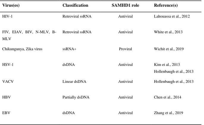
23 APOBEC3F are upregulated after infection, in an IFN-β mediated manner. Interestingly, APOBEC3G overexpression or knock-down does not influence HCMV replication, suggesting a possible viral immune evasion mechanism. In fact, under the selective pressure of APOBEC proteins, HCMV evolved an under-representation of cytidines throughout its genome, to avoid the APOBEC-driven restriction (Pautasso et al., 2018). As far as SAMHD1 is concerned, this is one of the most extensively studied RFs. It was firstly reported to limit HIV-1 infection, and then revealed to restrict also various RNA and DNA viruses. While this thesis was in preparation, in July 2019 “Cell Reports” published the first study of the role of SAMHD1 in HCMV infection, which appears to act in limiting the transcription of IE genes during the early phases of the infection (Kim et al., 2019). However, since the role of SAMHD1 during HCMV infection is the object of this thesis, SAMHD1 will be extensively described in the next chapter, and its implication in viral restriction will be discussed in detail.
3. SAMHD1
Sterile α motif (SAM) and histidine-aspartic domain (HD) containing protein 1 (SAMHD1) is a cellular deoxynucleotide triphosphates (dNTPs) triphosphohydrolase, that hydrolyses dNTPs into deoxynucleosides (dNs) and inorganic triphosphates (Goldstone et al., 2011). To date, it is not only one of the most known RFs against HIV-1, but it is the only cellular protein known to attend the triphosphohydrolase function and thus, the only one to negatively balance the activity of cellular ribonucleotide reductase (RNR), reducing the pool of available dNTPs for cellular and viral DNA synthesis.
a. Expression
SAMHD1 was discovered by Li and colleagues in 2000, as a human homologue of MG21, a mouse IFN-γ induced gene (Lafuse et al., 1995). In the article published by Li et al., SAMHD1 was firstly identified in an extensive screening of a cDNA library generated from human MDDC and was designated as dendritic cell-derived IFN-γ induced protein (DCIP) (Li et al., 2000).
24 Human SAMHD1 gene locates on chromosome 20 and the protein is expressed by most cell types. Expression levels can vary among different tissues: for example, small intestine, spleen and ovary express SAMHD1 at high levels, but adipose tissue, liver and muscle express low or irrelevant amounts of the protein (Schmidt et al., 2015). Supporting its role as an antiviral RF of HIV-1, the expression profile in human cells shows an abundant and constitutive expression in vagina, foreskin and rectum tissue, sites of viral entry during sexual contact, as well as in leukocytes, resident and/or infiltrating these tissues. Moreover, cycling and non-cycling monocytes, macrophages, dendritic cells, CD4+ and CD8+ T cells,
show high expression of SAMHD1 (Schmidt et al., 2015). Compared to proliferating primary human fibroblasts, the quiescent ones show an increased SAMHD1 expression, associated with a strong decrease of intracellular dNTPs (Franzolin et al., 2013).
Regarding SAMHD1 regulation of expression, CD4+ T cell lines and lung adenocarcinoma samples were used to reveal mechanisms of epigenetic regulation of the promoter, that contains CpG islands prone to methylation and subsequent transcriptional repression (de Silva et al., 2013; Wang et al., 2014). SAMHD1 expression can be upregulated in vitro in primary macrophages by IL-12 and IL-18 treatment and in an IFN-γ-independent way, leading to restriction of HIV-1 (Pauls et al., 2013). In lung fibroblasts, SAMHD1 expression can be upregulated by TNF-α treatment and it is IRF-1 dependent (Liao et al., 2008). IFN-I and IFN-II stimulation of primary monocytes can induce SAMHD1 expression, downregulating miR-181a and miR-30a (negative regulators of SAMHD1 translation), with no changes in the promoter activity (Riess et al., 2017). On the other hand, CD4+ T cells and DCs did not show an overexpression of SAMHD1 after IFN-I treatment, maybe due to SAMHD1 mRNA post-transcriptional regulation (St Gelais et al., 2012). SAMHD1 transcription can be induced also by phospho-IRF3, that directly binds SAMHD1 promoter and increases its expression after activation of intracellular PRRs (Yang et al., 2016).
b. Structure
SAMHD1 is a 72 kDa protein of 626 amino acids and it is characterized by the presence of two domains, the SAM and the HD domain, expressed in tandem (Liao et al., 2008) (Figure 5).
25 Figure 5: Schematic representation of SAMHD1 monomer
Starting from N-terminal portion (left), KRPR nuclear localization signal (NLS), the SAM and HD domain, T592 regulatory site and Vpx interacting domain are represented. The four enzymatic sites inside the HD domain are also shown (adapted from Sze et al., 2013).
The sterile α motif (SAM) domain is important for protein-protein interactions, it is dispensable for the triphosphohydrolase activity, but it is required for its maximal activity. SAM domain is so called because 4 out of 14 proteins identified by Ponting et al. that contain this domain are involved in yeast sexual differentiation, and it contains a secondary protein structure rich of α-helices (Ponting, 1995). In human cells, SAM domains are present in proteins with different biological functions: Tyr and Ser/Thr kinases, lipid kinases, scaffolding proteins, RNA binding proteins and transcription factors. In general, SAM domains are very much represented as protein-protein interaction domains (Qiao and Bowie, 2005).
The HD domain is responsible for the triphosphohydrolase activity (Beloglazova et al., 2013), exerted by three enzymatic sites, H167, HD206-207 and D311 (Sze et al., 2013). This domain is part of a superfamily of metal-ion-dependent phosphohydrolases rich of histidine and aspartic acid residues, that are evolutionary conserved in proteins involved in nucleic acid metabolism (Aravind and Koonin, 1998; Laguette and Benkirane, 2012). It extends from amino acid 162 to 335 and the crystal structure revealed the presence of both α-helices and β-barrel folds, that contain the key amino acids residues for its catalytic activity (Goldstone et al., 2011). The HD domain also provides the protein with the interface for enzyme oligomerization, binding dGTP and dNTPs to the allosteric sites, thus permitting the assembly of the tetrameric active complex (Powell et al., 2011) (see below and Figure 7). Notably, dNTPs, and particularly dGTP, are not only substrates of the enzyme but also allosteric activators, and this characteristic renders SAMHD1 both a regulator and a sensor of the cellular dNTP pool. Interestingly, this characteristic is shared
26 by the cellular enzyme RNR, responsible for dNTP production, thus acting on the other arm of the dNTP pool balance. Like SAMHD1, RNR is active in an oligomeric state, with allosteric sites that sense intracellular dNTP concentration and, in a similar manner, its activity is regulated by nucleotide binding (Ji et al., 2014).
SAMHD1 is generally described as a nuclear protein, and contains a nuclear localization signal (NLS) located on its N-terminal portion (Brandariz-Nuñez et al., 2012), although it was also reported a cytoplasmic localization in resting CD4+ T cells (Baldauf et al., 2012).
At the C-terminal portion, SAMHD1 contains a viral protein x (Vpx)-interacting domain, that is the site of interaction with HIV-2 and simian immunodeficiency virus (SIV)-encoded accessory protein Vpx (Laguette et al., 2012). Near the Vpx-interacting domain, there is the regulatory residue Threonine 592 (T592), an important site of phosphorylation and the most characterized mechanism of negative regulation of SAMHD1 activity and restriction (discussed later in the text, see page 29).
c. Metabolic activity and functions
As mentioned above, SAMHD1 is a regulator of dNTP pool and catalyzes the hydrolyzation of dNTPs into dN and triphosphates. This activity is tightly associated with three intertwined important functions of SAMHD1: cell cycle regulation, sensing of genome integrity and antiviral restriction (discussed later in the text, see page 33) (Mauney and Hollis, 2018). Substrate nucleotides allocate in the active sites and are stabilized by interactions with water molecules between them and the amino acids, leading to unspecific interactions that permit a “substrate promiscuity”. In the active sites of the HD domain, the core amino acids His167-His206-Asp207-Asp311 coordinate Mg2+ ions and α-phosphate nucleotides, while His210-His233-Asp288 catalyze the hydrolyzation of triphosphates (Mauney and Hollis, 2018). Hydrolyzation of dNTPs may serve as an important and at present the only known mechanism of negative regulation of dNTP pool enrichment, critical to ensure genome stability and improve DNA synthesis fidelity (Franzolin et al., 2013). The importance of its role is supported by the evidence that an unbalanced dNTP pool may stimulate cell cycle arrest at the S-phase (Chabes and Stillman, 2007).
Therefore, SAMHD1 is involved in cell cycle in two ways: it is regulated by cell cycle proteins and impacts cell cycle progression, through dNTP pool control (Franzolin et al.,
27 2013). One of the first observations of a possible involvement of SAMHD1 in cell cycle regulation was the evidence that non-cycling CD4+ T cells abundantly express SAMHD1 and are resistant to HIV-1 infection (Baldauf et al., 2012).
Later works revealed that SAMHD1 is active and negatively controls dNTPs during the G0
phase, when cells are quiescent and they do not need dNTPs for DNA duplication (Sze et al., 2013). When cells exit from G0 and enter in G1 phase, SAMHD1 is phosphorylated at
T592 and subsequently inactivated. This transition is facilitated by a mitogen-induced activation of Raf/Mek/Erk kinases and increase of cyclin A2/Cdk1 activity (Mlcochova et al., 2017) (Figure 6a). The p21waf1/cip1 cell cycle inhibitor has been reported to influence
SAMHD1 phosphorylation. In MDDCs growth in vitro with IFN-γ and CD40L, p21waf1/cip1
increase of expression is associated with SAMHD1 dephosphorylation and dNTP pools decrease (Valle-Casuso et al., 2017) (Figure 6b). To support this observation, topoisomerase inhibitors-induced DNA damage results in activation of p21waf1/cip1 and loss of SAMHD1 phosphorylation (Mlcochova et al., 2018). A recent work revealed that PP2A-B55α, a mammalian key mitotic exit phosphatase, can interact with and dephosphorylate SAMHD1 at T592 site in M/G1 transition on actively proliferating cells (Schott et al.,
2018) (Figure 6c). Moreover, it seems that SAMHD1 dynamic during cell cycle is finely regulated. Indeed, Tramentozzi and colleagues have recently shown that SAMHD1 is not simply activated/inactivated during the different phases of the cell cycle, but it is active and regulates dNTP pools during all cell cycle phases, controlling an excessive accumulation of DNA precursors even during the S-phase, when a too large amount of dNTPs could be detrimental for DNA replication fidelity (Tramentozzi et al., 2018).
28 Figure 6: SAMHD1 expression and activation during cell cycle progression
SAMHD1 is highly expressed and active during G0 phase but expressed at low level and inactive during S/G2
phase. (a) Raf/Mek/Erk activation increases cyclin A2/Cdk1 activity, that in turn phosphorylate and inactivate SAMHD1 at the entry of the S phase, increasing intracellular dNTP levels. (b) IFN-α mediates the induction of p21waf1/cip1 that decreases the levels of cyclin A2/Cdk1 and limits SAMHD1 phosphorylation. (c)
PP2A-B55α phosphatase interacts with and dephosphorylates SAMHD1 at T592 site during M/G1 transition
(adapted from Sze et al., 2013)
In fact, during DNA replication, errors in the incorporation of new deoxynucleotides can physiologically occur. In steady state conditions, exogenous and endogenous stressors can create DNA damages, that can be detrimental for the cells and the entire organism; to limit the effect of these modifications, cells developed the so-called DNA damage responses (DDRs). When a break of the double helix of the DNA molecule occurs, the double strand break (DSB) can be repaired by two mechanisms: the non-homologous end-joining recombination (NHEJR), that directly ligates the two broken ends, and the homologous recombination (HR), that utilizes the homologous sequence of a sister chromatid to accurately repair the damage. SAMHD1 can be directly involved in HR mechanisms, recruiting the C-terminal binding protein interacting protein (CTIP) and the MRE11-RA50-NBS1 (MRN) nuclease complex (Daddacha et al., 2017) and acting as a scaffold protein for the “molecular actors” of HR. Notably, MRE11 is a 3’-5’ exonuclease that
29 participates in resection of nucleotides at the stalled replication forks (Coquel et al., 2018), and it could contaminate SAMHD1 preparations for in vitro assays, and thus be responsible of the supposed, but controversial, exonuclease activity of SAMHD1 itself (Beloglazova et al., 2013; Seamon et al., 2015).
d. Mechanisms of regulation
SAMHD1 is active in a tetrameric form and the dynamic of oligomerization is complex and still under debate. One of the main features of tetramerization is the binding of dNTPs inside two allosteric sites, located at the interface between monomers, named A1 and A2 (Bhattacharya et al., 2016) (Figure 7). These two allosteric sites are located nearby: the A1 site binds specifically dGTP and GTP, while the A2 site is defined as a coactivator and it is more promiscuous, binding other dNTPs, despite it shows a preference for purines (Mauney and Hollis, 2018). Together, binding of dGTPs (or GTP) and dNTPs is fundamental for the formation of a long-lived tetrameric form. In their absence, SAMHD1 is inactive and monomers can only associate in a dimeric form, establishing a monomer-dimer equilibrium not enzymatically competent (Hansen et al., 2014).
The most characterized mechanism for formation and stabilization of active tetramer is phosphorylation of the SAMHD1 monomer at the C-terminal Threonine 592 (T592) residue. Human and murine SAMHD1 are substrates of phosphorylation at multiple sites, but T592 acquired, over time, a predominant importance for SAMHD1 activity and biology. The first observation of SAMHD1 phosphorylation at T592 was made by White and colleagues in 2013, when they identified a cyclin-dependent kinase target motif, ranging from amino acid 592 to 595 (592TPQK595), and described a differential phosphorylation status in cycling and noncycling THP-1 and CD4+ T cells. In non-cycling cells, SAMHD1 was unphosphorylated and active, and capable to restrict HIV-1 infection, while in cycling cells it became phosphorylated and unable to restrict HIV-1, despite the ability to hydrolyze dNTPs remained intact (White et al., 2013a).
30 Figure 7: Model for SAMHD1 regulation of stabilization and activity
SAMHD1 activity is controlled by activator nucleotides, essential for preservation of nucleotide homeostasis. In low dNTPs condition, SAMHD1 is present in a monomer–dimer equilibrium. GTP binding in A1 stabilizes the dimer, while dNTPs binding at A2 favor SAMHD1 tetramerization and stabilization. Tetrameric SAMHD1 catalyzes dNTPs degradation and prevents their accumulation. T592 phosphorylation destabilizes tetramer stability apparently without modifying catalytic efficiency. PP2A-BB5α mediates SAMHD1 dephosphorylation (adapted from Mauney and Hollis, 2018).
In the same year, two other research groups pointed to the influence of T592 phosphorylation on the negative regulation of SAMHD1 antiviral activity, without the affection of dNTP pool regulation (Welbourn et al., 2013), and the involvement of cyclin A2/Cdk1 activity for an efficient phosphorylation (Cribier et al., 2013). Mass spectrometry analysis of human SAMHD1 conducted in 2014 by Gelais et al. confirmed the interaction with cyclin A2 and Cdk1 and revealed an interaction with Cdk2 and S-phase kinase-associated protein 2 (SKP2). All these proteins were expressed in cycling U937 and THP-1 monocytic cells, that are permissive to HIV-1 infection, suggesting a role of these proteins in negative regulation of SAMHD1 restrictive activity and establishing a cellular environment permissive to HIV-1 replication (Gelais et al., 2014). Despite at that time the precise effect of T592 phosphorylation on SAMHD1 function was unclear, later studies revealed that this post-translational modification prevented the long-lived and enzymatically active tetramer formation, and reported a correlation between the destabilization of the active tetrameric form by phosphorylation and the impairment of retroviral restriction, despite the dNTPase function seemed to be lowered but not completely abolished (Arnold et al., 2015; Yan et al., 2015). In summary, T592
31 phosphorylation inhibits SAMHD1 antiviral activity, despite dNTPase catalysis is not completely abolished, and it occurs through destabilization of the active homotetramer. However, despite the extensive knowledge on SAMHD1 phosphorylation, little is known about its subsequent dephosphorylation. In a recent paper, it has been reported the only dephosphorylating enzyme known to date, the cellular serine/threonine protein phosphatase 2 A holoenzyme (PP2A), containing the regulatory subunit B55α (PP2A-B55α). The interaction with PP2A-B55α and SAMHD1 dephosphorylation has been characterized in vitro in non-cycling monocyte derived macrophages (MDMs), and it has been described to promote mitotic exit of the cells, through activation of SAMHD1 and subsequent reduction of dNTP pool at levels compatible with cellular quiescence (Schott et al., 2018).
In recent years, researchers focused on alternative mechanisms of SAMHD1 activity regulation, such as oxidation. This is a reversible post-translational modification that can be triggered by different stimuli and regulates the activity of proteins involved in cellular metabolism, cell signaling and cell cycle progression. SAMHD1 contains three important cysteines, Cys341, Cys350 and Cys522 that can be substrates of oxidation. When this happens, Cys341 and Cys350 can form a disulfide bond between themselves, and Mauney and collaborators proposed a model in which Cys522 act as a “switch” for protein oxidation, that causes conformational changes of allosteric sites, impairing dNTPs binding and destabilizing the formation of active tetramers (Mauney et al., 2017). Subsequently, Wang and colleagues reported that functional Cys341 and Cys522 are required for SAMHD1 restriction of HIV-1 infection, but that mutations in these sites did not affect dNTPs hydrolyzation and protein tetramerization. Given that, they proposed that oxidation of the protein is a key factor to discriminate dNTP metabolic activity and retroviral restriction (Wang et al., 2018).
Another mechanism proposed to regulate SAMHD1 activity is the acetylation at Lys405, a substrate for the acetyltransferase arrest-defective protein 1 (ARD1), which results in an increased dNTP hydrolyzing activity, during the G1 phase of the cell cycle (Lee et al.,
2017).
In summary, SAMHD1 phosphorylation, oxidation and acetylation may represent different means to control its catalytic activity and are a testimonial of the cellular need to precisely control and maintain the dNTP pool, in physiological conditions as well as in the context of viral infections.