SAPIENZA University of Rome Molecular Medicine Department
PhD Course in
Immunological, Hematologic and Rheumatologic Sciences
Curriculum: Immunology and Immunopathology
XXXI Cycle (A.A. 2017/2018)
Tumor-stroma interactions influence the response to PI3K targeted
agents in preclinical models of colorectal cancer (CRC)
PhD Student Chiara Bazzichetto
Tutor: Coordinator:
1
Contexts
1. Introduction 2
1.1 Colorectal cancer: epidemiology and genetic and molecular characterization 2 1.2 Druggable targets in CRC treatment: MAPK and PI3K pathways 4
1.2.1 MAPK signalling pathways 4
1.2.2 PI3K/mTOR signalling pathways 5
1.3 The PTEN tumour suppressor 7
1.3.1 PTEN regulation 7
1.3.2 PTEN functions 8
1.4TME 10
2. Aim of the study 12
3. Materials and Methods 13
3.1 Cell cultures 13
3.2 Drug treatments 13
3.3 Cell viability assay 14
3.4 CRC-fibroblast co-culture 14
3.5 Statistical analysis 15
3.6 Immunofluorescence 15
3.7 Western blot analysis 15
3.8 Immunoprecipitation 16
3.9 Detection and analysis of soluble factor 17
4 Results 18
4.1 Response to MEK inhibition is dictated by CRC genetic background 18 4.2 Response to PI3K/mTOR double inhibition is modulated by CRC/TME interactions 19 4.3 Pharmacologic interactions between MEK and PI3K/mTOR inhibitors are profoundly
modulated by CRC/TME interactions 20
4.4 Exposure to fibroblast CM selectively increases sensitivity of PTEN-competent
HCT116 cells to double PI3K/mTOR inhibition 21 4.5 Soluble factors released in fibroblast CM modulate PTEN function 24 4.6 Soluble factors released by stromal cells activate the mTORC1 complex and increase
sensitivity to PI3K/mTORi treatment 27
4.7 Stromal cell CM upregulates PI3K pathway and PI3K/mTOR double inhibitor response 29 4.8 Screening of cytokines and chemokines production by stromal cells 32
5 Discussion and conclusions 36
6 Future prospects 40
2
1. Introduction
1.1 Colorectal cancer: epidemiology and genetic and molecular characterization
Colorectal cancer (CRC) is the second main cause of cancer death, leading to over 700,000 deaths annually in worldwide and is the second most diagnosed tumour (accounting for 16% and 13% of newly diagnosed cancers in men and women, respectively) and principal cause of death in Italy (11%) [1, 2].
It is currently known that the risk of developing CRC is not only influenced by hereditary predisposition or medical conditions (i.e. chronic inflammatory bowel disease and type 2 diabetes), but is also strongly associated with a western lifestyle which can increase the risk of developing CRC. Indeed, the increase in incidence is associated with risk factors among which smoking, diet (with an excess of body weight), and alcohol [3, 4].
Given the multiplicity of factors underling its development, we can epidemiologically distinguish three types of CRC based on origin and expression: a sporadic form (65%), a familial type (30%) and a hereditary type (<5%); the latter can in turn be associated or not with familial adenomatous polyposis (FAP or HNPCC, respectively) [3, 5].
General principles of current CRC treatment include radical surgical resection and, in more advanced stages, surgery can be preceded or followed by chemotherapy (neoadjuvant therapy and adjuvant therapy, respectively) and radiotherapy. CRC chemotherapy treatment is based on the use of fluoropyrimidine for intravenous infusion (5-fluorouracil (5FU)) or for oral use (capecitabine), variously associated in doublets or triplets to irinotecan and oxaliplatin [6-8].
CRC is a specific multifactorial pathology, different from individual to individual; alterations of the genetic and epigenetic background, indeed, are specific to each patient and to predict the tumour progression steps, different classification methods are currently used [9]. Specifically, three main pathways of CRC development and progression have been defined: they include aberrations in global genomic status [Chromosomal INstability (CIN) status and MicroSatellite Instability (MSI) status] and epigenomic status [CpG Island Methylator Phenotype (CIMP) status]. These mechanisms play a significant role in determining characteristics of tumour [10]. CIN, MSI and CIMP status, indeed, are characterized by specific pathological precursors which are found in three fundamental categories of genes: tumour suppressor genes [e.g. Adenomatous polyposis coli (APC), and
3
tumour protein 53 (TP53)], protoncogenes (e.g. KRAS, NRAS), and DNA repair genes [e.g. mismatch repair (MMR) and mutY Homolog (MUTYH)] [11].
APC gene mutations were one of the earliest genetic events identified in the development of CRC and indicate a pivotal role for the WiNgless-Type MMTV integration site family member (Wnt) pathway in CRC tumorigenesis [12]. APC is considered a tumor-suppressor in CRC and it is dysregulated at both the germline and somatic level (APC mutations occurs in 80% of sporadic colorectal tumours and loss of heterozygosity (LOH) is reported in 30%–40% of CRC patients) [13].
Somatic mutations in the TP53 gene are one of the most frequent alterations in human cancers; in particular, in CRC, the frequency of TP53 mutations is around 45%. It is actually known that different mutations in TP53 correlate with different prognosis, moreover patients with TP53 mutations could derive more benefit from 5FU-based chemotherapy [14].
Phosphatidylinositol-3-Kinase (PI3K) signalling is deregulated in CRC through a variety of mechanisms, including loss of Phosphatase and tensin homolog deleted on chromosome 10 (PTEN); loss of PTEN expression occurs in 30% of sporadic cases, and is associated with lack of response to Epidermidal Growth Factor Receptor (EGFR) inhibitors (cetuximab) [15]. Sartore-Bianchi and colleagues demonstrated that combined profiling of KRAS, PI3K, and PTEN could identify patients with metastatic CRC (up to 70%) of who would fail to benefit from anti-EGFR antibodies [16].
The KRAS proto-oncogene encodes for a guanosine triphosphate (GTP)/guanosine diphosphate that exerts its role in Mitogen-Activated Protein Kinase (MAPK) pathway downstream of the EGFR. Mutations in KRAS may occur early in the development of pre-cancerous adenomas in the colon and rectum and are detected in 30-40% of CRC cases [17]. KRAS plays a critical role in CRC treatments, although is not a significant prognostic factor. Randomised clinical trials demonstrated that, for patients with KRAS wild-type, the addition of cetuximab to standard chemotherapy improves best overall response rate (ORR), progression free survival (PFS) and overall survival (OS) compared with those patients receiving chemotherapy alone [18].
Deficiencies in the MMR system lead to a hypermutator phenotype; the insertion or deletion of repetition units drive the mutation of genes involved in apoptosis regulation or implicated in cellular regulation of growth, such as BCL2-associated X protein (BAX),
4
transforming growth factor beta receptor II (TGFβRII), or insulin-like growth factor II receptor (IGFIIR), respectively [19, 20].
1.2 Druggable targets in CRC treatment: MAPK and PI3K pathways
The RAS-MAPK and PI3K signalling pathways play a central role in tumorigenesis, and in particular in CRC; for this reason, the analysis of mutations or other molecular aberrations in these pathways could have numerous clinical implications [21]. Extensive cross-talk interconnecting the two pathways and occurring at different hubs of signalling cascades through “vertical” and “lateral” feedback loops [22], form the bases for therapeutic strategies that exploit the single or combined action of molecular target inhibitors downregulating the two molecular pathways [23].
1.2.1 MAPK signalling pathways
The MAPK pathways incorporate proline-directed and protein-serine/threonine kinases, which regulate numerous processes in both normal and cancer cells, such as: embryogenesis, cell differentiation, cell proliferation, and cell death [24].
Different MAPK cascades have been identified: extracellular signal-regulated kinase (ERK) 1 and 2, c-Jun N-terminal kinase (JNK), p38, and ERK5; nevertheless, the ERK1/2 cascade was the first identified and most extensively characterized MAPK pathway [25, 26].
The signalling cascade is composed of a set of three evolutionarily conserved, sequentially acting kinases: a MAPK, a MAPK kinase (MAPKK), and a MAPKK kinase (MAPKKK). The MAPKKKs, could be activated through phosphorylation of tyrosine kinase receptors (RTKs) and/or as a result of their interaction with a small GTP-binding protein of the RAS/Rho family in response to extracellular stimuli [27, 28].
The activation of RTKs is transmitted to the small GTPase RAS, which is activated mainly at the plasma membranes. Three genes encode four different RAS proteins: HRAS, NRAS, KRAS-4A and KRAS-4B, the latter two being alternative splice variants of the KRAS gene, and differ according to the capability which they activate the transduction cascade: KRAS, indeed, activates with greater efficacy RAF/MEK/ERK signalling cascade, while HRAS more effectively activates PI3K/mTOR pathway [29, 30]. Activated RAS, recruits RAF to
5
plasma membrane leading to its activation through phosphorylation and homo- and hetero-dimerization between BRAF and CRAF [31]. RAF activates, in turn, MEK1 and MEK2 (MEK1/2) through phosphorylation of two Ser residues in the activation loop, allowing for the activation of ERK1/2, phosphorylated in turn by MEK1/2 [32]. Once activated, ERK can regulate numerous proteins in the cell: it can directly or indirectly phosphorylate and activate many transcription factors, such as c-Jun and cAMP (Adenosine 3'5' Cyclic Monophosphate) Response Element-Binding Protein (CREB) through the phosphorylation of 90 kDa ribosomal S6 kinase (p90rsk), respectively [33].
Target-based therapies are focused on developing RAS/RAF/MEK signalling pathways inhibitors and several MEK1/2 and RAF inhibitors have been tested clinically or are currently in clinical trial for CRC and other cancers [34]. KRAS and BRAF mutations should be able to predict sensitivity to RAF-targeted agents, but CRC resistance to BRAF inhibition has been demonstrated both preclinically and clinically and has been attributed to rebound activation of EGFR at the cell membrane. Phase III clinical trials demonstrated that BRAF and KRAS mutations were negative predictors of benefit to cetuximab and panitumumab [35]; in spite of this, in patients with PI3K wild-type/KRAS mutant CRC, the addition of a PI3K inhibitor (such as BKM120) may overcome cetuximab resistance [36].
1.2.2 PI3K/mTOR signalling pathways
The PI3K pathway plays a central role in cell growth, proliferation, survival, and angiogenesis in both normal and malignant cells and thus is an attractive target for targeted molecular therapy [37].
PI3Ks family includes three different classes of kinase: I, II, III; each class has different characteristics, both in the molecular structure and in substrate specificity. Following binding to membrane receptors, class I PI3K phosphorylate substrate phosphatidyl-inositol 4,5 bisphosphate (PIP2) to phosphatidylinositol 3,4,5 triphosphate (PIP3). PIP3 recruits’ proteins containing the pleckstrin homology domain (PH) to the cytoplasmic membrane, including the protein kinase dependent phosphoinositide (PDK1) and the serine/threonine protein kinase B (PKB, also known as AKT) [38]. Full activation of AKT is a multistep process consisting of the phosphorylation of two different residues, Thr308 and Ser473, via PDK1 and PDK2, respectively [39]. Given its pivotal role in cell proliferation and survival
6
through phosphorylation of different substrates, overexpression of activated AKT isoforms can transform cells and AKT alteration is common in different human cancer [40, 41], including CRC [42].
The mammalian target of rapamycin (mTOR) is a critical downstream regulator of the PI3K pathway, playing a fundamental role in the regulation of growth, proliferation, and cellular metabolism [43, 44]. mTOR can act as a substrate and effector of the AKT pathway: indeed, AKT indirectly activates mTORC1 by direct phosphorylation of the tumour suppressor Tuberous Sclerosis Complexes (TSC) 2 on Ser939 and Thr1462, but evidence show that mTORC2, can also phosphorylate directly AKT at Ser473 [45]. mTOR is a serine/threonine kinase that exerts its function through two functionally and structurally distinct multi-component complexes, mTORC1 and mTORC2 [46]. mTORC1 is composed by the core proteins mTOR (that can bind other proteins in a species- and condition-specific manner, such as: Proline-Rich AKT Substrate 40 (PRAS40), DEP domain-containing mTOR interacting protein (DEPTOR), GRp58, Tel2-interacting protein 1 (Tti1)- Telomere maintenance 2 (Tel2), Rac1, Regulatory-associated protein of mTOR (Raptor) and mLST8 [47].
mTORC2 complex includes the core proteins mTOR, Rapamycin insensitive companion of mTOR (Rictor), and mammalian Lethal with Sec13 protein 8 (mLST8), as well as various associated proteins, Proline-Rich Protein (PRR) 5, Heat shock protein (Hsp) 70, DEPTOR, GRp58, Tti1-Tel2, Rac1, mammalian Stress-activated protein kinase Interacting protein (mSIN) 1 and Protein observed with RICTOR (Protor) [47].
mTORC1 is activated by growth factors, amino acids, and energy levels and regulates different processes, such as growth, proliferation, biosynthesis of macromolecules, and angiogenesis, by regulating p70 ribosomal protein S6 Kinase 1 (p70S6K1) and Elongation Initiation Factor (EIF)-4E Binding Protein 1 (4E-BP1) [46]. mTORC2 is primarily responsive to growth factors and controls cell structure, cytoskeletal reorganization, and survival by activating Serum and Glucocorticoid Kinase (SGK), AKT, and Protein-Kinase C (PKC) [46].
Recently, preclinical and clinical studies for the treatment of CRC revealed the efficacy of targeting PI3K signalling pathway, and in particular dual PI3K/mTOR inhibitors, that overcome the feedback loop activation of PI3K [48]. Molecular inhibitors, such as
7
GSK2126458, DS 7423, XL765 e NVP-BEZ235 are currently being tested in clinical trials, although dual inhibition could lead to substantial toxicity [49].
1.3 The PTEN tumour suppressor
The signal transduction pathway mediated by the PI3K proto-oncogene is regulated negatively by PTEN. The PTEN tumour suppressor is the main negative regulator of the PI3K pathway and a common target for inactivation in sporadic cancers [50]. Somatic mutations, transcriptional silencing, deletions or protein instability in PTEN occur in different human tumours, and PTEN loss of function is found in 9-30% of CRC patients [51].
PTEN levels and/or function can be regulated through multiple mechanisms, including transcriptional and transcriptional regulation, subcellular localization, post-translational modifications, and proteins interaction [52, 53].
1.3.1 PTEN regulation
PTEN gene is negatively and positively regulated by different transcription factors. Negative regulators can directly (such as c-JUN) and indirectly (such as nuclear factor kappa B (NF-KB)) inactivate PTEN expression, while positive regulators include different protein such as TP53 and early growth response protein 1 (EGR-1) [52].
microRNAs (miRNAs) are the principal regulators of PTEN abundance at the post-transcriptional level. Different miRNAs include those derived from miRNA precursors containing a single hairpin structure (e.g. miR-21, miR-22 and miR-205), as well as those with a polycistronic structure (e.g. mir-17-92, mir-106b-25, mir-367-302b, and mir-221-222); they can play a role as a tumour suppressor or oncogenes leading to increased or decreased PTEN levels, respectively [54].
Numerous post-translational modifications (such as ubiquitination, acetylation, sumoylation, and phosphorylation) modify the proprieties of the PTEN tumour suppressor and impact on PTEN localization in the cell.
Monoubiquitination or ubiquitination of specific residues (such as Lys13 and Lys289) are involved in PTEN shuttling and nuclear import, while PTEN polyubiquitination leads to PTEN cytoplasmic retention and degradation. Although the acetylation of Lys402 does not
8
involve a change in the intracellular localization of PTEN, this modification enhances the interaction of PTEN with PDZ-domain proteins. While sumoylation is essential to PTEN tumour suppressive functions, by increasing the anchorage to the plasma membrane, the phosphorylation in C-terminal cluster of Ser and Thr maintains PTEN in a closed conformation, reducing its phosphatase activity and membrane localization. PTEN C-terminal tail is phosphorylated by different kinases, Casein Kinase (CK) 2, Glycogen synthase kinase 3 beta (GSK3), RAK, protein interacting with carboxyl terminus 1 (PICT-1) and Rho-associated kinase (ROCK) [55, 56].
1.3.2 PTEN functions
Besides PTEN main role as a negative regulator of the PI3K pathway, different functions associated with this tumour suppressor are currently known.
PTEN is involved in the control of metabolic pathways: indeed, it regulates different processes such as glucose uptake, by controlling the membrane expression of glucose transporter type 1 (GLUT1) and/or the levels of gluconeogenesis through inhibition of forkhead box O (FOXO) 1 [53].
PTEN is also involved in the regulation of cell motility and polarity; such PTEN role in migration is PI3K pathway-independent: indeed, PTEN can inhibit glioma cell migration independently of its phosphatase activity [57].
The absence of nuclear PTEN is a prognostic indicator in different aggressive cancers including CRC [58]. The importance of PTEN in the nucleus is associated with its role in controlling cell cycle progression, DNA repair and genomic stability [53, 59].
Furthermore, recent evidence highlight the role of PTEN in regulating the tumour microenvironment (TME). Experiment in mice demonstrate that deletion of PTEN in fibroblasts creates a tumour-permissive stroma in mammary gland tumours, with recruitment of innate immunity cells, remodelling of the ECM and tumour vasculature, and increased malignancy of tumours [60]. PTEN's regulation of the tumour microenvironment is also related to the modulation of cytokines in the microenvironment itself; indeed, a relationship between PTEN-loss and Interleukin (IL)-8 upregulation is known in prostate carcinoma and glioblastoma [61, 62]. Numerous studies show the active contribution of PTEN to the regulation of angiogenesis at different levels. PTEN loss in cancer cells leads to increased Vascular Endothelial Growth Factor (VEGF) expression, due to the
9
upregulation of Hypoxia-Inducible Factor (HIF) 1, while PTEN transfection in multiple myeloma cell line results in a decreased mRNA and protein expression of Matrix Metallopeptidase (MMP)2, MMP9 and FAK (Focal Adhesion Kinase), leading to decreased cell migration [63].
Recent studies highlight the therapeutic potential of agents that target Programmed death-1 (PD-1) or its ligand Programmed death ligand-1 (PD-L1) that have been identified as a factors associated with poor prognosis in different types of cancer [64]. Loss of the tumour suppressor PTEN may induce PD-L1 overexpression, thus inducing immunoresistance in CRC [65].
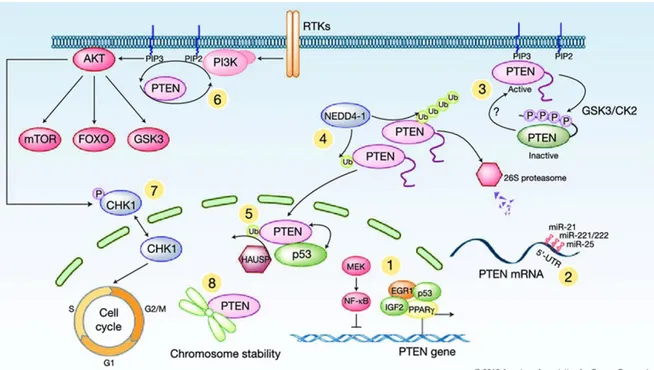
Figure 1. PTEN regulation and function. 1. Activation and/or inhibition of PTEN mRNA transcription; 2.
post-transcriptionally regulation of PTEN mRNA by miRNAs; Post-translational modifications: 3. phosphorylation of the PTEN C-terminal tail; 4. poly- and mono-ubiquitination of PTEN, NEDD4-1-mediated; 5. deubiquitination of PTEN in the nucleus and nuclear exclusion; 6. PIP3 dephosphorylation and
suppression of PI3K pathways; 7. regulation of the cell cycle PTEN-mediated, 8. PTEN association with centromeres in the nucleus for chromosome stability maintenance. (Modified from Zhang S., and Yu D. 2010 [66])
10
1.4 TME
TME is fundamental in different steps of tumour progression (Figure 2); indeed, tumour is not composed only by a mass of transformed cells, but also by different “genetically normal” host-derived cell types, and cancer development cannot be dissociated from its local microenvironment. Extracellular matrix (ECM), immune and inflammatory cells, and fibroblasts are the main components of TME [67].
ECM is composed by a complex network of macromolecules such as proteins, glycoproteins, proteoglycans, and polysaccharides that may serve to block or facilitate, not only cell migration, but also free diffusion of cytokines, growth factors, and hormones secreted by stromal and tumour cells; its dysregulation is one of the hallmarks of cancers and the enzymes involved in ECM remodelling are and produced by immune cells and cancer-associated fibroblasts (CAFs) [68].
Immunity cells and their reciprocal interaction with tumour cells are the principal elements involved in TME inflammatory process. Solid tumors are commonly infiltrated by immune cells, belonging to both adaptive (e.g. T cells, B cells, natural killer (NK) cells) and innate immunity (e.g macrophages, dendritic cells (DCs), neutrophils, eosinophiles, basophiles and mast cells) [69]. Macrophages are a major component of innate immune cells and particular interest has been attribuited to tumour-associated macrophages (TAM) [70]; indeed, TAMs accumulation is associated with poor clinical outcome.
CAFs are the major components of cancer stroma, their crucial role is associated with progression, growth and invasion. CAFs may derived from both normal fibroblast (through genetic alteration that leads to a constitutively activated phenotype) and epithelial cells (through epitelial-mesenchimal transition (EMT)); different markers of CAFs were actually identified including alpha smooth muscle actin (SMA), fibroblast activation protein (FAP), Thy-1, desmin, and S100A4 protein [71]. It’s actually known that fibroblast activation in CRC patients correlates with a high risk to develop metastases or recurrence and to have an aggressive disease progression [72].
The various cells that compose TME can modulate treatment response and are therefore potential candidates for therapeutic intervention. Currently, clinical studies that target TME components are in progress and different elements of TME are targeted; among them, siltuximab and ruxolitinib, that act by inhibiting IL-6 and JAK, respectively, block inflammatory pathway activation, whereas bevacizumab inhibits VEGF and is a mainstay
11
in the therapy of CRC [73]. Indeed, In KRAS wild type and mutant CRC, antibody treatment (such as bevacizumab) plus FOLFOX-4 show 9.3 and 8.7 months of PFS, respectively [74].
12
2. Aim of the study
Our group has recently demonstrated that PTEN plays a crucial role in determining the functional outcome of combined MAPK/PI3K inhibition in a panel human cancer cell lines of different histological origin, including CRC. These studies show that growth inhibitory synergism with combined MAPK/mTOR inhibition is almost invariably observed in cells with PTEN-loss, but not in PTEN-competent tumour cells [76]. Identification of the precise mechanisms of drug action in relationship to molecular disease drivers is crucial to the successful development of new therapeutic strategies and recently high-throughput molecular analysis is being used to identify putative biomarkers to provide personalized cancer therapy. However, such analysis is usually focused on tumour cell-autonomous molecular determinants of sensitivity to drug treatment and overlooks microenvironmental interactions.
The aim of the study is to investigate how PTEN expression/function in CRC cells modulates the response to signalling inhibitors in the context of complex microenvironmental interactions.
To this purpose, we have investigated the potential role of stroma in response to "horizontal" combinations of agents targeting MEK and PI3K/mTOR signalling in CRC cells differing for PTEN status in different culture conditions, the alternative responses generated by different elements of stroma-endothelial microenvironment, and finally the potential role of PTEN in regulating responses to microenvironmental cues.
13
3. Materials and Methods
3.1 Cell cultures
X-MAN™ HCT116 and HCT116 PTEN-/- were generated by Horizon from homozygous knock-out of PTEN by deleting exon 5 which encodes the active site of the protein in the CRC cell line HCT116 (Horizon Discover www.horizondiscovery.com). Cell lines were routinely maintained in RPMI 1640 medium supplemented with 10% fetal bovine serum (FBS), 2 mM L-glutamine, and antibiotics (Pen/Strep) in a humidified atmosphere with 5% CO2 at 37 °C. Fibroblast cell line HFF (human foreskin fibroblasts) GFP-labeled, HF and BJ were kindly provided by Dr. Maurizio Fanciulli. EA.hy926 was obtained from the American Type Culture Collection (ATCC). Fibroblast and endothelial cells were routinely maintained in DMEM medium supplemented with 15% fetal bovine serum (FBS), 2 mM L-glutamine, and antibiotics (Pen/Strep) in a humidified atmosphere with 5% CO2 at 37 °C.
Cell culture reagents were purchased from Euroclone and Invitrogen (Milan, Italy).
To obtain stromal cells conditioned medium (CM), HFF, BJ, HF and EA.hy926 cells were seeded and the media was replaced by serum-free medium (SFM) after 24 hours from plating; CM was collected after 72 hours.
3.2 Drug treatments
Trametinib (GSK1120212) was kindly provided by GlaxoSmithKline (Brentford, Middlesex, UK). Gedatolisib (PF05212384) and alpelisib (BYL719) were kindly provided from Selleck Chemicals (Huston, TX, USA). MK-2206 was obtained from by Merck and Co. (Kenilworth, NJ, USA). Trametinib, gedatolisib, alpelisib, and MK-2206 were dissolved in DMSO as a 1 mM (trametinib, gedatolisib and MK-2206) and 5mM (alpelisib) stock solution, and stored at -20°C or -80°C. Everolimus (RAD001) was obtained from Novartis Pharma (Basel, Switzerland) and was dissolved in 100% ethanol as a 10 mM stock solution and stored at -20°C.
14
3.3 Cell viability assay
Cell proliferation was evaluated by direct cell counting or Crystal Violet assay.
For cell counting, Thoma chamber was used. Effects on cell growth in response to different treatments were monitored by Crystal Violet assay. For Crystal Violet assay, a fixed number of tumour cells were dispensed into 24-wells (NEST Biotechnology), and the following day cells were treated at indicated concentrations of drugs. After 72 hours of treatment, the cells were washed with PBS, fixed with 4% formaldehyde for 10 minutes and then stained with 0.1% Crystal Violet for 40 minutes, at room temperature. Excess stain was removed with water and the plates were dried. Crystal violet stain was extracted with 95% acetic acid and the absorbance was measured at 570 nm.
The dose of drug that causes 50% of cell growth inhibition (IC50 value) was calculated according to the Chou-Talalay method using the Calcusyn software (Biosoft, Cambridge, United Kingdom).
3.4 CRC-fibroblast co-culture
HFF fibroblasts were used to develop a CRC/stroma co-culture model, in order to investigate the influence of stromal cells on CRC cell growth and drug sensitivity in correlation with the presence/absence of PTEN.
X-MAN™ HCT116 and HCT116 PTEN-/- cells and fibroblast cells GFP-labeled were seeded alone or with a ratio of 1:1 in plates 35 mm, in RPMI 1640 supplemented with with 10% FBS, 2 mM L-glutamine, and antibiotics (Pen/Strep) in a humidified atmosphere with 5% CO2 at 37 °C. After 24 h the medium was replaced with SFM containing different drug concentration alone or in combination. After 72 h cells were harvested and resuspended in ice-cold RPMI 1640 10% FBS. The total amount of cells was counted in the Thoma chamber and the percentage of CRC cells and fibroblast cells in co-culture were analysed by flow cytometry analysis that allows to differentiate between HFF cellular populations, fluorescently labeled in comparison to unlabeled X-man cells.
15
3.5 Statistical analysis
The Mann-Whitney U test was used for comparing quantitative variables. Results with two-tailed P values <0.05 were judged to be statistically significant. Synergism, additivity, and antagonism were assessed by isobologram analysis with a fixed-ratio experimental design using the Chou-Talalay method [77]. Results were analysed with the Calcusyn software (Biosoft, Cambridge, United Kingdom) and combination indexes (CI) were appropriately derived. By this method, an average CI at the ED50, ED75, and ED90 < 1 indicates synergism, = 1 indicates additivity, and > 1 indicates antagonism, respectively.
3.6 Immunofluorescence
Cells were seeded on on 22x22 mm coverslips and the media was replaced by SFM and HFF CM after 24 hours from plating. After 24 hours from the replaced medium the grown cells were fixed in 4% paraformaldehyde in phosphate-buffered saline (PBS) for 10 min, permeabilized in PBS containing 0.5% Triton X-100 for 5 min, and blocked with PBS containing 5% Bovine Serum Albumin (BSA) and 0.3% of Triton X-100 for 90 min. Coverslips were incubated overnight with primary antibodies in 1% BSA, 0.3% Triton X-100 in PBS. Coverslips were then washed three times in PBS and then incubated with one of the following secondary antibodies for 1 h at a dilution of 1:200: goat antirabbit Alexa Fluor 488 (A11034, Invitrogen) and goat antimouse Alexa Fluor 594 (A11032, Invitrogen). After washing, the nuclei were counterstained with DAPI. Cells were then washed twice with PBS and observed under Zeiss Axiovert 200 M fluorescence microscope. Specific fields were photographed with a digital camera equipped with Zeiss Axiovision acquisition software.
3.7 Western blot analysis
Whole cell extracts were obtained by Chaps Cell Extract buffer, containing 50 mM Pipes/Hcl (pH 6.5), 2 mM EDTA, 0.1% Chaps, 20 g/ml Leupeptin, 10 g/ml Pepstatin A, 10 g/ml Aprotinin, 5mM 1,4-Ditiotreitolo (DTT). Assay sample for protein concentration
16
used was Bradford (Protein Assay Dye Reagent Concentrate, Bio-Rad). An amount of total cell lysate was fractionated by SDS-polyacrylamide gel electrophoresis and transferred to nitrocellulose membrane (Amersham, Arlington Heights, USA). Membranes were probed with the following primary antibody: phosphorylated (Ser380/382/383) and total PTEN, phosphorylated (Thr389) p-p70S6K, phosphorylated (Thr202/Tyr204) ERK1/2, mTOR, RAPTOR, PRAS40 and Rictor, phosphorylated (Thr37/46) p-4E-BP1 (from Cell Signaling Technology Inc. Beverly, USA). Signal was detected using peroxidase-conjugated anti-mouse or anti-rabbit secondary antibodies (Jackson Immunoresearch Labs, Inc., Baltimore, USA). The enhanced chemi-luminescence (ECL) system (Amersham) was used for detection and image detection was performed with UVITEC Alliance 4.7 system (Cambridge, UK). To control the amount of proteins transferred to nitrocellulose membrane β-actin and GAPDH were used and detected by anti β-actin mAb (clone AC-15, Sigma, St. Louis, USA) and by anti GAPDH (Abcam, Cambridge, MA, USA) respectively.
3.8 Immunoprecipitation
Chip-Grade Protein A/G Magnetic Beads (Thermo Fisher Scientific, Rochester, NY, USA) were incubate with 2 g of FRAP (Santa Cruz Biotechnology, Santa Cruz, CA) over night at 4 °C. Precleared beads were than incubated over night at 4 °C with 1 g of protein. The immunoprecipitates were collected and after 2 wash in CHAPS buffer re-suspended in 35 µl of the same buffer and Ladder buffer 1X. The immune complexes and 20 μg of protein total cell extract were analysed by Western blot analyses with, mTOR, RAPTOR, PRAS40 Rictor and phosphorylated (Thr389) p-p70S6K. Signal was detected using peroxidase-conjugated anti-mouse or anti-rabbit secondary antibodies (Jackson Immunoresearch Labs, Inc., Baltimore, USA). The enhanced chemi-luminescence (ECL) system (Amersham) was used for detection and image detection was performed with UVITEC Alliance 4.7 system (Cambridge, UK). To control the amount of proteins in total cell lysate transferred to nitrocellulose membrane β-actin was used and detected by anti β-actin mAb (clone AC-15, Sigma, St. Louis, USA).
17
3.9 Detection and analysis of soluble factor
1x10⁶ HFF, HF, BJ and EA.hy926 cells were plated into 150mm dishes (Falcon BD). After 24 hours from the plating, the culture medium was replaced by SFM, and after 24 hours, media were collected and cells were counted. Cells culture media were analysed by Proteome Profiler (Antibody Arrays, R&D System) according to the manufacturer's protocol.
X-MAN™ isogenic HCT116 cell lines cell culture media were analysed in triplicate as per the manufacturer’s instructions using IL-8, IL-6 and MCP-1 specific ELISA, purchased from ENZO Life for IL-8 and IL-6 analysis and from R&D Systems for MCP-1 analysis. Absorbance was read at 450 nm. IL-8, IL-6 and VEGF expression was represented as pg/mL and then related to the control.
Recombinant human CXCL8/IL-8 and IL-6 were purchased from R&D System. For recombinant protein experiment 500 pg/mL of IL-8 and 1000 pg/mL of IL-6 were used in DMEM medium without the addition of FBS.
18
4 Results
4.1 Response to MEK inhibition is dictated by CRC genetic background
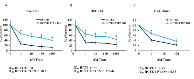
To investigate the role of PTEN in modulating the response of CRC cells to MEK inhibition (trametinib, MEKi), X-MAN™ isogenic HCT116 cell lines (HCT116 and HCT116 PTEN-/-) were treated with increasing concentrations of the MEK inhibitor trametinib (1-1000 nM) for 72 hours, in different culture conditions: isogenic HCT116 cells grown in SFM, isogenic HCT116 cells grown in the presence of HFF CM and isogenic HCT116 cells co-cultured with GFP-tagged fibroblasts (HFF). Effects on cell growth/viability were monitored by Crystal Violet Assay.
In order to evaluate the influence that microenvironment could exert in modulating response to treatment, the contribution of direct cell-to-cell contact and soluble factors produced by fibroblast cell was evaluated using co-culture and fibroblast CM experiments. The functional effects of MEKi on viability and cell growth is shown in Figure 3: regardless of cell culture conditions (isolated tumor cells, HFF CM, or HFF co-culture), the parental HCT116 cell line (PTEN-competent) was significantly more sensitive to MEK inhibition, as compared to its PTEN-loss counterpart (HCT116 PTEN-/-). IC50 was less than 1 nM in parental HCT116 under any culture condition, while it increased to 6 nM, 68 nM, and 215 nM in HCT116 PTEN-/-, under HFF co-culture, isolated culture, and HFF CM culture conditions, respectively.
Figure 3. PTEN influences the response to MEKi independently from microenvironmental elements.
19
were monitored by Cristal Violet assay for both SFM (w.o. FBS, A) and CM (HFF CM, B) conditions and by cytofluorimetric analysis for Co-culture (C). The percentage of viability was represented as % of control, assuming the levels in control cells as 100%. Results represent the average +SD of three independent experiments. Asterisks indicate statistically significant differences (p <0.01 by 2-tailed Student’s t test) for the comparison between * HCT116 and HCT116 PTEN-/- treated with MEKi.
4.2 Response to PI3K/mTOR double inhibition is modulated by CRC/TME interactions
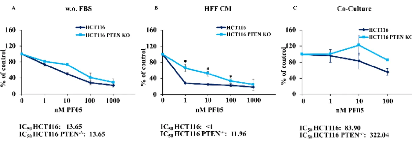
To investigate the role of PTEN status in modulating the response to PI3K/mTORi (gedatolisib), the same analysis was performed for PI3K/mTORi (at concentration ranging from 1 to 1000 nM for SFM and CM condition and from 1 to 100 nM for co-culture experiments). As opposed to MEKi treatment, the presence of stromal elements strongly influenced the response to PI3K/mTORi. As shown in Figure 4, response of isogenic HCT116 cell lines to PI3K/mTORi was independent of PTEN status (IC50 for both HCT116 and HCT116 PTEN-/-: 13.65 nM). However, sensitivity of parental HCT116 cells (PTEN-competent) to the growth inhibitory activity of PI3K/mTORi was significantly increased in the presence of fibroblast CM (IC50: <1), while was not modulated in the HCT116 PTEN-/- cell line (IC50: 11.96). Conversely, direct cell-to-cell contact in a co-culture setting, rendered both HCT116 and HCT116 PTEN-/- more resistant to PI3K/mTORi treatment (IC50: 83.90 and 322.04, respectively).
Figure 4. The PI3K/mTOR double inhibitor response is strongly influenced by microenvironmental soluble factors in PTEN competent cells. HCT116 and HCT116 PTEN-/- cells were treated with increasing
20
concentration of drugs, as indicated for 72 hours. Effects on cell growth were monitored by Cristal Violet assay for SFM (w.o. FBS, A) and CM (HFF CM, B) conditions and by cytofluorimetric analysis for Co-Culture (C). The percentage of viability was represented as % of control, assuming the levels in control cells as 100%. Results represent the average +SD of three independent experiments. Asterisks indicate statistically significant differences (p <0.01 by 2-tailed Student’s t test) for the comparison between * HCT116 and HCT116 PTEN-/- treated with PI3K/mTORi.
4.3 Pharmacologic interactions between MEK and PI3K/mTOR inhibitors are profoundly modulated by CRC/TME interactions
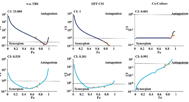
We further tested pharmacologic interactions between MEKi and PI3K/mTORi, using a fixed dose-ratio (1:1) experimental design over a wide range of concentrations (1–1000 nM) of each agent, under the culture conditions described above: isogenic HCT116 cells grown in SFM and isogenic HCT116 cells co-cultured with GFP-tagged fibroblasts (HFF). Figure 5 shows that under SFM culture conditions pharmacologic interactions between the two agents were strongly synergistic in HCT116 PTEN-/- cells (CI: 0.530) and antagonistic in PTEN-competent HCT116 cells (CI: 23.089), as previously described in FBS-supplemented culture conditions [76].
While the presence of soluble factors in HFF CM did not substantially modify pharmacologic interactions between MEKi and PI3K/mTORi in either cellular model (synergistic in HCT116 PTEN-/-: CI 0.301; additive in HCT116: CI 1), direct cell-to-cell completely subverted the type of interaction: indeed, under co-culture conditions, combined MEKi and PI3K/mTORi treatment became additive in HCT116 PTEN-/- (CI: 0.993) and strongly synergistic in parental HCT116 (CI: 0.001).
21
Figure 5. Effect of combined MEKi and PI3K/mTORi in isogenic CRC cell lines. HCT116 (top panel)
and HCT116 PTEN-/- (bottom panel) cells were exposed to MEKi and PI3K/mTORi in a fixed dose-ratio
combination (1:1) in SFM (w.o. FBS), with CM (HFF CM) and Co-culture (Co-Culture). CI were calculated by conservative isobologram analysis for experimental data and plotted against the fraction affected.
4.4 Exposure to fibroblast CM selectively increases sensitivity of PTEN-competent HCT116 cells to double PI3K/mTOR inhibition
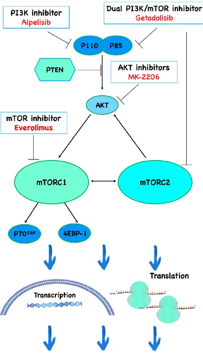
Among drug response-modulating effects of CRC/TME interactions, sensitization of PTEN-competent cells to the growth inhibitory action of PI3K/mTOR double inhibitor in the presence of HFF CM was one of the most prominent; in order to investigate which signalling hub along the PI3K cascade was responsible of the observed effect, we investigated the functional effects of different molecular target inhibitors (Figure 6) on cell viability under SFM and CM culture conditions.
22
Figure 6. PI3K/AKT/mTOR pathway inhibitors. (Ciuffreda L.)
X-MAN™ HCT116 and HCT116 PTEN-/- cell lines were exposed to increasing concentration of alpelisib (selective class I PI3K inhibitor, 5-5000 nM), MK-2206 (allosteric AKT inhibitor, 10-1000 nM) and everolimus (rapamycin-like mTORC1 inhibitor, 1-1000 nM) for 72 hours in SFM or HFF CM.
23
As shown in Figure 7, selective inhibition of a single step along the PI3K pathway was not sufficient to result in drug sensitization in the presence of HFF CM. Indeed, no differences were observed in the response of either HCT116 or HCT116 PTEN-/- to class I PI3Ki, AKTi, and mTORC1i in the presence of HFF CM, as compared to SFM, suggesting that exposure to fibroblast CM selectively increases the sensitivity of PTEN-competent HCT116 cells to double PI3K/mTOR inhibition.
Figure 7. Effect of different PI3K pathway inhibitors in response in isogenic colorectal cancer cell lines. Isogenic colorectal cancer cell lines HCT116 and HCT116 PTEN-/- were treated with increasing
24
as % of control, assuming the levels in control cells as 100%. Results represent the average +SD of three independent experiments. Asterisks indicate statistically significant differences (p <0.01 by 2-tailed Student’s t test) for the comparison between * SFM and HFF CM condition.
4.5 Soluble factors released in fibroblast CM modulate PTEN function
Since the PI3K/mTORi-sensitizing effect of fibroblast CM was selectively observed in PTEN-competent CRC cells, we set out to investigate whether CM influenced PTEN expression and function. Thus, we investigated PTEN expression and function in response to fibroblast CM in parental HCT116 cells.
Western blot analysis, shown in Figure 8, shows that PTEN expression was upregulated with HFF CM until 48 hours and SFM was used as control to calculate the variations of proteins expression levels as compared to HFF CM.
These results correlate with increased levels of p-PTEN Ser380/382/383 C-tail phosphorylation in the presence of HFF CM. It is currently known that phosphorylation of PTEN C-tail results in an hyperactivation of PI3K pathway; moreover, this post-translational modification does not allow the protein to bind the plasmatic membrane thus interfering with its localization, stability and activity [78, 79]. Consistently, our results showed that the upregulation of PTEN C-tail phosphorylation is associated with an increased level of p-p70S6K Thr389, suggesting an activation of PI3K pathway related to HFF CM.
25
Figure 8. The CM upregulates total PTEN and PTEN C-tail phosphorylation. HCT116 cells were
cultured with SFM (w.o FBS) and fibroblast CM (HFF CM). After 24 and 48, HCT116 cells were lysed and analysed by Wester Blotting using the indicated antibodies and -actin was used as protein loading and blotting control.
In order to understand how PTEN was distributed in the cell in presence of HFF CM, we investigated PTEN subcellular localization as compared to SFM using immunofluorescence analysis.
HCT116 and HCT116 PTEN-/- were seeded on coverslips and the media was replaced by SFM and HFF CM after 24 hours from plating; specific antibody for PTEN was incubated overnight to detect its distribution.
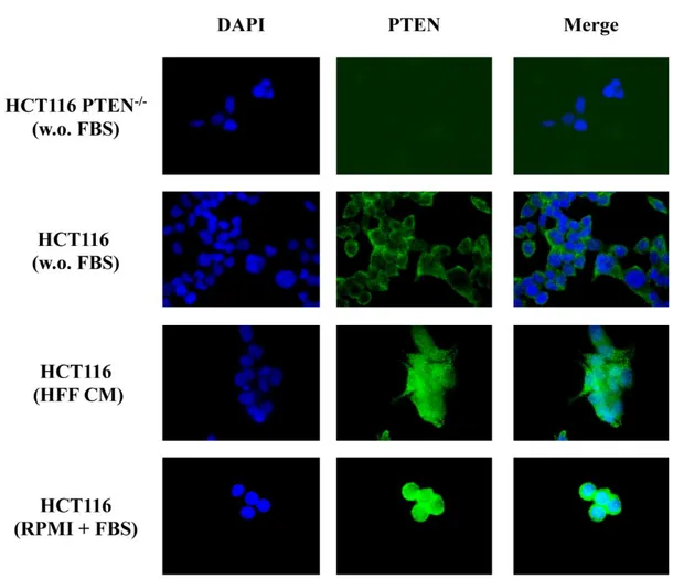
As shown in Figure 9, immunofluorescence experiments revealed that HFF CM upregulated PTEN expression and was associated with a diffuse, cytoplasmic distribution of tumour suppressor in the cells.
26
Figure 9. Fibroblast CM upregulates PTEN expression and a spread distribution of PTEN. Isogenic
CRC cells were exposed to different cell culture condition: SFM (w.o FBS), fibroblast CM (HFF CM) and complete medium (RPMI+FBS), for 24 h; PTEN levels was then assessed by Immunofluorescence using specific antibodies. Microphotographs were taken at ×63 magnification. Results from one experiment representative of at least three independent experiments performed with superimposable results are shown. Immunofluorescence of PTEN in HCT116 PTEN-/- is shown as PTEN negative control.
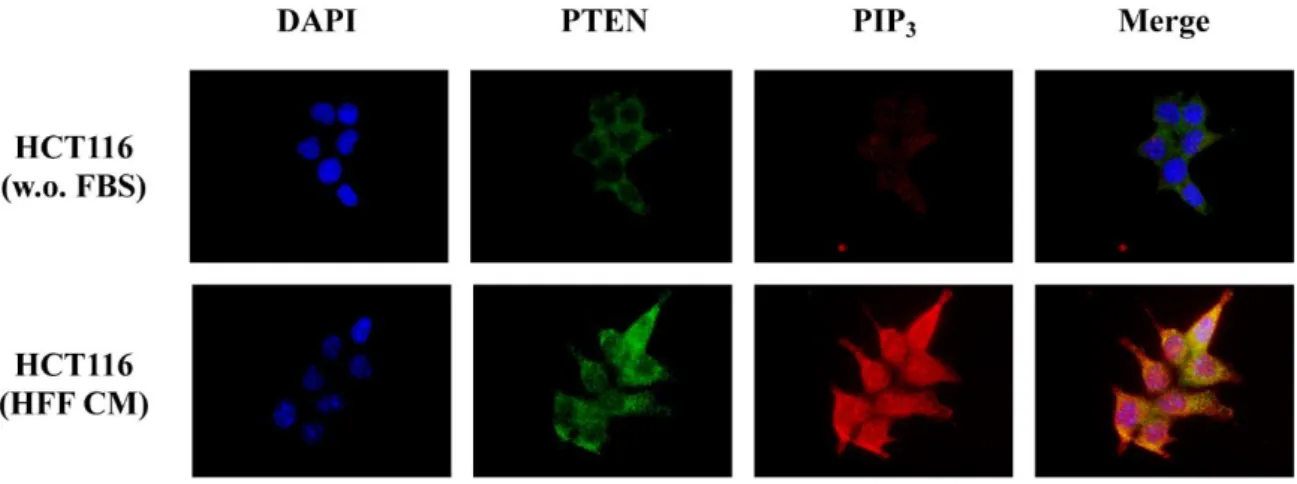
Further check the functional status of PTEN phosphatase activity, we also detected the main PTEN substrate, the phospholipid second messenger PIP3, which is dephosphorylated by PTEN to yield PIP2. PIP3 and PTEN protein levels and localization were analysed by Immunofluorescence assay in SFM and HFF CM (Figure 10). Protein detection showed an increase in PIP3 levels using HFF CM. Moreover, as shown in merge images, PTEN and PIP3 did not co-localize.
Figure 10. The CM leading to PTEN partial inactivation. HCT116 cells were cultured in SFM (w.o. FBS)
and HFF CM for 24 h. PTEN and PIP3 levels were then assessed by Immunofluorescence using specific
antibodies. Microphotographs were taken at ×63 magnification. Results from one experiment representative of at least three independent experiments performed with superimposable results are shown.
These data underline that soluble factors derived from fibroblast cells decreased PTEN phosphatase activity leading to PI3K pathway basal activation.
27
4.6 Soluble factors released by stromal cells activate the mTORC1 complex and increase sensitivity to PI3K/mTORi treatment
In order to understand whether PI3K pathway activation downstream of PTEN in response to fibroblast CM would result in a shift of the balance between mTORC1 and mTORC2 complexes, mTOR immunoprecipitation experiments were carried out in isogenic colorectal cancer cells in both SFM and HFF CM conditions.
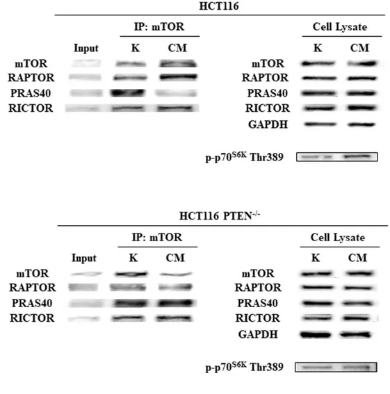
As shown in Figure 11, IP/WB experiments revealed that exposure of parental HCT116 cells (PTEN-competent) to fibroblast CM induced the association of mTOR with Raptor and the release of PRAS40 from the complex, while no difference in Rictor association with mTOR was observed in different culture conditions. Conversely, in the absence of PTEN (HCT116 PTEN-/-) no significant changes in mTOR complexes’ composition was observed. These results suggest specific activation of the mTORC1 complex in response to fibroblast CM in PTEN-competent CRC cells, which, in turn, resulted in downstream pathway activation, as demonstrated by p-p70S6K Thr389 phosphorylation in parental HCT116, but not HCT116 PTEN-/-.
28
Figure 11. HFF CM activates PI3K/mTOR pathway in PTEN-wt context leading to mTORC1, but not mTORC2, complex activation. Isogenic CRC cells were subjected to SFM and HFF CM culture condition
for 24 h; Endogenous mTOR was immunoprecipitated and the immunocomplexes were blotted for RAPTOR, PRAS40 and Rictor. RAPTOR, PRAS40, Rictor and p-p70S6K Thr389 levels in total cell lysates are also
shown. GAPDH was used as protein loading and blotting control in total cell lysate.
We then investigated the molecular effects of PI3K/mTOR double inhibitor in HCT116 and HCT116 PTEN-/- cell lines under different culture conditions. Cell lines were exposed to increasing concentration of the drug for 24 hours in SFM and in the presence of HFF CM (Figure 12). As expected from the formation and activation of the mTORC1 complex described above, exposure of HCT116 to fibroblast CM increased p-4E-BP1 Thr37/46 and p-p70S6K Thr389 levels, which were more effectively suppressed by PI3K/mTORi
29
treatment in parental HCT116 cells. No differences between SFM and HFF CM conditions were observed in the HCT116 PTEN-/- cell line, in which PI3K/mTORi treatment proved to be equally efficient in shutting down 4E-BP1 and p70S6K phosphorylation in both culture conditions (data not shown).
Exposure to fibroblast CM slightly increased basal p-ERK levels, but those were not modulated by PI3K/mTORi, independent of PTEN status.
Figure 12. The CM increases the HCT116 response to PI3K/mTORi. HCT116 cells were incubated with
increasing doses of PI3K/mTORi, as indicated, for 24 hours SFM and HFF CM. Protein lysate was analysed by Western Blotting using p-ERK, p-4E-BP1 and p-p70S6K specific antibodies. -Actin was used as protein
loading and blotting control.
4.7 Stromal cell CM upregulates PI3K pathway and PI3K/mTOR double inhibitor response
We next investigated the effects of CM derived from different stromal cells on PI3K/mTOR activation and response to the PI3K/mTOR double inhibitor gedatolisib in isogenic CRC cell lines. To this purpose, HCT116 and HCT116 PTEN-/- were exposed to SFM and CM derived from normal fibroblast cell lines (BJ and HF) or endothelial cells (EA.hy926).
As shown in Figure 13, p-4E-BP1 Thr37/46 and p-p70S6K Thr389 levels in SFM and with stromal CM derived from HFF, BJ, HF and EA.hy926 were detected using Western blot analysis. Even in this case, p-ERK Thr202/Tyr204 protein levels were analysed as control of the analysed system.
30
Figure 13. Stromal soluble factors upregulate PI3K pathway in PTEN competent tumor cells. Isogenic CRC (HCT116 top panel and HCT116 PTEN-/- bottom panel) cells were cultured with SFM and stromal cells
CM, as indicated, for 24h. Protein lysate was analysed by Western Blotting using ERK, 4E-BP1 and p-p70S6K specific antibodies. -Actin is used as protein loading at blotting control. SFM was used as control for
calculate system variations.
As already observed with HFF CM, p-4E-BP1 Thr37/46 and p-p70S6K Thr389 levels were
upregulated with all stromal cell CM in PTEN-competent parental HCT116 cells, whereas no striking differences were observed in HCT116 PTEN-/-. Conversely, p-ERK
31
Thr202/Tyr204 levels were slightly increased by exposure to stromal cell CM, regardless of PTEN status.
Overall, these results support the hypothesis that PTEN mediates paradoxical PI3K pathway activation in response to soluble factors released in the TME by stromal elements (fibroblasts, endothelial cells).
We further verified that PI3K/mTOR activation in response to different stromal cell CM would result in increased sensitivity to PI3K/mTORi in PTEN-competent CRC cells. To this purpose, HCT116 and HCT116 PTEN-/- were exposed to two different concentrations of PI3K/mTORi (1 and 10 nM) for 72 hours in SFM and stromal cell CM (Figure 14). Regardless of the stromal cell used as the source of CM, CM selectively increased sensitivity to PI3K/mTORi in parental HCT116. In particular, fibroblast-derived CM had greater influence on response to PI3K/mTORi, as compared to endothelial cell-derived CM. Conversely, HCT116 PTEN-/- did not respond to PI3K/mTORi, regardless of the source of CM.
Figure 14. Analysis of stromal-induced response to PI3K/mTORi treatment. XMAN isogenic HCT116
cells were treated with increasing concentration of PI3K/mTORi, as indicated, for 72h in SFM and stromal CM. Effects on cell growth were monitored by Crystal Violet assay. The percentage of viability was represented as % of control, assuming the levels in control cells as 100%. Results represent the average +SD of three independent experiments. Asterisks indicate statistically significant differences(**: p <0.01 or *: p < 0.05 by 2-tailed Student’s t test) for the comparison between SFM and stromal cells CM response.
32
4.8 Screening of cytokines and chemokines production by stromal cells
To preliminarily ascertain which soluble factors produced by stromal cells could be candidate mediators of the PI3K/mTOR pathway activation and, most importantly, of the PI3K/mTORi-sensitizing effects, CM from different fibroblasts (HFF, BJ and HF) and endothelial cells (EA.hy926) were analysed by Proteome Profiler Array after 24 hours of culture in SFM (Figure 15).
Figure 15. Analysis of cytokine/chemokine production by stromal cells reveals similar patterns of expression. Cell culture media of HFF, BJ, HF and EA.hy926 were analysed by Proteome Profiler Array.
As shown in Table 1, different cell lines of fibroblast and endothelial cells produce a similar pattern of cytokines and chemokines expression. Some of them are commonly produced by both fibroblast and endothelial cells (e.g. IL-8, IL-6, UPAR), while other were relatively specific for fibroblasts (e.g. MCP-1, SDF-1 and Angiopoietin-1).
33 Table 1. Profiling proteins in stromal cells CM
We also confirmed the presence of some of the detected cytokines by ELISA. As shown in Figure 16, IL-8 was produced at variable levels by all stromal cells tested; IL-6 was produced by fibroblasts and at very low levels by endothelial cells, while MCP-1 was selectively produced by fibroblasts.
34
Figure 16. IL-8, IL-6, and MCP-1 expression in stromal cells. Stromal cells, were analysed by ELISA
assay; the results are expressed as pg/mL.
To assess the potential role of two of the main soluble factors detected in stromal cell CM (IL-8 and IL-6), we tested the molecular effect of recombinant human IL-8 and IL-6, alone and in combination, when added to SFM. The concentration of recombinant proteins was chosen based on the levels of cytokine detected by ELISA assay in CM (500 pg/mL for IL-8 and 1000 pg/mL for IL-6).
As shown in Figure 17, both cytokines stimulated ERK phosphorylation at Th202/Tyr204, and to a lesser extent of 4E-BP1 Thr37/46, in parental HCT116 cells; conversely,
35
phosphorylation of p70S6K at Thr389 was selectively induced by HFF CM, while recombinant IL-8 and IL-6, either alone or in combination, were not able to induce mTORC1 activation.
Figure 17. IL-8 and IL-6 do not mediate mTORC1 activation in the HFF CM. HCT116 cells were
cultured with SFM, HFF cells CM, and SFM with recombinant Human protein of IL8, IL6 and the combination of two (500pg/ml, 1000pg/ml and 500pg/ml+1000pg/ml, respectively) as indicated, for 24h. Protein lysate was analysed by Western Blotting using p-ERK, p-4E-BP1 and p-p70S6K specific antibodies.
-Actin is used as protein loading at blotting control. SFM was used as control for calculate system variations.
These data underline that a similar profile of cytokine/chemokines was secreted by different stromal cells: IL-8 and IL-6 were produced, albeit at variable levels, by all fibroblast cells, but were not able to mediate mTORC1 activation in PTEN-competent CRC cells.
36
5 Discussion and conclusions
Recent advances in high-throughput technologies are responsible of the carachterization of numerous genes in different human cancers. Increased knowledge of cancer mutations, allowed not only to understand the evolution of the tumour in its various steps, but also to the development of personalized therapies for patients [80]. Indeed, prognostic and predictive biomarkers have been identified in several cancers, including CRC; for example, only patients lacking KRAS or NRAS mutations could benefit from cetuximab and panitumumab treatment [81].
The present study provides evidence that PTEN expression could influence the response to targeted therapies in the presence of stromal and microenvironmental elements.
We recently demonstrated that the lack of PTEN is a crucial determinant of synergistic interactions between MAPK and PI3K inhibitors in a panel of cancer cells of different histological origins [76]. Furthermore, several recent studies indicate that PTEN status influence tumour growth, survival, invasion, angiogenesis and metastasis in different tumour or surrounding cells, for example through the production of cytokine and chemokine [82, 83]. Indeed, recent data obtained in our lab indicate a correlation between the occurrence of BRAFV600E mutations and/or PTEN-loss and higher levels of IL-8 production in CRC cells [84]. Whether it occurs through the production of soluble factors or through direct cell-to-cell contact, it is currently recognized that TME plays a pivotal role in cancer progression and metastasis, but it also has a profound effect on therapeutic efficacy [75]. Bidirectional communication between tumour cells and their microenvironment is critical at different stages of tumour development and progression and the capability of stromal cells to modify the TME can exhibit opposite effects: tumour-promoting or tumoricidal capacities. Moreover, the importance of stromal elements led to a new, microenvironment-based CRC classification, developed in datasets obtained by profiling whole-tumour samples containing both tumour and stromal cells; this classification is composed by four different subtypes, in order to correlate each subtype with patient outcome [85].
CAFs represent one of the most abundant stromal cell types in several cancers and, at a difference from epithelial tumor cells, mutations and loss of heterozygosity are rare; recent evidence are actually focused on defining a "CAF signature", indeed, depending on stimuli
37
received by cancer cells, CAFs could exert suppressive or stimulatory effects in response to TME [86, 87].
Based on this evidence we set up multicellular culture models to study the interplay between the tumor genetic background (namely PTEN-inactivating events) and the surrounding TME and elucidate the molecular mechanisms of interactions, with specific regard to the growth-inhibitory effects of potentially therapeutic signalling inhibitors. From preliminary data, we observed that the presence or absence of serum in culture medium induces profound changes in the phosphoproteome profile, upregulating different signalling pathways. Thus, in order to standardize basal levels of pathway activation, we set up isolated CRC cell cultures, CM, and co-culture experiments in SFM. We investigated the response to MEKi, PI3K/mTORi and their combination in isogenic CRC cell lines, that only differ for PTEN status, under different conditions of growth (SFM, fibroblast CM and co-culture). The results obtained show that response to MEKi was independent from the presence of microenvironmental elements (Figure 3). Conversely, the impact of PI3K pathway inhibitors on stromal elements has been recently studied and the potential anti-tumor efficacy of their inhibition may also derive from interference with the capability of tumour cells to respond to different stromal signals, thus underpinning cell-specific roles of the PI3K/mTOR pathway both in cancer cells and in the tumor microenvironment [88]. At a difference from MEKi, in our system the presence of fibroblast-derived soluble factors strongly and selectively increased the response of PTEN-competent CRC cells (HCT116) to the PI3K/mTOR double inhibitor gedatolisib (Figure 4). As opposed to the results obtained in isolated tumor cell cultures [76], response to combined MEKi/PI3Ki treatment was dramatically changed by direct cell-to-cell contact between CRC cells and stromal fibroblasts (Figure 5): indeed, combination treatment in co-culture results in synergistic effects only in PTEN-competent cell line.
The signalling hub at which PI3K pathway inhibition takes place appears to be relevant: indeed, fibroblast CM-mediated drug-sensitizing effects were only observed with double PI3K/mTOR inhibition, whereas response to different single-step PI3K pathway inhibitors were not influenced by fibroblast-derived soluble factors (Figure 7). The first generation of PI3K pathway inhibitors (i.e wortmannin and LY294002), were associated with elevated toxicity and a limited therapeutic efficacy; increased interest, however, has grown over the years for PI3K/mTOR double inhibitors. BEZ235, GSK2126458, gedatolisib, apitolisib
38
and PQR309, have an apparently greater antitumor activity, although safety and tolerability remain of some concern [89]. Experiments conducted in spheroid models and transgenic mice suggest that dual PI3K/mTOR inhibition is a potential treatment strategy for APC- and PI3K-mutant colorectal cancers [90]; our data add to such evidence, indicating that PTEN-competent CRC cells might respond best to PI3K/mTOR double inhibition in the presence of microenvironmental cues, mediated by soluble factors.
Biallelic inactivation of PTEN occurs in 20-30% of case of CRC and is currently known that PTEN alterations and MSI are correlated [91]. However, the evaluation of PTEN functional state is cumbersome and requires expression and function analysis, in addition to sequencing of the entire gene; this is due to the fact that PTEN function is finely regulated through the control of its expression, through genetic and epigenetic mechanisms, post-translational modifications or changes in its subcellular localization [92]. Moreover, PTEN tumour suppressor function is not just limited to its lipid phosphatase activity: its multiple role, indeed, has recently been expanded to include different subcellular location where it performs distinct functions, such as in the nucleus, where it favours genomic stability and controls cell cycle progression, in the endoplasmic reticulum (ER) and mitochondria-associated membranes (MAMs), where PTEN interacts with the inositol 1,4,5-trisphosphate receptors (IP3Rs) and regulates the sensitivity to apoptosis [93].
Since the drug sensitizing effects of fibroblast CM were observed only in cells that express a functional PTEN protein, we analysed the effects of CM on PTEN expression and function. Despite increased PTEN protein levels, PTEN phosphorylation at Ser380/382/383 was increased, thus explaining the paradoxical functional inactivation of the PTEN tumour suppressor (Figure 8). Consistent with the key role of C-tail phosphorylation in determining an alteration of PTEN to an inactive state, resulting in a reduction of phosphatase activity and a lower association of plasmatic membrane association [94], immunofluorescence and WB experiments showed an apparently paradoxical response to CM stimulation: indeed, PTEN protein levels were increased upon CM stimulation, but its distribution within the cell became spread in the cytoplasm, instead of membrane-associated (Figure 9); indeed, PIP3 levels were increased upon CM stimulation and did not co-localize with PTEN (Figure 10), resulting in overall downstream pathway activation, as demonstrated by the upregulation of p-p70S6K Thr389.
39
Activation of the mTOR pathway has been described in intestinal polyp formation and mTORC1 activation, in particular, appears to be involved in both tumour initiation and progression in CRC, enhancing CIN levels [95]. Results presented here show that exposure to fibroblast CM shifts the balance towards mTORC1 activation, by stimulating the association of RAPTOR, but not PRAS40, with mTOR selectively in PTEN-competent contexts; conversely, mTORC1 and mTORC2 did not differ in HCT116 PTEN-/-, regardless of CM stimulation (Figure 11). Moreover, in PTEN-competent CRC cells, activation of the mTORC1 complex was associated with a sustained response to PI3K/mTOR double inhibition (Figure 12). These results are consistent with evidence that cells over-expressing p-p70S6K and p-4E-BP1 were more sensitive to PI3K/mTOR inhibitor treatment [96]. Functional interaction between tumor genetic background (PTEN status) and microenvironmental cues (soluble factors released in CM) specifically controls activation of the PI3K/mTOR pathway, while the MAPK pathway is upregulated in the presence of HFF CM independently of PTEN status. We previously demonstrated that constitutive ERK activation represses PTEN expression in melanoma and other cancer models, through c-Jun and miR-25 [97]; however, c-JUN and miR-25 were not modulated by HFF CM in parental HCT116 (data not shown).
It is becoming increasingly evident that TME is composed by a heterogeneous mixture of tumour cells plus endogenous stroma that differ for soluble factors production that influence not only homing, invasion and migration of tumour cells but also mechanisms of sensitivity/resistance to drug [98]. We thus analysed fibroblasts from different sources and endothelial cells, for their ability to produce soluble factors that would modulate PI3K pathway activation and sensitivity to PI3K/mTOR double inhibition. Effects observed with HFF CM were confirmed with different stromal cells; however, endothelial cells were less effective in increasing the response to PI3K/mTOR double inhibitor. The analysis of cytokine/chemokine production revealed a similar pattern in CM from different stromal cells (Table 1). However, although IL-8 and IL-6 were among the most prominent common factors produced by all stromal cells, molecular data revealed their relative inability to induce mTORC1 activation in PTEN-competent HCT116 cells (Figure 17).
40
6 Future prospects
Overall, the work conducted so far demonstrates that in a preclinical model of CRC: 1) sensitivity to MAPKi is dictated mostly by the tumor genetic background (PTEN status), while sensitivity to PI3K/mTOR inhibition and pharmacologic interactions with combined MAPK/PI3K inhibition is strongly influenced by interaction with the TME; 2) in PTEN-competent cells soluble factors released by stromal elements paradoxically impair PTEN function, leading to downstream mTORC1 complex formation and pathway activation; 3) paradoxical mTORC1 activation upon exposure to stroma-derived soluble factors results in functional hypersensitivity of PTEN-competent CRC cells to the growth inhibitory effects of double PI3K/mTOR inhibitors.
We recognize the fact that using immortalized fibroblasts to model tumor/stroma interactions represents a gross oversimplification of the in vivo situation and does not entirely reflect the real cancer tissue and TME. As a first step towards creating more physiological multicellular preclinical models, we are currently isolating autologous fibroblasts and cancer cells from patients affected by CRC at different stages. Nevertheless, our data provide solid evidence that factors contained in fibroblasts’ CM may trigger paradoxical mTORC1 activation, selectively in the presence of a functional PTEN protein. Regardless of the identification of the specific factor responsible for this effect, paradoxical PI3K/mTOR activation in a PTEN-competent context, may represent a novel therapeutic target, potentially amenable to pharmacological intervention (e.g. by PI3K/mTOR kinase inhibitors, as highlighted here).
We are also aware that many other factors, in addition to cytokines and chemokines, may influence the interactions between the tumor and its TME. Recent evidence show that communication between tumor and stromal cells can be also mediated by miRNAs [99] and numerous miRNAs have been identified in PTEN regulation in tumors of different histological origins [100, 101]. For this reason, to evaluate the putative role of miRNAs in stromal CM, we will isolate extracellular miRNA derived from stromal CM [102]. miRNA analysis will be carried out also through the isolation of exoRNA; indeed, exosomes are cell derived vescicles abundant in biological fluids, and miRNAs or RNA inside could be isolated using specific ultracentrifugation and analyzed to identify possible mediators of PTEN inactivation.
![Figure 2. Contribution of stromal cells to the Hallmarks of cancer [75]](https://thumb-eu.123doks.com/thumbv2/123dokorg/2893821.11425/12.892.244.708.248.699/figure-contribution-stromal-cells-hallmarks-cancer.webp)