1802
|
wileyonlinelibrary.com/journal/ccr3 Clin Case Rep. 2019;7:1802–1803.1
|
CLINICAL IMAGE
A 15‐year‐old female patient was referred to our hospital with a suspicion of acute pancreatitis and a previous clinical history of early satiety with postprandial fullness, epigastric pain, nausea, and several vomit episodes.
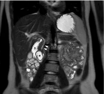
Since ultrasonography was not conclusive due to intes-tinal gas distension, the patient underwent magnetic reso-nance cholangiopancreatography (MRCP), which showed a fluid‐filled lesion (28 mm) arising from the medial du-odenal wall and protruding into the dudu-odenal lumen, con-sistent with a duodenal duplication cyst (DDC) (Figures 1-3).
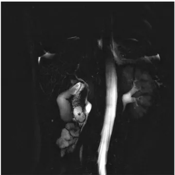
The patient underwent endoscopic marsupialization of the cyst, whose volume reduction was confirmed by a three‐ month follow‐up MRCP (Figure 4).
Duplication cysts of the gastrointestinal tract are rare congenital anomalies, which develop during embryonic life, usually involving, in descending order, jejunum (50%), ileum (44%), esophagus (10%‐15%), colon (7%), and stomach (4%‐9%).1
Received: 24 December 2018
|
Revised: 26 June 2019|
Accepted: 27 June 2019 DOI: 10.1002/ccr3.2327C L I N I C A L I M A G E
Recurrent acute pancreatitis caused by duodenal duplication cyst
in a young patient
Giuseppe Cicero
|
Francesca Catanzariti
|
Ugo Barbaro
|
Velio Ascenti
|
Silvio Mazziotti
This is an open access article under the terms of the Creative Commons Attribution‐NonCommercial‐NoDerivs License, which permits use and distribution in any medium, provided the original work is properly cited, the use is non‐commercial and no modifications or adaptations are made.
© 2019 The Authors. Clinical Case Reports published by John Wiley & Sons Ltd.
Section of Radiological Sciences, Department of Biomedical Sciences and Morphological and Functional Imaging, University of Messina, Messina, Italy
Correspondence
Giuseppe Cicero, Department of Biomedical Sciences and Morphological and Functional Imaging, University of Messina, Via Consolare Valeria 1, 98100 Messina, Italy.
Email: [email protected]
Abstract
Duodenal duplication cyst is a rare congenital anomaly that develops during the embryonic stage and could remain unknown until the adult age. Although often asymptomatic, duodenal duplication cysts can lead to various clinical scenarios with different degree of severity, from nonspecific abdominal pain up to cholestasis, in-tussusception or pancreatitis.
K E Y W O R D S
duodenum, duplication cyst, magnetic resonance cholangiopancreatography, magnetic resonance imaging
FIGURE 1 Coronal T2‐w TSE scan demonstrates the DDC (asterisk) arising from the medial duodenal wall and protruding into the duodenal lumen (D). The common bile duct (arrowheads) is also visible
|
1803CICERO Etal.
As regards the radiological differential diagnosis, duode-nal diverticula, and pancreatic pseudocyst can be respectively ruled out due to the lack of a connection with the duodenal lumen and an ab extrinseco compression of the duodenum.
Although more challenging, a misdiagnosis of choledo-chocele should be avoided due to the lack of communication with the biliary three.
After the histological confirm, DDCs are generally man-aged with surgical intervention through radical or partial resection.2
AUTHOR CONTRIBUTIONS
GC: drafted and reviewed the manuscript. FC: participated in manuscript writing. UB: reviewed the literature. VA: col-lected the images. SM: conceived the idea, reviewed and ac-cepted the final version of the manuscript.
ORCID
Giuseppe Cicero https://orcid.org/0000-0001-9414-4627
REFERENCES
1. Liu R, Adler DG. Duplication cysts: diagnosis, management, and the role of endoscopic ultrasound. Endoscopic Ultrasound. 2014;3(3):152‐160.
2. Salemis NS, Liatsos C, Kolios M, Gourgiotis S. Recurrent acute pancreatitis secondary to a duodenal duplication cyst in an adult. A case report and literature review. Can J Gastroenterol. 2009;23(11):749‐752.
How to cite this article: Cicero G, Catanzariti F,
Barbaro U, Ascenti V, Mazziotti S. Recurrent acute pancreatitis caused by duodenal duplication cyst in a young patient. Clin Case Rep. 2019;7:1802–1803.
https ://doi.org/10.1002/ccr3.2327
FIGURE 2 Coronal tick‐slab 3D T2‐w TSE RARE image clearly demonstrates the DDC (asterisk) within the duodenal lumen
FIGURE 3 Axial T2‐w TSE scan shows the DDC (asterisk). The common bile duct joins the Wirsung duct posteriorly to the cyst (arrows), allowing the differentiation of DDC from “type III” choledochal cyst
FIGURE 4 Axial T2‐w SPAIR scan performed three months after endoscopic marsupialization of the cyst. The DDC is significantly decreased in volume (arrow), without any compression on biliary or pancreatic ducts. The patient has been without symptoms for three months since the endoscopic intervention