UNIVERSITÀ DEGLI STUDI DELLA TUSCIA DI VITERBO
DIPARTIMENTO DI SCIENZE ECOLOGICHE E BIOLOGICHE
Corso di Dottorato di Ricerca in
Genetica e Biologia Cellulare – XXVII Ciclo
DISSECTING THE ROLE OF EXTRACHROMOSOMAL
HISTONE H2B IN CYTOKINESIS
s.s.d. BIO/11
Tesi di dottorato di:
Dott.ssa Laura Monteonofrio
Coordinatore del corso Tutore
Prof. Giorgio Prantera Dott.ssa Silvia Soddu
1
INDEX
INDEX 1
ABSTRACT 3
INTRODUCTION 5
1. The cell cycle 5
1.1 Different stages of Mitosis 6
1.2 Cytokinesis, the final stage of cell division 7
1.3 Abscission , the final steps of cytokinesis 12
1.4 Cytokinesis failure 16
2. Histone H2B 17
3. H2B and HPK2 localize at the midbody: a new extrachromosomal activity
of histone H2B 20
AIM 23
RESULTS 24
H2B LOCALIZATION DURING MITOSIS 24
CYTOKINESIS-SPECIFIC LOCALIZATION OF HISTONE H2B IS
INDEPLENDENT OF RNA 27
H2B DEPLITION CAUSES ABSCISSION DEFECTS AND DELAY 31
H2B DEPLETION ALTERS THE MIDBODY LOCALIZATION OF A FEW
ABSCISSION FACTORS 36
2
DISCUSSION 43
A NEW SUBCELLULAR AND SUB-STRUCTRAL LOCALIZATION
OF HISTONE H2B-S14P IN CYTOKINESIS 43
HISTONE H2B LOCALIZES AT THE MIDBODY INDEPENDENTLY
OF NUCLEIC ACIDS 44
HISTONE H2B IN THE CONTROL OF ABSCISSION 45
INTERPLAY BETWEEN HISTONE H2B AND PROTEIN
INVOLVED IN CYTOKINESIS: A DIRECT INTERACTION WITH CHMP4B 46
MATHERIAL AND METHODS 49
3
ABSTRACT
Histones are constitutive components of nucleosomes, the basic units of DNA packaging in eukaryotes and their post-translational modifications regulate chromatin structure. Among different modifications, specific phosphorylation of the N-terminal tail of H2B, at S14 in vertebrate and S10 in yeast, has been linked to DNA damage response (DDR) and chromatin condensation in apoptosis and meiosis. In addition to chromatin-related functions, a few extrachromosomal activities of specific histones have been described. Recently, our laboratory demonstrated that histone H2B localizes at the midbody, the intercellular bridge connecting two daughter cells during cytokinesis. The cytokinetic specific localization of histone H2B is independent of the presence of chromosome bridges at the cleavage plan, indicating a distinct role from chromatin organization of histone H2B. At the midbody, H2B is phosphorylated at S14 (H2B-S14P) by homeodomain interacting protein kinase 2 (HIPK2), a kinase involved in DDR and development, whose activity is required for cytokinesis and prevention of tetraploidization. In HIPK2-defective cells, expression of a phosphomimetic H2B-S14D mutant is sufficient to overcome cytokinesis defects and rescue cell division and proliferation. This unexpected finding uncovers new role of extrachromosomal histone H2B in cell division. However, the function exerted by histone H2B in cytokinesis is unknown.
Here we show that histone H2B-S14P localizes in specific areas, during different steps of cell division, from metaphase to telophase. Furthermore, in late telophase, at the abscission onset, H2B-S14P show an asymmetric distribution probably due to a specific sub-structural localization. Moreover, we show that the midbody localization of histone H2B is independent of both DNA and RNA. In addition, microscopic studies and live-cell imaging of HeLa cells depleted for histone H2B revealed that histone H2B is necessary for faithful cytokinesis. Indeed, depletion of H2B results in prevention of cell cleavage, accumulation of the connections between daughter cells with the formation of long intercellular bridges (LIBs), and abscission defects and delay. H2B depletion results in tubulin disorganization, presumably linked to LIBs formation and the stretching conditions triggered by abscission defects, thus supporting a role for histone H2B in cytokinesis. Furthermore, in HeLa cells, H2B depletion do not affect the spatio-temporal localization of proteins at the cleavage
4 furrow (e.g., Aurora B, INCENP, PRC1 and PLK1). In the subsequent stage, the midbody formation, PLK1, PRC1, Aurora B, Survivin, INCENP, ECT2, MKLP1 and HIPK2, are present at the midbody and follow the tubulin structures. In contrast, severe defects of localization and organization are observed for the abscission factors ALIX, ESCRT-III subunit CHMP4B, and Spastin, while the localization of Citron kinase is not affected. By in vitro biochemical assays, we show that H2B directly interacts with CHMP4B (Charged Multivesicular Body Protein 4B), a core subunit of the ESCRT-III (Endosomal Sorting Complex Required for Transport-III)-dependent filaments, recently identified as cortical constriction helices required for abscission.
Overall, our data indicate that histone H2B is an important regulator of the final step of cell division and reveal a novel function of extrachromosomal histone H2B for a faithfulness abscission.
5
INTRODUCTION
1.
The cell cycle
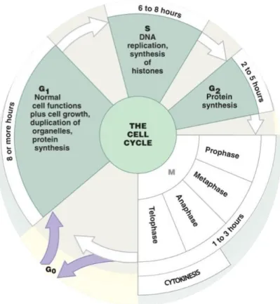
Cell growth and division follow a series of events known as the cell cycle. During cell cycle, chromosomes are duplicated and one copy of each duplicated chromosome segregates to each of two daughter cells. In eukaryotes, the cell cycle is divided into four different phases: gap 1 (G1), synthesis (S), gap 2 (G2) and mitosis (M) (Fig 1). The first three phases are collectively named interphases (I). During G1 phase, a cell confirms that internal and external conditions are favorable for a new round of DNA synthesis and division, and commits itself to the process. S phase comprises the duplication of chromosomes. A new copy of each chromosome is synthesized and the two identical DNA molecules are called sister chromatids. G2 phase represents further growth and preparation for entry into mitosis. M phase culminates with the generation of two identical daughter cells by portioning the chromatids to opposite poles of the cell so that each daughter cell receives a copy of each chromosome.An additional phase (G0) refers to a quiescent state in which the cell remains metabolically active, but no longer proliferates unless appropriate extracellular signals are received. Progression through the cell cycle is controlled biochemically by a network of molecular signal and stringent checkpoint that ensure the cell meets the requirements for passage into the next phase (Kops et al., 2005).
6
Figure 1. The eukaryotic cell cycle.
The cell cycle consists of four distinct phases: G1 phase, S phase (synthesis), G2 phase (all collectively known as interphase) and M phase (mitosis). M phase is itself composed of two tightly coupled processes: mitosis, in which the chromosomes are divided between the two daughter cells, and cytokinesis, in which the cytoplasm divides in half forming two distinct cells (modified by Enderson Education inc 2011).
1.1 Different stages of Mitosis
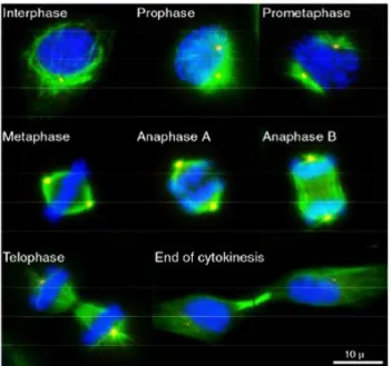
Mitosis can be subdivided into five morphologically distinct phases: Prophase, Prometaphase, Metaphase, Anaphase, Telophase and Cytokinesis (Fig. 2).
At the mitosis onset, animal cells show the most drastic alterations in cytoskeletal organization. During prophase, the largest phase of mitosis, after nuclear envelope breaks down (NEBD), adherent cultured cells become rounded. The centrosomes, which have also been duplicated during S phase, separate and migrate to opposite poles of the nucleus. The microtubule cytoskeleton is re-organized to assemble the bipolar mitotic spindle (a dynamic structure capable of aligning pairs of sister chromatids, sensing chromosome misalignment, and generating forces to segregate chromatids). Chromosomes condense and sister chromatids produced by DNA replication during S phases are captured by microtubule. In prometaphase, held together by a structure known as the centromeres, sister chromatids migrate to the
7 equatorial plane in the center of the cell known as the metaphase plate. At the metaphase, chromosomes align themselves along the metaphase plate. During anaphase sister chromatids separate and move to opposite poles of the mitotic apparatus, or spindle, segregating one of the two sister chromatids to each daughter cell. Mitosis end with telophase during which the nuclear envelope re-forms around the segregated chromosomes as they decondense, the spindle disassemble and physical division of the cytoplasm, called cytokinesis, occurs.
Figure 2. The different stages of M phase.
The immunofluorescence images illustrate HeLa cells in different cell cycle phases. The mitotic spindle is shown in green (α-Tubulin), the centrosomes in red (γ-Tubulin) and DNA in blue (DAPI staining).
1.2 Cytokinesis, the final stage of cell division
Cytokinesis is the final stage of the cell cycle through which a single cell divides into two daughter cells while partitioning its cellular content. The process begins at the onset of anaphase when the metaphase plane is specified and finishes when the sister cells are physically separated (GloTzer et al., 2005;Barr and Gurneberg, 2007). Cytokinesis is a highly regulated process, requiring an intricate interplay between cytoskeletal, chromosomal, and cell cycle regulatory pathways (Fededa and Gerlich 2012). Failure to complete cytokinesis has been proposed to promote tumourigenesis by leading to tetraploidy and ensuing chromosomal instability (Fujiwara et al., 2005; Caldwell et al., 2007).
8 Faithful inheritance of the genome requires tight temporal coordination of cytokinesis with chromosome segregation. This is achieved by a common molecular cue, the activation of the E3 ubiquitin ligase anaphase-promoting complex (APC), which initiates both chromosome segregation and cytokinetic furrow ingression. The APC triggers chromosome segregation by targeting securin, an inhibitor of the protease separase that destroys the cohesive link between sister chromatids, for proteasome-mediated destruction. Simultaneous targeting of the cyclin-dependent kinase 1 (CDK1) coactivator cyclin B for degradation leads to CDK1 inactivation, resulting in dephosphorylation of many CDK1 substrates by the counteracting phosphatases, which promotes cytokinetic furrow ingression and mitotic exit.
The cytokinesis can be divided into four stages:
Stage I: Positioning the Division Plane and Initiating Cytokinesis Stage II: Ingression of the Cleavage Furrow
Stage III: Formation of the Midbody Stage IV: Abscission
Each stages is dependent on the proper execution of the prior stage, and thus interference with any stage may result in cytokinesis failure (Fig. 3) (Normand and King, 2010; Miezwa and Gerlich, 2014).
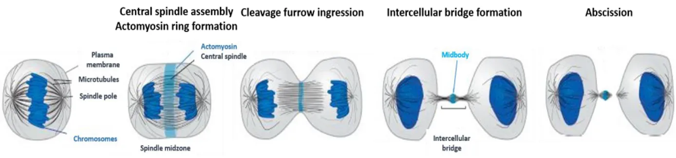
Figure 3. Stages of cytokinesis.
Cytokinesis initiates during anaphase when the two sets of sister chromatids segregate toward opposing spindle poles. Microtubules of the mitotic spindle then reorganize to form the central spindle. Contraction of the actomyosin ring ingresses the attached cell cortex to form the cleavage furrow. Completion of actomyosin ring contraction results in the formation of the intercellular bridge, which contains the midbody at its center. Abscission proceeds by disassembly of the microtubules adjacent to the midbody and membrane fission to physically separate the nascent daughter cells (modified by Mierwa and Gerlich, 2014).
9 The first stage of cytokinesis is characterized by a microtubules reorganization of the mitotic spindle that mediates division plane specification. Three populations of microtubules have been implicated in the regulation of cytokinesis. Equatorial astral microtubules, which emanate from the spindle pole to the site of cleavage, may be stabilized in the equatorial cortical region and deliver positive signals that stimulate formation and contraction of the cleavage furrow (Canman et al., 2003). Polar astral microtubules, which emanate from the spindle pole to sites away from the site of the furrow, may help position the cleavage furrow by inhibiting cortical contractility, perhaps by spatially biasing the pattern of myosin recruitment (Burgess et al., 2005). Central spindle microtubules, which form an overlapping network between the spindle poles following anaphase, send positive signals that become especially important during later steps of cytokinesis (Bringmann et al., 2005).
The reorganization of mitotic spindle is mediated by different protein. A key factor in the central spindle assembly is the microtubule-bundling protein regulator of cytokinesis 1 (PRC1), which binds the microtubules once CDK1 inhibitory phosphorylation has been removed at the anaphase onset (Mollinari et al., 2002). CDK1 also inhibits microtubules bundling by phosphorylating centralspindlin complex, which consists of two subunits of the kinesin-6 motor, also known as mitotic kinesin-like protein 1 (MKLP1), and the Rho-family GTPase-activating protein, Male germ cell Rac GTPase-activating protein (MgcRacGAP), respectively (Mishima et al., 2002). Central spindle assembly requires a third essential component, the chromosomal passenger complex (CPC) comprising the Aurora B kinase, the inner centromere protein (INCENP), borealin, and surviniv. The CPC relocates from centromeres to the spindle center at the anaphase onset, depending on the removal of a CDK1 phosphorylation from the INCENP subunit and directly contribute to microtubule bundling (Carmena et al., 2012). In animal cells, spindle reorganization and cleavage plane
specification is regulated by the small GTPase Ras homolog family member A (RhoA), which
accumulates at the site of the future furrow. Similar to other GTPase, RhoA is regulated by guanine-nucleotide exchange factors (GEFs) and GAP and GTPase activating proteins (GAPs). An important activator of RhoA is the GEF Epithelial cell transforming sequence 2 (ECT2), which targets to the central spindle by binding the phosphorylated subunit MgcRacGAP of centralspindlin. This latest interaction is mediated by a Polo Kinase 1 (PLK1), which localizes at the centrosomes (spindle poles) and thereafter relocalizes at midzone where acts as positive regulator of cytokinesis by phosphorylating MgcRacGAP (Petronczi et al., 2007).
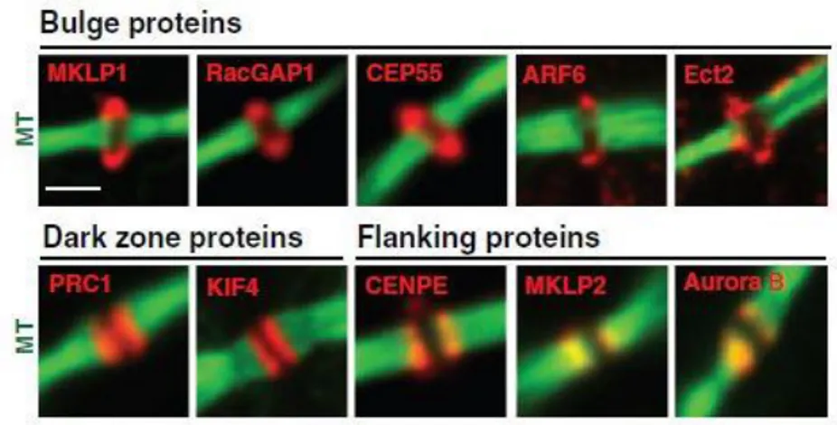
10 In the second stage of cytokinesis, RhoA leads to recruitment and activation of effectors that organize the furrow and stimulate its ingression. In particular, once at the equatorial plate, activated RhoA promotes actomyosin ring assembly and contraction through two regulatory pathways. On the one hand, RhoA mediates actin polymerization through activation of formins that stimulate nucleation of unbranched actin filaments. On the other hand, RhoA indirectly activates a motor protein myosin II by activating both myosin light chain kinase Rho-associated protein kinase (ROCK) and Citron kinase (Severson et al., 2002). The third stage of cytokinesis is characterized by formation of the midbody and stabilization of the cytokinetic furrow. As furrow ingression progresses, the constricting furrow compacts the midzone microtubule array and forms an intercellular bridge, or midbody, that is 1-2 microns in diameter. The midbody is a tightly packed antiparallel microtubule bridge that transiently connects the two daughter cells at the end of cytokinesis. These microtubules overlap at the midbody, which also contains an electron-dense matrix of unknown composition. During the formation of the intercellular bridge, more than 100 different proteins relocate from central spindle to distinct domains at the midbody (Fig. 4) (Skop et al., 2004). PRC1 remain associated with the microtubules of the central overlapping zone called “dark zone” in reference to its antibody-excluding characteristic, whereas the CPC proteins localize at microtubules adjacent to the midbody termed “flanking zone”. Several actomyosin ring components, including anillin, septins, Citron kinase, and RhoA, localize to a ring surrounding the midbody in a region called “bulge zone” (Hu et al., 2012).
Figure 4. Distinct domain of the midbody.
Midbody proteins were categorized into three groups according to their localizations on the midbody in immunofluorescence. (green microtubule; red different proteins) (Modified by Hu et al.,2012).
11 The midbody serves as a platform to orchestrate the cytoskeleton rearrangements, plasma membrane remodeling, and recruitment of the functional complexes needed for abscission, the last steps of cytokinesis. Mass spectrometry analyses have shown that midbdies contain not only proteins related to the cytoskeleton, but also factors involved in other pathways, such as lipid raft and vesicle trafficking (Skop et al., 2004). Indeed, plasma membrane of the midbody is enriched by specific lipids. Phosphoinositides and their phosphorylated derivatives play a particularly important role in cytokinesis. Phosphatidylinositol 4,5-bisphosphate (PtdIns(4,5)P2) accumulates at the equatorial cortex when the cleavage furrow ingresses. This specifies the localization of various cytokinesis proteins such as anillin, septin, RhoA, and MgcRacGAP (Echard, 2012; Emoto et al., 2005). Phosphatidylinositol 3,4,5-triphosphate (PtdIns3P) accumulates close to the midbody where it binds FYVE domain-containing centrosomal protein (FYVE-CENT) and tetratricopeptide repeat domain 19 (TTC19), two factors that mediate accumulation of the abscission factor charged multivesicular protein 4B (CHMP4B) subunit of the endosomal sorting complex required for transport-III (ESCRT-III) (Sagona et al., 2010). Endosomal vesicles also contribute to cytokinesis. At the midbody, upon complete ingression of the cleavage furrow, endosomal vesicles containing ras-related protein in brain 11 (Rab11) and Rab11-family of interacting protein-3 (FIP3) mediate depolymerization of cortical actin filaments of the actomyosin ring through the delivery of p50RhoGAP. Other than their function in remodeling actin filaments, Rab11/FIP3 vesicles also contribute to a gradual narrowing of the intercellular bridge, which precedes the formation of rippled constriction containing ESCRT-III of 17 nm filaments (Guizetti et al., 2011; Schiel et al., 2012).
Furrowing actomyosin ring disassembly, the midbody is stabilized by scaffold proteins such as anillin and septins. In particular, cortical anchorage of the intercellular bridge beyond the central midbody region depends on anillin, which localizes at the midbody, as well as at adjacent regions. Anillin binds to PtdIns(4,5)P2 through its C-term plekstrin homology (PH) domain (Liu et al., 2012) and links to underlying septin filaments (Kechad et al., 2012). Anillin is recruited and maintained at the intercellular bridge through binding to Citron kinase. Citron kinase further contributes to the stability of the midbody by binding to MKLP1 and another microtubule-associated kinesin, kinesin family member 14 (KIF14). Those kinesins interact with PRC1 at the central midbody (Gruneberg et al., 2006). Thus, the stability of the intercellular bridges depends on multiple factors that tether the midbody and the adjacent regions to plasma membrane.
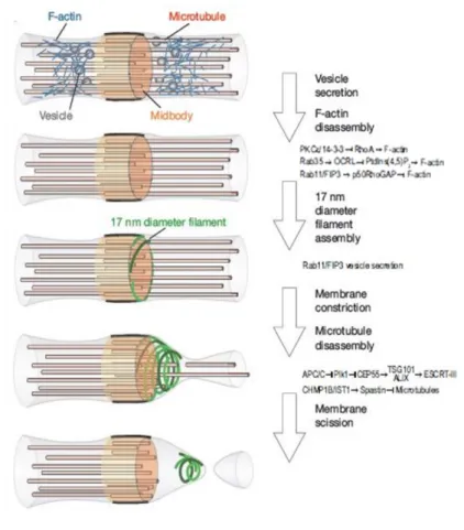
12 In the fourth and last step of cytokinesis called abscission, the two daughter cells are physically separated. A wide variety of proteins involved in vesicle and protein trafficking, membrane scission, and cytoskeleton rearrangement are required for abscission, suggesting that the final stage of cytokinesis is just as complex as earlier stages (Fig. 5).
Figure 5. Schematics of intercellular bridge maturation.
Complete ingression of the cleavage furrow is followed by disassembly of cortical F-actin. Fusion of vesicles correlates with gradual narrowing of the intercellular bridge on both sides of the midbody. Abscission proceeds by assembly and constriction of 17 nm filaments adjacent to the midbody and simultaneous disassembly of the microtubules lateral to the midbody (modified by Fededa and Gerlich, 2012; Miezwa and Gerlich, 2014).
1.3 Abscission, the final steps of cytokinesis
During abscission, the final steps of cytokinesis, the intercellular bridge the connects the two daughter cells is severed adjacent to the midbody. Three models of cell separation have been proposed (Fig. 6).
13 Figure 6. Schematic representation for Abscission models.
The mechanical rupture model (left) posits that as daughter cells move away from one another, the intercellular bridge is torn. The vesicle fusion model (center) proposes that vesicles delivered to the midbody fuse with each other and with the plasma membrane to separate the daughter cells. The membrane fission model (right) proposes that the midbody membrane is acutely constricted, and then sealed and severed (modified by Elia et al., 2013).
The first abscission model proposed was the mechanical rupture model. It was
suggested that the connection between the two daughter cells is stretched and eventually broken by mechanical rupture as the daughter cells pull in opposite direction. Mullins proposed that a wound healing mechanism seals the membrane opening. Evidence that cell adhesion molecules, such as integrins, and cellular wound healing component are required for successful cytokinesis is some cells support this model (Kanada et al., 2005; La Flamme et al., 2008). However, because the abscission occurs also in nonadherent cells (e.g., leukocyts and lymphocytes) and nonmotile cells that do not migrate away from one another under physiological condition, researchers began to look for alternate mechanism.
A second model of abscission proposes that vesicle may fuse with the plasma membrane
before abscission to establish separating membranes from within the intercellular bridge. Vesicle from Golgi-secretory pathway and from recycling endosome accumulate at the intercellular bridge together with several vesicles targeting and tethering factors, including
14 centriolin, the exocyst complex, Rab5, Rab11, v- and t-soluble N-ethylmaleimide-sensitive factor activating protein receptor (v- and t-SNARE). These factors were all shown to be required for abscission (Fielding et al., 2005; Gromley et al., 2005; Kouranti et al., 2006). However, electron microscopy studies showed that vesicle disappear from the intercellular bridge before abscission. Thus the failure in abscission upon depletion of these factors, however, seems more likely to result from perturbed intercellular bridge maturation rather than defects during the final abscission stage (Guizetti et al., 2011).
The third model proposes that midbody membrane is acutely constricted and then
severed. This model indicates the involvement of the endosomal sorting complex required for transport (ESCRT) machinery that mediate the final membrane scission event and separates the daughter cells. The ESCRT-III machinery mediates topologically equivalent constriction and scission events during both multivesicular body formation (MVB) and retrovirus budding from plasma membrane (McCullough et al., 2013). Once the midbody has thinned, about half of its initial width, a group of proteins component of the ESCRT machinery localize at the midbody by a sequential recruitment of its targeting factors. In particular, during late cytokinesis, PLK1 degradation leads to dephosphorylation of centrosomal protein CEP55 and its consequent relocalization from the spindle midzone to the dark zone at the midbody through interaction with MKLP1. CEP55 recruits the ESCRT-I subunit tumor susceptibility gene 101 (TSG101) and the ESCRT-associated protein apoptosis-linked gene-2-interacting protein X (ALIX) that also localize at the dark zone to the midbody (Morita et al., 2007). TSG101 and ALIX then recruit ESCRT-III subunit, also called CHMPs, to cortical ring at both sites of the midbody (Elia et al., 2011). Later in cytokinesis, a second and smaller pool of ESCRT-III proteins accumulates at the abscission site, approximatively 1 µm away from the center of the midbody, where acute membrane constriction and abscission occur. CHMPs recruitments at the secondary ingression is essential for cytokinetic specific localization of spastin, a microtubule severing AAA-ATPase. Indeed, spastin localizes at the midbody by binding the ESCRT subunits CHMP1B and IST1, and mediates microtubule scission at the end of the abscission process (Fig. 7) (Connell et al., 2009). ESCRT-III also interacts with membrane via PtdIns3P-binding protein FYVE-CENT and TTC19. During the final stages of abscission, the ESCRT-III depolymerization factor vacuolar protein sorting-associated protein 4 (VPS4) accumulates at the midbody where mediates the disassembly of the complex following the membrane scission (Agromayor et al., 2009).
15 Figure 7. The ESCRT complex in abscission.
The centralspindlin component recruits Cep-55 to the midbody. Cep-55 in turn recruits ESCRT-I protein Tsg101 or ALIX. Tsg101 and the ESCRT-III subunit CHMP4B (purple spheres) are sequentially recruited into the center of the intercellular bridge. Later in cytokinesis, CHMP4B concentrates at the narrow secondary abscission zones, closely followed by Vps4 (yellow circles). ESCRT-III subunits extend towards these sites of cortical constriction forming filaments, which extend towards the site of secondary ingression. ESCRT-III can then recruit the microtubule-severing enzyme spastin (scissors) (Modified by Neto and Gould, 2011).
About 10-20 minutes before abscission, the cortex adjacent to the midbody constricts. This secondary ingression of the ell cortex is essential for abscission. Guizetti and colleagues, by electron microscopy analysis of late stage of cytokinesis, revealed that cortical constriction zone contains membrane associated 17 nm diameter filaments forming large intertwined helices surrounding the intercellular bridge (Guizetti at al., 2011). The ESCRT-III machinery is a candidate of the 17 m diameters filaments because co-localizes with the constriction zones and is required for the formation of these filaments. Thus far, three models are proposed to clarify how ESCRT-III polymers may contribute to abscission: ESCRT-III may grow filament spirals that decrease their diameter when they extend away from the midbody to constrict the intercellular bridge (Guizetti et al., 2011). Alternatively, part of an ESCRT-III filament helix may be released, possibly by VPS4, to slide away from the midbody while constricting the cortex (Elia et al., 2012). Otherwise, an ESCRT-III-independent process leads to membrane constriction, which is then captured and stabilized by ESCRT-III (Schiel et al., 2012).
In summary abscission proceeds by a secondary ingression of the cell cortex, involving of 17 nm diameter filaments spanning the intercellular bridge. The prevailing model suggest the involvement of the ESCRT-III machinery in abscission. Moreover, the membrane scission model was enriched by the founding that spastin, a microtubule severing enzyme, directly
16 binds to ESCRT-III subunit at the secondary ingression. Thus, it was propose that the ESCRT machinery couples microtubule severing to the physical cleavage of the membrane. However, how ESCRT-III adapts to the large membrane tube of the intercellular bridge and how these large structures constrict remain important open questions. Moreover, understanding the mechanism by which the plasma membrane ultimately splits and how vesicle or wound healing contributes to abscission will require further investigation.
1.4 Cytokinesis failure
Cytokinesis, the last step of cell division, proceeds throughout subsequent stages each owing to the previous one, that requires a complex interplay among a still enlarging number of regulatory and effectors components related to cytoskeleton, chromosome, cell cycle, lipid raft, vesicle, and membrane trafficking factors. Cytokinesis failure can arise through defects in any of the four stages in cytokinesis. Inhibition or excessive activation of different cytokinesis components can give rise to distinct phenotypes, including precocious ingression before the chromosomes have been separated, regression of the furrow giving rise to binucleated cells, or stabilization of the cytoplasmic bridge where daughter cells remain connected. High fidelity cytokinesis is crucial to ensure that both daughter cells inherit a diploid set of chromosomes. Cytokinesis failure and the resulting tetra- and poly-ploidization promote chromosomal and genomic instability, both hallmarks of cancer (Ganem et al., 2007).
Inhibition or regression of the cleavage furrow induces both formation of binucleated and persistence of connections between daughter cells with formation of long intercellular bridges (LIBs) and syncytial-like structures (Normand and King, 2010). Faithful cell division requires tight coordination with chromosome segregation. Before cytokinesis start, chromosomes need to be cleared from the cleavage plane to avoid being damaged during furrow ingression (Eggert et al., 2006; Glotzer, 2005). The initiation of chromosomes segregation and cytokinesis both depend on the activation of E3 ubiquitin ligase anaphase-promoting complex/cyclosome (APC/C). The APC/C promotes mitotic exit by targeting securin, an inhibitor of the protease separase that destroy the cohesive link between sister chromatids, for proteosome-mediated degradation. APC/C also acts by targeting the CDK1 coactivator cyclin B for degradation leading to CDK1 inactivation. Thus, dephosphorylation of many CDK1 substrates promotes cytokinetic furrow ingression. While chromosome
17 segregation normally completes early after anaphase onset, it can be severely delayed by lagging or bridged chromosomes. Segregation defects have been estimated to occur in about 1% of dividing somatic cells, and at higher incidence in transformed cells (Cimini et al., 2003). Similary, abscission must occur only after the complete separation of sister chromatids. Chromosome bridges can result from dysfunctional telomeres (Maser and DePinho, 2002; Stewenius et al., 2005), DNA double-strand breaks (Acilan et al., 2007), or from misregulated chromosome cohesion (Cimini et al., 2003) or decatenation (Chan et al., 2007). Circumstances that cause chromosome segregation errors have shown to delay abscission. In animal cells, Aurora B kinase controls the abscission delay induced by chromosome bridges by a regulatory pathway termed “NoCut”. In the presences of chromosome bridges, at the midbody, Aurora B remains active for a much longer duration after cleavage furrow ingression, which may contribute to abscission delay (Mathieu et al., 2013; Bembenek et al., 2013; Steigemann et al., 2009). Aurora B mediates the phosphorylation of MKLP1 and this phosphorylation is maintained for a longer duration in the presence of chromosome bridges, thus stabilizing the interaction between the ingress furrow and the midbody and prevents the furrow regression in cells with chromosome bridge (Steigemann et al., 2009). Aurora B also regulates the abscission delay through the ESCRT-III subunit CHMP4C. It has been hypnotized that phosphorylated CHMP4C is recruited early at the midbody and may out-compete other ESCRT-III subunit in binding to their targeting factors such as ALIX and CHMP4B, thus inhibiting the assembly of the ESCRT-III filaments required for abscission (Agromayor et al., 2013; Capalbo et al., 2012; Carlton et al., 2012).
2 Histone H2B
Histones are constitutive component of nucleosomes. The nucleosome is the basic unit of chromatin packing in eukaryotes, consisting of a segment of DNA wound around histone proteins. In particular, the nucleosome core consists of about 147 base pairs (bps) of DNA wrapped around a histone octamer consisting of two copies of each core histones H2A, H2B, H3 and H4 (Fig. 8A). The nucleosomal structure repeats with a regular periodicity every 160-220 bps. This conformation is termed the 10 nm fiber or “beads on a string”, referring to the way chromatin appeared on electron micrographs when it was first visualized in 1973 (Olins and Olins, 1974). To compact DNA, multiple nucleosomes further assemble into higher-order
18 structures, which are stabilized by histone H1 (linker histone) that binds and rearranges the DNA between nucleosome units (Luger et al., 1997; Li and Reinberg, 2011).
Histones have long amino-terminal (N-term) tails protruding from the nucleosome, that are post-translational modified at several places. Modifications of the tails include acetylation, phosphorylation, methylation, ubiquitination, sumoylation, and ADP-ribosylation. These modifications can usually by read by other proteins (e.g., chromatin-associated proteins, transcription factors, regulatory proteins) and act in diverse biological processes such as gene regulation, DNA replication, DNA damage repair and chromosome condensation (Strahl et al., 2000; Jenuwein et al., 2001; Bannister et al., 2011).
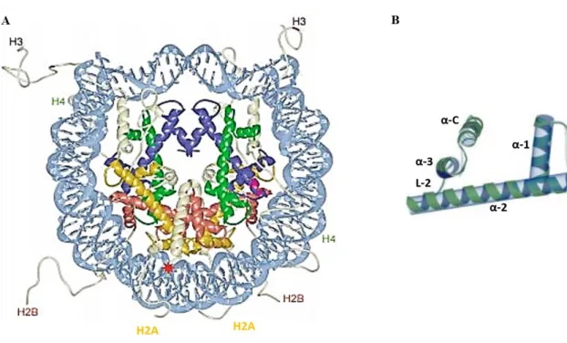
Figure 8. Nucleosome and histone H2B structure.
(A) The histone fold domains of H2A, H2B, H3, and H4 are colored in yellow, red, blue, and green respectively; histone tails and extensions are shown in white; DNA is shown in light blue (modified by Luger, 2003). (B) Histone H2B domain (α1, α2, α3 and αC connected by two loop L-1 and L-2) are shown.
Histone H2B is a small, highly basic protein component of the nucleosome core. The structure of histone H2B (126 aa) is characterized by a structured conserved domain, histone fold (HF), and a flexible non-structured tail at the N-term protruding outward from the nucleosome.
19 The HF is directly responsible for the formation of the octamer and DNA contacts and is formed by three α-helices (α1, α2, and α3) connected by two short loops (L1 and L2). In addiction to the helices of the HF domain, histone H2B is characterize by a C-term helix (αC) extremely well ordered (Fig. 8B). This helix is exposed on the surface of the histone octamer and plays an important role in defining the surface on the nucleosome core particle (White et al., 2001). The flexible N-term tail is exposed also on the nucleosome surface, protrudes away from the DNA, and is an acceptor for a variety of posttranslational modifications (Luger et al., 1997; Harp et al., 2000; Luger, 2001).
Among different modifications, it has been shown that phosphorylation of the N-term tail of histone H2B at serine 14 (H2B-S14P) by caspase-3-activated mammalian sterile twenty kinase 1 (Mst1) specifically correlates with the onset of apoptosis (Cheung et al., 2003). Ahn and colleagues extended these results to yeast by demonstrating that Sterile twenty kinase, a yeast orthologue of Mst1, directly phosphorylates H2B at serine 10 (H2B-S10P) in a hydrogen peroxide-induced cell death pathway (Ahn et al., 2005a). Phosphorylation of histone H2B at S14 in human and S10 in yeast has been also linked to chromatin condensation during meiosis (Ahn et al., 2005b). Furthermore, H2B phosphorylation or the H2B N-term tail, but not other histone tails, were shown to be essential for chromatin condensation in a Xenopus cell-free system (de la Barre et al., 2001).
In mammals, histone genes represent an unusual family of genes. Indeed, there are different functional copies of the genes for each of the four core histone proteins. The metazoan core histone genes are clustered together in the genome. There are two major histone gene clusters on chromosome 6p21–p22 (HIST1) and 1q21 (HIST2) and one minor cluster on 1q42 (HIST3). Each of these genes has a different nucleic acid sequence and produces different histone variants, which share aa sequence homology and core structural similarity to a specific class of major histones so-called canonical. Indeed, histone variants shown only a few aa substitutions (Marzluff et al., 2002). There are 21 histone H2B genes reported for human that produce 16 histone variants (Mariño-Ramírez et al., 2011; Khare et al., 2011). Among different H2B variants, it was found that both variants H2B.A (also called TH2B) (Zalensky et al., 2002) and H2B.F-WT (Churikov et al., 2004) are involved in a process that mediates chromatin compaction during spermatogenesis (e.g., when spermatogonia are initially converted into spermatocytes and subsequently into spermatids through meiosis). To date, it is unclear the role of the other histone variants and whether their expression varies between tissues, cell types, or different physiological conditions. Recently, it has been shown that particular H2B variants are expressed at different levels induced under
20 specific cellular circumstances, but further studies are necessary to elucidate their functions (Kari et al., 2013).
During S-phase, a large amount of histones must be rapidly synthesized, about 108 molecules of each core histone protein. Histone mRNA levels increase 35-fold as cells enter S-phase and decrease again at the end of the S-phase (Olsey, 1991). Studies on histone-exchange and -deposition kinetic revealed that H2B is the most rapidly and abundantly exchanged among the core histones (Kimura and Cook, 2001). Histone H2B transits in and out of the nucleosome more rapidly than other core histones, such as H3, H4, and H2A. Thus, about 3% of the total amount of H2B is exchanged within 6 min, ≈ 40% within 130 min, and ≈ 50% by 8.5 hours (Kimura, 2005).
In addition to chromatin-related functions, a few other extrachromosomal localization and activities of specific histones have been described. Extrachromosomal H2B was shown to be involved in the antiviral innate immune responses (Kobiyama et al., 2010). In apoptotic conditions, histone H1.2 is discharged from the nucleus into the cytoplasm and induces cytocrome C release from mitochondria (Konishi et al., 2003). Phosphorylated histone H1 has also been found in the cytoplasm of M phase HeLa cells (Bleher and Martin, 1999), while H3-S10P has been shown to co-stain with Aurora B in mitosis and cytokinesis (Song et al., 2007).
3 H2B and HPK2 localize at the midbody: a new extrachromosomal
activity of histone H2B
Recently in our laboratory, Rinaldo and colleagues have discovered a new extrachromosomal localization and activity of histone H2B. They found that both H2B and Homeodomain interacting protein kinase 2 (HIPK2) localize at the midbody, the intercellular bridge connecting two daughter cells during cytokinesis (Fig. 9). At the midbody, phosphorylation of histone H2B at serine 14, mediated by HIPK2 is essential for faithful cytokinesis (Rinaldo et al., 2012).
21
Figure 9. Localization of EGFP-HIPK2 and H2B-S14P in HeLa Cells.
EGFP-HIPK2 (green) and histone H2B-S14P (red) localize at the intercellular bridge. DNA is marked
with DAPI (blue) (modified by Rinaldo et al., 2012).
Serine/threonine kinase HIPK2 plays important roles in the regulation of proliferation and apoptosis during development and in cell response to DNA damage (reviewed in Rinaldo et al., 2007; D’Orazi et al., 2012). This kinase can either interact with homeobox proteins by acting as transcriptional corepressors or with other types of transcription factors by acting as coactivators or corepressors, depending on the promoters or the cellular context. Indeed, HIPK2 was shown to phosphorylate itself in vitro and binds and phosphorylates a still enlarging body of targets, including transcriptional regulators (D’Orazi et al., 2002; Hofmann et al., 2002; Zhang et al., 2003; Wiggins et al., 2004; Wee et al., 2008; Hikasa et al., 2010), chromatin modifiers (Zhang and Wang 2007; Bracaglia et al., 2009), signal transducers (Kanei-Ishii et al., 2004; Ritterhoff et al., 2010), and E3 components of SUMO ligases (Roscic et al., 2006; Swarup and Verheyen, 2011). In DDR, HIPK2 exerts its effects by binding and phosphorylating an increasing array of transcription factors and coregulators. The p53 tumor suppressor is among the first non-homeotic transcription factors identified as HIPK2 target. HIPK2 activity enhances the p53-mediated transcriptional activation of proapoptotic factor such as PIG3, BAX, and NOXA, as well as the repression of the anti-apoptotic factor Galectin-3 (D’Orazi et al., 2002; Hofmann et al., 2002; Di Stefano et al., 2004b; Cecchinelli et al., 2006a). In the presence of DNA damage induced by UV irradiation or antineoplastic treatments (e.g., doxorubicin or cisplatin), HIPK2 specifically phosphorylates human p53 at Ser46 and mouse p53 at Ser58 and this kinase activity is required for the induction of apoptosis (D’Orazi et al., 2002; Hofmann et al., 2002; Moller et al., 2003; Di Stefano et al., 2004b; Cecchinelli et al., 2006b). In addition, HIPK2 can promote apoptosis by targeting factors, such as the CtBP transcriptional co-repressor (Zhang et al., 2003), and by modulating the activity of other proteins directly or indirectly related to
22 apoptosis, such as the p53 family members p73 and p63 (Kim et al., 2002; Lazzari et al., 2011) and the p53 inhibitor MDM2 (Wang et al., 2001; Di Stefano et al., 2004 a).
In contrast to their association with DNA as kinase involved in DDR and as chromatin structural component, respectively, both HIPK2 and H2B localize at the midbody independently from the presence of chromosome bridges, DDR and/or apoptosis. The completion of cytokinesis by abscission needs to await complete clearance of chromatin from the cleavage plane. Several defects such as dysfunctional telomeres, DNA double strand breaks (DSBs), misregulated chromosome segregation, cohesion, or decatenation can result in the persistence of chromosomal bridges that are visualized by the presence of nucleosomal histones (Steigemann et al., 2009 and references therein). Interestingly, histone H2B localizes at the spontaneous chromosome bridges of HeLa cells, but it was not phosphorylated at S14 and was not immuno-stained for HIPK2. In contrast, all the apparently normal telophases have both H2B-S14P and HIPK2 colocalized with midbody markers, indicating an extrachromosomal function for the modified histone (Rinaldo et al., 2012).
Furthermore, cytokinetic-specific localization of H2B is independent of HIPK2 but the absence of the kinase results in loss of H2B-S14 phosphorylation, prevention of cell cleavage and accumulation of tetra- and poly-ploid cells that contribute to tumorigenicity (Rinaldo et al., 2012; Valente et al., 2015). Of relevance, the sole expression of a phosphomimetic H2B-S14D mutant in HIPK2-null cells is sufficient to overcome cytokinesis defects and restores cell division and proliferation without any induction of apoptosis (Rinaldo et al., 2012).
These data show that HIPK2 controls cytokinesis through extrachromosomal H2B-S14 phosphorylation, but the mechanisms through which H2B contributes to cell division is still unknown.
23
AIM
Cytokinesis is a complex process that requires the interplay of many components and regulatory factors. Its failure is known to lead to genetically unstable states, such as tetra- and poly-ploidization, considered a critical step in tumourigenesis. Histones are assembled into nucleosomes, the basic units of DNA packaging in eukaryotes, as core (H3, H4, H2A, and H2B) and linker (H1) histones. Histones have N-terminal tails that protrude from DNA and their post-translational modifications regulate chromatin structure. In addition to chromatin-related functions, a few extrachromosomal activities of specific histones have been described. Recently, our laboratory demonstrated that histone H2B localizes at the midbody, the intercellular bridge connecting two daughter cells during cytokinesis, in a DNA-independent manner. At the midbody, phosphorylation of H2B at serine 14 is mediated by HIPK2, a kinase involved in DNA damage response and development, whose activity is required for cytokinesis and prevention of tetraploidization. In HIPK2-defective cells, expression of a phosphomimetic H2B-S14D mutant is able to rescue cytokinesis defects, cell division, and proliferation, suggesting that H2B phosphorylation at serine 14 in required for cell division.
This unexpected finding uncovers a new role of extrachromosomal histone H2B in cell division and prevention of tetraploidization. However, the mechanism through which H2B contributes to cytokinesis is still unknown.
The aim of this work is to dissect the extrachromosomal functions and activity(ies) of histone H2B in cytokinesis. This will have clear implications for the understanding of cell division and of its failure as promoter of tetra- and poly-ploidization, conditions that can generate chromosomal instability.
24
RESULTS
H2B LOCALIZATION DURING MITOSIS
Histone H2B and its phosphorylated form at serine 14 (H2B-S14P) were previously reported
to localize at the midbody, the intercellular bridge that connects two daughter cells in the last step of cell division. In addition, both H2B and H2B-S14P co-localize with well-characterized midbody markers, such as α- and β-tubulin and Aurora B kinase. The midbody localization of both H2B and H2B-S14P has been confirmed by Western blot analysis of midbodies isolated and extracted from HeLa cells enriched in telophase (Rinaldo et al., 2012).
At the onset of this study, in search of the role of extrachromosomal H2B, we asked if localization of H2B is only restricted to the midbody. The subcellular distribution of histone was re-examined using a new rabbit monoclonal antibody that detects the H2B phosphorylated at S14. Phosphorylated histone H2B-S14 has been linked to DNA damage response (DDR) and chromatin condensation in apoptosis (Cheung et al., 2003; Ahn et al., 2005). Thus, we first evaluated the specificity of the new anti-H2B-S14P antibody (Ab) by testing its capability to detects apoptotic chromatin in ultraviolet (UV)-irradiated HeLa cells. Immunofluorescence (IF) analysis revealed that anti-H2B-S14P Ab recognize the apoptotic chromatin, as expected (Fig. 10).
25 Figure 10. H2B Antibody Specificity.
The anti-H2B Ab specificity was demonstrated in UV-irradiated HeLa cells. Immunostaining of HeLa cells was performed in the untreated (upper panels) or the irradiated (lower panels) with the anti-H2B-S14P Ab (green). DNA was stained with Hoechst (blue). Scale bar is 10 µm.
Next, we analyzed H2B-S14P localization during mitosis. IF analysis showed
uncharacterized areas of localization of H2B-S14P. During metaphase, H2B-S14P forms
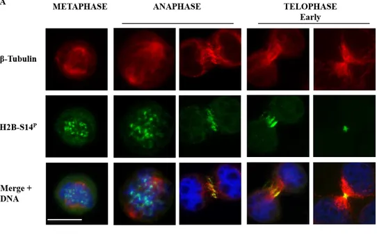
unclear aggregates that do not co-localize with kinetochores, as assessed by antibody anti-CREST (calcinosis, Raynaud's phenomenon, esophageal dysmotility, sclerodactyly, telangiectasia), a kinetochore marker (Williams et al., 1984) (data not show). In anaphase, H2B-S14P accumulates at the central spindle, remains associated with the spindle midzone during cleavage furrow contraction and thereafter accumulates at the midbody, in the flanking zone (Fig. 11A). Interestingly, in late telophase, H2B-S14P shows an asymmetric distribution, with a partially re-localization at the secondary ingression site, where acute membrane constriction and abscission occur (Fig. 11B).
26
Figure 11. Localization of endogenous histone H2B-S14P.
(A) Localization of H2B-S14P nuclear aggregates in metaphase, central-spindle in anaphase, flanking
zone at the midbody in telophase. Scale bar is 10 µm. (B) asymmetric localization of histone H2B-S14P in
late telophase. Scale bar is 5 µm. Microtubules were marked with anti-β-tubulin Ab (red); H2B was marked with anti-H2B-S14PAb (green), DNA was marked with Hoechst (Blue).
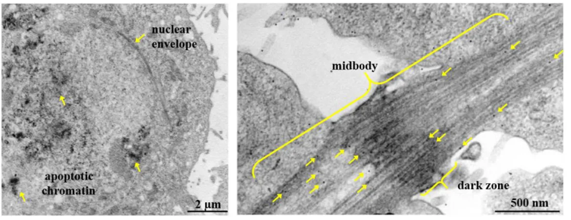
The H2B-S14P localization at the midbody was confirmed by transmission electron microscopy (TEM) analysis of telophase-enriched HeLa cells. Positive controls were obtained analyzing damaged HeLa cells characterized of apoptotic chromatin. Immunogold labeled anti-H2B-S14P Ab revealed that phosphorylated form of histone H2B at S14 localizes at the
27 midbody associated with vesicle-like structures at one arm of the midbody, the so-called flanking zone, and with microtubule (Fig. 12) at the opposite arm. This latter aspect may explain to the asymmetric distribution of H2B-S14P showed by IF.
Figure 12. TEM, localization of endogenous histone H2B-S14P in telophase.
(Left panel) positive Ctr: anti-H2B-S14P Ab gold-conjugated marks apoptotic chromatin. Scale bar is 2
µm; (right panel) asymmetric localization of histone H2B-S14P in telophase, associated with different
sub-structures at the opposite site of the midbody. Scale bar is 500 nm.
Taken together, these results show that histone H2B-S14P localizes in specific areas in
different steps of cell division, from metaphase to telophase. Furthermore, in late telophase, at the abscission onset, H2B-S14P show an asymmetric distribution probably due to a specific sub-structural localization.
CYTOKINESIS-SPECIFIC LOCALIZATION OF HISTONE H2B IS
INDEPLENDENT OF RNA
Different types of DNA and chromosomal dysfunctions can result in chromatin localization at the cleavage plane, the so-called chromosome bridges (Steigemann et al., 2009). Because of the role of histone H2B as structural component of chromatin, Rinaldo and colleagues have first evaluated the relationship between localization of histone H2B at the midbody and the presence of DNA. They found that the cytokinetic specific localization of histone H2B is independent of chromosome bridges (Rinaldo et al., 2012). Since DNA is not involved in
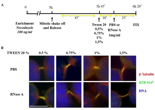
28 H2B midbody localization, we asked whether RNA might be responsible from the H2B cytokinetic localization. This hypothesis was also stimulated by recent observation that DDR focus formation requires structural support of site-specific DICER and DROSHA RNA products (Francia et al., 2012). Thus, to assess whether RNA contributes to H2B midbody localization and/or activity, mitosis-enriched HeLa cells were permeabilized with different percentages of TWEEN 20 and treated with ribonuclease A (RNase A) that catalyzes the degradation of RNA; PBS was used as negative control (Fig 13A). Positive control was obtained by assessing loss of DDR-induced 53BP1 foci in RNase A treated HeLa cells after γ-irradiation (Fig. 14), as previously reported (Francia et al., 2012). After treatments, we analyzed H2B localization by IF (Fig. 13B) and both treated and control cells showed H2B at the midbody indicating that H2B midbody localization is independent of single-strand RNA (ssRNA).
Figure 13. H2B-S14P localization in RNase A-treated HeLa Cells.
(A) Schematic representation of both mitosis-enrichment and RNase A treatment protocols. (B) Localization of H2B-S14P in telophase was assessed in HeLa cells treated with PBS or RNase A 1mg/ml.
Microtubules were marked with anti-β-tubulin Ab (red); H2B was marked with anti-H2B-S14PAb
29 Figure 14. 53BP1 localization in RNase A-treated HeLa Cells.
(A) Schematic representation of irradiation and RNase A treatment protocols; (B) localization of 53BP1 (green) at DDR foci was assessed in irradiated HeLa cells treated with PBS or RNase A 1mg/ml. Microtubules were marked with anti-β-tubulin Ab (red) and DNA was marked with Hoechst (Blue). Scale bar is 10 µm.
Because of the disappearance of histone H2B localization in early stages of cytokinesis already in control cells, it was not possible to analyze the relationship between RNA and H2B localization in metaphase or at the contractile ring in anaphase (Fig. 15).
Figure 15. H2B-S14P localization in RNase A-treated HeLa Cells.
H2B-S14P localization disappears in early stages of cytokinesis. Microtubules were marked with
anti-β-tubulin Ab (red); H2B was marked with anti-H2B-S14PAb (green), DNA was marked with Hoechst
(Blue). Scale bar is 10 µm.
To further analyze the relationship between nucleic acids and midbody localization of H2B, we evaluated the presence of histone H2B at the midbody in mitotic enriched HeLa cells. First, we confirmed that localization of H2B at the midbody is independent of DNA (Rinaldo et al., 2012) by using DNase I. As expected, midbody localization of H2B was
30 revealed both in control and treated cells. Next, we treated the mitotic-enriched HeLa cells with RNase H and RNase III, which respectively bind and cleave hybrid DNA/RNA and double-strand RNA (dsRNA). The cytokinetic specific localization of histone H2B was not affected after degradation of both hybrid DNA/RNA and dsRNA (Fig. 16).
Figure 16. H2B-S14P localization in RNase H/III and DNase I treated HeLa Cells.
Localization of H2B-S14P in telophase was assessed in HeLa cells treated with PBS or RNase H/ RNase
III/ DNase I. Microtubules were marked with anti-β-tubulin Ab (red); H2B was marked with anti-H2B-S14PAb (green), DNA was marked with Hoechst (Blue). Scale bar is 10 µm.
Eventually, we studied the role of non-coding RNA by using HCT116 cells wild-type or knockout for DICER, an enzyme that catalyzes the cleavage of non-coding RNA involved in gene silencing (Manni et al., 2009; Cummins at al., 2006). No defect in H2B localization was observed in DICER-knockout HCT116 cells respect to the wild-type counterparts (Fig 17).
31
Figure 17. Localization of endogenous H2B-S14P in DICER WT and DICER KO HCT116 cells.
Localization of H2B-S14P in HCT116 DICER WT and KO: nuclear aggregates in metaphase,
central-spindle in anaphase, flanking zone at the midbody in telophase. Microtubules were marked with anti-β-tubulin Ab (red); H2B was marked with anti-H2B-S14PAb (green), DNA was marked with Hoechst
(Blue). Scale bar is 10 µm.
H2B DEPLITION CAUSES ABSCISSION DEFECTS AND DELAY
To study the activity of extrachromosomal histone H2B in cytokinesis, HeLa cells were depleted for histone H2B. As constitutive component of chromatin, during S-phase it must rapidly synthesize large amounts of histones, approximately 108 molecules of each core histone protein. Histone mRNA levels increased 35-fold as cells enter S-phase and decreasing again at the end of S-phase (Olsey, 1991). Thus, first of all, we have optimized a protocol that ensures cell viability and, at the same time, an efficient depletion of extrachromosomal histone H2B. We have interfered histone H2B expression in HeLa cells by transfecting commercially available H2B short interfering RNAs (siRNA). There are 21 histone H2B genes reported for human that produced 16 histone variants (Mariño-Ramírez et al., 2011; Khare et al., 2011). We have tested combination of 9 H2B variant-specific siRNAs (i.e., H2B.B; H2B.D; H2B.E; H2B.G; H2B.K; H2B.H; H2B.I; H2B.J; H2B.2E) at different concentrations and times and in HeLa cells seeded at different confluence. Control depletion was obtained by universal negative control (Ctri) sequences. Among a variety of protocol (data not shown), because of the high transcription rate of histones and the requirement to maintain nucleosome integrity to allow cell division, two pulses of siRNA transfection, the
32 second 24 hours from the first one, was needed to obtain a successful depletion of extranucleosomal H2B (Fig. 17A) without affecting cell viability. HeLa cells were depleted with a double-pulse of a combination of H2B variant-specific siRNAs (H2Bi) and their related Ctri and analyzed by IF to assess cytokinesis failure. Compared to Ctri, depletion of H2B results in the accumulation of cytokinetic defects, such as prevention of cell cleavage, abscission defects, and accumulation of LIBs. These latter structures are long and often convoluted and resemble elongated midbodies. We also observe tubulin disorganization presumably linked to LIBs formation and the stretching conditions triggered by abscission defects (Fig. 17B).
Figure 17. H2B depletion in HeLa cells result in tubulin disorganization.
HeLa cells were depleted for histone H2B with a combination of 9 variant-specific siRNAs (H2Bi) or with universal negative control (Ctri). (A) A representative WB for the indicated proteins is shown. (B) Representative immunofluorescence imagines of Ctri and H2Bi HeLa cells stained with anti-β-Tubulin Ab (red) and Hoechst (blue). The presence of LIBs (H2Bi top panels), abscission defects (H2Bi middle panels) and tubulin disorganization (H2Bi bottom panels) is detectable in the H2Bi cells compared to the Ctri. Scale bar is 10 µm.
Microscopy analysis of fixed asynchronous HeLa cells (n=1,000) after H2B depletion reveal that about 34 ± 3.5% of telophases are aberrant in H2Bi cells compared to
B
33 Ctri (11 ± 3.5 %). Depletion of H2B also results in accumulation of cells that are in the midbody stage. Indeed, in asynchronous HeLa cells H2Bi the percentage of cells at the end of cytokinesis is about 15 ± 2.2% dissimilarity to the 9 ± 1.6% of telophases detectable in relative Ctri. In contrast, no defects are observed in accumulation of bi- and multi-nucleated cells nor increase in the number of chromosomal bridges (i.e., below 10% in both H2Bi and Ctri HeLa cells). We detected only a slight increase in multinucleated cells in HeLa H2Bi (3.4 ± 0.7 %) in respect to their Ctri (2.8 ± 1.2 %) (Fig. 18A, B and
C). Furthermore, the aberrant midbodies (n=100) are negative for staining with both
anti-H2B Ab (Fig. 18D) and anti-anti-H2B S14P Ab (Fig. 18E).
Figure 18A-C. H2B depletion in HeLa cells results in abscission defects.
HeLa cells were depleted for histone H2B with a combination of 9 variant-specific siRNAs (H2Bi) or with universal negative control (Ctri). Cells were stained with anti-H2B Ab or anti-H2B-S14P (green),
Hoechst (blue), and anti-β-tubulin Ab (red)to identify the cytoplasm in interphase and the midbodies or the LIBs in telophase.About 1,000 cells per sample were scored for (A) the presence aberrant telophases, (B) the number of cells in telophase, and (C) the presence of one or two nuclei/cell; data are represented as mean ± SD (** P-value < 0.01; *** P-value < 0.001; ns P-value > 0.05 by Student t test).
34 Figure 18D, E. H2B depletion in HeLa cells results in abscission defects.
HeLa cells treated and stained as above were analyzed for cytokinesis defects. Representative images of H2B (D) or H2B-S14P (E) at the midbody in Ctri and H2Bi HeLa cells. Scale bar is 10 μm.
Next, to elucidate the cytokinetic function of histone H2B, we analyzed asynchronous HeLa cells H2Bi and Ctri by time-lapse live-cell imaging for 24 hours, starting after the second pulse of siRNA (Fig. 19A). We evaluated the cytokinesis time by analyzing the timing from the round up, when cells became rounded after nuclear envelop break down, to cleavage ingression, the easiest recognizable phase of cytokinesis. We also analyzed the timing from cleavage furrow ingression to abscission, when the two daughter cells are physically separated, the so-called abscission time. The timing from the round up to cleavage furrow ingression is about 40 minutes for both H2Bi (n=100) and Ctri (n=150) HeLa cells (Fig. 19B). Furthermore, in Ctri cells (n=80), the midbody extend from each daughter cell into the intercellular bridge and abruptly break down approximately 2 hours after cleavage furrow ingression. In contrast, in H2B-depleted cells, this abscission event is severely delayed with the abscission time of H2Bi HeLa cells (n=100) being at least 1 hour longer than their related controls (Fig. 19C). Among the persistent intercellular bridges, some become progressively thinner and persist for many hours, often becoming overextended and resembling the LIBs we D
35 see by IF. Although abscission clearly failed in these cells, we did not see re-fusion of daughter cells, has previously observed in HIPK2 depleted HeLa cells (Rinaldo et al., 2012), confirming the observation made by IF and thus indicating that H2B depletion results only in abscission delay.
Figure 19. H2B depletion is associated with Abscission failure.
Ctri and H2Bi HeLa cells were employed as total populations. (A) Stills from time-lapse recording of HeLa Ctri and H2Bi cells after the second pulse of siRNAs are shown. Ctri cells divide and return mono-nucleated in about 3 hours while H2Bi cells remain connected by intercellular bridge for several hours before abscission occurs. Timing from round up to cleavage furrow ingression (B) and from cleavage furrow ingression to abscission (abscission time) (C) are reported in box-plot graph. (B) The median of the timing from the round-up to cleavage furrow is 39 minutes both in Ctri and H2Bi. (C) The median of the abscission time is 126 minutes and 174 minutes for Ctri and H2Bi cells, respectively. Scale bar is 10 µm.
Altogether, these data show that H2B depletion by siRNA induces accumulation of cytokinesis-dependent aberrations and impairs abscission that results in abscission delay. These results strongly suggest that histone H2B is required for completion of the late stage of cytokinesis.
A
36
H2B DEPLETION ALTERS THE MIDBODY LOCALIZATION OF A FEW ABSCISSION FACTORS
To begin investigating the mechanisms underlying cytokinesis activity of histone H2B, we compared the spatio-temporal localization of a series of structural and functional proteins sequentially recruited during cytokinesis to assure proper cell division. We analyzed the interplay between these proteins and H2B by analyzing their epistatic relationships in H2B-proficient (n=500) and -depleted HeLa cells (n=500). Using specific antibodies previously verified and characterized, we examined:
1) proteins associated with midzone formation, such as the CPC proteins (Aurora B and INCENP) PLK1, and PRC1;
2) proteins associated with midbody formation such as PLK1, CPCs, HIPK2, and Citron Kinase;
3) proteins associated with abscission events, such as CEP55, ALIX, CHMP4B subunit of ESCTR-III machinery, and Spastin.
In all cells analyzed at the early stages of cytokinesis, we observed that the localization patterns for all proteins examined are not affected during midzone formation and cleavage furrow ingression in the H2B depleted cells (Fig. 20), indicating that histone H2B is not mainly involved in the early events of cytokinesis.
37 Figure 20. H2B depletion effects on the localization of proteins associated at the early stage of cytokinesis.
Localization of different proteins involved in early stages of cytokinesis was evaluated in H2Bi and Ctri HeLa cells. Representative images of PLK1, PRC1, Aurora B, and INCENP are shown. The different proteins were marked in green; microtubules were marked with anti-β-tubulin Ab (red); DNA was marked with Hoechst (Blue). Scale bar is 10 µm.
In contrast, in the subsequent stage of midbody formation, PLK1, MKPL1, PRC1, ECT2, CPCs, HIPK2 and Citrone Kinase can be detected at the midbody but their distribution pattern is altered. In particular, MKLP1 and ECT2 appear less sharp than control cells while PLK1, PRC1, CPCs, and HIPK2 becoming dispersed along the midbody microtubules and following the aberrant tubulin structures. These distributions are in sharp contrast to their localizations at the flanking zone in the control cells. Furthermore, the midbody localization at the bulge zone of Citron kinase, a regulator for midbody formation, is not affected by the depletion of histone H2B (Fig. 21).
38 Figure 21A and B. H2B depletion effects on the localization of proteins at the midbody.
Localization of different proteins associated to the midbody was evaluated in H2Bi and Ctri HeLa. (A) Representative images of PLK1, PRC1, Aurora B, Survivin, and INCENP are shown. (B) The percentage of protein localized, delocalized or absent are indicated in the related graphic. The different proteins were marked in green; microtubules were marked withanti-β-tubulin Ab (red); DNA was marked with Hoechst (Blue). Scale bar is 10 µm.
A
39 Figure 21. C and D. H2B depletion effects on the localization of proteins at the midbody.
Localization of different proteins associated to the midbody was evaluated in H2Bi and Ctri HeLa cells. (A) Representative images of HIPK2, MKLP1, ECT2, and Citron Kinase are shown. (B) The percentage of protein localized, delocalized or absent are indicated in the related graphic. The different proteins were marked in green; microtubules were marked withanti-β-tubulin Ab (red); DNA was marked with Hoechst (Blue). Scale bar is 10 µm.
Even more interestingly, by analyzing the proteins involved in abscission, several defects of localization and organization were revealed in H2B depleted cells for the critical abscission factors ALIX, the ESCRT-III subunit CHMP4B, and Spastin, while the localization of CEP55 appeared less sharps than controls cells. Later in cytokinesis, a second and smaller pool of both CHMP4B proteins and Spastin accumulate at the abscission site, approximatively 1 µm away from the center of the midbody, where acute membrane constriction and abscission occur. This latter localization is completely missing in the H2B depleted cells (Fig. 22).
C
40
Figure 22. H2B depletion effects on the localization of proteins involved in abscission.
Localization of different proteins associated to the midbody was evaluated in H2Bi and Ctri HeLa cells. (A) Representative images of CEP55, ALIX, CHMP4B, and Spastin are shown. (B) The percentage of protein localized, delocalized or absent are indicated in the related graphic. The different proteins were marked in green; microtubules were marked with anti-β-tubulin Ab (red); DNA was marked with Hoechst (Blue). Scale bar is 10 µm.
Overall, these results indicate that histone H2B plays a role at the terminal stage of cytokinesis by controlling the morphology of the midbody and by organizing the localization of a few critical regulators of the abscission
H2B INTERACTS WITH THE ESCRT-III SUBUNIT CHMP4B
To get hints on the mechanisms through which H2B controls abscission, we start investigating the relationships between the ESCRT-III subunit CHMP4B and H2B. This finding was stimulated because of HIPK2 has been previously identified as a possible interacting partner of CHMP4B in a yeast two-hybrid screen (Tsang et al., 2006). This
A