UNIVERSITÀ DEGLI STUDI DI MESSINA
DIPARTIMENTO DI MEDICINA CLINICA E SPERIMENTALE
XXIX CICLO DI DOTTORATO IN SCIENZE BIOMEDICHE CLINICHE E SPERIMENTALIREACTIVE OXYGEN SPECIES AND CELL
INJURY
TESI DI DOTTORATO: Do:.ssa Federica GALFO RELATORE: Ch.mo Prof. Francesco Squadrito CO-RELATORE: Prof.ssa Alessandra Bi:o ANNO ACCADEMICO 2015/2016Faber est suae quisque fortunae
Table of contents Chapter I: Reac.ve Oxygen Species (ROS): overview of ROS involvement in disease pathogenesis Introduc.on 1 Superoxide Ion Radical, (O•2-/HO•2 ) 3 Mitochondrial ROS produc.on 6 Mitochondrial Electron Transport Chain 9 Oxyda.ve stress-induced cell death 14 Contribu.ons of ROS in burn 17 Contribu.ons of ROS in I/R Injury 19
Chapter II: Role of NLRP3 inflammasome in multy organ dysfunc.on following thermal injury Introduc.on 22 Cell-death pathwayy in burn 24 NLRP3 inflammasome in burn 26 Results 29 Conclusions 31 Materials and Methods 35 LIST OF FIGURES 37
Chapter III:
Protec.ve role of Ammonium Tretrathiomolybdate (ATTM) following Ischaemia /Reperfusion (I/R) Injury Introduc.on 41 Reperfusion Injury 44 Oxygen paradox 46 Biological role of hydrogen sulfide 49 Ammonium tetrathiomolybdate (ATTM) mechanism of ac.on 53 Results 55 Conclusions 57 Materials and Methods 59 LIST OF FIGURES 62 References 64
Chapter I
Reac5ve Oxygen Species (ROS): overview of ROS involvement in disease pathogenesis
IntroducOon
ReacEve oxygen species (ROS), such as superoxide anion (O2−),
hydrogen peroxide (H2O2), and hydroxyl radical (HO-), consist of
radical and non-radical oxygen species formed by the parEal reducEon of oxygen. Cellular ROS are generated endogenously as in the process of mitochondrial oxidaEve phosphorylaEon, or they may arise from interacEons with exogenous sources such as xenobioEc compounds. When ROS overwhelm the cellular anEoxidant defense system, whether through an increase in ROS levels or a decrease in the cellular anEoxidant capacity, oxidaEve stress occurs. OxidaEve stress results in direct or indirect ROS-mediated damage of nucleic acids, proteins, and lipids, and has been implicated in cancer (Trachootham D, et al., 2009), neurodegeneraEon (Redza-Dutordoir M, et al., 2016; Shukla V, et al., 2011), atherosclerosis, diabetes (Paravicini TM et al., 2006), and aging (Haigis M et al., 2010). However, ROS involvement in the pathogenesis of disease states is not confined to macromolecular damage. There is increasing
evidence that ROS signaling contributes to disease. For example, ROS have been shown to promote tumor metastasis through gene acEvaEon (Ishikawa K et al., Science 2008). While there exists ample evidence demonstraEng the role of ROS in regulaEng cellular signaling
p
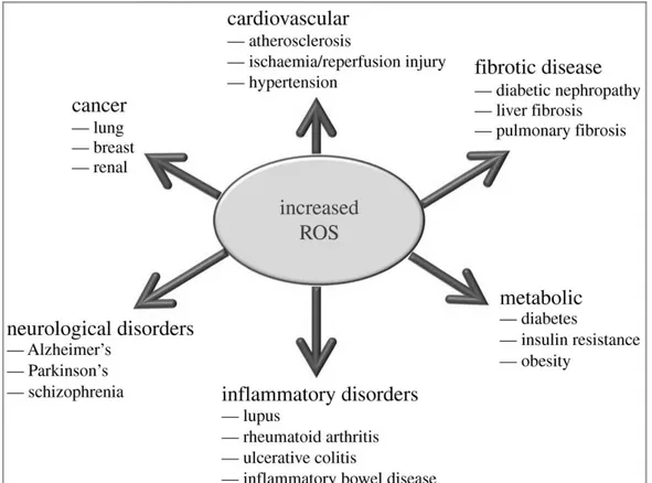
athways, the quesEon that is raised isexactly how do ROS iniEate cellular signaling? The “oxidaEve interface” is that boundary between ROS and the signaling molecules they acEvate; that is, the figuraEve region that describes how ROS directly acEvate oxidaEve stress-responsive pathways (Fig. 1).
Figure 1: ROS involvement in disease pathogenesis. SchemaEc representaEon of the effects of increased ROS producEon in the development of various pathologies.
We know that the balance between ROS is Eghtly regulated and extremely important for maintaining vital cellular and biochemical funcEons. This balance, oden referred to as the redox potenEal, is specific for each organelle and biological site, and any interference of the balance in any direcEon might be deleterious for the cell and organism. Changing the balance towards an increase in the pro-oxidant over the capacity of the anEpro-oxidant is defined as oxidaEve stress and might lead to oxidaEve damage. Changing the balance towards an increase in the reducing power, or the anEoxidant, might also cause damage and can be defined as reducEve stress.
Superoxide Ion Radical, (O2-/HO2)
Because most radicals are short-lived species, they react quickly with other molecules. Some of the oxygen-derived radicals are extremely reacEve with a short half-life. For example, OH can survive
for 10-10 sec in biological systems. Non-radical metabolites also
possess a relaEvely short half-life varying from parts of seconds to hours, as in the case of HClO. Obviously, the physiological environment, consisEng of such factors as pH and the presence of other species, has a great influence on the half-life of ROS.
Toxicity is not necessarily correlated with reacEvity. In many cases a longer half-life of a species might imply a higher toxicity of the compound by allowing it adequate Eme to diffuse and reach a sensiEve locaEon where it can interact and cause damage a long distance from its site of producEon. For example, the relaEvely long half-life of superoxide radicals permits them to move to locaEons where they can undergo interacEon with other molecules; these radicals can be produced in the mitochondrial membrane, diffuse towards the mitochondrial genome, and reduce transiEon metals bound to the genome. On the other
hand, a highly reacEve species with an extremely short life span, like OH, is produced in locaEons where it can cause damage (Barnese K et al., 1981) by interacEng with its immediate surroundings. If there is no essenEal biological target adjacent their producEon site,
radicals will not cause oxidaEve
damage.
The high reacEvity of radicals and their short life span illustrate the potenEal toxic effect and difficulEes in prevenEng oxidaEve damage. To prevent the interacEon between radicals and biological targets, the anEoxidant should be present at the locaEon
where the radicals are being produced in order to compete with the radical for the biological substrate.
The reducEon of oxygen by one electron at a Eme produces relaEvely stable intermediates. Superoxide anion (O2−), the product
of a one-electron reducEon of oxygen, is the precursor of most ROS and a mediator in oxidaEve chain reacEons.
DismutaEon of O2− (either spontaneously or through a reacEon
catalysed by superoxide dismutases) produces hydrogen peroxide (H2O2), which in turn may be fully reduced to water or parEally
reduced to hydroxyl radical (OH-), one of the strongest oxidants in
nature. The formaEon of OH- is catalysed by reduced transiEon
metals, which in turn may be re-reduced by O2−, propagaEng this
process (Liochev & Fridovich, 1999). In addiEon, O2− may react with
other radicals including nitric oxide (NO-) in a reacEon controlled by
the rate of diffusion of both radicals. The product, peroxynitrite, is also a very powerful oxidant (Beckman & Koppenol, 1996; Radi R et al. 2002).
In vivo, O2− is produced both enzymaEcally and non-enzymaEcally.
EnzymaEc sources include NADPH oxidases
located on the cell membrane of polymorphonuclear cells, macrophages and endothelial
cells (Babior, 2000; Vignais, 2002; Babior et al. 2002) and cytochrome P450-dependent oxygenases (Coon et al. 1992). The proteolyEc conversion of xanthine dehydrogenase to
xanthine oxidase provides another enzymaEc source of both O2− and H2O2 (and therefore
consEtutes a source
of OH-) and has been proposed to mediate
deleterious processes in vivo.
The non-enzymaEc producEon of O2− occurs when a single
electron is directly transferred to oxygen by reduced coenzymes or prostheEc groups (for example, flavins or iron sulfur clusters) or by xenobioEcs previously reduced by certain enzymes (for example, the anEcancer agent adriamycin or the herbicide paraquat).
The mitochondrial electron transport chain contains several redox centres that may leak electrons to oxygen, consEtuEng the primary source of O2− in most Essues.
Mitochondrial ROS produc5on
Mitochondria are an important source of ROS within most mammalian cells. This ROS producEon contributes to mitochondrial damage in a range of pathologies and is also important in redox
signaling from the organelle to the rest of the cell. Consequently, knowledge of how mitochondria produce ROS is vital to understand a range of currently important biomedical topics (Fig. 2). Figure 2: Overview of mitochondrial ROS producOon ROS producEon by mitochondria can lead to oxidaEve damage to mitochondrial proteins, membranes and DNA, impairing the ability of mitochondria to synthesize ATP and to carry out their wide range of metabolic funcEons. In addiEon, mitochondrial ROS producEon leads to inducEon of the mitochondrial permeability transiEon pore (PTP), which renders the inner membrane permeable to small molecules in situaEons such as ischaemia/reperfusion injury.
Mitochondria are double-membrane organelles involved in numerous cellular processes like apoptosis inducEon, reacEve oxygen species generaEon, adapEve thermogenesis, ion homeostasis and innate immune responses (W.J.H. Koopman, et al., 2012). Classically, mitochondria also act as key suppliers of
cellular energy by providing the machinery that
generates ATP from the energy stored in NADH and FADH2. The lajer two molecules are generated by the glycolysis
pathway in the cytosol (NADH) and the tricarboxylic acid (TCA) cycle in the mitochondria (NADH and FADH2), and are oxidized at complex I
(CI) and complex II (CII) of the Electron Transport Chain (ETC), respecEvely. The factors determining the rate of O2− producEon by
mitochondria are relaEvely straighkorward. The first is the concentraEon of the enzyme or protein [E] containing electron carriers that can exist in a redox form able to react with O2 to form
O2−. The second is the proporEon (PR) of this enzyme’s electron
carrier present in a redox form that can react with O2. As many redox-acEve groups exist only transiently in a state that can react with O2, PR is a Eme-average. The remaining factors are the local [O2-] and
the second-order rate constant (kE) for the reacEon of that electron carrier with O2 to form O2-.
This can be extended to consider several potenEal electron- donor sites within mitochondria, and also to take into account mulEple electron donor sites within a single protein. The concentraEon of the enzyme responsible for O2− producEon, [E], will vary with organism,
Essue, state, age or hormonal status, and may underlie many of the changes in maximum ROS producEon capacity between Essues (Barja, G. 1999); for example, complex I
content may explain the different maximum capaciEes of pigeon and rat heart mitochondria.
Mitochondrial Electron Transport Chain
The mitochondrial electron transport chain (ETC) has been recognized as one of the major cellular generators of reacEve oxygen species (ROS), which include superoxide (O2), hydrogen peroxide
(H2O2) and the hydroxyl free radical (OH-; Loschen et al. 1971; Boveris
et al. 1972; Chance et al. 1979). The released electrons are then transported to complex III (CIII) by coenzyme Q (CoQ) and subsequently to complex IV (CIV) by cytochrome c (cyt c). At the lajer complex, the electrons react with molecular oxygen (O2) to form
water. Electron transport is energeEcally coupled to the translocaEon of protons from the mitochondrial matrix across the mitochondrial
inner membrane (MIM) at CI, CIII and CIV. This results in an inward-directed proton-moEve force (PMF) across the MIM that consists of a chemical (ΔpH) and an inside-negaEve electrical gradient (Δψ). Protons are allowed to flow back into the mitochondrial matrix via the FoF1-ATP-synthase (CV) to drive the synthesis of ATP from ADP and inorganic phosphate (Pi). Together with CV, the four complexes of the
ETC consEtute the oxidaEve phosphorylaEon (OXPHOS) system. By reverse-mode acEon CV can also hydrolyse ATP and expel protons from the mitochondrial matrix thereby sustaining Δψ (Fig. 3) (D.G. Nicholls et al.,1972).
Figure 3: At the inner mitochondrial membrane, a high energy electron is passed along an electron transport chain. The energy released pumps hydrogen out of the matrix space. The gradient created by this drives hydrogen back through the membrane,
through ATP synthase. As this
happens, the enzymaEc acEvity of ATP synthase synthesiszes ATP from ADP.
This mechanism requires that ATP is transported from the cytosol to the mitochondrial matrix by reverse-mode acEon of the mitochondrial adenosine nucleoEde translocase (ANT) or is generated by mitochondrial substrate-level phosphorylaEon (C. Chinopoulos 2011). A sufficiently large PMF is not only required to sustain ATP producEon but also is of crucial importance for mitochondrial fusion, protein import, metabolite exchange with the cytosol, and apoptosis inducEon (F. Palmieri 2008, F. Fischer et al., 2012, J. Nunnari et al., 2012 and E.I. Rugarli e al., 2012). The proton pump of the electron transport chain (ETC) or mitochondrial respiratory chain establishes the mitochondrial membrane potenEal or proton moEve force (Δp). The protons are pumped from the matrix space to the intermembrane space, creaEng a substanEal pH and electrical gradient across the mitochondrial inner membrane.
These protons eventually re-enter the matrix space via the ATPase, driving the synthesis of ATP. This potenEal is dependent on a variety of other mitochondrial funcEons and is involved in the apoptoEc process. In this sense, Δψ can be considered as an important funcEonal readout of mitochondrial health.
In addiEon to being the source of energy that supports life under aerobic condiEons, mitochondria can also be the source of signals that iniEate apoptoEc cell death (Goelieb RA, 2000).
Mitochondria contain key regulators of caspases; a family of proteases that are major factors in many apoptoEc processes (Wang X, 2001).
Cytochrome c is released from the mitochondrial intermembrane space inducing the assembly of the apoptosome that is required for acEvaEng downstream caspases. Cytochrome c release from mitochondria is a key event in iniEaEng apoptosis, but the actual mechanism of its release is sEll debatable. In parEcular, the relaEon between mitochondrial physiology and the release of cytochrome c and other apoptogenic factors from mitochondria is not clear. Mitochondria uElize oxidizable substrates to produce a membrane potenEal in the form of a proton gradient across the mitochondrial inner membrane. It was shown that the supply of oxidizable substrates to mitochondria depends on the concentraEon of external growth factors (Vander Heiden MG et al., 2001). Withdrawal of growth factors or loss of the extracellular glucose supply will lead to a decline in mitochondrial membrane potenEal (MMP). If growth factor
or glucose deprivaEon persists, cells ulEmately undergo apoptosis that is iniEated by cytochrome c release from mitochondria.
Whether changes in mitochondrial physiology contribute to the iniEaEon of cell death in response to growth factor withdrawal remains a controversial issue.
The mitogen-acEvated protein kinase (MAPK) cascades consist of four major MAPKs; the extracellular signal-related kinases (Erk1/2), the c-Jun N-terminal kinases (JNK), the p38 kinase (p38), and the big MAP kinase 1 (BMK1/Erk5). These kinases are evoluEonarily conserved in eukaryotes and play pivotal roles in cellular responses to a wide variety of signals elicited by growth factors, hormones, and cytokines, in addiEon to genotoxic and oxidaEve stressors. FuncEon and regulaEon of the MAPK cascades have been comprehensively covered (Raman M et al., 2007; Cuadrado A et al., 2010; Weston CR et al., 2007; Ramos JW et al., 2008).
Among the members of the MAPK cascades, apoptosis signal-regulated kinase 1 (ASK1) is an upstream MAPKKK that regulates the JNK and p38 MAPK pathways leading to apoptosis through phosphorylaEon of MKK4, MKK3, and
MKK6 MAPKKs (Ichijo H et al., 1997). ASK1, once acEvated under various stress condiEons including
oxidaEve stress (Tobiume K et al., 2001), is homo-oligomerized by both C- and N-terminal coiled-coil domain interacEon and acEvaEon occurs through phosphorylaEon of a conserved threonine (Human: Thr-838, Mouse: Thr-845) residue in the acEvaEon loop of the human ASK1 kinase domain. ASK1-deficient mouse embryonic fibroblasts were shown to be less suscepEble to TNF or H2O2-induced
cytotoxicity along with decreased JNK and p38 MAPK acEvaEon, suggesEng that ASK1 plays a pivotal role in promoEng cell death under oxidaEve stress; however, ROS acEvated ASK1 mediates p38 signaling leading to non-apoptoEc outcomes also, such as differenEaEon (Takeda K et al., 2007) and immune signaling (Matsuzawa A et al., 2005), thus reinforcing the role of ROS signaling in cellular homeostasis.
Oxida5ve stress- induced cell death
Apoptosis plays an essenEal role in normal development and Essue homeostasis, and when dysregulated it contributes to various diseases including cancer, autoimmunity and neurodegeneraEve disorders. There is a direct interacEon between ROS levels and inducEon of cell death and mitochondria play a central
decision-making role in execuEng the cell-death program. The liberaEon of toxic factors together with the idenEficaEon of key regulatory proteins that influence mitochondrial funcEon, each suggesEng that the mitochondrion may be an important therapeuEc target for the design of new intervenEons to abrogate or at least miEgate inordinate cell death ader burn. How burn signals become integrated at the level of the mitochondria to acEvate the cell-death pathway is unknown and is an area of acEve invesEgaEon.
Apoptosis signal-regulaEng kinase 1 (ASK1) is a mitogen-acEvated protein (MAP) kinase kinase kinase (MAPKKK) family member that acEvates the JNK and p38 MAP kinase pathways and is acEvated by various stresses including oxidaEve stress, TNFα, calcium overload, and endoplasmic reEculum stress.
Recent analyses of ASK1-deficient mice have revealed that ASK1 is required for cell death induced by oxidaEve stress, TNFα, and endoplasmic reEculum stress (Guo X et al., 2010).
Oxidants have been demonstrated to regulate the acEvaEon of c-Jun N-terminal kinase (JNK), as well as the transcripEon factor NF-κB. JNK is a member of the family of mitogen-acEvated protein kinases, which is well known to be acEvated by
oxidants and a
variety of other stresses in many cell types. The
contribuEon of JNK to many phenotypic outcomes, including survival (J Wang, et al., 2011) and apoptosis (Li D, et al., 2015), appears to depend upon the cell type, sEmulus, the duraEon of JNK acEvaEon as well as the engagement of other signaling modules (JW Chambers, et al., 2011).ROS acEvaEon of JNK can induce extrinsic or intrinsic apoptoEc signaling (Win S et al., 2014). Upstream of JNK is the redox sensiEve MAPK kinase kinase, ASK1. ASK1 acEvity is inhibited by interacEons with redox proteins (Grx, Trx1), heat shock proteins (Hsp90, Hsp72) (Fig. 4). TNFα is a potent acEvator of MAPK cascade, and the ASK1-JNK pathway plays an important role in TNF-R1-mediated apoptoEc signaling in various cell types.
Figure 4: Summary our current understanding of the mechanism of ROS and redox modulaEon of ASK1/JNK signaling in cell apoptosis. This model proposes that ROS mediates the interacEon of Trx1 and the N-terminal domain of ASK1, prevenEng ASK1 acEvaEon and downstream propagaEon of an apoptoEc signal (Magdalena L.Circu, 2010).
Contribu5on of ROS in burn
During the early phase of a burn, significant neutrophil accumulaEon was demonstrated in various Essues such as gastric mucosa, liver and lung suggesEng that the source of ROS could be neutrophils sequestered in systemic organs as a result of the systemic inflammatory reacEon to a local burn insult. Neutrophil accumulaEon
in these Essues following
severe burn may be involved in the pathogenesis of remote organ damage by producEon of ROS. Normally, cells are able to defend themselves from damaging effects of oxygen radicals in the normal physiologic condiEon by way of their own anEoxidant mechanisms including enzyme systems, vitamins, elements, and some anEoxidant molecules.
Normally, there is an exquisite balance between producEon and destrucEon of ROS. When this equilibrium is destroyed, ROS are produced excessively and all Essues are exposed to oxidaEve injury. Severe burn is a trauma with high oxidaEve stress. Ader burn, generalized Essue inflammaEon is present in uninjured organs within hours. Evidence from animal models and human studies (Wiggins-Dholvic et al 2014) demonstrated involvement of oxidaEve stress in burn injuries.
Ader a burn trauma the necrosome formaEon iniEates the cascade of phosphorylaEon of several downstream target proteins including phospholipase A2, the proteases calpains and cathepsins, the cytoplasmaEc NOXA1/NADPH oxidase complex, and the mitochondrial complex I, thereby leading
to excessive ROS producEon, ATP depleEon, and opening of the mitochondrial
permeability transiEon pores. These events are accompanied by prolonged JNK acEvaEon and necrosome-induced sEmulaEon of glycolysis, glycogenolysis, and glutaminolysis as well as the sEmulaEon of Krebs cycle.
Contribu5on of ROS in I/R injury
OxidaEve stress has been suggested to be involved in a number of cardiac pathologies including heart failure (H Tsuitsui et al 2011). Evidence to support this claim has come from animals models of myocardial infarcEon, pressure overload and pacing-induced heart failure as well as in paEents with chronic led ventricular systolic failure (Y Azizi et al 2016). InteresEngly, oxidaEve stress-induced heart failure may be ajributed in part to increased cardiomyocyte apoptosis.
Several studies have shown a direct effect between ROS levels and apoptosis. InteresEngly, treatment of cardiac cells with H2O2 was
sufficient to trigger dissipaEon of inner mitochondrial membrane potenEal and PTP opening mitochondrial with ajendant cytochrome c release and caspase-3 acEvaEon.
Importantly, agents that inhibited the PTP opening suppressed H2O2-induced apoptosis suggesEng that
mitochondrial perturbaEons
were fundamental to the apoptoEc process.
Recently, apoptosis repressor with CARD domain (ARC), a novel caspase-inhibitory protein restricted to cardiac muscle cells, was shown to block cell death triggered by H2O2 in the myogenic cell line,
H9c2 (Neuss M et al 2001). ARC was also effecEve in blocking H2O2
-induced loss of membrane integrity and/or disrupEon of mitochondrial ΔΨm in two human cell lines in which it is not normally
expressed. These results demonstrate that, in addiEon to its ability to block caspase-dependent events in apoptosis, ARC also prevents cell death via the preservaEon of mitochondrial funcEon.
Notably, a study by (Akao et al. 2003) proposed that H2O2-induced
cardiomyocyte apoptosis is characterized by a specific pajern of mitochondrial defects. The model is defined by three phases: priming, depolarizaEon and fragmentaEon. During the priming phase, exposure to H2O2 sEmulates mitochondria to undergo specific
changes in inner mitochondrial membrane structure consistent with swelling and loss of cristae ultrastructure. Priming is associated with matrix calcium overload but not alteraEons in ΔΨm.
In the next phase, aptly named depolarizaEon, myocyte mitochondria undergo a sudden loss in ΔΨm, which is conEngent
upon PTP opening. Finally, myocytes are dismantled in the fragmentaEon phase where mitochondria undergo massive swelling and release cytochrome c quickly, and predictably apoptosis ensues. Whether this model applies globally to oxidaEve stress or to H2O2
specifically is undetermined, but it provides a novel perspecEve on the temporal and spaEal relaEonship of mitochondrial perturbaEons to cardiomyocyte apoptosis.
Chapter II
Role of NLRP3 inflammasome in mul5-organ dysfunc5on following thermal injury
Introduc5on
Burns are a serious global health problem, according to the World Health OrganizaEon, with over 195,000 related deaths each year. Burn injury alters host immune funcEons, predisposing paEents to opportunisEc and nosocomial infecEons, sepsis, and mulEple organ system dysfuncEon and failure. Burn injury oden leads to a systemic inflammatory state, which has been ajributed to the resulEng exacerbated innate immune response, referred to as systemic inflammatory response syndrome (SIRS; Hoover L et al 2006; Sauaia A et al 1996). In addiEon, the response to the iniEal burn is oden associated with secondary damage to Essues distant from the injured site.
Prolonged exposure to temperatures higher than 40°C leads to denaturaEon of proteins and finally loss of their plasma membrane integrity. The local changes result in the clinical picture of coagulaEon and necrosis. Temperature and duraEon of contact have also a synergisEc effect (Van Heren et al 2013).
The systemic pathophysiologic changes following thermal injuries affect mulEple organs and body systems leading to clinical manifestaEons including shock, intesEnal alteraEons, respiratory and renal failure, immunosuppression and
others. Evidence from animal models and human studies demonstrated the involvement of oxidaEve stress in burn injuries. Indeed, there is a direct correlaEon between ROS levels and inducEon of apoptosis and mitochondria play a central decision-making role in execuEng the cell-death program. Acute renal failure is one of the major complicaEons of burns and it is accompanied by a high mortality rate (Brusselaers, N et al 2010). Most renal failures occur either immediately ader the injury or at a later period. Two major mechanisms are involved in the pathophysiological changes in the kidney: filtraEon failure and tubular dysfuncEon caused by various factors and interacEng with each other (Aikawa N et al 1990). The renal failure that occurs in extensively burned paEents is usually associated with failure or dysfuncEon of other organs in a form of mulEple organ dysfuncEon syndrome which adversely influences the prognosis. Acute renal failure occurring immediately ader burns is mostly due to reduced cardiac output, which is mainly caused by fluid loss (Pruie BA Jr 2000).
Many mediators, including cytokines (TNF, IL-1, etc.), prostaglandins (PGs), thromboxane, leukotrienes, and
platelet-aggregaEng (acEvaEng) factor (PAF) are produced or released in the early post-burn period. They act variably to
increase vascular permeability and to induce Essue damage (Ansermino M et al 2004).
An intense affiliaEon has been demonstrated between the quanEty of lipid peroxidaEon and the degree of burn complicaEons such as remote organ damage and shock. A primary effect of lipid peroxidaEon is decreased membrane fluidity, which alters membrane properEes. The mechanism of this event is the deformaEon of cell membrane phospholipids by oxidazing radicals.
Cell-death pathway in burn
Inflammatory cells produce reacEve oxygen species (ROS) as part of the microbiocidal/cytocidal system. Several studies demonstrated that burn iniEates systemic inflammatory reacEons by producing burn toxin. At molecular level, both complement acEvaEon and intravascular sEmulaEon of neutrophils result in the producEon of cytotoxic ROS. Increased histamine acEvity, enhanced by the catalyEc
properEes of xanthine oxidase, causes progressive local increases in vascular permeability.
AcEvaEon of c-Jun NH2-terminal kinase (JNK) is based on the producEon of reacEve oxygen species (ROS) and acEvaEon of apoptosis signal-regulaEng kinase-1 (ASK1), a member of the MAP3K family (Ichijo H et al 1997).
The pro-inflammatory cytokines such as tumor necrosis factor-α (TNF-α), interleukin -1β (IL-1β) and cell adhesion molecules are overexpressed ader severe burns, leading to uncontrolled inflammatory response and organ injury. The excessive producEon of reacEve oxygen species (ROS) can lead to cell damage and finally result in organ failure. Furthermore, ROS overproducEon leads to mitochondrial dysfuncEon and adenosine triphosphate (ATP) depleEon (Dal Pizzol F et al 2010), triggering cytochrome c to leak from the mitochondria into the cytoplasm and ulEmately causing cell apoptosis (Yang Y et al 2015). Cells are able to defend themselves from damaging effects of oxygen radicals in normal physiological condiEons, because the exisEng balance between producEon and destrucEon of ROS. However, how burn signals become integrated at
the level of the mitochondria to acEvate the cell-death pathway is unknown and is an area of acEve invesEgaEon.
NLRP3 Inflammasome in burn
The acEvaEon of NLRP3 inflammasomes has been implicated in a growing number of diverse pathological condiEons, ranging from bacterial infecEons to cardiovascular dysfuncEon and metabolic syndrome (Benko S et al., 2008, Duewell et al., 2010, Overley-Adamson et al., 2014). Despite rapid and extensive efforts in idenEfying various agents that sEmulate the NLRP3 inflammasome, the underlying mechanisms by which these diverse danger signals acEvate the same molecular machinery remain poorly understood.
ROS, produced by many known acEvators of NLRP3 inflammasomes, are shown to be a criEcal mechanism triggering NLRP3 inflammasome formaEon and acEvaEon in response to many exogenous sEmuli as well as endogenously produced or secreted molecules from damaged cells such as DAMPs (Tschopp J et al., 2010).
Increased producEon of ROS results from trauma, including burn injury. These oxygen radicals are released from neutrophils ader inflammaEon or ischemic injury. The producEon of ROS contributes to
increased vascular permeability, Essue edema, systemic inflammaEon, and mulE organ dysfuncEon.
The hypothesis of ROS as an NLRP3-acEvaEng trigger arose when inhibiEon of NADPH oxidase-derived ROS prevented ATP-induced caspase-1 acEvaEon and IL-1beta producEon in alveolar macrophages (Cruz CM et al., 2007). Further substanEaEng this hypothesis, knockdown of the p22phox subunit of NADPH oxidase significantly suppressed IL-1beta release in THP1 cells in response to asbestos and MSU challenge (Dostert T et al., 2008 ).
The crystal structure of NLRP3 contains a highly conserved disulfide bond connecEng the PYD domain and the nucleoEde-binding site domain, which is highly sensiEve to altered redox states (Bae JY et al., 2011). The presence of this unexpected disulfide bond between Cys-8 and Cys-108 spans across six species, including humans, monkeys, and mice, and the strict conservaEon of this bond is indicaEve of a crucial redox role for NLRP3.
It has been shown that a burn causes a disEnct inflammatory cytokine expression profile in severely burned paEents (Finnerty CC et al 2006) and that the cytokine profile at the Eme of admission can predict which burned paEents will develop infecEous complicaEons
and sepsis during the hospital course (Finnerty CC et al., 2007). Although these studies enhance our understanding of the post-burn
inflammatory response and allow predicEon of paEent outcome, effecEve therapies for altering paEent outcome do not exist.
In order to invesEgate new drugs and intervenEons that will modulate post-burn hypermetabolism and hyperinflammaEon, geneEcally modified mice with either altered NLRP3 protein expression (knock-out or knock-in) have been used. Therefore, characterizaEon of the murine inflammatory response to burn is essenEal to not only allow modulaEon of the response, but also to allow comparison to the human response to determine the appropriateness of using mice to study the human post-burn response.
Results
Western bloRng and histological results
Apoptosis signal-regulaEng kinase (ASK) 1 is acEvated in response to various cytotoxic stresses including TNF, Fas and reacEve oxygen species (ROS), and acEvates c-Jun NH2-terminal kinase (JNK) and p38. However, the roles of JNK and p38 signaling pathways during apoptosis have been controversial.
Kidneys from KO animals had detectable expression of ASK1, ader 7 days (Fig.1), this expression was instead markedly reduced in KI mice, maybe due to their overexpression of NALP3 that likely processed most of the DAMPs.
The c-Jun NH2-terminal kinase (JNK) is acEvated when cells are exposed to mulEple forms of stress, and this signaling pathway has been implicated as a mediator of stress-induced apoptosis. In this animal model of thermal burn injury, as expected, an high JNK acEvaEon has been detected only in KO animals ader 7 days (Fig.3).
MAPKs comprise a family of serine/threonine protein kinases that funcEon as criEcal mediators of signal transducEon and include also the p38 MAPKs. As we know,
the JNKs and p38 MAPKs are acEvated
by proinflammatory cytokines and a variety of cellular stresses, including UV light, hyperosmolarity, heat shock, and microtubule disrupEng drugs. Because the lack of expression of ASK1 in KI animals, p38 acEvaEon in this experimental group might be due to the high inflammatory profile sEll present ader 7 days of being injured compared with WT-injured mice as show by the presence of proptosis in KI animals even under normal condiEons (Fig.5).
Bax (member of the proapoptoEc group of mulEdomain Bcl2-related proteins) are essenEal for the JNK and p38-sEmulated release of cytochrome c and apoptosis. Indeed, both KO and KI animals present high level of BAX ader 7 days as a sign of mitochondrial dysrupEon and extensive cell death as confirmed by histological results from kidney slices by TUNEL apoptosis assay (Fig.6).
Conclusions
It is generally accepted that burn injury and trauma primes the innate immune system to enhance inflammatory responses to pathogen associated molecular signals (PAMPs), however, the molecular pathways responsible for injury-induced immune system acEvaEon are not well defined. The inflammasome was idenEfied as a central feature of the danger response because most of the extracellular or intracellular signals that acEvate the inflammasome are considered danger molecules or alarmins. A number of theories have been proposed for the idenEty of the cellular signal responsible for NLRP3 acEvaEon, The finding that ER stress, like other NLRP3 acEvators, acEvates the NLRP3 inflammasome in a K+
efflux- and ROS-dependent manner suggests that it may also be sensed by mitochondria. Thus, it is conceivable that ER stress iniEates a signal that is transmijed to mitochondria and then relayed to the NLRP3 inflammasome.
Severe thermal injuries result in a wide array of stress-associated inflammatory and metabolic changes aimed at restoring homeostasis
uncontrolled, persisEng far past the iniEal trauma, they lead to a state of severe metabolic dysfuncEon.
This study provides evidence of a role of the NLRP3 inflammasome in systemic inflammatory response following thermal injury. Under this peculiar condiEons KO-animals displayed an elevated mortality rate (over 60%) during the first 4 days, and none of the KO mice survived ader day 7. On the other hand, the animals overexpressing the NLRP3 survived up to 14 days as the WT ones. The excessive triggering of the apoptoEc/inflammatory pathways in KI-animals was evident even under normal condiEons, in fact pyroptosis was detected in kidneys from sham animals. KO-mice showed an increased acEvaEon of apoptosis-related molecules ader 7 days, while in KI animals the inflammatory pathway was more acEvated compared to either WT and KO mice.
These results may suggest that the NLRP3 inflammasome is of fundamental importance in survival and recovery from systemic injury. Indeed, recent studies demonstrated that
mitochondrial dysfuncEon involving increased mitochondrial ROS producEon (Galley HF 2011, Zhou et al., 2011) and the release of mitochondrial DNA into the cytosol are criEcal events associated with NLRP3 inflammasome
acEvaEon. However, Jennifer R. Deuis and colleges demonstrated that NLRP3 and its downstream product caspase-1 have a limited role in the development of acute burn-induced pain and that pharmacological inhibiEon of NLRP3 is unlikely to be an effecEve treatment strategy for the treatment of acute procedural pain or inflammaEon in burn-injured paEents. Burned paEents oden develop also a form of stress-induced diabetes (with hyperglycemia, insulin resistance, and hyperlipidemia) which is linked to marked increases in morbidity and mortality. ParEcularly, in burned paEents, studies have demonstrated that the significant pathophysiological changes and extreme inflammatory responses are not only present during acute hospitalizaEon causing delays in their rehabilitaEon and reintegraEon. Although intensive efforts have long focussed on idenEfying the underlying mechanisms of these extreme metabolic alteraEons, few studies have managed to elucidate how thermal injury induces hypermetabolism, prolonged inflammaEon and stress responses, and insulin resistance, and whether these alteraEons are responsible for the increased morbidity and mortality (Stanojcic M et al., 2014). Others have also suggested that mitochondrial dysfuncEon and subsequent increases in reacEve oxygen species (ROS) may be
responsible for priming and acEvaEng the inflammasome (Sorbara MT et al., 2011; Zhou R et al., 2011) in metabolic disorders. It would be interesEng to invesEgate this aspect in future research.
Meterial and Methods
Animals
For these experiments we used 3 different strain of mice: the hyper expressing Nlrp3A350VneoR (k-in), the null Nlrp3L351PneoR mice (ko) and their relaEve wild type strain C57BL/6J. Animals were kept under standard condiEons at the Animal Facility of Messina University.
Thermal burn injury
To produce a second degree scald burn, a 6 cm2 area on the back of
each animal was shaved (30% of the body surface). Mice were anestheEzed and immersed in 80°C water for 10 seconds; saline soluEon was administered to allow recover. All animals (n=5 for experimental group) were allocated in single cages and killed ader 7 days for further analysis.
Western BloRng analysis
Protein samples from kidney were extracted to detect p-ERK 1/2, p-JNK 2/3, p-p38, p-ASK1 and BAX. Protein samples (30 mg) were
separated in SDS-PAGE and blojed on PVDF membranes. To prove equal loading, the blots were stripped and beta-acEn expression assessed.
Histologycal analysis
For light microscopy Essues were fixed in 10% buffered formalin, embedded in paraffin, secEoned at 5-um thickness and stained with hematoxylinn and eosin (H&E) or with a TUNEL assay kit (GenScript).
Sta5s5cal analysis
All data are expressed as mean ± standard deviaEon (SD). The significance of difference was assessed by Unpaired T-test. P values less than 0.05 was considered significant. Graphs were performed using GraphPad Prism (version 5.0 for Windows).
LIST OF FIGURES
Figure 1: Kidneys levels of ASK1 ader 7 days. KO vs KI *p<0.001; KO vs WT-inj #p<0.05; WT-inj vs KI §p=0.02; (Mann-Withney t-test).
Figure 2: p38 western blot levels in kidneys ader 7 days. KI vs WT-inj *p=0.01 (Mann-Withney t-test). ASK1 (155 KDa) β actin (42 KDa) P38 (38 kDa) β actin (42 KDa) sham wt sham ko wt in jured ki inj ured ko in jured 0 1 2 3 in te gr at ed in te ns it y sham wt sham k o wt inj ured ki inj ured ko inj ured 0 2 4 6 in te gr at ed in te ns ity * # § § *
Figure 3: Kidneys levels of JNK ader 7 days. KO vs KI *p=0.01; KO vs WT-inj #p<0.005; Sham ko vs Sham wt §p=0.02(Mann-Withney t-test).
Figure 4: Bax protein levels in kidneys ader 7 days confirmed acEvaEon of mitochondrial cell death pathway. KO vs KI *p<0.001; KO vs WT-inj #p<0.005; Sham KO vs Sham WT §p<0.001(Mann-Withney t-test). BAX (27 kDa) β actin (42 KDa) # p-JNK 1/2/3 (46-54 kDa) β actin (42 KDa) sham w t sham kowt injur ed ki inju red ko inj ured 0 2 4 6 in te gr at ed in te ns it y * § sham wt sham k o wt inj ured ki inj ured ko inj ured 0 2 4 6 in te gr at ed in te ns ity * # §
Figure 5: Histological evaluaEon by H&E of kidneys ader 7 days shows an excessive triggering of the inflammatory pathways in KI-animals even under normal condiEons, in fact
pyroptosis (black arrows) was detected in kidneys from sham animals .
Sham-WT
WT injured
Sham-KO
KO-injured
Figure 6: Histological evaluaEon of kidneys slices by TUNEL-apoptosis-assay shows an increased presence of apoptosis (black arrow) in KO injured animal animals and apoptoEc bodies in KI group even under normal condiEon (black arrow).
Sham-WT
WT injured
Sham-KO
KO-injured
Sham-KI
KI-injured
Chapter III
Protec5ve effect of Ammonium Tetrathiomolybdate (ATTM) following Ischemia Reperfusion (I/R) Injury
Federica Galfo1,2, Alex Dyson1 and Mervyn Singer1
1Bloomsbury InsEtute for Intensive Care Medicine,University College
London, UK. 2 Department of Clinical and Experimental Medicine,
University of Messina, Italy
Introduc5on
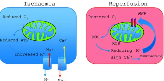
In Essue subjected to ischemia followed by reperfusion (I/R), pathologic mechanisms are elicited that produce reversible cell injury and dysfuncEon, which can progress to irreversible damage if the nature and extent of ischemia is prolonged or if the pathologic sequelae to reperfusion are of sufficient magnitude. This damage is referred to as I/R injury and can be divided into different phases (Hausenloy et al 2013, Moens AL et al 2005) (Fig. 1).
During ischemia (the first phase of injury), interrupEon of the blood supply to an organ causes a reducEon in oxygen and nutrient delivery to the affected Essues. This disrupts ATP generaEon via oxidaEve phosphorylaEon, causing cells to alter their metabolism and impairs energy-dependent cellular funcEon.
Figure 1: SchemaEc representaEon of I/R injury phases.
Reduced ATP availability limits ion pumps in cell membranes, resulEng in calcium overload, structural disorganizaEon, and apoptoEc, necroptoEc, and necroEc cell death. In addiEon, ischemia induces conformaEonal changes in enzymes such as xanthine oxidase and elicits the formaEon of pro-inflammatory mediators and expression of adhesion molecules that promote leukocyte/ endothelial cell adhesive interacEons (Eltzschig HK et al 2011, Perrelli MG et al 2011). These lajer processes do not directly contribute to injury during the ischemic phase, but rather set the stage for the second stage of I/R injury (ie, that due to reperfusion), wherein Essue injury is exacerbated when the blood supply is re-established. Paradoxically, the lack of oxygen during ischemia and the replenishment of oxygen during reperfusion both contribute to the total injury sustained by Essues subjected to I/R. The clinical outcome is also determined by a third phase of ROS producEon that occurs during post-reperfusion repair that is characterized by Essue remodeling and adaptaEon (Raedschelders K et al 2012).
Reperfusion injury
The double-edged sword effects of I/R-induced ROS generaEon may be related to species of ROS produced, the amount of oxidants generated, and the subcellular locaEon and cellular source of their producEon under a given set of condiEons, as well as at what Eme during the three phases of responses to I/R they are formed (Kalogeris T et al 2014).
Reperfusion represents the second phase of I/R injury and pre- cipitates the generaEon of ROS that is fueled by the reintroducEon of molecular oxygen to the Essues. Xanthine oxidase and phagocyte NADPH oxidase-derived oxidants can damage virtually every biomolecule found in cells and Essues (Robert AM et al 2014).
ROS induce Essue dysfuncEon by directly damaging cells via a number of mechanisms including peroxidaEon of cell membrane and organelle lipids, oxidizing DNA , acEvaEon of matrix metalloproteinases and calpains, producing osmoEc cell lysis, inducEon of no-reflow, and causing opening of the mitochondrial permeability transiEon pore (Zinkevich NS et al 2011, Kharbanda RK et al 2010, Seidlmayer LK et al 2015).
ROS may also induce cell dysfuncEon and death by indirect mechanisms by interacEng with NO, fajy acids or free iron to form peroxynitrite, peroxyl radicals, and hydroxyl radicals,
respecEvely, each of which are capable of producing even more cellular damage than superoxide or hydrogen peroxide (Fig.2).
Figure 2: IllustraEve scheme of the main mediators of lethal reperfusion injury. As shown in the right, restoring oxygen to ischemic myocardium causes ROS producEon.
Oxygen paradox
Oxygen-derived ROS also act to enhance the inflammatory response to reperfusion via formaEon of oxidant-dependent proinflammatory mediators and upregulaEon of cytokine/chemokine and adhesion molecule expression (Naha PC et al 2010, Harris J 2010, Naik E et al 2011) .
Thus, while there is cellular demand for replenishment of oxygen which is met by re-establishing the blood supply, the reintroducEon of molecular oxygen to the Essues results in ROS formaEon that is detrimental to the reperfused Essues. The divergent roles of oxygen in the first two phases of I/R injury are referred to as the oxygen paradox (Buia LM et al 2010).
A third ROS paradox arises in later phases of reperfusion, where ROS generaEon affects several Eghtly regulated processes that promote organ repair and survival. This third phase consEtutes the reparaEve phase of I/R injury and involves ROS-dependent generaEon of growth factors that promote angiogenesis, induce proliferaEon and differenEaEon of vascular smooth muscle cells to effect vascular remodeling, and promote the acEvaEon of matrix metallo-proteinases and other factors that contribute to
fibrosis, Essue
remodeling and formaEon of scar Essue (Buia et al 2010, Kawaguchi M et al 2011).
The mitochondria also produce ROS from respiratory chain components, as well as by acEvaEon of mitochondrially localized monoamine oxidase, the growth factor adaptor Shc (p66Shc), cytochrome b5 reductase, dihydroorotate dehydrogenase, mitochondrial ATP-sensiEve potassium (mKATP) and large-conductance, calcium acEvated potassium (BKCa) channels, and the Nox isoform designated Nox4. Superoxide normally produced in mitochondria is scavenged by manganese-superoxide dismutase (Mn- SOD or SOD-2) localized in the matrix. In addiEon, copper/zinc-SOD (Cu/ZN-SOD or SOD-1), which is typically considered a cytoplasmic isoform, is also located in mitochondria between its inner and outer membranes (Arany I et al 2011, Finkel T et al 2011, Daiber et al 2010). These SODs dismutase superoxide to less reacEve hydrogen peroxide, which can be further metabolized to water and oxygen by the catalyEc acEvity of catalase and glutathione peroxidase.
Mitochondrial uncoupling proteins also serve to reduce the producEon of ROS by causing mitochondrial depolarizaEon, which reduces the potenEal driving electron
transfer and by allowing
protons to reenter the matrix, thereby bypassing ATP synthase (Mailloux RJ et al 2011).
Short-term opening is involved in cardioprotecEon that involves transient ROS formaEon (Perrelli MG et al 2011).
In contrast, long-lasEng mPTP opening, which is facilitated by restoraEon of pH, calcium overload and the burst of ROS formaEon at the onset of reperfusion, is followed by profound and irreversible alteraEons in cellular bioenergeEcs. Sustained pore formaEon results in increased mitochondrial permeability to ions and other solutes up to molecular weights of 1.5 kD and collapse of the mitochondrial membrane potenEal. This is rapidly followed by ATP and NAD+
depleEon, release of accumulated mitochondrial calcium, matrix swelling and outer mitochondrial membrane rupture, which in turn results in loss of pyridine nucleoEdes, release of pro-apoptoEc factors, and further inhibits electron flow through the electron transport chain.
The massive release of ROS during reperfusion requires the involvement of the mPTP in a ROS-induced ROS release posiEve feedback loop (Zorov DB et al., 2014). It is widely believed that mPTP is thus a major causaEve event in reperfusion injury and cell death.
This concept is consistent
with the observaEon that cardioprotecEve intervenEons all seem to intersect at inhibiEon of the mPTP as an end-effector of enhanced tolerance to I/R.
Because O2− producEon could be mediated by complex IV during
ischemia, the goal of this study is to asses if the administraEon of ATTM, a new class of sulfide-releasing drugs, may confer protecEon by inhibiEng the complex IV of the respiratory chain, reducing both ROS producEon and membrane potenEal depolarizaEon.
Biological role of hydrogen sulfide (H2S)
H2S is a colourless, flammable gas with a characterisEc odour of rojen eggs. It is soluble in water (1 g in 242 ml at 20°C). In water or plasma, H2S is a weak acid which dissociates as follows: H2S ↔ HS− + H+. The pKa at 37°C is 6.76; therefore, when either sodium hydrosulfide (NaHS) or H2S is dissolved in physiological soluEon (pH 7.4, 37°C), it will form approximately 18.5% H2S and 81.5% hydrosulfide anion (HS), as predicted by the Henderson–Hasselbach equaEon. H2S is a highly lipophilic molecule and freely penetrates cells of all types. It is this property which endows H2S with, at least the potenEal for, biological acEvity.
H2S is formed in mammalian cells largely by the acEvity of two pyridoxal phosphate-dependent enzymes, cystathionine γ lyase (CSE, EC 4.4.1.1) and cystathionine β synthetase (CBS, EC 4.2.1.22) (Fig. 3). These enzymes are widespread in mammalian Essues and cells and also in many invertebrates and bacteria.
Figure 3: Scheme of representaEve pathways for H2S biosynthesis
(blue) and catabolism (red) in mammalian Essues.
The pathway(s) by which H2S is broken down in the body is/are
parEally known, although several alternaEves have been idenEfied. H2S is rapidly oxidized in mitochondria to thiosulfate, which is then
further converted to sulfite and sulfate (the major product). Whether this is how H2S is biologically deacEvated is not known. However, the
majority of sulfate in urine is believe to derive from the direct oxidaEon of cysteine by cysteine dioxygenase acEvity and, as such, sulfate cannot be used as a marker for the presence of H2S in the
same way, for example, that nitrate (and nitrite) are oden used as markers of NO.
H2S also undergoes methylaEon in the cell cytosol by thiol
S-methyltransferase to yield methanethiol and dimethylsulfide, and can bind to methaemoglobin to form sul†aemoglobin.
H2S interacts with membrane and cytosolic proteins to produce
reacEve and unstable persulfides. These persulfides can be further converted to other biochemical forms including thiocysteine, thiotaurine, protein–SSH, thiocysEne, mercaptopyruvate and others. Sulfide donors may impart structural changes in proteins through persulfide related sulfuraEon and sul†ydraEon reacEons of proteins.
It is interesEng to note that the physiological funcEons of H2S are comparable to that of NO, as well as controversy surrounding their cytoprotecEve roles. Throughout the literature on H2S it is reported that the cytoprotecEve and anEoxidant effects occur in the
micromolar range; whereas higher H2S exposures, i.e., in the
millimolar range, potenEate redox stress and are cytotoxic. In the coming future, it is most likely that the field will come to realize the cellular and signalling funcEon and physiological potency of low nanomolar concentraEons of H2S, and that various biochemical forms
of the molecule serve important roles in regulaEng H2S bioavailability
and cellular redox balance. From the informaEon above, it is safe to say that H2S serves as a proverbial “double-edged sword,” where it
can be extremely beneficial or harmful depending on its concentraEon and cellular locaEon. These observaEons also reveal how crucial it is moving forward to accurately determine and control for the levels of H2S in experimental se‡ngs, reinforcing the need for
rigorous and reliable measurement techniques to monitor the biological levels of H2S. Finally, increased clarity regarding sulfide
cellular signaling will also alleviate confusion and lead to a bejer understanding of the effects of H2S administraEon in biological
systems. The future of H2S biochemistry, chemical biology and
pathophysiology represent ferEle territory in which to bejer understand redox processes that will ulEmately be important for human health and disease.
Ammonium tetrathiomolybdate (ATTM) mechanism of ac5on
Ammonium tetrathiomolybdate (ATTM) was found to be a slow H2S
releasing agent, in a characterisEc Eme, temperature and pH dependent manner. First synthesis report was almost two centuries ago (Berzelius 1826) and since then has been used in men to treat Wilson’s, autoimmune, fibroEc and cancer diseases, but has been also used in animals as a copper chelator. However the real efficacy seems related to its anE-inflammatory and anE-angiogenic effects.
Hydrogen sulfide (H2S) represents the most recently idenEfied
endogenously produced gaseous messenger. Although long considered a noxious gas with wide-ranging cytotoxic effects, there is now an accumulaEon of scienEfic evidence that H2S plays a
prominent role in cellular signaling. In recent years, the cytoprotecEve effects of endogenous and exogenous H2S have been
invesEgated in models of in vitro and in vivo ischemic injury. H2S has
also been shown to increase KATP channel currents in isolated smooth muscle cells (Zhao W et al 2001) but the mechanism behind the cardioprotecEve effects of H2S is not limited to modulaEon of KATP channels and Ca2+ handling.
There is much evidence to suggest that H2S also has anE-apoptoEc
roles in the cell during M/IR trough acEvaEon of two important cell survival pathways, extracellular signal-regulated kinase (ERK1/2)/ mitogen-acEvated protein kinase (MAPK) and phosphaEdylinositol 3-kinase (PI-3-kinase) (Hu I et al 2008).
The life of a cell is also dependent on the degree of mitochondrial funcEonality. During I/R, mitochondria are subjected to oxygen deprivaEon, reacEve oxygen species (ROS) overproducEon, and mitochondrial membrane potenEal (ΔΨm) depolarizaEon. Has been shown that H2S at high levels (80 ppm) can induce a state of
hypothermia in mice by inhibiEng cytochrome c oxidase, decreasing their
metabolic rate and core body temperature trough the inducEon of a “suspended animaEon” state, that can prevent ischemic damage to cells (Roth MB et al 2005). During myocardial ischemia, ROS producEon is accelerated and all of the cell’s anEoxidants become depleted. H2S is a cytochrome C oxidase inhibitor and therefore (Hill BC et al 1984) inhibiEng respiraEon has been shown to decrease the producEon of ROS thereby avoiding lost of ΔΨm. Thus H2S, at low
concentraEons, can decrease the producEon of ROS and preserve mitochondrial funcEon.
Results
One parEcular aspect of cardiomyocyte injury during ischemia/ reperfusion is the disrupEon of mitochondrial funcEon, indeed, the maintenance of oxidaEve phosphorylaEon for prevenEng myocyte death ader ischemic injury has long been recognized as a criEcal event ader myocardial injury (MI). In the context of the conEnuously high energeEc demand of the heart, the loss of Mitochondrial membrane potenEal (Δψm) causes a rapid impairment of mitochondrial and cellular funcEon that can lead to necroEc or apoptoEc cell death. Thus, also the maintaining of the Δψm is of paramount importance.
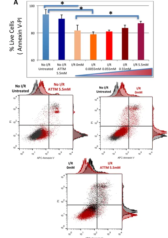
We conducted 4 experiments in order to evaluate the effects of ATTM on H9C2 cells, comparing by the use of Annexin V/ PI assay the percentage of viable cells with and without the drug ader 24hrs anoxia/ 2hrs reoxygenaEon. The results show that ATTM seems increase cell survival in a dose-dependent manner (Fig. 1) also reducing the percentage of late apoptoEc cells.
The postulated protecEve effect of ATTM on mitochondria is based on its anEoxidaEve effect, deacreasing reacEve oxygene species (ROS)
producEon by blocking CIV of the mitochondrial electron transport chain.
We then measured superoxide levels by MitoSOX Red in three separate experiments, using the same I/R protocol.
Histograms of FACS analysis showed marked increase of in mitochondrial fluorescence intensity of MitoSOX in H9c2 cells treated with AnEmycin A (Fig.2). QuanEtaEve measurements of the mean fluorescence intensiEes from the samples demonstrated fold increase in MitoSOX fluorescence intensity of 2 ± 0.5 with AntA, 0.7 ± 0.17 following 24h hypoxia/2h reoxygenaEon without drug and a significant reducEon with ATTM 5.5mM.
Conclusions In the current study we show that administraEon of ATTM at the Eme of reperfusion limits the extent of myocyte death in our in vitro I/R model. The study of the preservaEon of mitochondrial funcEon in myocardial cytoprotecEon is of paramount importance. Mitochondria play a central role in the development of reperfusion injury because the recovery of pH, oxidaEve stress, and calcium overload induce abrupt opening of mitochondria permeability transiEon pores (mPTPs), high conductance megachannels that are localized to contact sites between the inner and outer mitochondrial membranes (Baines CP, 2010). When opened, mPTPs permit communicaEon between the cytoplasm and the mitochondrial matrix. While low pH
during ischemia prevents opening of the megachannel, oxidaEve opening of the mPTP is criEcal to reperfusion injury. Depending on a complex balance among cellular inducers and antagonists, the open probability of the mPTP can be transient or long-lived.
H2S known to be a potent and reversible inhibitor of cytochrome c
oxidase (complex IV of the mitochondrial electron transport chain), in addiEon to the ability to modulate a whole organism metabolism inducing a
suspended animaEon-like state in mice, conducts also
cardioprotecEon by inhibiEng the mitochondrial respiraEon during reperfusion.
The inhibiEon of mitochondrial respiraEon has been shown (Chen Q et al 2006) to protect against myocardial I/R injury by limiEng the generaEon of reacEve oxygen species and diminishing the degree of mitochondrial uncoupling leading to decreased infarct size and preserved funcEon.
In conclusion ATTM shows protecEon against the increased ROS producEon and the drop of cell viability during Ischaemia/ Reperfusion Injury in our in vitro model. Because the majority of reacEve oxygen species derive from mitochondria both during ischaemia and reperfusion phases, leading to cell dysfuncEon and direct damage of lipids, proteins and DNA, sulfide’s ability to modulate metabolism is of parEcular interest due to the implicaEons this could have for modulaEon of inflammaEon and apoptosis, and oxidaEve stress reducEon.
The widespread synthesis of new sulfide’s donors with different releasing properEes reflects their wide range of applicaEons. ATTM characterized by a slow and consistent release of sulfide, is one the most effecEve sulfide donors moving towards clinical pracEce.
Meterial and Methods
Cell culture
Embryonic rat cardiomyoblast, H9C2, have been plated at density of ≈1 x 10⁶ in 75 cm2 flask, cultured at 37°C in 5% CO2 humidified
atmosphere in Dulbecco’s Modified Eagles Medium (DMEM 1X, Gibco, 41966-029) and supplemented with 10% heat-inacEvated foetal bovine serum (FBS, Sigma F9665). Cells have been passed regularly and sub-cultured at about 70% for 3 passages before experimental procedure.
Experimental protocol anoxia/reoxygenaOon
H9C2 cells have been plated 24hrs before the experiment at density of 60 x 103 in 12 well plates.
At the start of the experiment the culture medium have been replaced with 2 mL of DMEM (serum free, in order to mimic hypoxic state). Cells have than been placed into an anaerobic chamber saturated with 95% N2 and 5% CO2 for 24 hrs.
Simulated ischemia have been followed by a simulated reperfusion period during wich normoxic fresh culture medium have been added
at the anoxic medium, with and without MGC-0109 (0mM; 0.0055mM; 0.055mM; 0.55mM; 5.5mM) in a normoxic incubator for 120 min .
Flow cytometry
Cell viability apoptosis
Viability and apoptosis have been assessed using Annexin V- Propidium Iodide assay. Cells have been harvested, washed with PBS (no Ca/Mg) and resuspended in 100ul of APC-Annexin V (BD biosciences) binding buffer (150mM NaCl, 10mM HEPES pH7.4, 10mM CaCl2) and stained for 15min at RT in the dark. At the end of
the staining 200ul of binding buffer have been added in each sample. All data have been acquired on FACS Calibur using Cell Quest sodware and analyzed with Flow Jo (version IX).
Superoxide producOon
For the determinaEon of mitochondrial superoxide, cells have been loaded with MitoSOX™ Red mitochondrial superoxide indicator for live cells (ThermoFisher, M36008) prepared following the manufacturer’s protocol.