THE GLUTAMATE HYPOTHESIS OF
DEPRESSION: THE EFFECT OF STRESS AND
GLUCOCORTICOIDS ON GLUTAMATE
SYNAPSE AND THE ACTION OF
ANTIDEPRESSANTS
Doctorate thesis
Giulia Treccani
Coordinator: Prof. Filippo Drago
Tutor: Prof. Maurizio Popoli
Acknowledgements
I would like to thank many people, without whom my work would not be possible:
Prof. Maurizio Popoli, my tutor, who wisely supervised my work and gave me trust during the PhD program.
Prof. Filippo Drago who gave me the opportunity to take part to this PhD program that represented an important moment for my scientific growth.
Dr. Laura Musazzi whose experience, advices, supervision and help have been extremely important to me.
Prof. Giorgio Racagni, the Head of the Department of Pharmacological and Biomolecular Sciences, University of Milano. Drs. Daniela Tardito, Alessandra Mallei, Alessandro Ieraci, Mara Seguini, with whom I worked during the PhD program.
Dr. Carla Perego from University of Milano for all
immunofluorescence and total internal reflection fluorescence microscopy experiments.
Prof. Giambattista Bonanno and Dr. Marco Milanese from University of Genova, for all glutamate and GABA release experiments.
Prof. Antonio Malgaroli and Dr. Jacopo Lamanna from Scientific Institute San Raffaele, in Milano, for all the experiments of electrophysiology.
Prof. Maria Pia Abbracchio for her kind support after my graduation. Special thanks to Ella, Sandro, Marta, Carola, Davide, Chiara and Jan.
INDEX
Abstract ...
5Introduction
... 111 Depression ... 13
2 The stress response ... 16
2.1 The HPA axis ... 16
2.2 Corticosteroid receptors ... 19
2.2.1 Classical cytosolic corticosteroid receptors ... 21
2.2.2 Novel membrane-associated corticosteroid receptors ... 22
2.3 Rapid corticosteroid effects in the brain ... 23
2.4 Delayed effects of corticosteroids ... 25
2.5 Chronic corticosteroid exposure... 26
3 Stress as a risk factor for neuropsychiatric disorders ... 29
4 New approaches in neuropsychopharmacology: the glutamatergic hypothesis of depression ... 32
Chapter I
The action of antidepressants on the glutamate system:
regulation of glutamate release and glutamate receptors ... 35
Chapter II
C
orticosterone rapidly increases the readily releasable pool of glutamate vesicles in synaptic terminals of prefrontal and frontal cortex by acting on multiple local receptors ... 97General discussion and conclusions
... 159General list of references
... 167In the last years, a consistent number of clinical and preclinical studies have demonstrated that glutamatergic transmission has a primary role in the pathophysiology of mood and anxiety disorders (MADI). It has been shown that in depressed patients the levels of glutamate and its metabolites are altered in plasma and in selected brain areas and mRNA and protein levels of glutamate receptors are changed in brain areas. A number of preclinical studies on animal models of MADI have shown that different types of environmental
stress and glucocorticoid administration affect glutamate
transmission and exert structural brain remodeling in the same areas involved in human pathology. These effects of stress and glucocorticoids have been associated with the onset and exacerbation of neuropsychiatric disorders.
In previous studies we found that acute footshock (FS)-stress induces an increase of glutamate release from synaptosomes of prefrontal and frontal cortex (PFC/FC), via glucocorticoid receptor (GR) activation and SNARE complexes accumulation in synaptic membranes. Furthermore, we have demonstrated that the increase of glutamate release induced by acute stress is prevented by chronic antidepressants (ADs). Additional studies have also shown that ADs can regulate glutamate transmission through glutamate receptors; reducing the function of NMDA receptors, potentiating the function of AMPA receptors and affecting different subtypes of metabotropic glutamate receptors. Together, these findings have identified the glutamate synapse as a target for novel glutamatergic ADs.
Considering the importance of stress-induced alteration of presynaptic glutamate release in the pathophysiology of MADI, we
aimed to study whether the enhancement of depolarization-evoked glutamate release induced by acute stress was related to an increase of the readily releasable pool (RRP) of vesicles and whether this effect was mediated by a synaptic non-genomic action of corticosterone (CORT).
We found that FS-stress increased glutamate release evoked by hypertonic sucrose (which mobilizes exclusively the RRP), suggesting an increase in the RRP size. Then we found that this synaptic effect of stress was dependent on local CORT action. Indeed, CORT was able to directly affect the RRP size through the activation of GR and mineralcorticoid receptors (MR). The preincubation with RU486, a selective GR antagonist, and spironolactone, a selective MR antagonist, prevented the CORT-induced increase of RRP. Contrary to acute stress, CORT by itself did not promote vesicle fusion, since CORT application in vitro did not increase glutamate release evoked by depolarization in control synaptosomes, and did not affect excitatory post-synaptic potentials and paired pulse facilitation in medial PFC slices. Furthermore, we found that CORT increased vesicle mobilization towards the RRP via GR and MR activation, by using total internal fluorescence microscopy, a technique that allows the study of events occurring in a 100 nm-interval below the plasma membrane.
Finally we found that stress and CORT modulated synapsin I, a protein involved in vesicle mobilization and in vesicles docking, fusion and recycling at active zones. We found that both FS-stress and CORT induce an increase of synapsin I phosphorylation in synaptic membranes selectively at site 1. The preincubation with
both RU486 and spironolactone, prevented the CORT-induced synapsin I phosphorylation at site I, suggesting that this protein is involved in the pathway downstream of activation of the two receptors.
Together these results suggest that the increase of the RRP size induced by stress is promoted by a local action of CORT on synaptic receptors. We speculated that CORT is necessary to promote an increase of the RRP, but not sufficient to increase depolarization-dependent glutamate release, suggesting that additional mediators or neurotransmitters released during the stress response, are necessary to trigger vesicle release.
1- Depression
Depression is a complex neuropsychiatric pathology and the fourth leading cause of disability and disease worldwide. The World Health Organization projections indicate that depression will be the highest ranked cause of disease in the middle- and high-income countries by the year 2030 (Mathers and Loncar, 2006; Musazzi et al., 2012). The diagnosis of depression is subjective and essentially based on meeting criteria spelled out in the Diagnostic and Statistical Manual of Mental disorders (DSM-IV-TR). The symptoms that must persist for a 2-week period include depressed mood, anhedonia (reduced ability to experience pleasure from natural rewards), irritability, difficulties in concentrating, and abnormalities in appetite and sleep (neurovegetative symptoms) (Nesler et al., 2002, Krishnan and Nestler, 2008).
Despite the huge number of clinical and preclinical studies and considering the prevalence of depression and its social impact, there is a lack of knowledge regarding the pathophysiology of the disease. The first explanation is that many brain regions are involved in the pathology and responsible for the different symptoms of depression. Indeed, neocortex and hippocampus may mediate cognitive aspects of depression, such as memory impairments and feelings of worthlessness, hopelessness, guilt, doom and suicide. Striatum (ventral striatum and nucleus accumbens) and amygdala are involved in emotional memory and could mediate anhedonia and anxiety observed in patients. For neurovegetative symptoms, the involvement of the hypothalamus has been proposed (Nestler et al., 2002). The second reason comes from the complex etiology of the
pathology. It has been proposed that the depressive phenotype is the outcome of a combination of different genetic and environmental factors (Figure 1) (Wong and Licinio, 2011). Indeed, epidemiological and clinical studies support the hypothesis that a genetic predisposition associated with psychological perturbations, in particular during the neonatal period (see below), may result in a phenotype that is vulnerable to additional exposure to stress, leading to development of depression in adult life (Heim and Nemeroff, 2001; Heim et al., 2004).
Figure 1. Interaction between genetic and environmental factors at the basis of depression (from Wong and Licinio, 2001).
A known example of gene-environment interaction in depression is represented by the polymorphism in the promoter region of the serotonin transporter (5-HTT) gene (Caspi et al., 2003). The 5-HTT that is involved in the reuptake of serotonin at brain synapses, is blocked by some second-generation antidepressants drugs (selective serotonin reuptake inhibitors and serotonin and noradrenaline reuptake inhibitors). The 5-HTT is encoded by a single gene, which has an allelic variation in the promoter region: the shorter allele “s” is responsible of a reduced transcriptional efficiency, compared with the longer one “l”. Individuals with one or two copies of the “s” allele of the 5-HTT promoter exhibit more depressive symptoms, diagnosable depression, and suicidality in relation to stressful life events more frequently than individuals homozygous for the “l” allele (Caspi et al., 2003). Although the genetic background plays a key role in depression, environmental factors seem to work as the main triggering events (Caspi and Moffitt, 2006). A number of early-life stress events were related with the onset of mental disorders, particularly maternal stress and substance abuse during pregnancy, low birth weight, birth complications, deprivation of normal parental care during infancy, childhood physical maltreatment, childhood neglect, premature parental loss, exposure to family conflict and violence, stressful life events involving loss or threat, substance abuse, toxic exposures and head injury (Caspi and Moffitt, 2006).
For these reasons the study of the mechanisms activated within the brain in response to environmental factors, in particular to stress and
to the stress hormones corticosteroids, has received high relevance to understand the etiopathogenesis of depression.
2- The stress response
All living organisms are in a dynamic metabolic equilibrium, which is called homeostasis. This equilibrium could be altered by physical and psychological events that are known as “stressors”, which are defined as events or experiences that interfere with the ability of an individual to adapt and cope (de Kloet E. R. et al., 2005, Popoli et al., 2012). As a consequence, the stressor evokes a physiological stress response, which involves the release of hormones and mediators that can promote adaptation, when the response is promptly turned off. However, if the response is dysregulated, it may promote pathological processes (Popoli et al., 2012; McEwen, 1998). Two systems are primarily involved in the stress response: the first one involves the rapid activation of the sympathetic nervous system, which leads to the release of adrenaline and noradrenaline from the adrenal medulla. The parasympathetic nervous system is consequently activated to prevent overshooting (Joëls et al., 2012). The second system involved in the stress response is the HPA axis. 2.1 The HPA axis
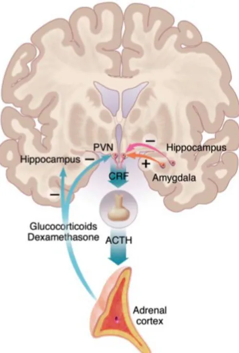
The main mechanism by which the brain reacts to stress is the activation of the HPA axis (Nestler et al., 2002) (Figure 2).
Figure 2. The HPA axis Parvocellular neurons of the PVN produce
CRH and vasopressin, which upon stress are released into the portal vessels. CRH and vasopressin reach the anterior pituitary gland, where their actions lead to secretion of ACTH into the circulation. In turn, this gives rise to the synthesis and release of glucocorticoids from the adrenal cortex, which exert a negative feedback on CRF and ACTH synthesis and release. The HPA axis is controlled by hippocampus (inhibitory effect) and amygdala (excitatory input) (from Nestler et al., 2002; see text for details).
Parvocellular neurons in the middle part of the paraventricular nucleus (PVN) produce corticotrophin-releasing hormone (CRH) and vasopressin, which upon stress are released in high amount from terminals at the median eminence into the portal vessels. Through these vessels, CRH and vasopressin reach the anterior pituitary gland, where their actions lead to secretion of adrenocorticotropin
hormone (ACTH) into the circulation (Joëls et al., 2012). In turn, ACTH stimulates the synthesis and release of glucocorticoids (cortisol in humans, corticosterone, CORT, in rodents) from the adrenal cortex. Blood concentration of adrenal glucocorticoids rise to peak level after 15-30 minutes and then decline slowly to pre-stress levels 60-90 minutes later (de Kloet et al., 2005).
The activation of the HPA axis is controlled by different brain areas including hippocampus and amygdala. The hippocampus exerts an inhibitory influence on hypothalamic CRF-containing neurons via a polysynaptic circuit, while amygdala gives a direct excitatory input (Nestler et al., 2002; see Figure 2). On the other hand, glucocorticoids exert a powerful negative feedback effect on the HPA axis, by regulating hippocampal and PVN neurons. In mammals, these hormones are released from the adrenal glands in a daily circadian cycle. Indeed, in humans the circulating levels of cortisol are low in the morning and progressively increase, reaching a peak at the end of the resting phase. However, high resolution blood sampling methods have shown that these circadian fluctuations overlay a highly oscillatory ultradian pattern, with a periodicity of approximately 60 minutes (Joëls et al., 2012). In animal models, it has been recently proposed that this ultradian hormone secretion
induces glucocorticoid receptor-mediated pulses of gene
2.2 Corticosteroid receptors
In the brain, glucocorticoids exert their function through the activation of two types of receptors: mineralocorticoid receptors (MR) and glucocorticoid receptors (GR) (Reul and de Kloet, 1985). The name of the two receptors derive from to the main peripheral process in which they are involved: mineral balance and gluconeogenesis, respectively. Classic MR and GR belong to the superfamily of nuclear receptors, which act as transcriptional factors. The human GR gene is constituted by nine exons, among which exons 2 to 9 encode for the GR protein (Joëls et al., 2012). Exons 1 and 9, can be alternatively spliced generating different mRNAs. Alternative splicing of the non-coding exon 1 produces different variants responsible for the region- and tissue-specific expression patterns, while the splicing of exon 9 produces two isoforms, GR and GR . Also the MR mRNA can be alternatively spliced, but less is known about the different isoforms of the receptor (Joëls et al., 2012). The expression of MR and GR varies in different brain regions and cells (Reul and de Kloet, 1985): GR are expressed both in neurons and in glial cells, and they are particularly expressed in PVN, in CA1 and dentate gyrus of hippocampus, in amygdala and in lateral septum, while MR are mainly expressed in neurons of hippocampus and lateral septum (Joëls et al., 2012). Moreover, GR and MR show a different affinity for endogenous hormones (corticosterone, cortisol and aldosterone) which in turn lead to a variation in MR and GR activity depending on hormone concentrations in the brain. MR have higher affinity for the
0.5 nM (Reul and de Kloet, 1985), while GR have a 10-fold lower affinity for CORT, and cortisol and many-fold lower for aldosterone. Considering the high affinity of MR for cotisol/CORT and the physiological higher level of cortisol/CORT in respect to aldosterone, in the brain MR in basal conditions are substantially occupied by cortisol/CORT, even during the intervals between ultradian pulses and in the absence of stress (Reul and de Kloet, 1985). In contrast, GR are partly occupied when corticosteroid levels are low and gradually become occupied when hormone levels rise (e.g. after stress) (Joëls et al., 2012).
As reported in table 1, the two receptors have different affinity also for synthetic compounds (selective and non-selective agonists and antagonists) used both in clinic and research.
Table 1. Pharmacological profile of human GR and MR
RECEPTOR AGONISTS (Affinity) ANTAGONISTS
GR Dexametasone>Triamcinolone> Prednisolone> Cortisol> Corticosterone= Deoxycorticosterone Mifepristone MR Deoxycorticosterone= Progesterone> Fludrocortisone> Aldosterone>Cortisol>Dexametasone Drospirenone Eplerenone Spironolactone
2.2.1 Classical cytosolic corticosteroid receptors
The most common mechanism of action of GR and MR implies the activation of genomic pathways. Circulating glucocorticoids diffuse through cell membranes and bind intracellular MR and GR. The receptors bind to a multiprotein complex of heat shock proteins (Hsp90, Hsp70, Hsp56, and Hsp40) and form activated receptor complexes. This induces a conformational modification allowing homodimerization or heterodimerization of activated receptors with the consequent dissociation from heat shock proteins. Dimerized MR and GR translocate into the nucleus, where they bind to consensus sequences in the promoter of responsive genes, directly modulating their transcription. On the other hand, GR or MR monomers can interact with other transcription factors (nuclear factor-kappa B (NFkB), activator protein 1 (AP1) or cyclic AMP response element-binding (CREB)), inhibiting their activity leading to transrepression (Datson et al., 2008). Genomic effects are slow in onset, and the initial physiological responses start at least with a 15 min-delay, and sometimes even hours after the beginning of stress (Groeneweg et al., 2012). However, a growing number of recent evidence demonstrated that, in neurons, corticosteroids, through GR and MR activation, have also fast effects, which have been observed within seconds to minutes, and which can not be explained through the activation of genomic pathways (Karst et al., 2005; reviewed in Groeneweg et al., 2012).
2.2.2 Novel membrane-associated corticosteroid receptors
To explain the rapid effect of corticosteroids, recent studies have shown that CORT also produces rapid non-genomic effects on neuronal excitability through the activation of the same GR and MR described above, but presumably associated to the plasma membrane. The presence of MR and GR has been shown by Western blot analysis in synaptosome extracts (Komatsuzaki et al., 2005; Wang and Wang, 2009) and at neuronal membranes using electron microscopy (Johnson et a., 2005; Prager et al., 2010). In particular, it seems that, in amygdala, MR are more expressed pre-synaptically, whereas GR are more expressed post-synaptically (Prager et al., 2010). The involvement of classical MR and GR in mediating rapid nongenomic effect on neuronal excitability was proved in different brain areas (see below) and using both antagonists and knockout mice for the MR and GR (Karst et al., 2005, 2010, see also above). However, the molecular mechanism by which the receptors interact or translocate to the plasma membrane is still unknown.
For another steroid receptor (the estrogen receptor ), it was demonstrated that it can be inserted into membrane through palmitoylation (Pedram et al., 2007). Starting from this evidence, it has been shown that GR have a conserved palmitoylation motif (Pedram et al., 2007), which could be responsible for the trafficking and binding of the receptor toward neuronal membranes.
However, other authors, starting from the experimental evidence that G-protein inhibitors can prevent many corticosteroid effects
independently from MR and GR activation, support the hypothesis that CORT can also bind to a G-protein coupled receptor (Di et al., 2009; Groeneweg et al., 2012, Olijslagers et al., 2008). Nevertheless, literature data are conflicting and this novel receptor has not been yet identified and cloned yet (Groeneweg et al., 2012).
2.3 Rapid corticosteroid effects in the brain
Different studies have been performed to understand the fast effects of CORT on neuronal excitability and in cognitive processes. The three brain regions in which these effects have been more characterized are hippocampus, amygdala and hypothalamus.
The high majority of studies in hippocampus focus on the rapid effect of CORT on neuronal excitability at pre- and postsynaptic level. It has been shown that CORT, after binding to presynaptic MR and activating the ERK1/2-MAP kinase signaling, promotes a rapid increase in the frequency of the miniature excitatory postsynaptic currents (mEPSCs), indicating an increase in the release probability of glutamate (Karst et al., 2005; Olijslagers et al., 2008). Moreover, it was shown that the hormone activates also postsynaptically located MR, which seem to be G-protein-coupled, which activate ERK1/2 that, in turn, via phosphorylation of potassium channels, inhibit the
repolarizing postsynaptic IA-currents. Since the current work to
dampen excitatory inputs, the CORT-induced decrease of IA-currents
could lead to an increase in the probability of the generation of the postsynaptic action potential (Olijslagers et al., 2008). It has been shown that, at postsynaptic level, CORT also changes glutamate
transmission increasing lateral movement of GluA2 subunits of the AMPA receptors, via MR activation, therefore modulating the plastic range of glutamatergic synapses through a change in the AMPA receptor surface trafficking (Groc et al., 2008).
As for hippocampus, neurons in the basolateral amygdala (BLA) show an increase in mEPSCs frequency dependent on MR after acute CORT exposure (Karst et al., 2010). Moreover, selectively in BLA, the effects of CORT are long lasting and greatly affect the responsiveness to CORT, since a subsequent pulse of CORT induces a GR-dependent decrease of the mEPSCs frequency. Similarly, it was shown that the acute in vitro stimulation with CORT of BLA slices from acute stressed animals (20 minutes restraint stress) activates an endocannabinoid signaling pathway that suppresses mEPSC frequency via GR-dependent pathway (Karst et al., 2010).
On the contrary, in parvocellular neurons of the PVN, the acute exposure to CORT decreases the release probability of glutamate, via activation of GR and involvement of retrograde endocannabinoid signaling. At the same time, CORT enhances GABAergic miniature inhibitory postsynaptic currents (mIPSCs) through retrograde nitric oxide signaling (Di et al., 2003; Di et al., 2009).
Compelling evidence suggests that the fast effect of glucocorticoids also plays a role in cognitive processing. Interestingly, it has been shown that a GR agonist affects memory functions mainly acting on the medial PFC (mPFC), via the activation of a membrane-bound steroid receptor, increasing levels of cAMP-dependent protein kinase
(PKA) through modulation of noradrenergic activity (Barsegyan et al., 2010).
2.4 Delayed effects of corticosteroids
Although there is an increasing knowledge regarding the non-genomic effects of CORT, the high majority of studies reported in literature concerns the gene-mediated effect of CORT. These effects have been particularly investigated in hippocampus, BLA and mPFC. As an example, in hippocampal slices, a pulse of CORT enhances the amplitude but not the frequency of mEPSCs recorded 1-4 hours after corticosteroid exposure, via cytosolic GR activation (Karst and Joëls, 2005; Martin et al., 2009). The effects on mEPSCs amplitude were shown to peak between 150 and 200 minutes and were not seen earlier than 1 h after CORT delivery (Karst and Joëls, 2005; Martin et al., 2009). A similar enhancement in mEPSC amplitude has been also reported for prelimbic neurons in the PFC after acute stress exposure. Indeed, it has been shown that, more than 1 hour after cessation of stress there is an increase in NMDA and AMPA receptors-mediated synaptic currents, sustained for 24 hours and mimicked by short-term CORT treatment in vitro. The delayed enhancement of glutamate transmission induced by acute stress and CORT was related with an increased surface expression of NMDARs and AMPARs at the postsynaptic plasma membranes, consequent to intracellular GR activation, and induction of serum- and glucocorticoid-inducible kinase (SGK) and Rab4 (Yuen et al., 2009; Yuen et al., 2011).
2.5 Chronic corticosteroid exposure
As reported above, acute corticosteroid exposure (minutes to hours) modulates glutamate transmission and enhances synaptic function. On the other hand, chronic exposure to corticosteroids (days to weeks) mediates adaptative plasticity, involving spine synapse turnover and dendritic shrinkage (Popoli et al., 2012; McEwen, 1999). These effects are strongly depending from the considered brain regions: many studies have been performed in hippocampus, amygdala, and to a minor extent, in PFC. For example, it has been shown that chronic unpredictable stress (CUS), an animal model of depression, causes a reduction in the length and branching of apical dendrites and decreases the number and function of spine synapses of pyramidal neurons in layer V of the mPFC (Li et al., 2011). Other chronic stress paradigms (such as restraint stress) cause similar reductions in dendrite complexity and spine density in PFC neurons and CA3 pyramidal cells of the hippocampus (Duman and Aghajanian, 2012; Joëls et al., 2012). Chronic stress also suppresses adult neurogenesis in the adult hippocampus and significantly reduces the number of glial cells in the mPFC (Duman and Aghajanian, 2012). On the other hand, increased dendritic complexity has been reported in principal cells of BLA and in the orbital cortex (Joëls et al., 2012, Roozendaal et al., 2009) (Figure 3), suggesting a different role of chronic corticosteroid exposure in these areas.
These morphological alterations seem to be reversible with the cessation of stress (Conrad et al., 1999; Radley et al., 2005) except
for the BLA, where changes persisted for at least 30 days after chronic stress (Vyas et al., 2004). These processes were also linked to the age of animals, indeed the mPFC of aged animals show a slower recovery than for younger animals (Bloss et al., 2010).
Structural plasticity process can also be activated after acute stress in amygdala. A single traumatic stressor causes BLA neurons to grow new spines over the next 10 days, with no growth of dendrites (Mitra et al., 2005). Moreover, a single, high dose of injected CORT causes delayed dendritic growth over the next 10 days (Mitra and Sapolsky, 2008), even if no data are available on possible effects on the number of spines.
Figure 3. Structural and morphological changes induces by stress in prefrontal cortex, hippocampus and amygdala (from
These morphological changes are expected to affect neuronal activity. In particular, in hippocampus, it was shown that chronic overexposure to stress hormones causes a reduction in the ability to induce or maintain long-term potentiation (LTP) and enhances the probability to induce long-term depression (LTD) (Joëls et al., 2012). Two different models have been proposed to explain the complex effects of chronic stress on neuronal morphology and excitability in hippocampus. The first one proposes that, at least in the CA3 hippocampal area, the increase of glutamatergic transmission consequent to chronic stress exposure leads to excitotoxic effects inducing dendritic atrophy and reduction in spine number (McEwen, 1999). This mechanism could be interpreted as a “protective” mechanism by which cells, through reduction in the number of synaptic contacts, tries to counteract the enhanced excitatory input. In support of this theory, treatment of animals with NMDA receptor blockers was found to prevent dendritic remodeling in the CA3 area of HC and in mPFC (McEwen and Magarinos, 1997; Christian et al., 2011; Li et al., 2011). According to this theory, enhanced excitatory transmission would precede dendritic retraction rather than occur simultaneously or as a consequence (Joëls et al., 2012).
Conversely, the second model, mainly based on experimental and mathematical evidence, suggests that dendrite remodeling and altered synaptic excitability, observed in the hippocampus after chronic stress, lead to atrophy-induced synaptic hyper excitability that could tilt the balance of plasticity mechanisms in favor of synaptic potentiation over depression. Indeed, it has been shown that chronic stress enhances NMDA receptor-mediated EPSCs in HC
CA3 neurons (Kole et al. 2002; 2004) and that DG granule cells from chronic stressed animals, when exposed to CORT in vitro, show an increase in the amplitude of AMPA receptor-mediated synaptic currents (Karst and Joëls, 2003). These larger currents were not found in cells after chronic stress only or acute CORT treatment. Similarly, it has been demonstrated using biophysical models of hippocampal CA3 neurons that dendritic atrophy leads to an amplification of intrinsic and synaptic excitability, suggesting that stress may impair learning and memory through a facilitation of intrinsic synaptic excitability and the consequent imbalance of bidirectional hippocampal synaptic plasticity (Narayanan and Chattarji, 2010).
The effects of chronic stress and corticosteroid exposure have been recently studied also in PFC. In this area, it has been shown that while acute stress enhances glutamate transmission and related cognitive function, chronic stress impairs these processes. Indeed, 5 to 7 days of restraint or unpredictable stress in young rats causes a reduction of both AMPA and NMDA receptor-dependent synaptic responses in pyramidal PFC cells, in association with ubiquitin/proteasome mediated degradation of selective subunits (Yuen et al., 2012).
3 Stress as a risk factor for neuropsychiatric disorders
The different effects of CORT depending on the age of the animal, the time of exposure and the duration and type of stressor experienced, underline how difficult is to clearly understand the
mechanisms by which CORT is able to modulate neuronal excitability and plasticity. A number of preclinical studies suggest that stress can produce in animals some of the cognitive and emotional disturbances that are also observed in patients with depression. Several animal models involving different forms of stress have been used for studying the etiopathogenesis of depression. One of the more recent models is chronic unpredictable stress CUS, where animals are exposed to a variable sequence of mild, unpredictable stressors (Willner, 2005). CUS was shown to increase blood levels of CORT and behavioral abnormalities, including core symptoms of depression, such as anhedonia (Li et al., 2011).
The importance of the functionality of the HPA-axis comes also from animal models in which components of the HPA axis were modified by mutagenesis (de Kloet et al., 2005). For example, GR-knockout mice generated by deletion of limbic GR (except for the hypothalamic PVN), exhibit a robust depression-like phenotype (Boyle et al., 2005). It was also clearly demonstrated that the correct functionality of the HPA-axis plays an important role in the etiology of depression also in humans. First, it has been shown that several individuals with depression exhibit hyperreactivity of the HPA-axis, even before the onset of any clinical symptom (Joëls et al., 2010). Second, several depressed patients show elevated circulating corticosteroid levels (especially during the circadian trough) and relative insensitivity to dexamethasone-induced suppression of the HPA-axis. The normalization of these functions generally precedes relief of depressive symptoms and the degree of normalization predicts the probability of relapse (Joëls et al., 2011). Third, it has been shown
that antiglucocorticoids (given in addition to antidepressants) to patients with psychotic depression accelerate and increase chances of successful treatment (Joëls et al., 2011). Since the functionality of corticosteroid hormones and receptors, as well as the HPA axis in general can be affected by mutations, the genetic background may also predict the probability that individual patients will respond to pharmacotherapy. For instance, it was demonstrated that a specific mutation in the FKBP5 gene confers a faster response to antidepressants compared with the wild type (Binder et al., 2004). This gene encodes for a co-chaperone of HSP90 and contributes to the folding of cytosolic GR, determining the affinity of cortisol for its receptor.
Other genetic factors might also cause individuals to be resilient in the developing of affective disorders. An example is the polymorphism in the ER22/23 EK allele, which is located at the beginning of exon 2 of the GR gene and confers a healthier metabolic profile and a better cognitive function than the general population. The polymorphism is also associated with a better treatment outcome in individuals with depression (de Kloet et al., 2005; van Rossum and Lamberts, 2004). It remains a challenge for the future to study the consequences of these genetic variants on corticosteroid-dependent modulation of neuronal activity (Joëls et al., 2012).
4 New approaches in neuropsychopharmacology: the glutamatergic hypothesis of depression
As previously shown corticosteroid and stress exert crucial effects on neuronal excitability and brain functions through rapid and delayed mechanism. Moreover, it has been demonstrated that the modulation of excitatory, glutamatergic transmission by CORT plays a critical role in the stress response and has a specific effect depending on the brain region involved. These effects on glutamate transmission are predominant in hippocampus, amygdala and PFC, all regions involved in depression. Abnormal function of glutamatergic transmission has been reported also in neuropsychiatric diseases, including depression. Indeed, it has been shown that the levels of glutamate and its metabolites are altered in plasma and in selected brain areas of patients affected by mood and anxiety disorders (Hashimoto et al., 2007; Küçükibrahimoglu et al., 2009, Yüksel and Öngür, 2010). Moreover, post-mortem studies have found alterations of mRNA and protein levels of glutamate receptors in brain areas from depressed individuals (Beneyto et al., 2007). Several studies have also investigated the role of glial cell that participate in the uptake, metabolism and recycling of glutamate and that have been proposed to be involved in the alterations of glutamate neurotransmission observed in depression. It has been shown that the expression of the glial excitatory amino acid transporters was reduced in individuals with mood disorders (Choudary et al., 2005), and that glial cell number and/or density is reduced in the brain
regions with glutamatergic predominance from patients with major depression (Rajkowska et al., 1999).
All these findings suggest that mood disorders are associated with
abnormal function and regulation of the glutamatergic
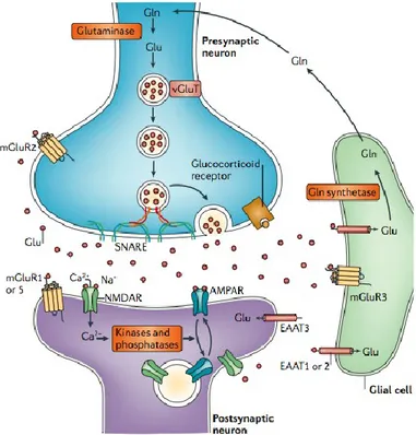
neurotransmitter system (Sanacora et al., 2008). For this reason the study of the mechanisms by which glutamate transmission can be modulated, particularly by stress and glucocorticoids, could play an important role in the development of new fast-acting antidepressants. Considering the glutamatergic synapse as a tripartite structure (Figure 4), corticosteroid and stress can affect different mechanisms including glutamate release, glutamate receptors and glutamate clearance and metabolism (Popoli et al., 2012). The identification of the mechanisms by which corticosteroids regulate the functions of the glutamate synapse and the mechanisms by which antidepressants can modulate glutamate transmission will provide the opportunity to use novel pharmacological interventions to improve and preserve neural function and to treat and possibly prevent some psychiatric disorders (Popoli et al., 2012).
Figure 4. The glutamate tripartite synapse The figure shows the
organization of glutamate synapse. Within the synapse glutamatergic transmissions is controlled at different points: glutamate release, postsynaptic receptor trafficking and function, transporter-mediated uptake and recycling of glutamate through the glutamate-glutamine cycle (from Popoli et al., 2012)
Biological Psychiatry (in press)
The action of antidepressants on the glutamate system: regulation of glutamate release and glutamate receptors
Laura Musazzi*, Giulia Treccani*, Alessandra Mallei, Maurizio Popoli
Laboratory of Neuropsychopharmacology and Functional Neurogenomics - Department of Pharmacological and Biomolecular Sciences and Center of Excellence on Neurodegenerative Diseases, University of Milano, Milano, Italy
* These authors contributed equally to this work
Address correspondence to: Maurizio Popoli, PhD
Laboratory of Neuropsychopharmacology and Functional
Neurogenomics-Department of Pharmacological and Biomolecular Sciences, University of Milano
Via Balzaretti 9 - 20133 Milano (Italy) phone: +39 02 5031 8361
fax: +39 02 5031 8278 email: [email protected] Keywords
Antidepressant, Glutamate neurotransmission, Glutamate release, Glutamate receptor, ketamine, drug development.
No. of words (Abstract): 249 No. of words (Article body): 3998 Number of figures: 2
Number of tables: 1 Suppl. Information: 0
Abstract
Recent compelling evidence has suggested that the glutamate system is a primary mediator of psychiatric pathology and also a target for rapid acting antidepressants. Clinical research in mood and anxiety disorders has shown alterations in levels, clearance and metabolism of glutamate, and consistent volumetric changes in brain areas where glutamate neurons predominate. In parallel, preclinical studies with rodent stress and depression models have found dendritic remodeling and synaptic spines reduction in corresponding areas, suggesting these as major factors in psychopathology. Enhancement of glutamate release/transmission, in turn induced by stress/glucocorticoids, seems crucial for structural/functional changes. Understanding mechanisms of maladaptive plasticity may allow to identify new targets for drugs and therapies.
Interestingly, traditional monoaminergic-based antidepressants have been repeatedly shown to interfere with glutamate system function, starting with modulation of NMDA receptors. Subsequently, it has been shown that antidepressants reduce glutamate release and synaptic transmission, in particular it was found antidepressants prevent the acute stress-induced enhancement of glutamate release. Additional studies have shown that antidepressants may partly
reverse the maladaptive changes in synapses/circuitry in stress and depression models. Finally, a number of studies over the years have shown that these drugs regulate glutamate receptors, reducing the function of NMDA receptors, potentiating the function of AMPA receptors, and, more recently, exerting variable effects on different subtypes of metabotropic glutamate receptors. The development of NMDA receptor antagonists has opened new avenues for glutamatergic, rapid acting, antidepressants, while additional targets in the glutamate synapse await development of new compounds for better, faster antidepressant action.
Introduction
Glutamate neurotransmission dysfunction is increasingly considered a core feature of stress-related mental illnesses. For over half a century the conceptual framework of research on these disorders has been dominated by the monoamine hypothesis, on which most of the drugs developed for clinical therapy are based. Although it was not acknowledged as a neurotransmitter until the early 1980s (1), glutamate has been later recognized as the major excitatory neurotransmitter in the brain, with glutamatergic neurons representing about 80% of total neurons in neocortex (2,3). In the
past decade it has become increasingly acknowledged that maladaptive changes in the structure and function of excitatory/inhibitory circuitry (representing the vast majority of neurons and synapses in brain) have a primary role in the pathophysiology of mood and anxiety disorders (MADI), particularly major depression (3-8). Hereafter, we briefly summarize the large and increasing body of evidence that links dysfunction in the glutamate system with MADI.
Evidence linking dysfunction in the glutamate system with mood and anxiety disorders
Clinical evidence: glutamate levels, magnetic resonance and neuroimaging studies
First, changes in glutamate levels have been found in plasma, cerebrospinal fluid (CSF) and brain of MADI patients. A number of studies reported elevated glutamate content, and decreased plasma glutamine/glutamate ratios, in the plasma of depressed patients (9-11). CSF studies found higher glutamate content in depressed patients (12) and lower glutamate in patients with refractory affective disorder (13). Interestingly, although an early study did not find any significant difference in frontal cortex (FC) (14), recent postmortem
studies showed significant increases in glutamate levels in FC and dorsolateral prefrontal cortex (PFC) of depressed and bipolar patients, respectively (15,16).
Second, in vivo proton magnetic resonance spectroscopy studies of MADI have reported consistent alterations in Glx, a combined measure of glutamate, glutamine and GABA (see 17,18 for reviews).
Overall, these results suggested abnormalities in
glutamate/glutamine/GABA cycling are involved in pathophysiology of MADI, although they do not provide information as to how glutamatergic transmission is changed (4,15,19,20).
Third, a large number of clinical neuroimaging studies of MADI have consistently shown regional volumetric changes in brain areas where glutamate neurons/synapses predominate, such as hippocampus (HPC), cortical regions (including PFC) and amygdala (see for meta-analyses: 21,22). In HPC, volumetric reduction was found particularly associated with repeated depressive episodes (23,24), suggesting correlation with illness duration. Volumetric reduction has been found also for cortical areas, while volumetric enlargement, at least in the early course of illness, has been found for amygdala, suggesting the volume of this stress-related area is dependent on the phase of illness (22).
Preclinical evidence showing structural/functional changes in brain
Although the reasons for volumetric reductions of HPC and PFC in MADI have not yet been clearly identified, it has been proposed that atrophy/remodeling of dendrites is a major factor (Fig. 1) (5-7,25,26). The evidence for this hypothesis comes mostly from experiments with rodents. Numerous studies with stress paradigms and models of depression (for a discussion see: 6,26) have shown that repeated stress exposure, in some cases even a few stress episodes (27), induce atrophy, retraction and simplification of dendritic arbor in CA3 HPC region and medial PFC (28-31). This phenomenon was detected in regions corresponding to those where volumetric reductions have been observed in humans. Dendritic remodeling in rodents was found to be reversible after cessation of stress (32), and antidepressants or mood stabilizers (as well as physical exercise) were shown to partly reverse the structural changes induced by stress (see below). However, the connection between dendritic remodeling and volumetric changes is at present inferential, because it links together complementary evidence obtained from clinical and preclinical studies, and needs further evidence (Fig. 1). Additional factors likely to be primarily involved in brain volumetric changes are
loss of glial cells (particularly in cortical areas) and impairment of neurogenesis (limited to HPC) (33,34).
The role of glucocorticoids and their effects on glutamate release/transmission
A primary role in stress-related dendritic remodeling has been attributed to glucocorticoid hormones, major mediators in the stress response, and to changes in glutamate release/transmission, in turn potently regulated by glucocorticoids. Numerous lines of evidence have shown that both stressors/glucocorticoid treatments acutely increase glutamate release in HPC, PFC/FC, and amygdala (35-40; see 6,26, for a discussion). Interestingly, it has also been shown that enhancement of miniature excitatory postsynaptic currents (EPSCs) in CA1 neurons by corticosterone is mediated by a mineralocorticoid receptor (MR), while acute stress-induced enhancement of depolarization-evoked glutamate release in PFC/FC is mediated by a glucocorticoid receptor (GR) (37,38). Accordingly, it has been argued that stress-induced rise of corticosterone and binding to synaptic MR/GR in turn enhance glutamate release and transmission (3,41). Therefore, combined evidence has suggested that changes in glutamate release/transmission, elicited by stress/glucocorticoids,
with time induce dendritic remodeling and morphological changes. Remodeling of synapses and neuronal networks is a constant, dynamic and physiological process (42). It can be envisaged that, when glutamate neurotransmission is repeatedly and abnormally overactivated, building up excessive synaptic or extrasynaptic levels of glutamate, physiological mechanisms of synaptic/dendritic remodeling can be transformed into maladaptive changes (7,25,26). However, the effects of chronic stress on glutamate release have been little investigated thus far; future studies are warranted, particularly to investigate how the response to acute stressors is modified by a previous period of chronic stress. The reduction of dendritic arbor by stress corresponds to a reduction of dendritic spines and synaptic boutons, with a shift in PFC from mature mushroom-type to thin- and stubby-type spines (43). Quantitative and qualitative changes in the synapses/circuitry morphology correspond to marked changes in synaptic transmission, likely involved in the ‘pathological’ behavioral phenotypes observed in stress models. Understanding this maladaptive plasticity may allow to gain insight into the processes whereby adverse environmental factors contribute to the pathogenesis of MADI (3,5), and may open new avenues for the development of novel therapies.
It is noteworthy that monoaminergic-based antidepressants have been repeatedly shown to interfere with the glutamate system, starting with the observation that they modulate the function of the N-methyl-D-aspartate receptor (NMDA-R) for glutamate (44,45). A growing number of studies in recent years have investigated the effects of antidepressants on the glutamate system, unveiling previously unknown effects, likely involved in their therapeutic action (6).
In the following sections we analyze in detail the action of antidepressants on the glutamate system, in particular the release of glutamate and the different classes of glutamate receptors (Fig.2). Antidepressant agents and the glutamate system
Antidepressants modulate synaptic transmission mediated by AMPA and NMDA receptors
A number of studies have shown that chronic antidepressant treatments of rats reduce excitatory synaptic transmission. Several drugs, including imipramine, citalopram, tianeptine (a neuroprotective antidepressant, serotonin reuptake enhancer), and electroconvulsive shock, attenuated synaptic transmission in FC (46,47). The magnitude of both α–amino-3-hydroxy-methyl-4-isoxazole propionic
acid receptor (AMPA-R) and NMDA-R dependent components of the field potential were reduced, the latter to a larger extent. These findings were in line with previous studies, which showed that antidepressants dampen NMDA-R function (see below). It was also found that imipramine reduced mean frequency and amplitude of spontaneous EPSCs in cortical pyramidal neurons, suggesting reduced presynaptic glutamate release (47).
Antidepressants dampen presynaptic glutamate release
As suggested by the reports showing that chronic antidepressants reduce glutamatergic transmission, a few studies have shown that these drugs actually reduce the presynaptic release of glutamate in HPC and cortical areas. An early study showed that both acute and chronic imipramine and phenelzine significantly reduced glutamate release evoked by depolarization. In PFC, but not striatal slices (48). In a later study, glutamate/GABA release was measured from HPC purified synaptosomes in superfusion, a technique allowing ex vivo, quantitative detection of endogenous neurotransmitter (3,49). Chronic antidepressant treatments (fluoxetine, desipramine, reboxetine) significantly reduced depolarization-evoked release of glutamate (50). Basal (non-stimulated) glutamate release and
basal/depolarization-dependent GABA release were not changed by drugs. Also acute treatments did not change glutamate/GABA release. Adaptive changes in presynaptic machinery were found altered by antidepressants. In synaptic membranes of drug-treated
rats, phosphorylation of -calcium/calmodulin-dependent protein
kinase II ( CaMKII), previously involved in the mechanism of
antidepressants (51,52), was markedly decreased. CaMKII
dephosphorylation in turn reduced its binding to syntaxin-1, one of the three SNAREs, and increased syntaxin-1/Munc-18 interaction. As a result, this shift of binding reduces the assembly of syntaxin-1 into the SNARE complex and depolarization-evoked glutamate release (50,53).
A further study showed that acute in vitro application of fluoxetine to cerebrocortical synaptosomes reduced 4-aminopyridine–evoked glutamate release, by inhibition of P/Q-type calcium channels (54). However, it is difficult to correlate the in vitro drug effect on synaptosomes with the outcome of chronic drug treatments in animals.
These results suggested that reduction of activity-dependent glutamate release by antidepressants could exert a protective action
when the synapse is overactivated by stress-related mechanisms. Regarding the mechanism, it was speculated that chronic antidepressants down-regulate monoamine autoreceptors but also partly affect NA and 5-HT heteroreceptors on glutamatergic terminals, reducing glutamate release (see 55 for a discussion). Moreover, the effects of antidepressants on synaptic glutamate could also be related with a direct modulation of the expression levels of glial-specific glutamate transporters, regulating the neurotransmitter homeostasis (56).
Antidepressants prevent the enhancement of glutamate release induced by acute stress
As addressed above, acute stress and glucocorticoids in rodents consistently induced enhancement of glutamate release in select brain areas. On the other hand, chronic antidepressants reduce presynaptic glutamate release in basal conditions. If stress-induced enhancement of glutamate release/transmission is a causal factor in the induction of synaptic/dendritic remodeling, and antidepressants are able to modulate glutamate release, this effect could be part of the antidepressant action. One of the first studies investigating the effect of psychotropic drugs on the efflux of glutamate induced by
stress showed that diazepam, administered 30 min prior to tail pinch stress, reduced the stress-induced glutamate efflux in HPC and PFC. This effect, explained with the allosteric stimulation of GABA-A receptors, is likely related with the acute anxiolytic action of benzodiazepines (57). However, chronic treatment with lorazepam was shown to reduce glutamate release from PFC slices in superfusion and to increase glutamate release in HPC slices (58), indicating that chronic effects of benzodiazepines can be different from acute effects.
A more recent microdialysis study showed that acute tianeptine or fluoxetine exert different effects on the increase of glutamate efflux induced by acute restraint stress in basolateral amygdala (BLA) and central amygdala (CA) (39). While acute tianeptine completely abolished the stress-induced increase of extracellular glutamate in the BLA, fluoxetine increased basal extracellular glutamate before stress application and potentiated glutamate levels in BLA/CA when stress was applied. Although the acute effect of tianeptine on stress-induced increase of glutamate awaits explanation, the result with fluoxetine provided a potential mechanism for the anxiogenic properties of selective serotonin-reuptake inhibitors, often observed in the initial phase of clinical treatment. The same authors showed
that also acute agomelatine (a new antidepressant with a partially non-monoaminergic mechanism) blocks the stress-induced increase of extracellular glutamate in BLA, CA and HPC (59).
It was recently shown that several chronic antidepressants (fluoxetine, desipramine, venlafaxine, agomelatine) significantly block the enhancement of glutamate release from PFC/FC synaptosomes in superfusion induced by acute footshock (FS) stress (38). Patch-clamp recordings in PFC slices prepared from FS-stressed rats showed that previous chronic desipramine normalized the increase of spontaneous EPSCs amplitude, as well as the marked reduction of paired-pulse facilitation (a measure of glutamate release) and its calcium-dependence. It was also shown that antidepressants did not block the stress-induced rise of circulating corticosterone, which increased glutamate release via GR. Moreover, it was found that FS-stress induced an increase of presynaptic SNARE complexes and of the pool of vesicles ready for release (RRP) in presynaptic membranes (38,60). These findings suggested that the action of antidepressants must be downstream of corticosterone rise and RRP increases. The novelty of this study was that it showed for the first time with the superfusion technique that chronic antidepressants can modify the acute stress response. This newly discovered effect of
antidepressants likely represents a component of the therapeutic effect, particularly the anxiolytic action. Further research is warranted to understand whether other classes of drugs share this long-term action of antidepressants, and whether compounds reducing stress-induced glutamate release in cortical areas could be efficient anxiolytic drugs.
Antidepressants and mood stabilizers reverse stress-induced reduction in dendritic arborization
If antidepressants reduce the enhancement of glutamate release/transmission induced by stressors, they should also be able to prevent or reverse the dendritic remodeling induced by repeated stress. Indeed, a number of studies have shown that antidepressants or mood stabilizers may reverse the structural changes observed in stress/depression models. Both tianeptine and lithium prevented dendritic remodeling caused by chronic restraint stress in HPC (29,61). A different animal model of depression, the olfactory bulbectomy, displays reduced spine density in CA1, CA3 and DG HPC areas; chronic amytriptiline reversed the spine reduction in all three areas, while mianserin reversed the reduction in DG only (62). More recently, subchronic (6 days) administration of desipramine
was shown to rescue the reduction of spines in HPC and the escape deficit in the Learned Helplessness (LH) model of depression (63). Moreover, different antidepressants have been shown to reverse the reduction in dendritic arborization and spine density induced by chronic mild stress (64).
Antidepressants regulate glutamate receptors
Glutamate released in the synaptic cleft can bind ionotropic postsynaptic glutamate receptors, including AMPA-R, NMDA-R and kainate receptors, or metabotropic glutamate receptors (mGluRs), localized at both pre- and postsynaptic sites. A number of studies have consistently shown that chronic antidepressants selectively regulate glutamate receptors expression and function (Tab. 1). In particular, it was demonstrated that antidepressants differentially modulate ionotropic receptors, leading to a reduction of NMDA-R function and a relative potentiation of AMPA-R-mediated transmission. Different antidepressants were shown to reduce radioligand binding to rat cortical NMDA-R (44,65) and decrease both mRNAs and protein expression levels of NMDA-R subunits (55,66). It was also shown that early-life stress increased the expression of GluN1 subunit of NMDA-R in HPC of the Flinders rat model of
depression, while chronic mild stress increased both transcriptional and protein expression of NMDA-R subunits in HPC. Chronic treatment with respectively escitalopram and duloxetine was able to normalize these alterations (67,68).
On the other hand, antidepressants also exert a positive regulation of AMPA-R expression and phosphorylation. Chronic antidepressants increased immunoreactivity of AMPA-R subunits GluA1 and GluA2/3 in both rat HPC and cortical areas (69-71): fluoxetine increased GluA1 phosphorylation at Ser845 (associated with increased GluA1 insertion), and imipramine increased synaptic expression of GluA1 in HPC (72,73). Intriguingly, it was also recently demonstrated that tianeptine, in addition to increasing GluA1 synaptic expression, also stabilizes membrane exposure of AMPA-R, by reducing their surface diffusion, favoring long-term synaptic plasticity (74).
Moreover, chronic antidepressants also induce specific changes in the expression and function of individual mGluR subtypes (75). mGluRs are separated into three groups based on sequence homology, second messenger coupling, and agonist selectivity. Group I mGluRs (mGlu1/mGlu5) are postsynaptic excitatory receptors positively coupled with phospholipase C. Group II mGluRs (mGlu2/mGlu3) are autoreceptors localized mainly at extrasynaptic
sites on glutamatergic neurons and modulate glutamate release (76). Group III mGluRs (mGlu4/mGlu6/mGlu7/mGlu8) are heterogeneous: mGlu4, mGlu7 and mGlu8 are mainly presynaptic and function as autoreceptors to inhibit glutamate or GABA release, while mGlu6 is a postsynaptic receptor mediating synaptic transmission at retinal cells. Repeated electroconvulsive therapy and prolonged imipramine treatment were shown to increase significantly the expression of group I mGluRs in HPC (77). It was argued that upregulation of group I mGluRs may reflect a compensatory mechanism caused by receptor subsensitivity consequent to antidepressant treatment. Moreover, chronic imipramine increased the expression of mGlu2/3 subunits in the rat HPC, cortex, caudate nucleus and nucleus accumbens (78).
Finally, mGlu7 (but not mGlu4) expression levels were decreased by citalopram, but not imipramine, both in rat HPC and cortex (79). It was recently found that chronic fluoxetine did not change mGlu4 expression in HPC of control rats, while restored mGlu4 levels reduced by early-life stress. On the other hand, mGlu7 and mGlu8 were up-regulated by fluoxetine in control rats but not in stressed rats (80).
Targeting glutamate ionotropic receptors in the treatment of mood disorders
Given the role of impaired regulation of glutamatergic neurotransmission in the pathophysiology of MADI, and the long-term
modulation of synaptic function induced by traditional
antidepressants, growing interest addressed the development of drugs directly targeted on glutamatergic system. This new pharmacological approach mainly aims at shortening the onset of therapeutic effects, reducing the number of non-responders, and avoiding the side effects of monoaminergic antidepressants. Indeed, early studies showed that NMDA-R antagonists exert antidepressant-like action, paving the way for more recent studies on this class of drugs. Moreover, positive modulators of AMPA-R have shown antidepressant effects. Also a number of mGluR modulators were proposed as alternative approach for regulation of glutamatergic function.
A number of recent, exciting studies reported rapid antidepressant action, both in preclinical and clinical studies, of low doses of high-affinity non-competitive NMDA-R antagonists (particularly the dissociative anesthetic ketamine). It has been shown that a single subanesthetic dose of ketamine induces rapid (within hours) and
sustained (up to 1 week) antidepressant efficacy in treatment-resistant patients, although the actual response to ketamine might be longer (81-83). These findings, particularly robustness, rapidity and durability of the antidepressant effect, were unanticipated. Ketamine appears to target directly the core depressive symptoms such as sad mood, suicidality and helplessness, rather than inducing a nonspecific mood-elevating effect.
The drug has also been shown antidepressant properties in different rodent models of depression. It rapidly reversed depressive-like behavior in two well-established models, the forced swim test (FST) and LH, as well as in a model of anxiety, the novelty suppressed feeding test (84), and reversed anhedonic/anxiogenic behaviors and functional/morphological alterations induced by chronic unpredictable stress (CUS) (85). However, the long-lasting effect of ketamine was
not replicated in a different study, where a higher dose (50-160
mg/kg) was used (86).
Although the mechanism has not been completely elucidated, it was consistently found that ketamine rapidly activates the mammalian target of rapamycin (mTOR) pathway, inducing a sustained increase in the expression levels of synaptic proteins and in the number of excitatory spine synapses in PFC. This suggests that the behavioral
effects of ketamine could be related with changes in synaptic plasticity and synaptogenesis (84,85). Somewhat comparable effects have been described after chronic treatments with traditional antidepressants; it is not know at present whether ketamine induces these restoring effects by-passing the monoamine-mediated effects or by a completely different mechanism.
Unfortunately, the adverse effects of ketamine, especially psychotomimetic consequences and cognitive impairment, limit a wide clinical use of this drug (83). Therefore, a number of other NMDA-R modulators (competitive NMDA-R antagonists, partial antagonists of glycine site, high- and low-affinity non-competitive NMDA-R antagonists) are currently under scrutiny as potential antidepressants. An emerging research approach is directed to the development of compounds selectively acting on specific NMDA-R subunits, particularly GluN2B. Ro25-6981, a GluN2B selective antagonist, showed antidepressant activity similar to ketamine in preclinical models, inducing significant dose-dependent reduction of immobility in the FST in mice (87), and rapidly ameliorating CUS-induced anhedonic behaviors in rats (86).
A complementary pharmacological approach consists in targeting AMPA-R to modulate glutamatergic transmission. Interestingly, it was
shown that the antidepressant action of ketamine, but not of a different NMDA antagonist (88), requires rapid AMPA-R activation (87), and that an increase in the AMPA-R/NMDA-R ratio plays a key role in the induction of antidepressant effects (89). It may appear counterintuitive that AMPA potentiators have antidepressant effect, knowing that antidepressants have been shown to dampen glutamate release (see above). However, AMPA potentiators work by increasing the response of AMPA-R, which is independent on the actual level of glutamate release. Several classes of AMPA-R potentiators, including nootropic agents and ampakines, have shown antidepressant efficacy in preclinical studies (90,91), and a number of compounds are currently under clinical development.
Targeting metabotropic glutamate receptors modulators in the treatment of mood disorders
Targeting metabotropic glutamate receptors represents an interesting alternative for modulation of glutamatergic transmission. Indeed, signaling via mGluRs is slower and longer-lasting than for ionotropic glutamate receptors, allowing a fine tuning of the cellular response to glutamate signaling. Accumulating evidence suggested anxiolytic and antidepressant effects of selective agents acting on mGluRs
(92,93). Group I mGluR, especially mGlu5, selective antagonists and mGlu7 antagonists showed antidepressant efficacy in preclinical studies (92,93) and two clinical studies of mGlu5 antagonists have
been concluded
(http://www.clinicaltrials.gov/ct2/show/record/NCT00809562?term=R O4917523&rank=9,
http://www.clinicaltrials.gov/ct2/show/NCT01145755?term=AZD2066 &rank=9). Intriguingly, both selective mGlu2/3 receptor agonists and antagonists exhibit antidepressant-like activity. Agonists of presynaptic mGlu2 receptors likely act by reducing excess of glutamate release. On the other hand, mGlu2/3 antagonists may increase synaptic levels of glutamate, potentiating AMPA-R-mediated transmission, and firing rates of 5-HT and DA neurons, whereby increasing extracellular levels of monoamines. Moreover, it was shown that, as for ketamine, the behavioral effects of selective mGlu2/3 antagonists are dependent on mTOR signaling (94, 95). Taken together, these results suggest that modulation of mGluRs may be of value in the treatment of depression.