Clinical Case Seminar
A5(1-5)
Abnormal
99m
Tc-MIBI Uptakein left lung mimicking a
neoplastic lesion in a young woman with nodular goiter. A
rare pitfall of
99m
Tc-MIBI.
1
Alfredo Campennì,
1Salvatore A. Pignata,
2Massimiliano Siracusa,
3Simone
Fogliani,
4Rosaria M. Ruggeri,
1Sergio Baldari.
1
Department of Biomedical Sciences and of Morphological and Functional Images, Nuclear
Medicine Unit, University of Messina, Messina, Italy.
2
Dipartimento Servizi Diagnostici, UOC di Medicina Nucleare-ARNAS Civico Di Cristina
Benfratelli, Palermo, Italy
3
Radiological Sciences Unit, Hospital of Milazzo, Messina, Italy
4Department of Clinical and Experimental Medicine and Pharmacology, Unit of Endocrinology,
University of Messina, Messina, Italy
KeyWords: 99mTc-MIBI scintigraphy; 99mTc-MIBI
pitfall thyroid ; 99mTc-MIBI scan.
Introducing Member: Prof. Alfredo Campennì
Corresponding Author: Alfredo Campennì - [email protected]
Abstract
99m
Tc-MIBI scintigraphy is a useful diagnostic tool in patients affected by thyroid disease, as already
described. We report on a young female patient with a thyroid nodule that largely occupied the right lobe.
The nodule showed suspicious findings at thyroid ultrasound thus fine needle ago-cytology was
performed. The result was conclusive for a class III nodule (i.e. indeterminate lesion), according to
Bethesda system. Laboratory evaluations demonstrated an euthyroidsm state but serum TSH level was in
the lower part of normal range. The patient underwent thyroid scintigraphy performed using both
99mTc-pertechnetate and
99mTc-MIBI. The nodule was “cold” at
99mTc-pertechnetate but showed increased MIBI
uptake (
99mTc-MIBI >
99mTc-pert.) at qualitative analysis. However, quantitative evaluation according to
Wash-out index method was consistent with a benign lesion. At MIBI-scintigraphy a well defined and
intense tracer uptake was noted into left thorax suspected to be a focal lung lesion. Targeted Computed
Tomography did not show any lung lesion. However, the patient referred on an important car accident
occurred six months before in which she have reported a severe contusive left lung trauma. At the time of
car accident, a Computed Tomography study had shown an irregular opacification in the lingular division,
caused by left chest contusion. Presently, the patient is regularly followed for her thyroid disease and,
mainly, no evidence of lung disease is noted. The present case confirms the usefulness of
99mTc-MIBI in
the work-up of cold thyroid nodules but highlights as an important contusive lung trauma should be taken
into account as potential pitfall at
99mTc-MIBI scintigraphy. Morpho-functional imaging can improve the
diagnostic performance of functional imaging alone.
Case Report
We present a 34-year-old woman who was referred to our University Hospital because of a
sudden, painless enlargement of the right thyroid lobe.
Thyroid ultrasound showed a nodule that largely occupied the lobe and presented suspicious
findings.
according to Bethesda system(1). At the time of initial diagnosis, serum FT3 and FT4 levels were
within normal ranges but TSH level was in the lower part of normal range (1.03 mIU/ml, normal
range: 0.27-4.2). In order to exclude a hot nodule, patient underwent
99mTc-pertechnetate thyroid
scintigraphy obtained 20 minutes after tracer administration (2). Planar image showed a “cold”
nodule that occupied the right lobe (Fig. 1, Panel A). Immediately after thyroid scintigraphy, we
performed a
99mTc-MIBI scan of the neck-chest regions to evaluate mitochondrial function of the
thyroid nodule (Fig.1, Panel B, C). The utility of
99mTc-MIBI scintigraphy in patients affected by
differentiated thyroid cancer or thyroid nodular disease with indeterminate cytology has already
been described (3-9).
At visual analysis (qualitative evaluation), planar images showed increased MIBI uptake into
cold nodule (
99mTc-MIBI >
99mTc-pert.) in early image (Fig.1, Panel B) that decreased in late
image (Fig. 1, Panel C) (white arrows).
On MIBI images, quantitative analysis was also performed according to the method already
proposed by Campennì(8) (i.e. Wash-out Index) (Fig 1, Panels D, E).
MIBIWOind was >19% and the patient did not undergo total thyroidectomy, as already described
(7-9).
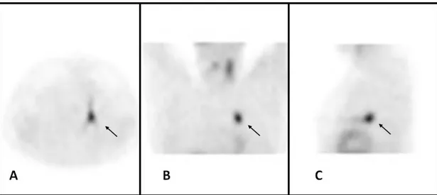
Surprisingly, images showed a well-defined and intense (target to background ratio: 3.4:1) MIBI
uptake into left thorax consistently with a lung lesion (Fig 1, Panels B-E). Single photon emission
tomography (SPET) of the chest was performed 60 minutes after tracer administration using the
same gamma-camera that has been already employed for planar images. Tomographic images
confirmed an abnormal MIBI uptake located in left lung (lingular division) (Fig. 2, black arrows).
The patient did not complain of any symptom or sign consistent with neoplastic or inflammatory
disease.
Thorax computed tomography (CT) without contrast media administration was obtained a few
days later MIBI scan to define the morphological characteristics of MIBI abnormal uptake.
The CT study did not show any lung lesion (Fig.3, Panel A, B). In particular, no focal lesion was
appreciated in the lingular division of left lung, but the patient remembered an important
contusive left lung trauma due to a car accident occurred six months before.
At the time of car accident, the patient had undergone CT study that had shown slight left pleural
effusion (Fig. 3, Panel C, black and white arrow-head) and, mainly, an irregular opacification (15
mm in maximum diameter) in the lingular division (Fig. 3, Panel D, black arrow-heads), directly
caused by left chest contusion.
on the same nodule one years ago) was conclusive for a class II nodule, according to Bethesda
system (1), confirming MIBI scan result. In addition, no evidence of lung disease was noted at
CT study performed some days after FNAC study.
Conclusion
In conclusion, our case confirms the utility of quantitative MIBI-scintigraphy using WOind
method already proposed by Campennì(8) in the work-up of cold thyroid nodules with
indeterminate cytology, in order to identify patients with benign lesions thus reducing the number
of unless surgery.
Finally, an important contusive lung trauma should be taken into account as potential pitfall at
99m
Tc-MIBI scintigraphy. Morpho-functional imaging can improve the diagnostic performance of
functional imaging alone.
Figure 1.Panel A: 99mTc-pertechnetate thyroid scintigraphy was obtained 20 minutes after tracer administration (111 MBq) using dual headed gamma-camera [Brightview-X (Philips, Cleveland, USA)] equipped with Low Energy High Resolution Parallel-hole collimators (LEHRPAR). Planar image (anterior projection; frame counts, 100 Kcounts; magnification, 1; matrix, 256x256; energy peak, 140±20 KeV) showed a “cold” nodule that occupied the right lobe. No tracer uptake was appreciated into thorax.
Panel B,C: 99mTc-MIBI thyroid scintigraphy was performed 10 (early image) and 60 minutes (late image) after tracer administration (370 MBq) using the same gamma-camera employed to obtain 99mTc-pertechnetate scintigraphy. Planar images (anterior projection, frame time: 10 minutes; magnification 1, matrix 256x256) showed increased MIBI uptake into cold nodule (99mTc-MIBI >99m Tc-pert.) in early image (B) that decreased in late image (C) (white arrows; qualitative analysis). At quantitative MIBI analysis, WOindx was >19% (D, E) (red and yellow circles) .A well-defined and intense MIBI uptake consistent with a lung lesion was appreciated into left thorax (B-E).
Figure 2.Single photon emission tomography of chest (magnification 1.4; matrix 256x256; acquisition modalities, step and
shoot; angular range, 3 degrees; frame time, 30 seconds). Axial (A), coronal (B) and sagittal images (C) confirmed an abnormal MIBI uptake located in left lung (lingular division) (black arrows).
Figure 3.Thorax computed tomography (CT) was obtained using a 16-slice TC scanner (Brilliance 16; Philips
Medical Systems, Eindhoven, Netherlands). The study was also compared with the thorax CT obtained with the same scanner at the time of car accident (C, D).
Panel A, B: Axial (A) and coronal (B) images (thickness: 3mm; increment: 1,5; Collimation: 16x1,5; Pitch: 0,938; kV: 120; mAs/slice: 200), obtained without contrast media administration, did not show any lung lesion.
Panel C, D: Axial images (thickness: 3mm; increment: 1,5; Collimation: 16x1,5; Pitch: 0,938; kV: 120; mAs/slice: 200) had shown slight left pleural effusion (C, black and white arrow-head) and, mainly, an irregular opacification (15 mm in maximum diameter) in lingular division (D, black arrow-heads), directly caused by left chest contusion.
Conflicts of Interest: There is not potential conflict of interest, and the authors have nothing to disclose.
This work was not supported by any grant.
References
1. Cibas, E.S., Ali, S.Z., NCI Thyroid FNA State of the Science Conference. (2009) The Bethesda System
For Reporting Thyroid Cytopathology. Am J Clin Pathol,132:658-665.
2.Giovanella, L., D’Aurizio, F., Campenni’, A., et al. (2016)Searching for the most effective thyrotropin
(TSH) threshold to rule-out autonomously functioning thyroid nodules in iodine deficient regions.
Endocrine, 54(3):757-761.
3.Hurtado-López, L.M., Arellano-Montaño, S., Torres-Acosta, E.M., et al. (2004) Combined use of
fine-needle aspiration biopsy, MIBI scans and frozen section biopsy offers the best diagnostic accuracy in the
assessment of the hypofunctioning solitary thyroid nodule. Eur J Nucl Med Mol Imaging, 31:1273–1279.
4. Hurtado-López, L.M., Martínez-Duncker, C. (2007) Negative MIBI thyroid scans exclude differentiated
and medullary thyroid cancer in 100% of patients with hypofunctioning thyroid nodules. Eur J Nucl Med
Mol Imaging, 34:1701-1703.
5. Giovanella, L., Suriano, S., Maffioli, M., et al. (2010) (99m)Tc-sestamibi scanning in thyroid nodules
with nondiagnostic cytology. Head Neck, 32:607-611.
6. Campennì, A., Violi, M.A., Ruggeri, R.M., et al. (2010) Clinical usefulness of
99mTc-MIBI scintigraphy
in the postsurgical evaluation of patients with differentiated thyroid cancer. Nucl Med Commun,
31:274-279.
7. Giovanella, L., Campennì, A., Treglia, G., et al. (2015) Molecular imaging with
99mTc-MIBI and
molecular testing for mutations in differentiating benign from malignant follicular neoplasm: a prospective
comparison. Eur J Nucl Med Mol Imaging,43(6):1018-1026.
8. Campennì, A., Giovanella, L., Siracusa, M., et al. (2016) (99m)Tc-Methoxy-Isobutyl-Isonitrile
Scintigraphy Is a Useful Tool for Assessing the Risk of Malignancy in Thyroid Nodules with
Indeterminate Fine-Needle Cytology. Thyroid, 26(8):1101-1109.
9. Campennì, A., Siracusa, M., Ruggeri, R.M., et al. (2017) Differentiating malignant from benign thyroid
nodules with indeterminate cytology by
99mTc-MIBI scan: a new quantitative method for improving
diagnostic accuracy. Sci Rep, 7(1):6147.
©2018 by the Author(s); licensee Accademia Peloritana dei Pericolanti (Messina, Italy). This article is an open access article distributed under the terms and conditions of the Creative Commons Attribution 4.0
International License (https://creativecommons.org/licenses/by/4.0/).
![Figure 1.Panel A: 99m Tc-pertechnetate thyroid scintigraphy was obtained 20 minutes after tracer administration (111 MBq) using dual headed gamma-camera [Brightview-X (Philips, Cleveland, USA)] equipped with Low Energy High Resol](https://thumb-eu.123doks.com/thumbv2/123dokorg/4591880.39226/3.892.118.760.734.1155/pertechnetate-scintigraphy-obtained-administration-brightview-philips-cleveland-equipped.webp)