PhD in Chemical Sciences
XXX Cycle
Structural characterization and semi-synthetic
modification of bacterial glycolipids
PhD Student: Marcello Ziaco
Tutor: Supervisor:
Prof. M. M. Corsaro Dr. A. Guaragna
Index
ABBREVIATIONS ………... V
SUMMARY ……...………. VIII
SECTION I: Introduction
Chapter 1: Gram-negative bacteria ... 2
1.1The Kingdom of Life ... 2
1.2 Bacterial cell envelope ... 3
1.3 Lipopolysaccharides (LPSs) ... 5
1.3.1 The Lipid A moiety: structure and activity ... 8
1.3.2 The core oligosaccharide: structure and activity ... 12
1.3.3 The O-polysaccharide moiety: structure and activity ... 13
1.4 LPS and Elicitation of Host Immune response ... 14
1.4.1 Innate immunity and Adaptive Immunity ... 14
1.4.2 LPS and Innate Immunity ... 19
References ... 22
Chapter 2: Structural characterization of LPS and LOS ... 24
2.1 Extraction and Purification of LPS and LOS ... 24
2.2. Structural determination of the saccharide moieties ... 25
2.2.1 Chemical analyses of the saccharide moieties ... 28
2.2.2 NMR Spectroscopy analysis of the saccharide moieties ………..………...…… 30
2.2.3 Mass spectrometry analysis of the saccharide moieties ... 33
2.3 Structural determination of the lipid A moiety ... 35
2.3.1 Chemical analyses of the lipid A moiety ... 35
2.3.2 Mass spectrometry analysis of the lipid A moiety ... 36
References ... 38
SECTION II: Lipid As isolated from Extremophiles Chapter 3: Extremophiles, an overview ... 45
3.1 Life-Forms in inhospitable environments ... 45
3.2 Psychrophiles ………..………... 47
3.3 Molecular and physiological adaptation ... 49
3.4 Industrial and biomedical applications ………..……… 53
References .………... 55
Chapter 4: Colwellia psychrerythraea strain 34H ... 57
4.1 Lipopolysaccharides and lipid A structures ..………... 58
4.1.1 Isolation and compositional analysis of lipid A ….…... 59
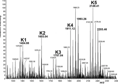
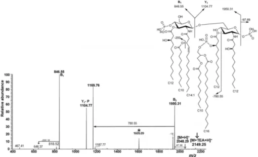
4.1.2 Mass spectrometry analysis of lipid A ..………..…... 60
4.1.3 NMR spectroscopy of lipid A ………...…...……… 69
4.1.4 Evaluation of biological activity ………... 72
4.2 Conclusions ... 73
4.3 Experimental section ... 75
References ... 80
Chapter 5: Psychrobacter arcticus strain 273-4 …... 82
5.1 Lipopolysaccharides and lipid A structures ..………... 82
5.1.1 Isolation and compositional analysis of lipid A ..………. 84
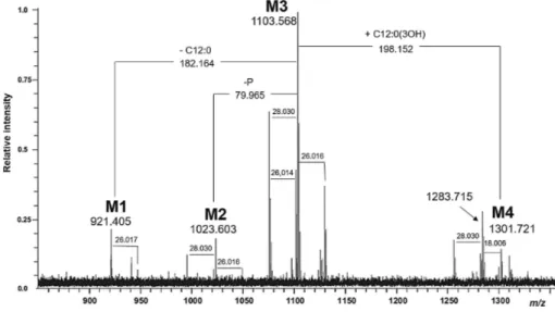
5.1.2 Mass spectrometry analysis of lipid A ..……...…….... 85
5.1.3 Evaluation of biological activity ………. 90
5.2 Conclusions ... 91
5.3 Experimental section ... 91
References ... 94
SECTION III: Semisynthetic derivatives from Escherichia coli lipid A Chapter 6: Multigram-scale extraction and purification of lipid A from E. coli ………... 97
lipid A from E. coli K4 fermentation ………..…... 99
6.2 Selective de-O-phosphorylation of lipid A ... 102
6.3 Structural characterization and biological assays ………. 103
6.4 Conclusions ……….………... 106
6.5 Experimental section ... 107
References .……….…… 112
Chapter 7: Functionalization on the C-6 of GlcN II of E. coli monophosphoryl lipid A ... 114
7.1 Site-selective chemical reactions ……….. 115
7.2 Insertion of a clickable moiety ………... 119
7.3 Semisynthetic “self-adjuvant” anticancer vaccine candidates ... 133
7.3.1 E. coli MPLA - Tn antigen conjugate ………….…...…... 137
7.3.2 E. coli MPLA - TF antigen conjugate ..…..……..….…… 141
7.4 Biological assays ……….…….……… 146
7.5 Multivalent Tn antigens conjugates - MPLA ……... 152
7.6 Conclusions ……….………....…. 155
7.7 Experimental section ... 156
References .………...…… 170
Chapter 8: Lipid pattern modification of E. coli monophosphoryl lipid A ... 173
8.1 Selective deacylation reactions ……….… 173
8.2 Selective re-acylation with shorter chains ………... 177
8.3 Biological assays ……….….…… 185
8.4 Conclusions ……….………... 187
8.5 Experimental section ... 187
References .………...… 191
Chapter 9: Fluorescent derivatives of E. coli lipid A ………..…….. 192
9.1 Flippases ...………..….………. 193
9.2 Fluorescent mono/diphosphoryl lipid As and ABC transporters ... 194
9.4 Conclusions ……….……….. 205
9.5 Experimental section ... 205
References .………...…… 207
Appendix PhD Course Activity Summary ………...…….. 210
Attended Courses ………..….………... 210
Attended Seminars ... 210
Publications ... 212
Attended congresses/workshops/summer schools/contribution ...…. 213
Other Activities ……….……….………….. 214
Portions of this work have been adapted from the following articles that were co-written by the author:
•! A. Casillo, M. Ziaco, B. Lindner et al., Unusual Lipid A from a cold adapted bacterium: detailed structural characterization. ChemBioChem 2017, 18, 1 – 11.
•! D. D’Alonzo, M. Cipolletti, M. Ziaco et al., A Semisynthetic Approach to New Immunoadjuvant Candidates: Site-Selective Chemical Manipulation of Escherichia coli Monophosphoryl Lipid A. Chem. Eur. J. 2016, 22, 1-12.
•! M. Ziaco, S. Górska, S. Traboni et al., Development of clickable monophosphoryl lipid As towards semi-synthetic conjugates with tumor-associated carbohydrate antigens. J. Med. Chem. 2017, 60, 9757-9768.
ABBREVIATIONS
AFGPs = Antifreeze glycoproteins AFP = Antifreeze proteins
AMG = Acetylated methyl glycoside APC = Antigen presenting cells
BMDMs = Bone marrow-derived macrophages CDMT = 2-chloro-4,6-dimethoxy-1,3,5-triazine COSY = Correlation spectroscopy
CS = Chondroitin sulfate CSPs = Cold-shock proteins DCC = Dicyclohexylcarbodiimide DIPEA = N,N-diisopropylethylamine DIPC = N,N’-diisopropylcarbodiimide DMAP = 4-(N,N-dimethylamino)pyridine DMF = N,N-dimethylformamide
DMSO = Dimethyl sulfoxide DO = Dissolved-oxygen
DPAP = 2,2-dimethoxy-2-phenylacetophenone DPLA = Diphosphoryl lipid A
EI = Electron ionisation
ELISA = Enzyme-linked immunosorbent assay EPSs = Exopolysaccharides
ESI = Electro Spray Ionisation
ESI FT-ICR MS = Fourier transform ion cyclotron resonance mass spectrometry
GlcN = Glucosamine
GlcNAc = N-Acetyl-glucosamine HEK = Human Embryonic Kidney
HMBC = Heteronuclear multiple bond correlation HOBt = 1-hydroxybenzotriazole
HSQC-DEPT = Heteronuclear Single Quantum
Coherence-Distorsionless Enhancement by Polarization Transfer IBX = 2-iodoxybenzoic acid
Kdo = 3- deoxy-D-manno octulosonic acid
LBP = LPS- binding protein LOS = Lipooligosaccharide LPS = Lipopolysaccharide
MALDI = Matrix Assisted Laser Desorption Ionisation MHC = Major histocompatibility complex
MPLA = Monophosphoryl lipid A NBD = 4-Chloro-7-nitrobenzofurazan
NOESY = Nuclear Overhauser enhancement spectroscopy NMM = 4-methylmorpholine
OM = Outer membrane
PAMPs = Pathogen Associated Molecular Patterns PMN = Polymorphonuclear neutrophilic leukocytes PRRs = Pattern Recognition Receptors
PyBOP® = (benzotriazol-1-yloxy)tripyrrolidinophosphonium hexafluorophosphate
RAFT = Regioselectively Addressable Functionalised Template SEC = Size exclusion chromatography
THP-1 = Human monocytic cell line TLRs = Toll-Like Receptors
TOCSY = Total correlation spectroscopy TBAB = Tetrabutylammonium bromide TBDMS = tert-butyldimethylsilyl TBDPS = tert-butyldiphenylsilyl
TBTU = O-(benzotriazol-1-yl)-N,N,N′,N′-tetramethyluronium tetrafluoroborate
TEMPO = 2,2,6,6-tetramethylpiperidine-1-oxyl TMSOTf = Trimethylsiliyl trifluoromethanesulfonate
! ! ! !
SUMMARY
The present PhD thesis aims to discover new natural lipid A structures and to obtain semi-synthetic derivatives thereof to be tested as candidates for the pharmacological use as vaccine adjuvants.
The term lipid A indicates a family of glycolipids representing the inflammation-inducing moiety of lipopolysaccharides (LPSs) and lipooligosaccharides (LOSs), that are amphiphilic molecules embedded in the Gram negative bacterial outer membrane, of which they are the major constituents and very often the major virulence factor.
The general structure of LPSs is characterized by three distinct portions:
•! The lipid A, generally composed of a !"
D-GlcN-(1→6)-D-GlcN
disaccharide (GlcN=2-amino-2-deoxy-glucopyranose) with a variable number (from three to seven) of acyl chains linked to the positions 2 and 3 of both glucosamine residues through amide or ester bonds, respectively. The structure is generally completed by one or two phosphate groups, linked at position O-4 of the non-reducing unit (GlcN II) and/or at position O-1 of the pseudo-reducing residue (GlcN I) with an #"configuration.1
•! The core oligosaccharide, distinguishable in an inner core and an outer core. This oligosaccharide is linked to the lipid A and is well conserved among different strains of the same bacteria and consists of uncommon sugar residues such as heptoses (L
-glycero-D-manno-heptose and, less commonly, D-glycero-D
-manno-heptose) and Kdo (3- deoxy-D-manno octulosonic acid).
•! The O-chain polysaccharide built up of oligosaccharide repeating units. The O-chain polysaccharide is the most variable portion of the LPS also within bacteria belonging to the same genus; it consists of up to 50 identical repeating units composed of two to eight different glycosyl residues (heteroglycans) or, in some bacteria, of identical sugars (homoglycans). This moiety can be absent and in that case LPSs are named LOSs.
Furthermore, LPS elicits host immune response through the Toll-like receptors-4 (TLR-4) and Myeloid differentiation factor-2 (MD-2) receptorial complex expressed on macrophages and neutrophils.
Toll-like receptors (TLRs) play an important role in the recognition of molecular signals of microbial invasion (Microbial Associated Molecular Patterns, MAMPs) from virus, bacteria, protozoa and fungi. In particular, TLR-4 ligands, such as lipid A, trigger innate immunity response in the host through chemokine and cytokine signalling cascades.2
This response is highly modulated by several structural features of lipid A,3
such as the number of phosphate groups and acyl chains as well as the length and the distribution of the latters.4
Indeed, hexa-acylated diphosphoryl lipid As possess strong inflammatory activity, that may be associated to a number of severe pathophysiological symptoms (fever, diarrhoea, blood pressure drop, septic shock), whereas underacylated lipid As can block or inhibit TLR-4, thus acting as TLR-4-antagonists. The deletion of the phosphate group at the anomeric position of GlcN I can furnish the 1-O-dephosphorylated form of lipid A, known as monophosphoryl lipid A (MPLA), was revealed to have significantly reduced endotoxicity but retain the strong immunostimulatory activity. MPLA has been proved to be clinically safe as a vaccine adjuvant to treat infectious and neoplastic diseases. The development of new TLR-4-stimulators to be used as immuno-adjuvants has attracted much effort in the last years. To this aim, the research is focused on the obtainment of new example of lipid A structures, both through isolation from wild-type or engineered bacterial extracts1, 5
and by synthetic6
and semisynthetic approaches.7
In the former frame, extremophiles may be a promising source of non-toxic LPSs and lipid As. In particular, cold environments, due to the still low contamination by pathogenic microorganisms, may represent an untapped reservoir of immunomodulating molecules. The outer membrane of Gram-negative bacteria is mainly constituted by LPSs, and it was demonstrated that structural changes could be present in these macromolecules isolated from cold-adapted bacteria.
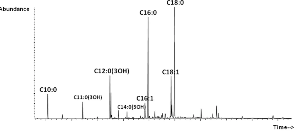
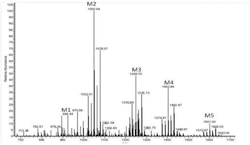
In particular, the analysis of C. psychrerythraea strain 34H genome, evidences the features that allow the live at freezing temperatures and for this reason this bacterium is considered as a model for the study of cold adapted life-style. The structures of the lipid A moiety from C. psychrerythraea strain 34H and its partially deacylated derivative were completely characterized in this Ph.D. work by high-resolution mass spectrometry (HR-MS), NMR spectroscopy, and chemical analysis. The high heterogeneity of the lipid A structure, showed by the fatty acids
analysis, was confirmed by the complexity of MS and MS/MS spectra. These experiments indicated a complex acylation pattern ranging from tetra- to hepta-acylated glycoforms. Furthermore, this lipid A displayed a structure that is quite new among known LPSs. An unusual structure with a 3-hydroxy unsaturated tetradecenoic acid as a component of the primary acylation pattern was identified. In addition, the presence of a partially acylated phosphoglycerol moiety on the secondary acylation site at the 3-position of the reducing 2-amino-2-deoxyglucopyranose unit caused tremendous natural heterogeneity in such lipid A structure.
Structure of C. psychrerythraea 34H lipid A.
The double bond is arbitrarily shown in the cis configuration.
The peculiar structural features showed by C. psychrerythraea 34H lipid A could be related to the cold-adaptation mechanisms adopted by this bacterium.8
Following the same strategy, during this Ph.D. work the lipid A moiety derived from the LPS of Psychrobacter arcticus 273-4 grown at 4°C, a Gram-negative bacterium isolated from Kolyma region of Siberian permafrost core9
, was completely elucidated. P. arcticus 273-4 is considered a psychro-tolerant microorganism, capable to grow at both low, even subzero temperatures, and even more than 20°C. The lipid A
and its partially deacylated derivatives were completely characterized by chemical analysis, electrospray ionization Fourier transform ion cyclotron (ESI FT-ICR) and Orbitrap mass spectrometry. As the chemical analysis and the mass spectrometry showed, the lipid A of P. arcticus is characterized by the presence of shorter acyl chains than lipid A from C. psychrerythraea 34H; decanoic acid (C10:0) as secondary acyl groups and mainly hydroxy-dodecanoic acid [C12:0(OH)], hydroxy-tetradecanoic acid [C14:0(OH)] and minor amount of 3-hydroxy-undecadecanoic acid [C11:0(3-OH)], 3-hydroxy-tridecanoic acid [C13:0(3-OH)] as primary acyl groups. Therefore, the lipid As moiety from both the studied psychrophilic bacteria display several structural features that could be related to the adaptation mechanisms and in particular an increased incorporation of unsaturated, short, and/or branched fatty acids to maintain membrane fluidity as part of a set of physiological actions necessary to survival in cold conditions.10
Preliminary biological activity assays were performed to characterize the effect of C. psychrerythraea 34H and P. arcticus 273-4 lipid As on TNF secretion in the presence of a serial dilution of Escherichia coli O111:B4 LPS. The elucidation of new lipid A structures is fundamental for the comprehension and the enlargement of the number of structure–activity relationships necessary for the design of new immunomodulatory molecules. Actually, these components are crucial for the development of modern formulations of recombinant or synthetic vaccines, in order to obtain strong and long-lasting responses also in elderly and immunocompromised patients.11
These molecules are usually obtained by purification of wild-type or engineered bacterial extracts without very good results for the purity,1
as well as through total chemical synthesis,that requires lengthy procedures with a high number of synthetic steps and very difficult to apply on a large-scale production.
An emerging approach is the use of semi-synthetic strategies that combine the advantages of a fermentative access to complex natural products with the development of site-selective chemical reactions modifying these structures.
In particular, Prof. Corsaro and co-workers developed a semi-synthetic approach relying upon the use of lipid A in gram quantities from fed-batch fermentation of E. coli, after de-O-phosphorylation at O-1 position
of GlcN I, as a scaffold for the obtainment of several other lipid As and derivatives thereof through few, tailored chemical modifications.7
These site-selective reactions were developed in this Ph.D. work, with a special focus on the functionalization of the primary hydroxyl group at position C-6 of GlcN II and on the modification of the lipid pattern. The former was attractive not only for the expected higher reactivity of the primary alcohol with respect to the secondary hydroxyls, but also because it could be biologically well tolerated since lipid As and the core part of LPSs are linked through a glycosidic bond between this position and the Kdo or, much more rarely, 2-octulosonic acid (Ko). In order to insert a chemical handle that would be easily and orthogonally derivatizable with respect to the other functionalities present on lipid A structure, the installation of a carboxylic acid function at position 6 of GlcN II was performed. This derivatization could not only serve as a suitable moiety for further functionalization, but also mimics the Kdo structure (lipid A-Kdo structures have been demonstrated to induce stronger immune responses than their parent lipid As).6, 12
In order to insert a clickable moiety on such oxidized lipid A structure, this derivative was treated with diverse amines carrying a suitable functionalization at their $-position (double or triple bond, azide, S-trityl as protected thiol). The first aim was to find the best conditions for the amide formation reaction using a model amine such as 4-penten-1-amine. The coupling was tested under several typical peptide coupling conditions. After having detected PyBOP®
and TBTU as the best coupling reagent under appropriate conditions, these were employed to install the other clickable functionalities indicated above.
O O NH O P O %O HO O O O O O O HO NH O O HO O O O HO OH O OH R= CH CH2 R= CH2C CH R= CH2CH2CH2N3 R= CH2CH2SCPh3 O O NH O O O O O O O HO NH O O HO O O O HO OH O P O %O HO H N R
DIPC, DMAP, DIPEA or CDMT, NMM, DIPEA
or TBTU, DIPEA
or PyBOP, HOBt, DIPEA
4%pentene%1%amine or 6%amino%1%hexyne or 6%azido%hexylamine or 5%(tritylthio)%pentan%1%amine O O NH O P O %O HO O O O O O O HO NH O O HO O O O HO OH OH I) TEMPO, NaOCl II) NaClO2
The production of these clickable lipid A derivatives opens a straightforward access to their conjugation with other interesting biomolecules, such as tumor-associated carbohydrate antigens (TACAs). In order to demonstrate the feasibility of this approach, the thiol-equipped derivatives of Tn and TF antigens were synthesized and then conjugated with a clickable lipid A derivative carrying a double bond moiety, through a UV-mediated thiol-ene reaction, to give two potential self-adjuvant anticancer vaccine candidates. A preliminary evaluation of the immunological activity of these lipid A-TACA antigen conjugates, as well as of other semi-synthesized lipid A derivatives, was performed.16
It was shown that most of them ameliorated production of proinflammatory cytokines IL-6, TNF-#, and IFN-% compared to control. The tested derivatives also induced chemokine CXCL8 (IL-8) and immunoregulatory cytokine IL-10 thus highlighting the promising immunobiological applications. Additionally, the described compounds exhibited very low toxicity which could facilitate their use in vaccines and give them an advantage over the currently used adjuvants.
Following this strategy, a collaboration with Prof. Renaudet (Univ. Grenoble Alpes, CNRS, France) has been started, in order to develop the first Regioselectively Addressable Functionalised Template! (RAFT) scaffold-MPLA conjugates as potent multivalent immunogenic vaccine candidates.
Moreover, we finely tuned the conditions suitable for the semisynthesis of other new lipid A derivatives, by selective modification of the lipid pattern (selective deacylation and re-acylation with shorter chains). In this way, a small collection of new lipid A derivatives was rapidly prepared, avoiding lengthy total synthetic approaches. Notably, potential access to several other modified lipid A structures could be opened by this strategy, because the reactions developed on E. coli lipid A scaffold should also be applicable on lipid A substrates from different bacterial sources. Some of the new compounds were assayed for preliminary in vitro immunological tests, showing somewhat reduced inflammatory activity and a cytokine production profile similar to the already used immunomodulant MPL& Salmonella Minnesota lipid A, thus highlighting the promising immunobiological applications.
Finally, in the last part of this PhD work, two different fluorescent lipid A derivatives were semi-synthesized, with the aim to improve the
understanding of the potential role of lipid A in transmembrane protein stability, in mediating drug export and in the mechanisms of ABC transporters. O O NH O P O HO HO O O O O O O HO NH O O HO O O O HO O O P O OH OH O O O NH H N O HO O NH O O O O O P O O HO O O O O HO HO OH O NH N O N NO2 HO NH N O N NO2 NBD-MPLA NBD-DPLA
Semi-synthetic fluorescent mono- (left) and diphosphoryl (right) lipid A derivatives.
In collaboration with Prof. Tampé (Institute of Biochemistry, at the Goethe-University in Frankfurt, Germany) these two derivatives were exploited in several biological assays in order to develop some in vitro fluorescence-based lipid A flippase assays, focused on the ABC transporters mechanisms comprehension (data still under investigation).17
Finally, these two fluorescent derivatives, in collaboration with Prof. Huser (Department of Physics, University of Bielefeld, Germany), were subjected to a preliminary cell imaging test aimed to observe their staining with two different cell lines on a deconvolution microscope (experiments still in progress).
References
1.! Molinaro, A. et al., Chem. Eur. J. 2015, 21 (2), 500–519. 2.! Medzithov, R. Nature 2008, 454, 428-435.
3.! Park, B.S. et al., Nature 2009, 458, 1191-1196.
4.! (a) Li, Y. et al., Mar. Drugs 2013, 11, 3197-3208; (b) Rietschel, E.T. et al., FASEB J. 1994, 8, 217-225.
5.! (a) Needhama, B.D. et al., Proc. Natl. Acad. Sci. USA 2013, 110, 1464-1469; (b) Carillo, S. et al., Chem. Eur. J. 2011, 17, 7053-7060. 6.! Gao J., Guo Z., Med. Res. Rev. 2017, (in press)
doi: 10.1002/med.21447.
7.! (a) D’Alonzo, D. et al., Chem. Eur. J. 2016, 22, 11053-11063; (b) Pieretti, G. et al., Appl. Microbiol. Biotechnol. 2014, 98, 7781-7790. 8.! Casillo A. et al., ChemBioChem 2017, 18, 1 – 11.
9.! Vishnivetskaya, T. et al., Extremophiles 2000, 4,165–173. 10.! Carty S.M. et al., J. Biol. Chem. 1999, 274, 9677 – 9685.
11.! (a) Kwissa, M. et al., Expert Rev. Vaccines 2007, 6, 673 – 684; (b) Marciani, D.J. Drug Discovery Today 2003, 8, 934 – 943.
12.! (a) Zughaier, S.M. et al., Infect Immun. 2004, 72, 371-380; (b) Yoshizaki, H. et al., Angew. Chem. Int. Ed. 2001, 40, 1475-1480. 13.! Saito, Y. et al., Tetrahedron 2008, 64, 3578−3588.
14.! Lee, J. W. et al., Tetrahedron Lett. 2001, 42, 2709−2711. 15.! Jagadish, B. et al., J. Med. Chem. 2012, 55, 10378−10386. 16.! Ziaco M. et al., J. Med. Chem. 2017, 60, 9757-9768. 17.! Bechara, C. et al., Nat. Chemistry 2015, 7, 255 – 262.
SECTION I
Chapter 1: Gram-negative bacteria
1.1 The Kingdom of Life
The three-domain system, which classifies organisms on Earth into three distinct kingdoms – Archea, Bacteria and Eukarya – was designed by the American microbiologist Carl Woese in the 1990. Otherwise, an alternative biological classification system currently used is known as “the six-kingdoms system of life” and include Archaebacteria, Eubacteria, Protista (comprising one-celled organisms), Fungi, Plantae and Animalia. While Archaebacteria and Eubacteria constitute the Archaea and Bacteria domains respectively (both representing the Prokarya kingdom), Protista, Fungi, Plantae and Animalia together form the Eukarya kingdom of life. Most prokaryotic cells have diameters in the range of 1-10 μm, much smaller than most eukaryotic cells (typically 10-100 μm). Prokaryotes have a cellular organization different from that of eukaryotes; these latter have a membrane-enclosed nucleus and numerous membrane-enclosed organelles, whereas prokaryotic cells lack these structural features. Typically, Archea domain includes all the extremophiles prokaryotic organisms that live and thrive in inhospitable, from a human viewpoint, environments such as alkaline and acidic waters, boiling hot springs, high pressure waters, ultra-saline brines, without excluding a combination of aforesaid chemical and physical extremes which is typical of polyextremophilic microbes (Mesbah and Wiegel, 2008). Bacteria comprise a wide group of microorganisms characterized by a bewildering assortment of size, shapes and arrangements reflecting the diverse environments in which they grow and reproduce (Pommerville, 2010). Despite the high structural variability,
Bacteria can be divided on the basis of their basic cell morphologies: a bacterial cell with a rod shape is called “bacillus” whereas spherical and curved cells are called “cocci” and “spirilli” respectively (Raven and Johnson, 2001). Some rod-shaped and spherical bacteria form colonies adhering end-to-end after their cellular division forming chains, whereas spirilla bacteria generally do not form associations with other cells. However, early systems for classifying bacteria relied on differential stains such as the colorimetric assay developed by the Danish physician Hans Gram, in use to date, that allows the distinction of bacteria in Gram-positive and Gram-negative on the basis of the different response to the test. The differences between Gram-positive and Gram-negative bacteria are related to diversities in the structure and chemical composition of their cell wall: Gram-positive bacteria have the thicker cell envelope, nearly uniformly dense layers, and stain the purple color of the crystal-violet typically used in the Gram stain, whereas Gram-negative bacteria contain just a single or two layers and lose the purple-colored dye (Staley et al., 2007).
!
1.2 Bacterial cell envelope
The complex system of layers that surrounds bacterial cells is referred collectively as the cell envelope. It consists of a cytoplasmic membrane (or plasma membrane), cell wall, and for some bacteria, capsule and glycocalyx from inner to outer surface. Both positive and Gram-negative bacteria present the cytoplasmatic membrane, namely a phospholipid bilayer surrounding the cytoplasm and representing a physical semi-permeable barrier that regulates the transport of nutrients and metabolic products in and out the cell. This membrane is, in turn,
cellular shape and protects the cell from swelling and rupturing (Raven and Johnson, 2001). The bacterial cell wall differs markedly from archaeal and eukaryotic cells walls in containing peptidoglycan (or murein) which is a network of saccharide molecules connected by polypeptide cross-links. In detail, it is composed of β-(1→4)-linked disaccharide chains, alternating N-acetylglucosamine and N-acetylmuramic acid residues in turn cross linked by short peptide stems composed of alternating L- and D-amino acids (Figure 1.1). It is possible to classify peptidoglycans on the basis of the third amino acid residue of the stem peptide; indeed, in Gram-negative bacteria this residue is the unusual meso-diaminopimelic acid (m- DAP), while in Gram-positive bacteria is commonly a lysine (Figure 1.1).
Figure 1.1 Peptidoglycan structure in Gram-positive (left) and Gram-negative (right) bacteria.
Furthermore, in Gram-positive bacteria the external portion of the cell is composed by 95% of rigid peptidoglycan cell wall that may be one of the reason why they retain the crystal-violet of Gram stain (Figure 1.2); on the contrary, in Gram-negative cells there is a thin layer of peptidoglycan, almost 5-10% of the cell wall, surrounded by an additional asymmetric phospolipid bilayer, termed outer membrane (OM) (Figure 1.2). This
external membrane is a unique feature of Gram-negative bacteria and represents the first barrier that protects bacterial cell from environmental stress factors, thus it is essential for bacterial survival. The outer membrane is separated by a gel-like compartment, termed periplasm, from the cytoplasmatic membrane and shows to have a structure similar to that of the plasma membrane, from which nonetheless differs for important physical and chemical properties. The inner portion of the OM is mainly composed of phospholipids, while the external leaflet is occupied for the 75% by glycolipids known as lipopolysaccharides (LPS) (Alexander and Rietschel, 2001). Furthermore, the outer membrane is also constituted by a large number of proteins, among which there is the Braun’s lipoprotein (BLP or murein lipoprotein), that acts as a bridge between the outer membrane and the peptidoglycan layer.
Figure 1.2 Cell-wall structure of Gram-positive (left) and Gram-negative bacteria (right).
1.3 Lipopolysaccharides (LPSs)
Lipopolysaccharides are heat-stable amphiphilic molecules indispensable for viability and survival of Gram-negative bacteria, as they heavily contribute to the structural integrity of the OM and to the protection of the bacterial cell envelope (Holst et al., 1996).
Being intercalated into the outer membrane, they form hydrophobic interactions with the phospholipid bilayer composing the inner layer, giving rise to the typical asymmetry of Gram-negative bacteria OM (Holst et al., 1996). The highly ordered structure and low fluidity of the LPS monolayer, stabilized by electrostatic interactions of divalent cations (as Ca2+
and Mg2+
) with negatively charged groups present on LPS molecules, are responsible for the increase of permeability to hydrophobic and higher molecular weight hydrophilic compounds, but also for the bacteria resistance to external stress factors. Indeed, most of the commonly used antibiotics directed against Gram-negative bacteria, such as polymyxin B, are able to destabilize abovementioned ionic interactions leading to the disruption of membrane integrity (Cardoso et al., 2007). In addition, since they are exposed toward the external environment, LPS molecules participate in crucial mechanisms of host-bacterium interactions like colonization, virulence in the case of pathogen and opportunistic bacteria, adhesion and symbiosis (Silipo et al., 2010). Among all these activities, LPS has been shown to be the most potent immunostimulant molecule playing a key role in the pathogenesis of Gram-negative infections and triggering the immune system in a wide range of eukaryotic organisms ranging from insects to humans (Alexander and Rietschel, 2001). LPSs belonging to different bacterial species possess different structures, but also LPS of an individual bacterial strain is not a single molecule, possessing a specific chemical structure, but rather a blend of various molecules characterized by an intrinsic size and structural heterogeneity (Raetz, 1990; Raetz and Whitfield, 2002). Furthermore, bacteria of the same species producing diverse LPS molecules under different growth conditions were also found. Despite the high structural heterogeneity, all LPSs broadly
comply with a common basic architecture composed of well-conserved domains in which three distinct regions encoded by different gene clusters, can be distinguished (Figure 1.3). Indeed, they consist of a polysaccharide (also known as O-antigen, O-side chain or O-specific polysaccharide) characterized by a highly variable structure which constitutes the chemical basis for the serological classification of bacterial strains. O-antigen is covalently linked to an oligosaccharide part (core), in turn, linked to a glycolipid portion (lipid A) that is the most conserved region of the entire macromolecule, even among different species belonging to the same genus (Holst et al., 1996).
Figure 1.3 Schematized structure of Gram-negative LPSs.
The lipid A moiety is embedded in the outer leaflet of the OM whereas the sugar chain is oriented outwards (Raetz and Whitfield, 2002). The O-polysaccharide chain is not ubiquitous, as it can be absent or partly truncated in some Gram-negative strains. Bacterial colonies can be catalogued by gross colony morphology as rough or smooth on the basis of the occurrence of the O-side chain, being absent in the former and present in the latter (Lüderitz et al., 1966). The terminology currently used to designate the different LPS types, namely with or without the O-polysaccharide portion, is S- LPS or R-LPS (or lipooligosaccharide, LOS), respectively (Raetz and Whitfield, 2002). !
1.3.1 The lipid A moiety: structure and activity
Lipid A represents the hydrophobic and endotoxic principle of the LPS molecule since it acts as a potent stimulator of the host innate immune system (see paragraph 1.4.2). It possesses a highly conservative structure consisting of a β-(1→6)-linked D-glucosamine disaccharide backbone substituted with a number of amide- and ester-linked 3-hydroxy fatty acids at the positions 2 and 3 (Figure 1.4), respectively. The acyl groups that are directly linked to the sugar backbone are defined primary and some of them are further acylated at the hydroxy groups by secondary acyl chains. Furthermore, the sugar backbone is generally α-phosphorylated at position O-1 of the reducing glucosamine (GlcN I) and at position O-4' of the non-reducing glucosamine (GlcpN II) (Figure 1.4) (Zahringer et al., 1999). The firstly characterized lipid As were from Escherichia coli (Figure 1.4) and Salmonella enterica LPSs in 1983. E. coli lipid A is built up of the following sugar backbone [P→4-β-D -GlcN-(1→6)-α-D-GlcN-1→P] acylated at position 2 and 3 of both GlcNs by
four C14:0 (3-OH) (Figure 1.4). The primary fatty acids located on the GlcNII were both esterified at their hydroxyl group by two secondary acyl chains: one C14:0 (3- OH) was esterified by a C12:0 while the other by a C14:0 (Figure 1.4). O O NH O P O HO HO O O O O O O HO NH O O HO O O O HO OH O P O OH OH GlcN II GlcN I
Despite their general architecture, lipid As also present a micro-heterogeneity due to the presence of subtle chemical differences depending on a wide number of factors including bacterial adaptation, incomplete biosynthesis, changing of environment, presence of external stimuli and chemical modifications resulting from the procedures used for lipid A extraction from bacterial cells. Micro-heterogeneity has been observed in the acylation (number, type and distribution of acyl chains) and phosphorylation patterns, and, less commonly, also in the disaccharide backbone as in the case of bacterial species as Aquifex pyrophilus (Hellerqvist et al., 1971), Brucella abortus (Hellerqvist et al., 1969), whose GlcN residues may be replaced with 2,3-diamino-2,3-dideoxy-D-glucopyranose (GlcN3N) residues. Moreover, phosphate groups can be derivatized with further phosphate groups, producing a pyrophosphate, but also by other polar substituents such as 4-amino-4-deoxy-L-arabinopyranose (4-amino-arabinose, Ara4N) and 2-amino-ethanol group (EtN), or by acid residues such as galacturonic acid (GalA); phosphate groups can be absent, as in the case of Bdellovibrio bacteriovorus lipid A, characterised by the replacement of phosphate groups with two mannose residues generating a totally neutral lipid A. Concerning the acylation pattern, fatty acids can be attached to the disaccharide backbone either symmetrically (3+3, e.g. Neisseria meningitidis) or asymmetrically (4+2, e.g. Escherichia coli). Finally, lipid A fatty acids present less frequently further peculiar structural features such as a methyl branch, different functional and hydroxyl groups, length of the chains up to 28 carbon atoms, odd numbered carbon chains and unsaturation. Even the subtlest variation in chemical structure of lipid A can affect the LPS molecule bioactivity, since this latter is strictly
responsible for its agonistic and antagonistic activity on innate immune system (Brandenburg et al., 1993; Rietschel et al., 1994; Seydel et al., 2000; Fukuoka et al., 2001; Oikawa et al., 2004).
A plethora of studies aimed to the elucidation of the LPS structure-activity relationship have highlighted that the hexa- acylated bisphosphorylated lipid A with an asymmetric 4:2 fatty acid distribution, found in the majority of enterobacteria, as the aforesaid E. coli lipid A, is considered to have the highest immunostimulatory capacity in mammalian cells. In contrast, the hypoacylated synthetic precursor of E. coli lipid A, the tetra- acylated lipid A (Lipid IVa), showed to have weak agonistic effects for some species of mammals and is well known to possess a strong antagonistic activity on human cells (Golenbock et al., 1991). O O NH O P O HO HO O O O O O O HO NH O O HO O O O HO OH O P O OH OH E. coli O O NH O P O HO HO O O O O O O HO NH HO O O O O O OH O P O OH OH P. aeruginosa
cystic f ibrosis f orm
O O NH O P O HO HO O O HO O O HO NH O O O HO O OH O P O OH OH P. aeruginosa
non-cystic f ibrosis form
O HO O O NH O P O HO HO O O O O O O HO NH O O HO O O O O OH O P O OH OH S. Typhimurium O O O NH HO P O HO HO O O O O O HO NH O O HO O HO OH OH H . pylori
These different biological effects are correlated to two interconnected structural parameters: the molecular shape of the lipid A and the tilt angle between the di-glucosamine backbone and fatty acid chains that is the inclination of lipid A hydrophilic moiety respect to the hydrophobic portion (Netea et al., 2002). In details, the molecular shape possessed by lipid A influences its ability to be recognized by host immune system receptors, indeed it has been shown that at 37°C, in aqueous solution and in physiological conditions, the most agonistic lipid A has a truncated cone form that drives to a hexagonal supramolecular structure, while lipid As presenting an antagonistic activity assume a cylindrical shape leading to a lamellar structure (Netea et al., 2002). Regarding the tilt angle, it has been found that the most active form has a conical structure with a tilt angle >50°, while the antagonist structures present smaller values of tilt angles (Seydel et al., 2000): species with a tilt angle <25°, such as lipid IVA and the penta-acylated and symmetrically (3+3) hexaacylated lipid As, act as antagonists; species with an angle between 25-50°, as monophosphorylated lipid A, have a lower bioactivity (Figure 1.6) (Seydel et al., 2000).
These physical parameters are reflected in the different binding- mode of the LPS molecule to its physiological receptor, the TLR4/MD-2 complex, since they regulate the lipid A affinity to the MD-2 hydrophobic binding pocket that is pivotal for the innate immune receptor activation.
1.3.2 The core oligosaccharide moiety: structure and activity
Core oligosaccharide is a complex component of the LPS molecule since it can be characterized by up to fifteen monosaccharides which can be organised giving either a linear or a branched structure (Holst, 1999). In the core LPS portion two different regions can be distinguished on the basis of the monosaccharide composition, termed inner core and outer core (Holst, 1999). The inner core region is directly linked to the lipid A structure is well conserved among different strains of the same bacteria and consists of uncommon sugar residues such as heptoses (L -glycero-D-manno-heptose and, less commonly, D-glycero-D -manno-heptose) (De Soyza et al., 2008) and Kdo (3- deoxy-D-manno
octulosonic acid) (Figure 1.7). This latter is considered a diagnostic marker for all Gram-negative bacteria. It connects the core oligosaccharide to the lipid A backbone with an α-configured ketosidic linkage in almost every LPSs investigated to date.
O HO HO OH O OH OH OH
The L-glycero-D-manno-heptose residues are often decorated with
charged substituents like phosphate, pyrophosphate, Ara4N or uronic acids, as found in bacteria belonging to Pseudomonas genus and in many other species. It is speculated that all the substituents bearing a positively charged free amino group, as Ara4N residues, might play a role in pathogenesis since they reduce the negatively charged surface on the outer membrane rendering it positively charged or in an isoelectric state that, in turn, confers resistance to antibiotic compounds and antimicrobial peptides (Raetz and Whitfield, 2002; Hamad et al., 2012). The outer core region is the most exposed portion, often branched, and characterised by a higher structural variability than the inner core region. It is typically characterised by common hexoses such as glucose, galactose, N-acetyl glucosamine and N-acetyl galactosamine and it may also contain residues as 6-deoxy-L-mannose (L-Rha) and N-acetyl-2,6-dideoxy-D-glucosamine (D- QuiNAc).
1.3.3 The O-polysaccharide moiety: structure and activity
The O-chain polysaccharide is the most variable portion of the LPS also within bacteria belonging to the same genus; it consists of up to 50 identical repeating oligosaccharide units composed of two to eight different glycosyl residues (heteroglycans) or, in some bacteria of identical sugars (homoglycans). A single bacterium produces LPSs with O-chains characterised by a wide range of lengths as a result of incomplete synthesis of the polysaccharide chain (Raetz and Whitfield, 2002); this different degree of polymerization is responsible for the “ladder-like” pattern, showed by SDS-PAGE (Kittelberger and Hilbink, 1993) typical of a smooth LPS. The high structural variability of the O-polysaccharide is ascribable to the large number of sugar residues (in
both pyranose and furanose rings, anomeric and absolute configurations) that can build up the repeating units as well as to the glycosidic sequence and to the presence of non-carbohydrate substituents such as phosphates, amino acids, sulphates, acetyl or formamide groups, often present in a non-stoichiometric fashion (Adinolfi et al., 1996). The function of these substituents is frequently unknown although it can be speculated that bacteria can modify their LPSs to mask themselves to the host immune system.
1.4 LPS and elicitation of host immune response
1.4.1 Innate immunity and adaptive immunity
From the moment of birth and throughout our lifetime, humans are continuously exposed to a multitude of potential pathogen bacteria, through inhalation, direct contact and ingestion. The ability to avoid most of these infections depends on the adaptive immune system which remembers the previous encounters with specific pathogens and destroys them when they attack again (Alberts et al., 2007). Adaptive immune responses, however, are slow to develop on the first exposure to a new pathogen, since it is required the activation and expansion of specific cells that take approximately seven days prior to give an effective response. For this reason, the first days of exposure to a new pathogen see the intervention of the innate immune system (Alberts et al., 2007). Indeed, innate immune system is the first line of defence against invading organisms while the adaptive immune system acts as a second line of defence and also affords protection against re- exposure to the same pathogen; moreover, the innate immune responses are not specific to a particular pathogen in the way that the adaptive immune responses are.
The strategy of recognition typical of the innate immune system relies on the detection of constitutive and conserved microbial molecular targets, that are common to many pathogens but are absent in the host; these latter are known as PAMPs (Pathogen Associated Molecular Patterns) and are recognised by specific receptors, the PRRs (Pattern Recognition Receptors) present on the surface of some phagocytic cells that rapidly destroy them. It is worth to note that in vertebrates, the skin and other epithelial surfaces, including those lining lung and gut, provide a physical barrier between the inside of the body and the outside world, thus can be considered part of the innate immune system (Alberts et al., 2007). The microorganisms that pass this first barrier are recognised by the sentinels of innate immunity which are phagocytes, or white blood cells, or leukocytes, that are distinguishable in: neutrophils, polymorphonuclear neutrophilic leukocytes (PMN), macrophages and monocytes (Figure 1.8).
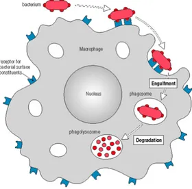
Macrophages and neutrophils show a variety of cell surface receptors, which make them able to recognise different types of microorganisms; the attack of a ligand to these receptors induces actin polymerization that allows the plasma membrane of phagocytes to surround the pathogens and to incorporate them forming a phagosome which becomes acidified (Figure 1.9). In addition to being phagocytic, macrophages and neutrophils have granules, called lysosomes, that contain lysozyme and hydrolases; the phagosome fuses with one or more lysosomes to generate a phagolysosome in which the lysosomal contents are released to degrade bacterial cell wall (Figure 1.9). After degradation, phagolysosomes exhibit the antigenic determinants on their cell surface with the consequent triggering of the mechanism of specific immune response (Figure 1.9) (Janeway et al., 2001).
Figure 1.9 Phagocytosis mechanism as starting point of the innate immunity.
Compared to the innate immunity, specific immunity (or acquired or adaptive immunity) represents a more sophisticated system of defence. A peculiarity of this second line of defence is the ability to discriminate between exogenous and autologous antigens (Janeway et al., 2001).
Therefore, acquired immunity strengthens the role of the innate immune response, amplifying, focusing and directing this response against foreign agents. It is possible to schematically distinguish two types of specific immunity: humoral and cell-mediated immunity (Figure 1.10). The main components of acquired immunity are two other types of white blood cells that are called lymphocytes, which are divided into B lymphocytes (expression of humoral immunity and developed in the bone marrow) and T lymphocytes (expression of cell-mediated immunity and developed in the thymus). The acquired immune response consists in a first step of antigen-processing by macrophages and dendritic cells (termed “antigen presenting cells” or APC) and a second step of presentation to T and B lymphocytes which ensure the recognition by specific receptors (TCR and BCR, T and B cell receptors) expressed on their cell membrane. The binding of the antigen to these receptors determines the activation, proliferation and differentiation into effector cells by a process of clonal selection. In details, in the humoral immune response, activated B cells (or plasma cells) secrete antibodies (or immunoglobulins) that circulate in the bloodstream and permeate other body fluids, where they bind specifically to the foreign antigen that stimulated their production, causing its destruction (Figure 1.10). In cell-mediated immune response, some T cells, termed cytotoxic, attack the leukocytes which present the antigenic determinants complexed to proteins of the major histocompatibility complex of class I (MHC I), and destroy them (Figure 1.10).
Figure 1.10 The humoral and cell-mediated immune responses.
Other T cells, the helper T immature cells, recognise the complex formed between the antigen and MHC class II proteins, and produce cytokines, known as macrophage colony-stimulating factor and gamma interferon, which promote the activity of macrophages. Moreover, the helper T cells secrete interleukin-2, which stimulates the proliferation of abovementioned cytotoxic T cells (Alberts et al., 2007). Following expulsion of the antigen, the immune response gradually comes to an end. Some cells, produced during clonal expansion, undergo a phase of differentiation into memory cells, which are capable, in case of a possible new encounter with the same antigen, to respond more quickly and with higher efficiency (Alberts et al., 2007).
1.4.2 LPS and innate immunity
As described in the above paragraph 1.4.1, innate immune response is activated after recognition of bacterial conservative molecular targets termed PAMPs, by specific innate immune receptors, termed PRRs (Janeway, 1989). PAMPs have three common features that make them ideal targets for innate immune recognition (Medzhitov, 2001):
•! are produced only by microbes and not by host •! are invariant between microorganisms of a given class •! are essential for microbial survival
In case of Gram-negative bacteria, lipopolysaccharides respond to all aforesaid three features, being recognised as PAMPs by a component of a family of PRRs known as Toll-Like Receptors (TLRs). Upon recognition of the real endotoxic principle of LPS, the lipid A moiety, the host innate immune system is immediately activated. The core oligosaccharide or, in smooth bacterial colonies, the O-chain, are the antigenic determinants recognised by the adaptative immunity. TLRs comprise a family of ten transmembrane receptors in humans, which are characterised by an extracellular leucine-rich repeat (LRR) domain and an intracellular Toll/IL-1 receptor (TIR) domain (Hashimoto et al., 1988; Medzhitov et al., 1997). Among all TLRs, TLR4 is the receptor designated to recognise the LPS macromolecule, but its detection requires several accessory molecules. The first host protein involved in LPS recognition is a serum protein, the LBP (LPS- binding protein) that forms a high affinity complex with lipid A released from bacterial lysis or replication, or still bound to the outer membrane of the intact microbial
LBP is responsible for lipid A binding while the C-terminal domain is specific for further recognition by CD14 (Figure 1.11). This latter can be found in two forms, soluble (secreted into serum) and membrane bound (as glycophosphoinositol-linked protein on the surface of macrophages) (Haziot et al., 1996), and has a crucial role in the enhancement of host response to endotoxins since it facilitates the recognition of LPS by the final receptor complex TLR4/MD-2. It was demonstrated that CD14-deficient mice have a profound defect in responsiveness to LPS, thus showing the pivotal role of CD14 in LPS-recognition process (Haziot et al., 1996). Once CD14 binds to the LPS, it has been shown that CD14 can chaperone LPS from LBP to TLR4 at the cell surface. This latter is linked, through its extracellular domain, to a small glycoprotein, MD-2 (myeloid differentiation factor 2), which turns out to be absolutely necessary for the proper functioning of TLR4 receptor; indeed, the same MD- 2 protein is able, by itself, to recognise lipid A with a good affinity, and also to discriminate different lipid A types. Therefore, TLR4 might be considered as a "pseudo-receptor", since, in the absence of MD-2, does not recognise its ligand. The TLR4/MD-2 complex has been shown to bind LPS with higher affinity than soluble MD- 2 (Akashi et al., 2003). However, the binding affinity of MD-2 or the MD-2/LPS complex for TLR4 is the same (Visintin et al., 2005). TLR4 with the MD-2 protein forms an heterodimer that recognizes a common structural motif in different LPS molecules. The binding of LPS to TLR4/MD-2 receptorial complex can activate two different immune response pathways (Figure 1.11) (Park et al., 2009).
Figure 1.11 Schematized model of LPS signaling.
In the first case, it results in activation of the transcription factor NF-κB, which regulates the expression of genes encoding for inflammatory cytokines; whereas the second pathway results in activation of MAP-kinases which regulate the transcription of genes involved in increasing the stability of particular regions of the mRNA. In both cases, the final result is the amplification of the transduction signal with the consequent massive production of inflammatory proteins thus eliciting the inflammatory process (Figure 1.11) (Park et al., 2009). If the inflammatory response is amplified and uncontrolled, due to the high toxicity of the LPS from the infecting pathogen, there may occur a fulminating septic shock syndrome.
References
!! Adinolfi M. et al., Carbohydr. Res., 1996, 284, 119-133. !! Akashi S. et al., J. Exp. Med., 2003, 198, 1035-1042.
!! Alberts B. et al., Molecular Biology of the Cell, 4th edition (Ed.: Garland Science), New York, chapter 24, 2007.
!! Alexander C., Rietschel E.T. J. Endotoxin Res., 2001, 7, 167-202.
!! Brandenburg K. et al., Eur. J. Biochem., 1993,218, 555-563. !! Cardoso L.S., et al., Microb. Cell Fact., 2007,6, 1.
!! De Soyza A. et al., Innate Immun., 2008, 14, 127-144.
!! Fukuoka S. et al., Biochim. Biophys. Acta., 2001, 1510, 185-197. !! Golenbock D.T. et al., J. Biol. Chem., 1991, 266, 19490-19498. !! Hamad M.A. et al., Mol. Microbiol., 2012, 5, 962–974.
!! Hashimoto C. et al., Cell, 1988, 52, 269-279. !! Haziot A. et al., Immunity, 1996, 4, 407-414.
!! Hellerqvist C.G. et al., Acta Chem. Scand., 1971, 25, 955-961. !! Hellerqvist C.G. et al., Carbohydr. Res., 1969, 9, 237-241. !! Holst O. et al., FEMS Immunol. Med. Microbiol., 1996, 16,
83-104.
!! Janeway C.A. Jr. Cold Spring Harb. Symp. Quant. Biol., 1989, 54, 1-13.
!! Janeway C.A. Jr. et al., Immunobiology, 5th edition (Ed.: Garland Science), New York, 2001.
!! Kittelberger R., Hilbink F., J. Biochem. Biophys. Methods, 1993, 26, 81-86.
!! Lüderitz O. et al., Ann. NY Acad. Sci., 1966, 133, 347-349. !! Medzhitov R., Nat. Rev. Immunol., 2001, 1, 135-45.
1997, 388, 394- 397.
!! Mesbah N.M., Wiegel J., Ann. NY Acad. Sci., 2008, 1125, 44-57.
!! Netea M.G. et al., Trends Immunol., 2002, 23, 135–139. !! Oikawa M. et al., Org. Biomol. Chem., 2004, 2, 3557- 3565. !! Pålsson-McDermott E.M., O'Neill L.A., Immunology, 2004,
113,153-162.
!! Park B.S. et al., Nature, 2009, 458, 1191-5.
!! Pommerville J.C., Alcamo’s foundamental of microbiology, 9th
Edition, (Eds.: Jones & Bartlett Publishers), Burlington, Chapter 4, 2010.
!! Raetz C.R., Annu. Rev. Biochem., 1990, 59, 129-70.
!! Raetz C.R., Whitfield C., Annu. Rev. Biochem., 2002, 71, 635-700.
!! Raven P.H., Johnson G.B., Biology 9th
Edition, (Ed.: McGraw Hill), Chapter 34, 2011.
!! Rietschel E.T. et al., FASEB J., 1994, 8, 217-225.
!! Seydel U. et al., Eur. J. Biochem., 2000, 267, 3032-3039.
!! Silipo A. et al., Prokaryotic cell wall compounds structure and biochemistry (Eds. Konig H., Herald C., Varma A.) Springer-Verlag, Berlin, Germany, 133-154, 2010.
!! Staley J.T. et al., Microbial Life 2nd
Edition, chapter 4, 2007. !! Visintin A. et al., J. Immunol., 2005, 175, 6465-6472.
!! Zahringer U., Lindner B., Rietschel E.T., Endotoxin in Health and Disease (Eds.: H. Brade, S. M. Opal, S. N. Vogel, D. C. Morrison), Marcel Dekker, New York, Basel, 93- 114, 1999.
Chapter 2: Structural characterization of LPS and
LOS
2.1 Extraction and purification of LPS and LOS
The first approach aimed to the structural elucidation of LPS/LOS is represented by their extraction from intact microbial cells. This is conventionally achieved through two complementary procedures, that lead to the selective isolation of R-LPS (LOS) and S-LPS, exploiting their different amphiphilic character; indeed, the latter has higher hydrophilicity, due to the presence of the polysaccharide moiety of the O-chain, whereas the former, which lacks such portion, has a more pronounced hydrophobic character. Thus, these different physico-chemical properties allow to perform a selective extraction of the LOS and LPS, with a high degree of purity. Lyophilized bacterial cells are firstly washed with acetone, water and ethanol to remove cell contaminants. After this initial step of purification, the procedure of extraction can take place. Typically, rough-type LPSs are extracted with a phenol-chloroform-petroleum ether procedure (PCP) (Galanos et al., 1969), while smooth-type LPSs are extracted with the hot phenol-water protocol (Westphal and Jann, 1965). The PCP extraction consists in the treatment of dried bacterial cells with phenol/chloroform/light petroleum ether in proportions 5:8:2 v/v/v; LOS is precipitated from pure phenol adding drops of water. After this extraction, cells undergo a second treatment with 1:1 v/v 90% phenol/water at 68°C. The two phases are then extensively dialysed and liophilised. As previously mentioned, typically the presence of the long O-chain moiety increases the hydrophilic nature of endotoxins, thus LPS molecules are extracted in
the water phase even though several factors, as the occurrence within the repeating unit of hydrophobic groups as well as charged monosaccharides or the length of the polysaccharide chains, may modulate LPS solubility in water. Thus, it can be detected either in the water or in the phenol extract.
After the extraction procedure, enzymatic treatments to remove proteins and nucleic acids are executed. The typical protocol consists in LPS/LOS purification with nucleases (DNAse and RNAse) and protease followed by dialysis in order to remove digested material. The screening for detection of LPS is realised by SDS polyacrylamide electrophoresis gel (SDS-PAGE) followed by silver nitrate staining which allows to define the typology of the extracted material and its purification degree (Kittelberger and Hilbink, 1993). The presence of LPS is testified by the observation of the typical “ladder-like” pattern of the electrophoretic migration, due to the presence of molecules differing in their structure for the number of repeating units composing the O-polysaccharide moiety. Conversely, LOS appears as a dark band that quickly migrates at the bottom of the gel, denoting the lower molecular weight of LOS lacking O-chain domain.
2.2 Structural determination of the saccharide moities
Structural investigation of LPS/LOS is a difficult task since their amphiphilic nature determines the tendency to form micelles with low solubility in both aqueous and apolar organic solvents. Several strategies have been developed to overcome this obstacle, including the usage of solvent mixtures and chemical modifications or degradation of the molecules to improve dissolution in simple solvent systems. The
common protocol consists in the analysis of the lipid portion and saccharide moiety separately by using several hydrolysis techniques, as aqueous 1% acetic acid or acetate buffer. The above mild conditions are sufficient to cleave selectively the acid-labile glycosidic linkage between the Kdo residue and the non-reducing glucosamine of the lipid A, thus releasing the oligo-/polysaccharide moiety in the water solution.
This linkage is very labile due to:
I.! the absence of a whichever electron withdrawing group at position adjacent to anomeric (C-3 in this case since anomeric is C-2) that favors the formation of the reaction intermediate oxonium ion;
II.! the passage from chair to half-chair conformation during the formation of the oxonium ion fastened by the presence of non-substituted carbons;
III.! the presence of a hydroxyl group in axial configuration at C-5 that contributes to a steric energy release in the formation of the intermediate ion.
The insoluble lipid A moiety can then be recovered by centrifugation and several washes with distilled water, whereas the supernatant is constituted by O-antigen still linked to core region. This does not complicate the structural investigation of the O-antigen since, in terms of monosaccharide residues number, the contribute of the core fraction is negligible respect to the O-chain. A disadvantage of the above described methodology, based on an acid treatment, is that it produces a Kdo reducing unit with a structural microheterogeneity (α and β anomers of pyranose and furanose rings, condensed or anhydro forms) that can
render the study of the core oligosaccharide of LOS by NMR spectroscopy particularly difficult. Thus, typically, the procedure to determine the primary structure of the core oligosaccharide portion considers the de-lipidation of the intact LOS by means of alkaline treatment in two steps: first, a mild hydrazinolysis in anhydrous conditions to remove ester-linked fatty acids from lipid A; second, a strong alkaline hydrolysis is performed to obtain the cleavage of amide-linked acyl chains too (Holst, 2000). The obtained product is represented by a homogenous phosphorylated oligosaccharide, comprehensive of the lipid A disaccharide backbone, that can be easily studied through chemical and spectroscopical investigations. Obviously, base-labile substituents of glycidic or non-glycidic nature are lost during this procedure. Therefore, it is possible to use the acid approach as a complement to the alkaline degradation, in order to evaluate the occurrence of base-labile groups lost during the alkaline treatment (i.e. pyrophosphate groups).
The primary structure determination of the saccharide portions so far obtained currently exploits state-of-art NMR experiments and soft-ionisation mass spectrometry techniques, together with compositional and linkage chemical analyses that allow the determination of:
•! the quali-quantitative composition of sugar residues •! the absolute configuration of each monosaccharide •! the cyclisation ring sizes
•! the glycosylation points of each monosaccharide •! the anomeric configuration of the glycosidic linkages •! the monosaccharide sequences
Additional information on the nature and the sequence of monosaccharides can be obtained from ad hoc selective cleavages of the polysaccharide such as partial acid hydrolysis, acetolysis, Smith degradation, β-elimination and solvolysis.
2.2.1 Chemical analyses of the saccharide moieties
Chemical analyses are useful to obtain important preliminary information regarding primary structure of poly/oligosaccharides. The procedure that provides the usage of Gas-Chromatography coupled with Mass-Spectrometry (GC-MS), can only be realized after conversion of the monosaccharides into apolar and volatile derivatives. There is a plethora of protocols that can be employed to identify the monosaccharide type as well as the glycosylation positions, thus the appropriate choice of the derivatisation method can highlight specific features of the sugar residues within the native LPS/LOS structure. Typically, the qualitative analysis by GC-MS is achieved by treating the oligo- /polysaccharide with MeOH/HCl leading to solvolysis of the molecule and to the formation of the O-methyl glycoside for each monosaccharide. Subsequent acetylation with acetic anhydride in pyridine produces the per-acetylated O-methyl glycosides (AMG), that can be injected and analysed by GC-MS. Comparison of the retention times from the GC analysis and the fragmentation pattern from the MS spectra with opportune standards allows to the identification of the monosaccharide residues. Quantification analysis can then be obtained by using an internal standard, that usually is per-acetylated inositol. Since the AMG protocol provides a solvolysis in acid conditions, several isomers for each monosaccharide may form (i.e. pyranose and furanose,
both as α and β anomers) resulting in the occurrence of many peaks in the corresponding chromatogram. Although not severely influencing the identification of the monosaccharides, this can lead to their misquantification. Therefore, an alternative and complementary approach that can be used leads to the formation of acetylated alditol derivatives. In this case, after strong acid hydrolysis with trifluoroacetic Acid (TFA), the carbonyl moiety of the monosaccharide residues is reduced with NaBH4, thus providing a single molecule from each
monosaccharide. The alditols are then acetylated and finally analysed via gas-chromatographic techniques.
A further procedure of chemical analysis that delivers the distinction between enantiomers provides that the solvolysis is executed with an enantiomerically pure alcohol as 2-(+)-octanol or 2-(+)-butanol. Thus, the absolute configuration of each monosaccharide residues can be determined (Leontein and Lönngren, 1978). Indeed, after acetylation and injection to GC-MS, the comparison of the retention time of the acetylated 2-(+)-octyl glycosides with the ones of a mixture of per-acetylated 2-(±)-octyl glycosides of standard monoses in D or L
configuration allows the assignment of the monosaccharide configuration.
The determination of ring size and glycosylation site of the monosaccharides is a further information obtainable through GC-MS analysis. The procedure consists in an extensively O-methylation of the oligo/polysaccharide with CH3I in strongly alkaline conditions. Then, the
permethylated oligo-polysaccharide is hydrolysed in acid conditions and reduced with a marked boro hydride (NaBD4). The alditols so obtained
have free hydroxyl groups only at the positions previously involved in glycosidic linkages and cyclisation, and can be then acetylated. These