Acute sensory
neuronopathy:
Report of a child
with remarkable
clinical recovery
Article abstract-A 9-year-old boy presented with acute sensory neu ronopathy, a condition generally associated with poor recovery. Although sensory nerve action potentials were abolished and sural nerve biopsy showed marked loss of myelinated fibers, he eventually made an almost complete clinical recovery.
NEUROLOGY 1994;44:762-764
J.M. Fernández, MD; A. Dávalos, MD; l. Ferrer, MD; C. Cervera, MD; A. Codina, MD; and J.L. Ochoa, MD, DSc
In 1980, Sterman et al1 described a new clinical
entity: acute sensory neuronopathy, a monophasic disorder characterized by rapid onset of general ized paresthesias, ataxia, and areflexia which "pro gresses rapidly to a severe, mainly proprioceptive sensory deficit."1 Sensory nerve action potentials are markedly reduced or absent, but motor function remains intact. The site of the lesion is thought to be at the dorsal root ganglia and, in the similar cases reported to date, ultimate recovery has been very poor.1·5
We present a patient with the typical clinical and electrophysiologic features of this syndrome who made a good functional recovery.
Case report. A previously healthy 9-year-old boy pre sented with fever, headache, and generalized exanthema 10 days before admission. Eight days later he developed vomiting, abdominal distension, and constipation. He had not been exposed to toxic substances and had not been given pyridoxine.
On adroission, arterial blood pressure was 110/60 mm Hg, heart rate 106 per minute, and respiration rate 40 per minute. Rectal temperature was 39 ºC. He was fully conscious and neurologic examination was normal. Abdominal x-rays revealed a distended small intestine. There was mild leukocytosis with shift to the left. Laparotomy disclosed mesenteric adenitis. He was given clindamycin and co-trimoxazole, and the fever subsided in 2 days. On his fifth day in the hospital he acutely developed numbness of the left side of the face and tongue that rapidly extended to the entire body. He com plained of dry eyes and mouth and generalized paresthe sias. In addition, he developed acute bladder paresis re quiring catheterization.
On examination, his mental status was normal and his pupils were mydriatic (7 mm), equal, completely unreactive to light, and near accomodation. Light touch and pinprick sensations of the face were reduced bilater ally, and corneal reflexes were absent. Ocular move ments and the rest of the cranial nerves were intact. Muscle bulk and strength were normal. There was mild loss of pain and temperature sense, severe iropairment of position and vibration sense in all limbs, and astereogno sis. He had profound trunca! and limb sensory ataxia which prevented hiro from walking or standing unaided. Limb tendon reflexes were absent, but jaw jerk was nor mal. Plantar responses were flexor.
Blood pressure fluctuated between 120/70 and 100/60 762 NEUROLOGY 44 April 1994.
mm Hg, but no orthostatic hypotension was detected. Normal values were obtained for CBC, chemistry, serum
lead and mercury, liver and thyroid functions, serology,
and immunology. Urine screen for porphobilinogen and porphyrins was negative. Cultures of peritoneal liquid, blood, and stools were negative. Small-intestine biopsy revealed no pathologic changes. EEG and brain CT were normal. CSF was clear and acellular; the glucose content was 60 mg/dl and the protein was 65 mg/dl with no dis crete bands on electrophoresis.
Clinical follow-up. Intestinal and bladder paresis improved within a week. Neurologic status remained un changed for 2 months. At 6 months, pinprick and touch sensations and ataxia had mildly improved, but marked loss of position and vibration sensation was still present.
N eurologic status continued to improve gradually over time. Three years after onset he had fully reactive pupils, normal gait, and no ataxia. At the last follow-up, 6 years after onset, he had only tendon areflexia, mild facial hypesthesia, depressed cornea\ reflexes, and impaired stereognosis of the ulnar aspect of the right hand.
Neurophysiologic investigations. These were per
formed on severa\ occasions, the first on the 13th day after the onset of neurologic symptoms and the last 6 years later. Concentric needle exaroination showed no abnormal spontaneous activity on any occasion. The vol untary pattern on maximal effort was full in proximal and distal muscles with motor unit potentials of normal configuration. In addition, turns/amplitude analysis of the interference pattem6 of severa\ muscles was always normal. Single-fiber EMG examination of the extensor digitorum communis muscle on the 13th day and again 1 month later showed normal fiber density (1.6), normal jitter values (less than 55 µsec), and no blocking phenom
ena. Motor nerve conduction velocities, compound muscle action potential amplitudes (CMAPs), and F-wave laten cies in the median, ulnar, peroneal, and posterior tibial nerves were normal at all evaluations. Repetitive stimu lation of the ulnar nerve at 3 and 50 Hz, recorded with surface electrodes in the hypothenar eminence, was nor mal on the 13th day. However, sensory nerve action potentials of the median, ulnar, radial, and sural nerves were unobtainable, as were somatosensory evoked poten tials and blink reflexes. H reflexes and the proprioceptive silent périod of the soleus muscle were absent in all exaroinations and reroain so. Pattem-shift visual evoked potentials and brainstem auditory evoked potentials were normal. Masseter reflex elicited by an oscilloscope triggering hammer was normal.
Follow-up. Blink reflexes were abolished for 3 years.
E Control- 7years E 10 O" 10 � o 8 w 8 e,: o z ::J 6 :I: 6 >- 1-� 4 4 z w o 2 2
°'
w CD u. 2 4 6 8 10 12 14 DIAMETER (µm)ranging from 70 to 85 rosee. On the last examination, 6 years after onset, early and late responses were elieited with increased lateneies (Rl, 25 rosee; R2, 55 to 65 rosee). Sural sensory response was 2 µ V (sural eonduetion veloe ity [SCV], 32.4 rn/see), and right median sensory poten tial (third digit) was 5 µ V (SCV, 39.4 rn/see).
Patient-9years
2 4 6 8 10 12 14
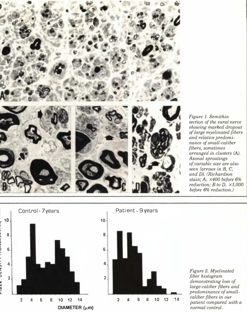
Figure l. Semithin section of the sural nerve showing marked dropout of large myelinated fibers and relative predomi nance of small-caliber fibers, sometimes arranged in clusters (A).
Axonal sproutings of variable size are also seen (arrows in B, C, and D). (Richardson stain; A, X400 before 6% reduction; B to D, Xl,000 before 6% reduction.) Figure 2. Myelinated fiber histogram demonstrating loss of large-caliber fibers and predominance of small caliber fibers in our patient compared with a normal control.
One year after onset of symptoms, a biopsy was obtained from the left sural nerve. The speeimen was fixed in 2.5% glutaraldehyde in Na+ eaeodylate buffer, postfixed in 1 % osmium tetroxide, and embedded in Aral dite. Semithin seetions were stained with Riehardson stain (toluidine blue, methylene blue, and borax).
Sections of the nerve showed marked loss of both myelinated and unmyelinated fibers (figure lA). Several sections revealed the presence of axonal sproutings (fig ure 1, B, C, and D). There were no cellular infiltrates in the epineurium or endoneurium, and no amyloid or metachromatic materials. In figure 2, a histogram of myelinated fibers shows loss of large-caliber fibers and predominance of small myelinated fibers in our patient compared with a normal control.
Discussion. This patient presented with general ized dysesthesias, autonomic dysfunction, ataxia, and profound proprioceptive deficit, but with nor mal motor power. Conventional and computerized EMG, motor conduction studies, repetitive nerve stimulation, and single-fiber EMG were normal, ruling out a subclinical motor disturbance or a neu romuscular transmission disorder (ie, botulism). These findings, combined with the absence of sen sory potentials and of blink and H reflexes, are diagnostic of acute sensory neuronopathy.1•7•8 This
diagnosis implies massive loss of neurons in the dorsal root ganglia, and therefore predicts poor recovery, as actually happened in the patients described previously.1•5 However, in this patient sensory functions improved considerably over sub
sequent years. Furthermore, there was electro physiologic and histologic evidence of partial senso ry regeneration over the following years. These findings suggest that a subpopulation of neurons underwent distal axonal degeneration-which manifested itself by a "dying back" neuropathy but not perikaryal death. In this situation, a cer tain degree of functional recovery might occur by way of distal sprouting, particularly in children. More severe lesions would result in death of the dorsal ganglion cell bodies (neuronopathy), leading to irreversible degeneration of the axon. 7•8 Such a
dose-dependent mechanism occurs in the clinical and experimental sensory neuropathy produced by pyridoxine9•10 and other neurotoxic drugs that may
involve selectively the dorsal root ganglion cells. 8
This patient's case demonstrates the clinical and pathologic overlap between the generally reversible acute sensory neuropathies and the usually irre versible acute sensory neuronopathies.
764 NEUROLOGY 44 April 1994
Acknowledgments
We are grateful to Mrs. Rita Murugarren and Mrs. Fátima Roca for their technical assistance.
From the Departments of Clinical Neurophysiology (Dr. Fernández) and Neurology (Drs. Dávalos, Cervera, and Codina), University Hospital Valle Hebrón, Universiqad Autónoma de Barcelona, Barcelona, Spain; the Neuropathology Unit, Department of Pathology (Dr. Ferrer), Hospital Príncipes de Espaí'la, Hospitalet de Llobregat, Spain; and the Neuromuscular Unit, Department of Neurology (Dr. Ochoa), Good Samaritan Hospital and Medica! Center, Portland, OR.
Supported by Fondo de Investigaciones Sanitarias grant 90/1152 (J .M.F.). Presented in part at the VIIth International Congress on Neuromuscular Diseases, Munich, Gennany, September 1990.
Received December 4, 1992. Accepted for publication in final fonn Octo ber 7, 1993.
Address correspondence and reprint requests to Dr. José M. Fernández, Department of Clinical Neurophysiology, University Hospital Valle Hebrón, 08035 Barcelona, Spain.
References
l. Sterman AB, Schaumburg HH, Asbury AK. The acute senso ry neuronopathy syndrome: a distinct clinical entity. Ann Neurol 1980;7:354-358.
2. Sacks OW. The disembodied lady. In: Sacks OW. The man who mistook his wife for a hat. New York: Summit, 1985:42-52.
3. Colan RV, Snead OC, Oh SJ, Kashlan MB. Acute autonomic and sensory neuropathy. Ann Neurol 1980;8:441-444.
4. Fagius J, Westerberg CE, Olsson Y. Acute pandysautonomia and severe sensory deficit with poor recovery. A clinical, neurophysiological and pathological case study. J Neurol Neurosurg Psychiatry 1983;46:725-733.
5. Knazan M, Bohlega S, Berry K, Eisen A. Acute sensory neu ronopathy with preserved SEPs and long-latency reflexes. Muscle Nerve 1990;13:381-384.
6. Nandedkar SD, Sanders DB, Stálberg EV. Automatic analy sis of the electromyographic interference pattern. Part 11: findings in control subjects and in sorne neuromuscular dis eases. Muscle Nerve 1986;9:491-500.
7. Asbury AK. Sensory neuronopathy. Semin Neurol 1987;7: 58-66.
8. Asbury AK, Brown MJ. Sensory neuronopathy and pure sen sory neuropathy. Curr Opin Neurol Neurosurg 1990;3: 708-711.
9. Albin RL, Albers JW, Greenberg HS, et al. Acute sensory neuropathy-neuronopathy from pyridoxine overdose. Neurol ogy 1987;37:1729-1732.
10. Xu Y, Sladky JT, Brown MJ. Dose-dependent expression of neuronopathy after experimental pyridoxine intoxication. Neurology 1989;39:1077-1083.