Contents lists available atScienceDirect
Pharmacological Research
journal homepage:www.elsevier.com/locate/yphrsReview
A metabolic perspective of late onset Alzheimer
’s disease
Miren Ettcheto
a,b,c,d,1, Amanda Cano
d,e,f,1, Oriol Busquets
a,b,c,d, Patricia Regina Manzine
a,c,d,g,
Elena Sánchez-López
d,e,f, Rubén D. Castro-Torres
a,c,d,h,i, Carlos Beas-Zarate
i, Ester Verdaguer
c,d,h,
María Luisa García
d,e,f, Jordi Olloquequi
j, Carme Auladell
c,d,h, Jaume Folch
b,d,
Antoni Camins
a,c,d,⁎aDepartament de Farmacologia, Toxicologia i Química Terapèutica, Facultat de Farmàcia i Ciències de l’Alimentació, Universitat de Barcelona, Barcelona, Spain bDepartament de Bioquímica i Biotecnologia, Facultat de Medicina i Ciències de la Salut, Universitat Rovira i Virgili, Reus, Spain
cInstitut de Neurociències, Universitat de Barcelona, Barcelona, Spain
dBiomedical Research Networking Centre in Neurodegenerative Diseases (CIBERNED), Madrid, Spain
eUnitat de Farmàcia, Tecnologia Farmacèutica i Fisico-química, Facultat de Farmàcia i Ciències de l’Alimentació, Universitat de Barcelona, Barcelona, Spain fInstitute of Nanoscience and Nanotechnology (IN2UB), Universitat de Barcelona, Spain
gDepartment of Gerontology, Federal University of São Carlos (UFSCar), São Carlos, SP, Brazil
hDepartament de Biologia Cel·lular, Fisiologia i Immunologia, Facultat de Biologia, Universitat de Barcelona, Barcelona, Spain
iLaboratorio de Regeneración y Desarrollo Neural, Instituto de Neurobiología, Departamento de Biología Celular y Molecular, CUCBA, Mexico jInstituto de Ciencias Biomédicas, Facultad de Ciencias de la Salud, Universidad Autónoma de Chile, Talca, Chile
A R T I C L E I N F O
Keywords: Alzheimer’s disease Type 2 diabetes mellitus Insulin
c-Jun N-terminal kinase inhibitors Licochalcone A
Neuroinflammation Reticulum stress
A B S T R A C T
After decades of research, the molecular neuropathology of Alzheimer’s disease (AD) is still one of the hot topics in biomedical sciences. Some studies suggest that soluble amyloidβ (Aβ) oligomers act as causative agents in the development of AD and could be initiators of its complex neurodegenerative cascade. On the other hand, there is also evidence pointing to Aβ oligomers as mere aggravators, with an arguable role in the origin of the disease. In this line of research, the relative contribution of soluble Aβ oligomers to neuronal damage associated with metabolic disorders such as Type 2 Diabetes Mellitus (T2DM) and obesity is being actively investigated. Some authors have proposed the endoplasmic reticulum (ER) stress and the induction of the unfolded protein response (UPR) as important mechanisms leading to an increase in Aβ production and the activation of neuroin-flammatory processes. Following this line of thought, these mechanisms could also cause cognitive impairment. The present review summarizes the current understanding on the neuropathological role of Aβ associated with metabolic alterations induced by an obesogenic high fat diet (HFD) intake. It is believed that the combi-nation of these two elements has a synergic effect, leading to the impairement of ER and mitochondrial func-tions, glial reactivity status alteration and inhibition of insulin receptor (IR) signalling. All these metabolic alterations would favour neuronal malfunction and, eventually, neuronal death by apoptosis, hence causing cognitive impairment and laying the foundations for late-onset AD (LOAD).
Moreover, since drugs enhancing the activation of cerebral insulin pathway can constitute a suitable strategy
https://doi.org/10.1016/j.phrs.2019.104255
Received 16 November 2018; Received in revised form 11 March 2019; Accepted 30 April 2019
Abbreviations: Aβ, amyloid β; AD, Alzheimer’s disease; AChEI, anticholinesterase inhibitors; AMPAR, α-amino-3-hydroxy-5-methyl-4-isoxazolepropionic acid re-ceptor; AβPP, amyloid precursor protein; AβPP/PS1, AβPPswe/PS1dE9; APOE, Apolipoprotein E; ATF4, activating transcription factor 4; ATF6, activating tran-scription factor 6; BACE1, beta-secretase enzyme 1; BDNF, rain-derived neurotrophic factor; BIP, binding immunoglobulin Protein; BMI, body mass index; CHOP, C/ EBP homologous protein; CNS, central nervous system; eIF2α, eukaryotic Initiation Factor 2; ER, endoplasmic reticulum; GADD34, growth arrest and DNA damage-inducible protein 34 kDa; GCN2, general control non-depressible 2; GRP78, glucose regulated protein 78; HFD, high fat diet; HRI, heme-regulated inhibitor kinase; HO-1, Heme oxygenase-1; IDE, insulin degrading enzyme; IL-6, interleukin 6; IR, insulin receptor; IRE1α, inositol requiring enzyme 1; IRS1, insulin receptor substrate 1; ISR, integrated stress response; LOAD, Late-onset Alzheimer’s disease; MAPs, microtubule-associated proteins; MMP-9, matrix metallopeptidase 9; MnSOD, manganese-dependent superoxide dismutase; MPTP, 1-methyl-4-phenyl-12,3,6-tetrahydropyridine; NFT, neurofibrillary tangles; NMDAR, N-methyl-D-aspartate receptors; NORT, novel object recognition test; NQO1, quinone oxidoreductase 1; p-CREB, phosphorylated cAMP response element-binding; PGC-1α, PPARγ-coactivator-1α; PERK, PKR-like endoplasmic reticulum kinase; PHF, paired helical filaments; PKR, RNA-activated protein kinase; PS1, presenilin; PS2, presenilin 2; PTP1B, protein-tyrosine phosphatase 1B; RAGE, The receptor for advanced glycation end products; T2DM, Type 2 diabetes mellitus; T3D, type 3 diabetes; TLE, temporal lobe epilepsy; TRAF2, tumour necrosis factor receptor associated factor-2; TNFα, tumour necrosis factor α; UPR, unfolded protein response
⁎Corresponding author at: Unitat de Farmacologia i Farmacognòsia, Facultat de Farmàcia i Ciències de l’Alimentació, Universitat deBarcelona, Barcelona, Spain.
Av. Joan XXIII 27/31, E-08028 Barcelona, Spain. E-mail address:[email protected](A. Camins).
1Both authors have contributed equally.
Available online 07 May 2019
1043-6618/ © 2019 Elsevier Ltd. All rights reserved.
for the prevention of AD, we also discuss the scope of therapeutic approaches such as intranasal administration of insulin in clinical trials with AD patients.
1. Introduction
The study of Alzheimer’s disease (AD) began in the early twentieth century, when the German physician Alois Alzheimer described thefirst case of this pathology in a 51-year old woman called Auguste Deter [1]. Her symptoms included remarkable memory loss, language difficulties and personality changes. After the post-mortem examination, the brain autopsy revealed specific neuropathological changes in the cerebral cortex [1–4], now known as the classical morphological symptoms of AD: amyloid β (Aβ) peptide plaques derived of the activity of the amyloidogenic pathway and neurofibrillary tangles (NFTs), composed by hyperphosphorylated tau protein [3–8].
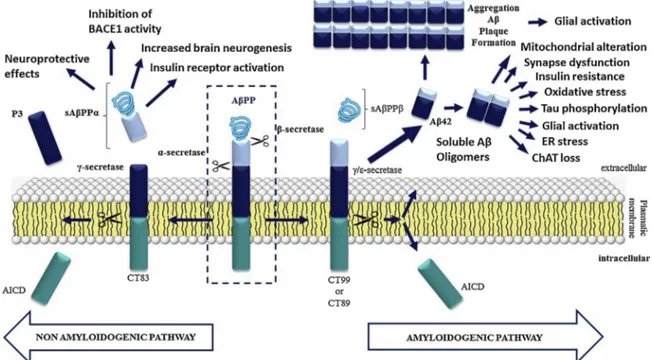
At present, the aetiological hypothesis most supported by the sci-entific community is the “amyloid cascade hypothesis”, which is sum-marized inFig. 1. According to this hypothesis, under physiological conditions amyloidβ protein percussor (AβPP) is cleaved by the en-zyme α-secretase, following the non-amyloidogenic pathway. This precludes formation of amyloidogenic peptides and leads to a release of secreted AβPP alpha (sAβPPα), which has neuroprotective properties [5]. On the contrary, in the amyloidogenic pathway, AβPP is cleaved by β-secretase (BACE-1) at N-terminus and, in turn, γ/ε-secretase cleaves it at the C-terminus to yield secreted AβPP (sAβPP), Aβ40/42 fragments (which remain in the extracellular space) and a C-terminal fragment with 99 amino acids (C99) that can be translocated to the nucleus. Here, it may induce expression of genes that promote neuronal death by apoptosis. Moreover, the soluble Aβ oligomers generated in this way affect synapse function, decrease neuronal plasticity, alter energy and glucose metabolism, induce oxidative stress and mitochondrial dys-function, and disturb celular calcium homeostasis [2,5].
During the early 1990s, some genetic factors -apart from AβPP-were shown to increase the risk of developing the disease, such as apolipoprotein E (APOE) or presenilin 1 and 2 (PS1 and PS2) [7–11].
These factors were also involved in the amyloid cascade. For instance, the allele for APOEε4 was been shown to impair Aβ clearance and promote its aggregation, leading to increased severity of this amyloid pathology [7–12]. In spite of this, the existence of other factors that can contribute to the pathogenesis of AD emphasizes its complexity. Some of these risk factors are shown inFig. 2.
Against this background, AD patients were classified into two groups. Thefirst one was formed for those subjects that developed the pathology due to genetic causes leading to the production of classical biomarkers like Aβ. This group covered about 3% of AD patients and was dubbed“familial or early-onset AD”. The remaining 97% of pa-tients were categorized as“sporadic or late-onset AD” (LOAD), whose progression was associated with advanced age, hypertension, hyperli-pidaemia, coronary disease, obesity and type 2 diabetes mellitus (T2DM) [12–14].
T2DM is a complex disease with a chronic evolution that requires continuous medical care, mainly focused on the reduction of global cardiovascular risk, peripheral complications and cognitive loss [15]. Unlike type 1 diabetes mellitus (T1DM), an autoimmune disorder characterized by the selective destruction of insulin-producingβ-cells [16,17], in T2DM there is an alteration in the mechanisms of uptake and/or secretion of insulin. This leads to a chronic increase in blood levels of glucose, resulting in a higher risk of macro and microvascular complications. T2DM is also associated with insulin resistance (IR), which is characterized by lower insulin activity at the cellular level, and affects the metabolism of carbohydrates, lipids and proteins [15]. As we have mentioned, in addition to a risk factor for cardiovascular pathol-ogies, T2DM is also an independent risk factor for LOAD [11–14]. Specifically, it is widely recognized that T2DM and AD share several kinds of abnormalities, including increased oxidative stress, impaired glucose metabolism and insulin resistance characterized by continuous hyperinsulinemia [12,12,13,14,18]. Likewise, some studies have
Fig. 1. Schematic representation of the APP’s protein processing in the cytoplasmic membrane. Both the amyloidogenic (right) and non-amyloidogenic (left) pathways are depicted. Thefirst one leads to the generation of Aβ oligomers that cause glial activation, mitochondrial alterations, synapse dysfunction as well as ER stress and insulin resistance. The second one favours the generation of the sAβPPβ peptide, which has been described to have neuroprotective effects: it promotes neurogenesis, favours insulin receptor activation and inhibits BACE1.
focused on the role of insulin receptor (IR), which might be an im-portant player in the pathology of AD, by contributing to the bio-chemical, molecular and histopathological characteristics of the pa-thology [13,19–27].
Given that the origin of the pathology is still unknown, and that there seem to be many players involved in its development, AD has been defined as a multifactorial disease (Fig. 2). Consequently, there are different research areas, in addition to neuroscience, trying to elu-cidate the origin of this pathology. In fact, prospective epidemiological studies have identified metabolic syndrome and T2MD as risk factors for multiple diseases of the nervous system [18–20]. Furthermore, an-imal studies have shown that hypercaloric diets affect the structure and functions of the hippocampus, although the specific mechanisms are unclear [28,29]. Thus, it has been reported that AβPPswe/PS1dE9 (AβPP/PS1) transgenic mice fed with a diet enriched in palmitic acid, showed reduced IR and increased insoluble Aβ peptide levels, as well as cognitive deficits [30]. Moreover, Ho and colleagues found evidence linking insulin resistance and increased relative risk for AD neuro-pathology development, by demonstrating that IR signalling can in-fluence Aβ production in the brain [31]. These results evidenced the relationship between metabolic alterations and progression of AD fea-tures, thus reinforcing the hypothesis of a metabolic aetiology of AD. Indeed, it has been proposed to re-name AD as“type 3 diabetes mel-litus” (T3DM) or brain-specific diabetes [32].
The present review is a state-of-the-art about the relation among obesity, Aβ oligomers and the IR modulation. In addition, we discuss the potential application of drugs modulating the brain insulin receptor pathway as targets for AD prevention.
2. An historical overview of AD’s hypotheses and available pharmacological treatments
Altghough over a century has passed since AD wasfirst described, the pathogenesis of this complex disease is still unclear. A number of theories about AD origin have been postulated so far and several drugs have been tested in accordance. Thefirst one, proposed in the 80 s, was the “cholinergic hypothesis”, which suggested that a dysfunction of acetylcholine-containing neurons in the brain contributes substantially
to the cognitive decline observed in AD patients [33–35]. This para-digm led to the development of the anticholinesterase inhibitors (AChEI), which are currently in the market and provide a symptomatic treatment of the disease [34–38]. However, these drugs only achieve a temporary improvement and they do not slow down or cure AD.
In 1992, a series of discoveries opened the door to the birth of the “amyloid cascade hypothesis”, which we have already discussed. This hypothesis stressed the role of Aβ peptide deposition in AD pathogen-esis, leading to neurofibrillary tangles, cell loss, vascular damage, and dementia [39–42]. Ostensibly, the appearance of plaques and the onset of AD would be related to the most common isoforms of Aβ, (1–40) and (1–42) [40–43]. However, some current preclinical discoveries pointed to Aβ as a cofactor or aggravator involved in a complex network of pathological changes in the brain, instead of considering Aβ as the main neurotoxin causing AD per se[44–48]. In any case, most drugs designed to inhibit amyloid synthesis have failed to modify the evolution of the disease. Notwithstanding, Verubescetat, a drug targeting Aβ or in-hibiting the β-site amyloid precursor protein cleaving enzyme 1 (BACE1), was reported to be more effective than placebo in treating AD [47,48].
Besides Aβ plaques, NFTs constitute the other main intracellular hallmark of AD. It was demonstrated that NFTs were composed of tau [48,49], a microtubule-associated protein (MAP) which acts as a major regulator of microtubule formation [50–53]. Interestingly, it was ob-served that the hyperphosphorylation of tau led to unconstructed mi-crotubules and the appearance of helically crossedfilaments that ag-gregate to form the NFTs [50–53]. In some cases, the presence of NFTs was correlated with the degree of dementia in patients with AD [53]. All these observations paved the way for the“tau hypothesis”, which postulates that the origin of AD is associated with an early conforma-tional change in the structure of tau [54]. Indeed, it has been demon-strated that the reduction of hyperphosphorylated tau alleviates the cognitive alterations induced by Aβ in a transgenic mice model of AD [55]. Although in terms of research the tau hypothesis has been a “supporting actor” when compared to the amyloid hypothesis, some anti-tau therapies have shown promising results [48,51]. For instance, TRx0237 (LMTX) is being currently tested in a phase III clinical trial as an inhibitor of tau aggregation, capable of reducing tau-mediated
Fig. 2. Some risk factors for the development of sporadic AD. Age, APOEε4 mutation, diet and lifestyle, gender, mental activity, cranioencephalic traumatisms and others factors such as lack of social engagement have been associated to the onset and/or progression of AD.
neuronal damage (clinical trial NT02245568). By this mechanism, this drug could modify the course of AD [48]. On another front, it has been reported that soluble oligomers of Aβ are able to accelerate the hy-perphosphorylation of tau [4,50]. Nevertheless, although the two markers are closely related neuropathologically, a mechanism linking both theories is still missing.
Evidence showing inflammatory microglia consistently associated with senile plaques in AD led to the“inflammation hypothesis” [56]. Indeed, it is known that local reactivity responses, such as microgliosis and astrogliosis, could be involved in the development of AD by fos-tering a severe neuroinflammation through the release of cytokines [42,56–62]. However, while it is accepted that neuroinflammation contributes actively to the development of AD, the fundamental ques-tion remains: is it the cause or the consequence of the underlying pa-thology? [60–64]. In this sense, the drug TTP488 (azeliragon), a re-ceptor for advanced glycation end products (RAGE) antagonist with anti-amyloid and anti-inflammatory properties is in a phase III trail. It has been shown that this compound reduces amyloid uptake in brain and lowers the inflammatory reaction in glial cells (NCT02080364) [48].
Finally, another major hypothesis about AD is the“excitotoxic hy-pothesis”, which describes how over-activation of glutamatergic transmission, especially of N-Methyl-D-aspartate receptors (NMDARs),
leads to a massive influx of calcium (Ca2+
) that damages neurons [65]. In mammals, NMDARs are distributed throughout the brain and mainly in the hippocampus. The hyperactivation of these receptors leads to the activation of a wide variety of intracellular pathways, inducing homeostatic alterations and diminishing neuronal viability, eventually leading to neuronal death [66–68]. However, the mechanism by which this process is carried out remains unclear since, even though this hy-pothesis advocates for an over-activation of NMDARs, it has been ob-served that patients who developed AD actually had a reduction of the number of these receptors in the cellular membrane [68]. Apparently, the lack of receptors leads to an increase in their sensitivity, causing a continuous activation leading to a progressive neurodegeneration due to the excitotoxic damage [68–70]. Interestingly, this also seems to be related to Aβ, since it has been demonstrated that Aβ causes an increase of the endocytosis of the NMDARs in cortical neurons, hence decreasing their expression in the membrane, which leads to an inhibition of sy-naptic plasticity [70]. Regarding the excitotoxic hypothesis, meman-tine, a NMDAR antagonist, was approved for the treatment of moder-ately severe to severe AD in 2002 by the European Agency for the Evaluation of Medical Products (EMEA). Unfortunately, as with any other approved treatment for AD so far, mematine only have shown symptomatic effects.
3. Mechanisms linking obesity and cognitive decline: results from preclinical models
Nearly 20 years ago, the Rotterdam study reported that T2DM pa-tients had increased risk to suffer dementia [71,72]. Today, obesity and diabetes, two T2DM-related disorders, are well established risk factors for AD [11,23,71–74]. In fact, it is of general concern that accumulation of fat in the adipose tissue favours the emergence of metabolic syn-drome and T2DM, due to IR signalling deficits in peripheral tissues. What is, perhaps, not so widely known is that obesity also affects the central nervous system (CNS) and it is associated with an exacerbation of cognitive decline [75]. In this respect, data from the clinical study Whitehall II and other research projects established a relation between body mass index (BMI) values (> 30 kg/m2), aging (> 50 years old) and the onset of cognitive deficits[73–78]. Nowadays, the molecular mechanisms at fault are still being studied. For instance, many authors suggest that vascular risk factors and inflammatory responses could also have significant roles [79–82].
Since the pathogenesis of AD remains unclear, it is necessary to use mixed models covering different hypothesis of the disease, evaluating
neuronal dysfunction, synapse loss and cell death, as well as po-tentiating Aβ effects in order to better understand its role in these neuropathological events [80–86]. For now, it seems that Aβ (1–42) oligomers would have a synergistic effect, accelerating the mechanisms related to cognitive impairment[86–89].
From a preclinical level, diet-induced obesity in in vivo models re-presents a suitable and reproducible method for identifying and un-derstanding potential mechanisms involved in T2DM or IR-induced cognitive loss. Using this experimental approach, the research group of Marta Di Carlo reported that rodents under a high fat diet (HFD) (60% kcal from fat) during 7 months exhibited brain abnormalities similar to the hallmarks of AD, such as the increase in tau phosphorylation, neuroinflammation and memory loss [90]. In addition, they demon-strated that those mice showed an increase in Aβ levels, which was associated with an increase in AβPP and BACE1 expression, both ac-companied by an increase in oxidative stress and mitochondrial dys-function. Our research group also demonstrated that mice fed with a HFD (45% kcal from fat) for 12 months, displayed insoluble Aβ peptide depositions in the brain, thus confirming the relation between the triad obesity, Aβ and cognitive loss [91].
Moreover, other studies showed that HFD-induced brain insulin resistance could decrease synaptic vesicle recycling, thus diminishing synaptic strength and leading to deleterious effects on cognitive beha-viour [92]. In a preclinical study, Hao and co-workers demonstrated that only three months of HFD feeding lead to a reversible deterioration of hippocampal synaptic plasticity, dendritic spine density and spatial memory in mice [93]. The authors propose that HFD activated a versible microglial synaptic phagocytosis process. In this line of re-search, Osborne and co-workers provided keyfindings on the implica-tion of Aβ in memory loss in HFD fed rodents [94]. In this study, they showed that intrahippocampal infusion of Aβ33–42 antibodies, with preferential affinity for aggregated Aβ oligomers, improved the memory process in rodents with HFD-induced cognitive loss. In addi-tion, the authors identified a potential mechanism involved in the process of cognitive impairment through the alteration of glutamate transmission by dysregulation of hippocampal α-amino-3-hydroxy-5-methyl-4-isoxazolepropionic acid receptors (AMPAR). However, other authors reported a detrimental cognitive effect of obesogenic HFD in 3xTgAD mice that was not associated with an increase in the brain le-vels of plaques, Aβ and tau [95]. Contrarily, Vandal and co-workers reported that an obesogenic diet induced insulin resistance in both non-transgenic and 3xTgAD mice, which worsened due to the HFD-amyloid pathology exacerbation [96]. This suggests that HFD exerts a sy-nergistic effect with AD-like pathology in 3xTgAD mice. Moreover, it has been described that the noxious effects of Aβ in brain can also contribute to pancreatic cell degeneration and insulin secretion, and, conversely, abnormal systemic changes might not only develop sec-ondary to brain dysfunction but might also affect AD progression [97]. This suggests that the interactions between the brain and the periphery act as a vicious circle with a crucial role in the development and pro-gression of AD [97]. These data are in line with other studies showing that brain Aβ could bind to IR in the liver and favour peripheral insulin resistance [98,99].
In another study, Ho and colleagues reported that HFD-induced insulin resistance was associated with a decrease in neuronal IR sig-nalling and insulin degrading enzyme (IDE) expression in Tg2576 mice, thus promoting cognitive loss and increased Aβ plaque formation [31]. These results were confirmed by other researchers [100]. In this regard, it is important to emphasize that IDE does not only degrade insulin, but also Aβ [30]. Thus, a decrease in its brain levels may contribute to both insulin signalling dysfunction and accumulation of Aβ. Likewise, other studies using animal models of obesity revealed that HFD induced an alteration in brain GLUT1 and GLUT3/GLUT4 glucose transporters, decreasing its up-take into the CNS. This was linked to cognitive deficits in the CA1 region of the hippocampus, which indicates an impaired synaptic plasticity [101]. Moreover, Ruiz and co-workers added
another interesting point by reporting that HFD increased peripheral glucose levels in AβPP/PS1 mice, probably due to hypothalamic al-terations [102]. Other studies provided data about the molecular me-chanisms involved in obesity-mediated neurodegeneration. For in-stance, previous results from our group reported an important down-regulation of PPARγ-coactivator-1α (PGC-1α) in AβPP/PS1 transgenic mice, a preclinical model of AD, under a HFD [30]. HFD caused a dropdown of PGC-1α levels, which was mediated by a decrease of phosphorylated cAMP response element-binding (p-CREB) protein le-vels [30]. The importance of these results is highlighted by the fact that PGC-1α has a key role in the mitochondrial biogenesis process and, therefore, in the mitochondrial function, which is essential to neuronal activity. Our study also demonstrated acceleration in the process of memory loss in AβPP/PS1 transgenic mice under a hypercaloric diet, as shown by short-term recognition memory values obtained in the novel object recognition test (NORT). This memory impairment was probably mediated by alterations in the brain IR and mitochondria [30]. Inter-estingly, a correlation between the down-regulation of PGC-1α ex-pression and AD has been also demonstrated, and the low exex-pression of PGC-1α in the brain of transgenic mice models of AD increases the formation of Aβ through the increase in BACE1 activity across the amyloidogenic route [103–107]. Accordingly, Katsouri and co-workers reported that the stereotaxic administration of a lentiviral vector to express human PGC-1α in the hippocampus and cortex of AβPP23 transgenic mice improved memory and showed neuroprotective effects, hence demonstrating the efficacy of this gene therapy as a potential treatment of AD [104]. In turn, Martins and co-workers reported that HFD impairs memory in 3xTgAD mice. This cognitive alteration was associated with changes in mitochondrial energetic balances and loss of synaptic contacts [107]. Since synapses are sites of high-energy de-mand, some authors correlate the synaptic degeneration process with dysfunctional mitochondria, which are transported to synaptic term-inals where high levels of ATP production are required [107].
In the same line, Sah and co-workers found that 3xTg mice fed with HFD suffered from memory impairment associated with a significant decrease in the expression of antioxidant enzymes, such as Heme oxy-genase-1 (HO-1) and manganese-dependent superoxide dismutase (MnSOD), through the inactivation of protein kinase B (AKT)- Nuclear factor (erythroid-derived 2 (NRF2) signalling pathway [108]. There-fore, obesity decreases antioxidant enzymes required for brain neuro-protection.
On another front, results obtained by Lin et al. revealed that 5XFAD mice fed with HFD exhibited cognitive impairment and hippocampal oxidative stress. Cognitive impairment is also believed to occur due to decreased levels of brain-derived neurotrophic factor (BDNF), which is a central molecule in synaptic plasticity [109]. Furthermore, this study demonstrated that brain alterations are independent of peripheral me-tabolic alterations, since glucose tolerance was not impaired in the transgenic mice. In turn, Thériault and co-workers showed that the HFD induces deterioration on cognitive performance in AβPP/PS1 mice, which was associated with a decrease in the enzymatic activity of matrix metallopeptidase (MMP)-9 and a reduction of mRNA and protein levels of BDNF in the brain [110]. These results reinforce the hypothesis of a metabolic aetiology in the molecular basis of the development of AD at the preclinical level [111]. In addition, others demonstrated a down-regulation of hypothalamic BDNF protein levels in HFD-fed transgenic mice compared with wild type HFD-fed mice [112].
In summary, all these preclinical studies showed the existence of an initial memory impairment process associated to obesogenic diets, which could be partially explained by increased levels of soluble Aβ and the inhibition of long term potentiation (LTP) linked to synapse loss. In addition, the down-regulation of BDNF levels mediated by PGC-1α, together with mitochondrial dysfunction, contribute to the decrease of synaptic connections, a marker that correlates with dementia in AD. Overall, these and other studies suggest that some improvement could be achieved through proper lifestyles, by avoiding metabolic alterations
and designing therapies targeting one or some of these biomarkers. 4. Are ceramides the bridge between peripheral type II diabetes and neurodegeneration in Alzheimer’s disease?
As discussed above, preclinical studies demonstrated that HFDs enhance cognitive loss, stressing its central effects. However, HFDs can also favour cognitive loss through peripheral alterations, specifically in the liver and adipose tissue. In an interesting study, Lyn-Cook and co-workers reported that HFDs induce liver damage (non-alcoholic stea-tohepatitis), by increasing the levels of pro-inflammatory cytokines (mainly TNFα) and ceramides [113]. Ceramides are toxic lipids which play key roles in brain oxidative stress, insulin resistance and cytos-keletal alterations, mechanisms involved in LOAD apparition [113]. In this respect, Dr. De la Monte suggested that chronic obesity induces insulin resistance associated with non-alcoholic steatohepatitis (NASH) [11–14,113]. In addition, T2DM-related obesity increases hepatic in-sulin resistance, thus favouring inflammation and metabolic dysfunc-tion, which leads to dysregulated lipolysis and the generation of cer-amides [113]. A significant increase in ceramide levels promotes ER stress in the liver, which exacerbates insulin resistance, inflammation, and oxidative stress [11–13].
In addition to these peripheral harmful effects, ceramides originated in adipose tissue or in the liver reach the bloodstream and pass through the blood-brain barrier, exerting toxic effects in neurons [13]. In this respect, we hypothesize that ceramides in association with Aβ 1–42 could exacerbate brain insulin resistance, oxidative stress, mitochon-drial alterations and neuroinflamation. Thus, there could be a vicious circle between brain and peripheral tissues: peripheral damage would increase the risk of cognitive loss and the neurotoxic effects of Aβ 1–42 in the hypothalamus would lead to an altered regulation of peripheral metabolism [114–116]. This idea is supported by the fact that Aβ 1–42 binds directly to the insulin receptor both in the periphery and in the hypothalamus, inducing a peripheral glucose intolerance and insulin resistance and eventually leading to T2DM [116]. Furthermore, it has been demonstrated that Aβ 1–42 induces hepatic insulin resistance through the activation of the Janus Kinase 2 (JAK2) in a preclinical model of familial AD [99]. In addition to the liver, some authors have shown that amyloid can accumulate in other peripheral tissues such as pancreas and skeletal muscle [117,118]. Therefore, the Aβ 1–42 pep-tide originated at the brain may constitute a key factor involved in insulin resistance and T2DM [97].
Likewise, Shinohara and Sato proposed the involvement of an X Factor underlying the bidirectional interactions between diabetes and LOAD. Hence, they suggest the existence of a vicious circle between diabetes and LOAD, which could be initiated by peripheral ceramides [119]. According to this idea, the authors put forward a potential equation where Aβ1–42 levels would be multiplied by an X factor. This unknown factor could be ceramides, which may amplify the neuro-toxicity of Aβ1–42 peptide. Consequently, cognitive loss could be pre-vented by breaking this circle, and antidiabetic treatments could be beneficial against LOAD.
5. Endoplasmic reticulum (ER) stress: a link between obesity and cognitive loss
Some research studies have indicated that the ER stress is involved in the appearance of degenerative diseases in the brain [120,121]. Obesity seems to exacerbate this situation, favouring cognitive loss and promoting AD-like neuropathology [122–125]. As we have discussed above, the accumulation of disease-specific misfolded proteins is a hallmark of AD, and ER could have a key role in this process.
In eukaryotic cells, such as neurons, the ER is a cellular compart-ment involved in calcium homeostasis, lipid biosynthesis and protein folding and maturation. Under normal physiological conditions, ER activity is paramount for most cellular mechanisms. Glucose-regulated
protein 78 or binding immunoglobulin protein (GRP78/BiP) molecules are bound to three different ER transmembrane receptor proteins, known as the PKR-like endoplasmic reticulum kinase (PERK), inositol requiring enzyme 1 (IRE1α) and activating transcription factor 6 (ATF6) [122]. Under ER stress, there is a release of BiP, which triggers the activation of ER stress response. The activation of PERK pathway leads to a signalling cascade which begins with the phosphorylation of the eukaryotic initiation factor 2 alpha (eIF2α) and eventually attenu-ates global protein synthesis [125]. Subsequently, this signalling pathway also promotes the activation of several downstream proteins like the activating transcription factor 4 (ATF4), proapoptotic C/EBP homologous protein (CHOP) and growth arrest and DNA damage-in-ducible protein 34 KDa (GADD34) [121,122].
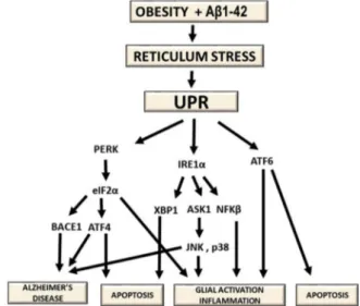
In neurons, an appropriate ER response is necessary for the control of protein synthesis and synaptic and neuronal function [125–127]. In AD, alterations in this response could lead to defects to long-term memory formation. Specifically, disruptions on the homeostasis of protein folding lead to the accumulation of misfolded proteins in the ER and upregulation of proapoptotic proteins like CHOP [124]. Hence, conditions inducing a sustained brain ER stress and leading to activa-tion of the unfolded protein response (UPR), such as obesity, could clearly result in deleterious effects, although the underlying mechan-isms are still not well understood. Some research groups have reported that palmitate could be one of the elements responsible for the acti-vation of these mechanisms in mice under HFD [128]. Thus, it was hypothesised that the association of palmitate and Aβ could increase even further the burden on the ER (Fig. 3) [129,130].
Other previous studies have shown that phosphorylation of eIF2α is increased in the hipocampus of preclinical mice models of AD. For in-stance, O’Connor and coworkers reported that PERK-eIF2α signalling activation increases AβPP processing and Aβ formation via direct up-regulation of BACE-1 [126]. This data was replicated in the 5xFAD mice model by other groups, which also combined heterogeneous knockouts for PERK+/− and the 5xFAD transgenic model (PERK haploinsuffi-ciency) [131–133]. The results showed improvements in memory def-icits in PERK+/−mice compared with 5xFAD mice. Furthermore, under insulin-deficiency conditions, PERK-eIF2α ER stress signalling pathway is dramatically activated in the brain of 5XFAD mice, thus increasing AD risk [133]. Likewise, the PERK pathway effector ATF4 is a repressor of CREB [121], which is required for synaptic plasticity as well as for learning processes.
Complementarity, eIF2α could also be phosphorylated by three different kinases associated with stressors. These kinases are the RNA-activated protein kinase (PKR), the heme-regulated inhibitor kinase (HRI) and the general control non-depressible 2 kinase (GCN2) [87,88,134]. They belong to the integrated stress response (ISR) and some of them have been linked to synapse loss and cognitive deficits in human and preclinical mice models of AD. For example, Mouton-Liger et al. reported that in human AD brains there is significant correlation between increase in PKR and BACE1 protein levels [135]. Strangely, Hwang and co-workers reported that PKR inhibition improves memory without affecting Aβ load in the hippocampus in 12-month-old 5xFAD mice [130]. The authors concluded that this PKR inhibition without affecting Aβ levels might provide a promising alternative strategy for developing a potential AD treatment. In another study, Ma and co-workers crossed PERK and GCN2 knockouts with AβPP/PS1 transgenic mice. They observed that eliminating either enzyme reduced brain le-vels of Aβ and prevented memory deficits associated with the AβPP/ PS1 phenotype [136].
On another research line, Sims-Robinson and co-workers evidenced an increase of ER stress activation on the hippocampus of B6-HF mice fed with HFD. They concluded that this was due to an inhibition of the IR signalling, probably mediated through the activation of the c-Jun N-terminal kinases (JNKs), which are direct inhibitors of the IR substrate 1 (IRS1) [137]. Indeed, an increase in the phosphorylation rates of serine amino acids of the IRS1 would induce desensitization of the
hippocampal IR and it is believed that the JNK1 isoform plays a pro-minent role in this mechanism [138]. In the same line, Liang and co-workers demonstrated a significant increase in the phosphorylation of both PERK and eIF2α, as well as IRE1α and JNK, in hippocampus and frontal cortex of HFD rats [139]. Therefore, they concluded that UPR activation was increased in the brain of obese rats, and that this me-chanism was involved in insulin resistance and cognitive loss through JNKs activation and IRS1 inhibitory phosphorylation. This data sup-ports the metabolic hypothesis of the appearance of cognitive deficits independent of Aβ production.
The administration of ursolic acid is a potential strategy tofind a therapeutic approach for these alterations. This natural triterpenoid compound exerts many pharmacological actions such as anti-oxidant, anti-inflammatory and anti-tumoral effects. Thus, Lu and co-workers demonstrated that ursolic acid has a protective effect preventing ER activation in the hippocampus, hence improving cognitive responsive-ness [128]. Furthermore, quercetin, a polyphenolic flavonoid com-pound present in a variety of fruits and vegetables, exerts beneficial effects on obesity and cognitive processes by preventing the activation of PERK-eIF2α signalling pathway and avoiding phosphorylation of ATF4 through GADD34 induction [140,141]. Likewise, it has been shown that the administration of the polyunsaturated fatty acid α-li-nolenic acid to aged female Sprague-Dawley rats fed with a HFD im-proved the cognitive process [142]. Its beneficial effects are mediated by the inhibition of UPR response through the down-regulation of brain ATF4 levels, thus leading to the increase of p-CREB phosphorylation rates [142].
6. Synaptic loss mediated by neuroinflammation in obesity Obesity has been associated with chronic inflammatory processes derived from the activity of hypertrophic adipocytes, free fatty acids and reactive oxygen species [143–145]. In the CNS, microglia have significant roles in the control of these mechanisms.
Under physiological conditions, microglial cells present a branched morphology and are responsible for the production of anti-in-flammatory and neurotrophic factors (inactive or unreactive state (M2) [146]. However, in some conditions such as obesity, microglial cells become reactive and change their morphology to amoeboid. This is followed by an enhancement of pro-inflammatory responses, both on microglia itself and in other cells such as astrocytes, through the release
Fig. 3. Chain of events relating obesity alterations, the accumulation of Aβ1–42 and the stress on the reticulum. Activating stimuli on the ER cause release of BiP and the initiation of UPR signalling. These mechanisms have been related to the appearance of pathologies like AD through the upregulation of apoptotic and glial reactivity biomarkers.
of certain cytokines (M1 state) [147]. Eventually, M1 microglia and astrocytic activity lead to neuronal death and pathologies like AD [148,149]. These assumptions have been supported by a meta-analysis of cytokines, which revealed increased concentrations of interleukin 6 (IL-6) and tumor necrosis factor α (TNFα) in peripheral blood of AD patients [150].
As discussed above, when any of the pathways of the UPR activates, there is an up-regulation of nuclear factor kappa-light-chain-enhancer of activated B cells (NF-kB), a key modulator of pro-inflammatory genes [151]. Likewise, several studies indicate that inflammation and ab-normal ER activity are critical events in the establishment of hy-pothalamic and peripheral insulin resistance in metabolic disorders. Thus, in animal models of T2DM and obesity, inflammation in the hy-pothalamus is an important part of the underlying pathogenic me-chanism, especially the activation of TNF-α and the kinase inhibitor of nuclear factor kappa-B kinase subunit beta (IKKβ)/NF-kB signalling axis [87,88,116,152]. Specifically, under a HFD, IKKβ becomes activated and phosphorylates IRS1 inhibitory serine sites, resulting in a desensi-tization of IR signalling [57]. In addition, cortical and hippocampal dysfunction seems to share common pathways in AD.
JNKs constitute another important element regulating inflammation and IR. JNKs pathways control several processes, including insulin re-sistance in peripheral tissues, such as liver or pancreas, and also in the brain [153–155]. Usually, these pathways are upregulated by cytokines and ER stress. Hence, it is known that neuroinflammation increases brain levels of TNFα, which activates JNKs -mainly JNK1- and leads to a decline of insulin signalling [56,57]. In addition, the activation of ER/ UPR response, specifically IRE1α pathway, triggers the splicing of X-box binding protein 1 (XBP1) mRNA. The products of this splicing also activate JNK. This process has important effects on the development of apoptotic processes accounting for cognitive decline [156]. Ad-ditionally, IRE1α can also be phosphorylated by JNKs, leading to a recruitment of tumour necrosis factor receptor associated factor-2 (TRAF2), which would enhance further apoptotic responses through caspase 12. Therefore, it seems that the UPR and JNKs are both in-volved in neuronal apoptosis processes, which are favoured by obesity [125,152].
Interestingly, the expression of protein-tyrosine phosphatase 1B (PTP1B) also becomes up-regulated in obesity models using HFD [157]. This phosphatase is located in the ER membrane and it is a major ne-gative regulator of insulin, leptin and BDNF receptors (Tyrosine Re-ceptor Kinase B; TRKB). Under physiologic conditions, the upregulation of PTP1B is promoted by TNFα and it has significant effects on cogni-tion and memory formacogni-tion. Elevated levels of PTP1B dephosphorylate tyrosine residues and inhibit downstream signal transduction of the
previously mentioned receptors, but activate microglia [158]. In addi-tion, it has been demonstrated that PTP1B is a regulator of the UPR response through the modulation of PERK/eIF1α signalling in periph-eral and neuronal cell culture studies [157]. Likewise, PTP1B inhibition attenuates neuronal toxicity in the ER mediated by the mitochondrial neurotoxin rotenone and tunicamycin [159].
Finally, soluble Aβ peptide oligomers can activate microglial cells and increase TNF-α brain levels [56,57]. Therefore, obesity and Aβ can together overstimulate an aberrant TNFα signalling, leading to activa-tion of the stress kinases that block insulin signalling in the brain. Following this line of thought, obesity and neurodegeneration may run in parallel in a complex mechanism where the activation of NF-kB would lead to the expression of inflammatory cytokines, hence pro-moting a neuroinflammatory state.
7. Pharmacological approaches for metabolic late onset Alzheimer’s disease treatment
According to the metabolic hypothesis of AD, therapies capable of restoring normal brain insulin signalling in the CNS may have bene-ficial effects on brain function. In this sense, a growing body of evidence suggest that insulin receptor have multiple brain functions related to cognition, neuroprotection through the activation of Akt, modulation of AβPP and Aβ levels, neuroinflammation and synapsis formation. Hence, brain insulin dysregulation could contribute to AD pathogenesis, and drugs involved in the modulation of insulin receptor could have po-tential application in the treatment of AD. This could be achieved not only by improving brain cognition at the hippocampal level, but also modulating the hypothalamus and peripheral tissues such as the liver, which would improve metabolic diseases such as T2DM. Consequently, bearing in mind that AD can be considered a metabolic alteration, its treatment should not only focus in brain processes such as memory, but also in peripheral organs related to insulin resistance (Table 1). 7.1. Intranasal insulin
Mao and co-workers reported that the administration of intranasal insulin for 6 weeks in AβPP/PS1 mice improves cognitive function by activating non-amyloidogenic pathways that favour a decrease in brain Aβ levels and Aβ plaque deposits [160]. Therefore, activation of brain insulin receptors improves Aβ pathology at preclinical level and in-tranasal insulin may be a potential strategy to modify the course of AD (Table 1).
Several clinical studies have also examined the effects of intranasal-administered insulin in AD patients. For instance, Craft and co-workers
Table 1
Clinical trials that are being conducted with drugs that act by activating the insulin receptor and the signaling pathway for the treatment of Alzheimer’s disease.
Drug Phase CT number Disease
Rosiglitazone II NCT00381238 Mild to moderate AD
Rosiglitazone II NCT00334568 Examine the drug response in patients with AD
Rosiglitazone I NCT00688207 AD (the study is designed to assess the pharmacokinetics of Rosiglitazone) Rosiglitazone III NCT00428090 Mild To Moderate AD
Rosiglitazone III NCT00550420 Mild to moderate AD Rosiglitazone III NCT00348309 Mild to moderate AD
Pioglitazone II NCT00982202 AD
Pioglitazone III NCT01931566 Mild to moderate AD
Pioglitazone III NCT02284906 AD
Intranasal Insulin Glulisine II NCT01436045 Mild To Moderate AD
Intranasal insulin detemir II NCT01595646 AD or amnestic mild cognitive impairment Metformin II NCT00620191 Amnestic Mild Cognitive Impairment Liraglutide Not Applicable NCT01469351 AD
Exendin-4 II NCT01255163 AD
Exendin-4 III NCT02847403 Cognitive Decline in Dysglycemic Patients
HM15211 I NCT03374241 Obesity
GIP/GLP-1 Co-Activity Not Applicable NCT03526289 Overweight and Type 2 Diabetes Insulin NCT00438568 Mild To Moderate AD
evaluated the effects of intranasal administrations of either insulin (10 or 20 IU twice a day for a total dose of 20 or 40 IU per day) or placebo (saline twice a day) for 4 months in AD patients (clinical trial number NCT00438568) [161,162]. They assessed changes in cognitive para-meters, brain glucose metabolism and Aβ levels in cerebrospinal fluid (CSF). The results of the trial suggested that the administration of in-tranasal insulin could improve cognition and brain glucose metabolism in patients with mild cognitive impairment or AD.
After the potential positive effects of intranasal insulin in AD, dif-ferent pharmaceutical preparations of insulin have been developed to improve its bioavailability. For example, insulin detemir incorporates fatty acids to its chemical structure, thus increasing its binding to al-bumin and allowing a slower elimination, which lengthens its half-life [162]. Craft and co-workers reported that the positive effects of insulin detemir on memory are associated with an increased volume in brain regions affected by AD neuropathology [162]. However, the authors reported that the efficacy of insulin detemir decreased over longer-term administration in these patients, whereas regular insulin continued to provide beneficial effects on memory. The explanation of these results is still unclear, although it has been proposed that long-acting insulins may desensitize the insulin receptor and thereby increase insulin re-sistance [56].
Finally, is worth to mention that cognitive processes in AD patients are influenced by gender and APOE4. Interestingly, cognition function in APOE4-negative patients is usually improved after intranasal insulin, while APOE4-positive AD patients showed contradictory results [26,163]. Likewise, men respond better to insulin than women [26]. 7.2. Incretins
This group includes some synthetic long-acting analogs such as Glucagon-like peptide-1 (GLP-1), receptor agonists such as exendin-4, as well as liraglutide and lixisenatide, which possess insulinotropic activity, among others.
At preclinical level, intraperitoneally administration of liraglutide or lixisenatide improves neuropathological markers of AD, such as de-crease in the number of plaques and glial activation, and also improves cognition parameters in AβPPSwe/PS1dE9 mice [164]. However, sev-eral clinical studies are now evaluating the potential efficacy and safety of GLP-1 mimetics in AD, such as liraglutide. For instance, the clinical trial NCT01469351 evaluated the efficacy of liraglutide in AD patients, reporting that 6-month treatment with this drug improved brain glu-cose metabolism with a slight improvement on cognition (Table 1). Another clinical study, the NCT02140983, evaluated the effects of lir-aglutide on memory and attention of elder patients with insulin re-sistance. Worth to mention, the patients had pre-diabetes and half of all subjects had a family history of dementia. However, no data from this study has been reported yet. Exendin-4 is another incretin-mimetic long-acting GLP-1 receptor agonist approved for T2DM treatment, which also have shown neuroprotective effects in preclinical models of AD [165]. Unfortunately, clinical trials with exendin-4 in early-stage AD, such as NCT01255163, did not reported beneficial effects on cog-nition.
Likewise, glucose-dependent insulinotropic polypeptide (GIP) ana-logues such as D-Ala2-GIP also showed neuroprotective effects on
sy-naptic plasticity and cognition in animal models. Thus, it has been re-ported that these compound significantly reduces Aβ42 plaques and neuroinflammation in preclinical AD mice models [166]. Since these peptides were effective at the preclinical level, the next strategy was the development of dual peptide agonists GLP-1/glucagon and GLP-1/GIP combined. Moreover, triagonists GLP-1/GCG/GIP are in early stages of development. Thus far, two studies in murine AD models have reported that triple agonists improve cognitive processes by increasing BDNF levels and p-CREB signalling pathway, and by decreasing plaques and neuroinflammation [167]. However, most research studies with tria-gonists have been carried out in metabolic disorders such as T2DM and
obesity, where they are more effective than the single administration of each peptide alone. Regarding clinical studies, only the triagonist HM15211 is under investigation as a treatment for obesity (NCT03374241). In this sense, the next aim is to evaluate the efficacy of this triagonist, which integrates the actions of the three endogenous hormones, in T2DM and weight decrease, as well as in other potential diseases.
7.3. Metformin
It has been shown that the anti-diabetic drug metformin is effective in preclinical models of AD [168]. Thus, Ou and co-workers reported that metformin improves cognitive process, decrease the Aβ production and neuroinflammatory response in the hippocampus of AβPP/PS1 mice [169]. The authors suggested that the neuroprotective effects of metformin are mediated by the modulation of the AMPK/mTOR/S6K/ BACE1 pathway. However, these neuroprotective benefits of metformin in AD are contradictory, since a recent study in transgenic models of tauopathy with cognitive deficits has shown that metformin can ag-gravate the risk of tauopathy in diabetic patients [170].
In turn, Infante-Garcia and co-workers studied in a mixed murine model of AD and T2DM (AβPP/PS1x db/db mice) the effects of the anti-T2DM polypill (PP), which contains several drugs used to treat anti-T2DM, including metformin, aspirin, a generic statin, and an angiotensin-converting enzyme inhibitor [171]. They reported that PP could be a suitable strategy for the treatment of serious complications of T2DM, such as cognitive alterations.
Recently, Koenig and co-workers published the results of a clinical trial with metformin (NCT01965756) in AD patients with mild cogni-tive impairment or mild dementia. They reported that metformin slightly improved learning and memory processes [172]. In another clinical trial (NCT00620191), the effects of metformin in Amnestic Mild Cognitive Impairment (MCI) were evaluated. Specifically, the aim of this study was to assess the changes in a memory and general cognitive function test (the Alzheimer’s Disease Assessment Scale-cognitive sub-scale-ADAS-Cog used in clinical trials.). The study also aimed to com-pare brain function through mean changes in PET scan between the patients treated with metformin and those treated with placebo. So far, no results from this study have been reported yet.
7.4. Thiazolidinediones
Thiazolidinediones are agonists of peroxisome-proliferator activated receptors (PPARs). They possess antidiabetic activity and have also shown neuroprotective effects in preclinical models of AD [48]. These compounds are nuclear hormone receptors that induce physiological responses through the regulation of gene expression, and they are in-volved in the metabolic regulation of carbohydrates, proteins and li-pids. They also reduce neuroinflammation by inhibiting glial activation, since it has been reported that rosiglitazone significantly improves cognition in rats after Aβ injection in the hippocampus through in-hibition of microglial cytokine release [173]. According to these results, the regulation of microglia constitutes a key target involved in cogni-tive improvement in preclinical AD models.
Regarding the use of rosiglitazone in clinical studies, Gold and colleagues reported that administration of 2-mg or 8-mg of this com-pound during 24 weeks in APOE-ε4 negative patients does not show any statistical difference when compared to placebo treatment [174]. The current clinical study NCT00334568 aims to evaluate the effects of rosiglitazone on i) cerebral glucose utilization measured by [18F] FDG uptake and ii) cognitive process in AD. No results have been reported yet. In turn, the study NCT00348309 assessed the effects of a 54-week treatment of rosiglitazone (extended release tablets) combined with donepezil on cognitive parameters of patients with mild to moderate AD. Again, no clinical improvement was achieved with this treatment [175].
On another front, previous studies have reported neuroprotective effects of pioglitazone in preclinical models of AD, through a decrease in neuroinflammation and also in mRNA and protein levesl of BACE1 [176]. This compound was able to significantly decrease the cerebral Aβ levels, and could hence constitute a potential drug with the capacity to modify the course of the disease. Interestingly, Fernandez-Martos and co-workers reported that acute 2-week treatment with combined leptin and pioglitazone improves cognition and decrease neuropatho-logical parameters of AD in AβPP/PS1 mice [177]. In turn, Geldmacher and colleagues evaluated the safety of long-term treatment with pio-glitazone (15-mg tablets) in elderly nondiabetic AD patients [178]. The authors reported that pioglitazone was well tolerated and safe during the 18-month treatment trial. In the same line, the NCT01931566 study evaluated the effects of pioglitazone compared with placebo in AD patients with mild cognitive impairment (MCI). The aim of this study was to delay the onset of memory loss in cognitively normal partici-pants who were at high-risk for developing MCI within the next 5 years. Currently, no results have been reported. However, the clinical trial NCT02284906 aims to evaluate the safety and effectiveness of piogli-tazone on cognitive function in participants who have completed the previous study.
Regarding thiazolidinediones, we can summarize that, in spite the promising results observed in preclinical studies, no clinical study has reported successful results yet [179–181]. Therefore, additional clinical studies with PPARγ agonists are still required, either in monotherapy or in combination with other drugs, in order to evaluate the efficacy in improving brain insulin/IGF-1 resistance and thus improving cognition and preventing neurodegeneration in initial stages of AD.
7.5. c-Jun-N-terminal kinase inhibitors
Although SP600125 is the best characterized inhibitor of JNK ac-tivity, its application has been impeded by its low target selecac-tivity, and the clinical efficacy of this compound is also limited by its poor aqueous solubility [157].
Chalcones are phenolic naturally compounds highly widespread in fruits, vegetables, spices, tea and soy-based foodstuff. Some of these compounds have been shown to inhibit JNK pathway and they are
considered important secondary metabolites, precursors offlavonoids and isoflavonoids in plants [182]. Moreover, these molecules are in-teresting due to their simple chemistry, easy synthetic procedures, multiplicity of substitutions and diverse pharmacological potentials, such as anti-cancer, antioxidants, anti-inflammatory, adenosine re-ceptor ligands, antimalarial, antimicrobial, anti-HIV or anti-protozoal [183–187]. Over the years, different chalcones have been isolated, such as isoliquiritigenin, echinatin, licochalcone A (Lic-A), licochalcone C and licochalcone E (Lic-E).
Lic-E exhibits cytotoxicity to human tumor cell lines and endothelial cells, as well as cutaneous anti-inflammatory potential [186]. More-over, Lic-E activates the NRF2-Antioxidant Response Element (ARE) system and up-regulates downstream NAD (P) H: quinone oxidor-eductase 1 (NQO1) and HO-1, suggesting a therapeutically relevant effect to oxidative-stress-related neurodegeneration [182]. In addition, it has been evidenced that Lic-E has a neuroprotective effect against 1-methyl-4-phenyl-1,2,3,6-tetrahydropyridine (MPTP), which induces nigrostriatal dopaminergic neurodegeneration in mice [186].
Lic-A could be a promising molecule for the treatment of obesity-derived complications associated with cognitive impairment. It is one of the major bioactive constituents of the roots of liquorice and it has been shown to have anti-inflammatory and anti-microbial activities, as well as anti-tumour effects [187,188]. Lic-A is a specific inhibitor of JNK1 and studies using JNK1 knockout mice revealed that these animals showed lower body weight and increased insulin sensitivity [(Busquets et al. 2018). Moreover, Lic-A has been identified as a PTP1B inhibitor, hence increasing its potential effect for the treatment of T2DM at per-ipheral level and enhancing the cognitive process in the brain. Of note, our group has reported beneficial effects of this molecule in a murine model of epilepsy [135,184,185], by demontsrating that JNK1 inhibi-tion by Lic-A reduced cell death derived of excitotoxic damage, as well as neuroinflammation.
Additionally, several research studies have synthesized and eval-uated hydroxychalcones as inhibitors of human acetylcholinesterase. These new compounds were found to be effective and could be poten-tial new disease-modifying drug candidates for the treatment of AD. Thus, Jeon and co-workers synthesized hydroxychalcones with potent BACE1 inhibiting effect, preventing the formation of insoluble Aβ
Fig. 4. Graphical representation of some of the mechanisms described. JNKs are mediators of proliferative, inflammatory and apoptotic mechanisms. Lic-A, a chalcone extracted of liquorice roots, behaves as a specific inhibitor of the JNK1.
peptide, as well as inhibition of MAPK signalling and TAU phosphor-ylation [186,187]. In addition, novel selective water-soluble and brain-penetrant JNK inhibitors have been tested [188]. Likewise, some ex-periments have evaluated the effects of i.p. and i.c.v. administrations of SR11935 and SR3306, brapenetrant JNK2/3 isoform-selective in-hibitors [188]. The results showed similar anorectic effects for both isoforms, suggesting that JNK2 and JNK3 mediate aspects of the an-orectic effect observed in pan-JNK inhibition.
8. Concluding remarks
It has been described that obesity enhances the loss of neurons [189]. Therefore, it is paramount to investigate how this condition is associated with soluble Aβ and how it promotes age-related pathologies [190,191]. Ceramides are generated in peripheral tissues during the obesogenic process. In the brain, these toxic lipids could amplify and potentiate the neurotoxic effects of Aβ1–42 [192]. Hence, drugs with antidiabetic peripheral effects are expected to be capable of preventing the cognitive loss in LOAD, by inhibiting the enhancing effects of cer-amides on Aβ1–42 [193–199].
In addition to obesity and altered insulin receptor activity, there is mounting data pointing to ER strees and neuroinflammation as key mechanisms in sporadic AD development [200]. Indeed, obesity causes ER stress by promoting a pro-inflammatory state through the activation of molecules like PTP1B and JNK, as well as by increasing Aβ levels through BACE1 activation in the amyloidogenic pathway [201,202]. Moreover, PTP1B and JNK are also relevant in the regulation of IR signalling, pleading to its inhibition and the appearance of insulin re-sistance. In this sense, JNKs emerge as molecular targets that could restore homeostasis alterations related to insulin resistance, ER stress and neuroinflammation [203–205]. In order to modulate these me-chanisms, different pharmacological approaches that may act in a combined and, potentially, synergistic manner have been proposed. Efforts should be addressed to achieve a better understanding of the precise role of each JNK isoforms in brain and during cognitive im-pairment, as well as to test effective and specific pharmacological in-hibitors.
Finally, drugs modulating one or several of these mechanisms could be a pharmacological strategy to prevent AD. Licochalcones are po-tential candidates, since these compounds have shown different neu-roprotective properties. Specially, Lic-A is a promising compound due to its inhibitory activity on JNK1 and PTP1B [203–205]. Thus, Lic-A is capable of reducing neuroinflammation and insulin resistance, two of the pathological hallmarks of sporadic cognitive loss related to AD (Fig. 4). Likewise, preclinical studies and clinical trials indicate that the intranasal administration of insulin, the administration of incretins and other antidiabetic drugs can also have a therapeutic application in the prevention of AD [206–214]. However, as to other diseases such as AIDS or the prevention of coronary heart disease, a combined treatment could be necessary for the successful treatment of AD.
Conflict of interest
The authors do not have any current or potential conflict of interest, including any financial, personal or other relationships with other people or organizations. All authors have reviewed the contents of the manuscript being submitted and approved its content.
Acknowledgements
This work was supported by the Spanish Ministry of Science and InnovationSAF2017-84283-R, PI2016/01, CB06/05/0024 (CIBERNED), the European Regional Development Founds and MAT2014-59134-R project. NIA 1R15AG050292 from Generalitat de Catalunya. Research team from UB and URV belongs to 2014SGR-525 from Generalitat de Catalunya. ESL and MLG belong to 2014SGR-1023. CBZ is supported by
grants from CONACyT Mexico (No. 0177594) and RDCT from Postdoctoral fellowship CONACYT No. 298337 and the Doctoral Program in Sciences in Molecular Biology in Medicine, LGAC Molecular Bases of Chronic Diseases-Degenerative and it’s Applications (000091, PNPC, CONACyT).
References
[1] A. Alzheimer, Über eine eigenartige Erkrankung der Hirnrinde, Allgemeine Zeitschrift Fur Psychiatrie Und Psychisch-Gerichtliche Medizin 64 (1907) 146–148.
[2] J. Hardy, N. Bogdanovic, B. Winblad, E. Portelius, N. Andreasen, A. Cedazo-Minguez, H. Zetterberg, Pathways to Alzheimer’s disease, J. Intern. Med. 275 (2014) 296–303.
[3] N. Nukina, Y. Ihara, One of the antigenic determinants of paired helicalfilaments is related to tau protein, J. Biochem 99 (1986) 1541–1544.
[4] H. Zempel, E. Mandelkow, Lost after translation: missorting of tau protein and consequences for Alzheimer disease, Trends Neurosci. 37 (2014) 721–732. [5] C.A. Lane, J. Hardy, J.M. Schott, Alzheimer’s disease, Eur. J. Neurol. 25 (2018)
59–70.
[6] M.A. Pericak-Vance, J.L. Haines, Genetic susceptibility to Alzheimer disease, Trends Genetics 11 (1995) 504–508.
[7] S. Craft, Insulin resistance syndrome and Alzheimer’s disease: age- and
obesity-related effects on memory, amyloid, and inflammation, Neurobiol Aging, Suppl. 1 (2005) 65–69.
[8] P.G. Ridge, R.B. Hoyt, K. Boehme, S. Mukherjee, P.K. Crane, J.L. Haines, R. Mayeux, L.A. Farrer, M.A. Pericak-Vance, G.D. Schellenberg, J.S.K. Kauwe, Alzheimer’s disease genetics consortium (ADGC). Assessment of the genetic var-iance of late-onset Alzheimer’s disease, Neurobiol. Aging 41 (2016) 200 E13-200.e20.
[9] S. Craft, E. Peskind, M.W. Schwartz, G.D. Schellenberg, M. Raskind, Jr.D. Porte, Cerebrospinalfluid and plasma insulin levels in Alzheimer’s disease: relationship to severity of dementia and apolipoprotein E genotype, Neurology 50 (1998) 164–168.
[10] V.A. Moser, C.J. Pike, Obesity accelerates Alzheimer-related pathology in APOE4 but not APOE3 mice, eNeuro 13 (2017) 4.
[11] S.M. de la Monte, Insulin resistance and neurodegeneration: progress towards the Development of New therapeutics for Alzheimer’s disease, Drugs 77 (2017) 47–65. [12] S.M. de la Monte, Insulin resistance and Alzheimer’s disease, BMB Rep. 42 (2009)
475–481.
[13] S.M. de la Monte, Type 3 diabetes is sporadic Alzheimer’s disease: mini-review, Eur. Neuropsychopharmacol. 24 (2014) 1954–1960.
[14] S.M. De la Monte, M. Tong, N. Lester-Coll, M. Plater Jr, J.R. Wands, Therapeutic rescue of neurodegeneration in experimental type 3 diabetes: relevance to Alzheimer’s disease, J. Alzheimer’s Dis. 10 (2006) 89–109.
[15] R.A. DeFronzo, E. Ferrannini, L. Groop, R.R. Henry, W.H. Herman, J.J. Holst, F.B. Hu, C.R. Kahn, I. Raz, G.I. Shulman, D.C. Simonson, M.A. Testa, R. Weiss, Type 2 diabetes mellitus, Nat. Rev. Dis. Primers 1 (2015) 15019.
[16] M. Ben Nasr, F. D’Addi, V. Usuelli, S. Tezza, R. Abdi, P. Fiorina, The rise, fall, and resurgence of immunotherapy in type 1 diabetes, Pharmacol. Res. 98 (2015) 31–38.
[17] R. Bassi, P. Fiorina, Impact of islet transplantation on diabetes complications and quality of life, Curr. Diab. Rep. 11 (2011) 355–363.
[18] M. Hokama, S. Oka, J. Leon, T. Ninomiya, H. Honda, K. Sasaki, T. Iwaki, T. Ohara, T. Sasaki, F.M. LaFerla, Y. Kiyohara, Y. Nakabeppu, Altered expression of diabetes-related genes in Alzheimer’s disease brains: Hisayama study, Cereb. Cortex 24 (2014) 2476–2488.
[19] S. Kang, Y.H. Lee, J.E. Lee, Metabolism-centric overview of the pathogenesis of Alzheimer’s disease, Yonsei Med. J. 58 (2017) 479–488.
[20] W.Q. Zhao, M. Townsend, Insulin resistance and amyloidogenesis as common molecular foundation for type 2 diabetes and Alzheimer’s disease, Biochim. Biophys. Acta 1792 (2009) 482–496.
[21] Y.F. Chuang, Y. An, M. Bilgel, D.F. Wong, J.C. Troncoso, R.J. O’Brien, J.C. Breitner, L. Ferruci, S.M. Resnick, M. Thambisetty, Midlife adiposity predicts earlier onset of Alzheimer’s dementia, neuropathology and presymptomatic cer-ebral amyloid accumulation, Mol. Psychiatry 21 (2016) 910–915.
[22] L. Devi, M.J. Alldred, S.D. Ginsberg, M. Ohno, Mechanisms underlying insulin deficiency-induced acceleration of β-amyloidosis in a mouse model of Alzheimer’s disease, PLoS One 7 (2012) e32792.
[23] S.M. De la Monte, Therapeutic targets of brain insulin resistance in sporadic Alzheimer’s disease, Front. Biosci. 4 (2012) 1582–1605.
[24] C.A. Grillo, G.G. Piroli, R.C. Lawrence, S.A. Wrighten, A.J. Green, S.P. Wilson, R.R. Sakai, S.J. Kelly, M.A. Wilson, D.D. Mott, L.P. Reagan, Hippocampal insulin resistance impairs spatial learning and synaptic plasticity, Diabetes 64 (2015) 3927–3936.
[25] E.M. Ribe, S. Lovestone, Insulin signalling in Alzheimer’s disease and diabetes: from epidemiology to molecular links, J. Intern. Med. 280 (2016) 430–442. [26] N. Zhao, C.C. Liu, A.J. Van Ingelgom, Y.A. Martens, C. Linares, J.A. Knight,
M.M. Painter, P.M. Sullivan, G. Bu, Apolipoprotein E4 impairs neuronal insulin signaling by trapping insulin receptor in the endosomes, Neuron 96 (2017) 115–129.
[27] S. Hoyer, Glucose metabolism and insulin receptor signal transduction in Alzheimer disease, Eur. J. Pharmacol. 490 (2004) 115–125.