UNIVERSITA’ POLITECNICA DELLE MARCHE FACOLTA’ DI SCIENZE
Dottorato di Ricerca in Biologia ed Ecologia Marina XV° ciclo
Development of innovative approaches for
assessing the impact of personal care products and
other micro-contaminants in the marine
environment
Tesi di Dottorato di: Tutor:
Francesca Marcellini Prof.ssa Cinzia Corinaldesi
Co-Tutor aziendale: Prof. Antonio dell’Anno
INDEX
INTRODUCTION ... 4
REFERENCES ... 13
CHAPTER 1 ... 22
Impact of sunscreen products on Paracentrotus lividus and Anemonia viridis.... 22
1.1INTRODUCTION ... 22
1. 2 OBJECTIVES ... 24
1. 3 MATERIALS AND METHODS ... 25
1. 4 RESULTS ... 33
1. 5 DISCUSSION ... 47
1. 6 CONCLUSIONS ... 54
1. 7 REFERENCES... 55
Chapter 2 ... 65
Impact of different brands of sunscreen products on Acropora sp. ... 65
2.1. INTRODUCTION ... 65
2. 2. OBJECTIVES ... 66
2.3. MATERIALS AND METHODS ... 67
2.4. RESULTS ... 74
2. 6 CONCLUSIONS ... 85
2. 7 REFERENCES... 86
Chapter 3 ... 93
Impact of inorganic UV filters on Acropora sp. ... 93
3. 1. INTRODUCTION ... 93
3.2. OBJECTIVES ... 95
3.3. MATERIALS AND METHODS ... 95
3. 5 DISCUSSION ... 108
3. 6 CONCLUSIONS ... 110
Chapter 4 ... 117
Effect of pharmaceutical products on Corallium rubrum ... 117
4.1 INTRODUCTION ... 117
4.4. RESULTS ... 131
4.5 DISCUSSION ... 142
4.6 CONCLUSIONS ... 146
4.7. REFERENCES... 147
Chapter 5 ... 158
Impact of pharmaceuticals and other micro-contaminants on Acropora sp. ... 158
5.1. INTRODUCTION ... 158
5.2 OBJECTIVES ... 159
5. 5 DISCUSSION ... 167
Chapter 6 ... 178
EcolCareTM Protocol (Ecoreach ltd) ... 178
6. 1 ECOREACH LTD: A LINK BETWEEN SCIENCE AND SOCIETY .... 178
6. 2 RATIONAL ... 178 6. 3 ECOLCARETM PROTOCOL... 180 6. 5 RESULTS ... 183 6. 6 CONCLUSIONS ... 187 CHAPTER 7 ... 190 7.1 CONCLUSIONS ... 190 7.2 REFERENCES ... 193
INTRODUCTION
Human activities alter biodiversity and composition of the ecosystems worldwide, endangering their capacity to deliver ecosystem services (Mouillot et al., 2016). Coastal marine environments are disappearing rapidly (ca. 20% annual loss of coastal areas, Millennium Ecosystem Assessment, 2005), indeed about 35% of the mangroves have been lost or converted, while more than 80% of coral reefs are severely threatened (Claire et., al. 2006). In addition, it has been estimated that only less than 15% of the European coastline is still considered in good conditions (Airoldi & Beck, 2007) with a regression of seagrasses exceeding 50% (and peaks above 80% in the Mediterranean) of those present in the pristine area. In this context, coastal areas appear to be among the most threatened ecosystems by anthropogenic and global change impacts (Neumann et al., 2015; UNEP, 2015).
The reasons for this ecosystem degradation are complex, but there is evidence that demographic factors play a significant role (Creel et al., 2003). Today, about 3 billion people live within 200 km of coastline and this number is expected to increase up to 4.2 billion by 2030 (UNEP, 2005). Furthermore, coastal areas are the major destinations for tourism, which represents the fastest-growing sector of the global economy (grew by 4.4% in 2015). It has been estimated that 85% of all tourism worldwide takes place in coastal areas, generating an annual revenue of US $1,050 billion in in 2014 (UNWTO, 2015). Only the Mediterranean coastal area, hosted approximately 250 millions of visitors in 2008 and this number could increase to 312 million by 2025 (UNWTO, 2011).
greater proportion of people visiting for coastal and marine areas than other parts of the world with 33.5 million international visitors only in 2014 (WWF-Pacific, 2015). In the Caribbean, for instance, official estimates indicate that 70,000 tons of waste are generated annually from tourism activities (UNWTO, 2015).
In the last decade, the study of the impact of micro-pollutants (i.e., synthetic organic and inorganic compounds) on marine ecosystems has increased considerably (Quiles & Tovar-Sánchez, 2015; Sánchez-Quiles et al., 2014; Danovaro et al. 2008) parallel with the rapid expansion of tourism (Sánchez-Quiles & Tovar-Sánchez, 2015). Pharmaceuticals and personal care products (PPCPs), defined as "emerging contaminants" (Hoyett et al., 2016), including antibiotics, industrial compounds/products, sunscreens and other micro-contaminants, are increasing in terms of quantities produced and the number of new molecules entering the market are attracting increasing interest for their potential impact on organisms and ecosystems (Lapworth et al., 2012). Pharmaceuticals and some personal care products are released directly or indirectly into marine environment by human activities (Arpin-Pont et al., 2016). Several chemicals are not necessarily new pollutants, as they may have been present in the environment since several years, but their presence and significance are only now being evaluated (Daughton, 2001). Recent studies have found a wide presence of these compounds in marine organisms (Martinez Bueno et al., 2014; Picot-Groz et al., 2014; Nakata et al., 2012; Gomez et al., 2012; Wille et al., 2011; Gatidou et al., 2010) and various environmental compartments such as sea water (Liang et al., 2013; Na et al., 2013; Wille et al., 2010; Knee et al., 2010) and sediments (Beretta at al., 2014; Liang et al., 2013; Na et al., 2013; Amine et al., 2012; Zeng et al., 2008). The main concern
is that these contaminants can act as endocrine disruptors able to interfere with the reproductive system and the normal development of living organisms (Molins-Delgado et al., 2015) and many of them also have adverse ecological effects at low concentrations (Danovaro et al., 2008; Kümmerer et al., 2004). In particular, previous studies, indeed, have shown that the sunscreen products and their ingredients (e.g., UV filters such as butylparaben, ethylhexylmethoxycinnamate, benzophenone-3, TiO2 and ZnO nanoparticles) can harm marine plankton and coral
reefs worldwide (Downs et al., 2015; Sánchez-Quiles et al., 2014; Danovaro et al., 2008; Danovaro & Corinaldesi, 2003).Sunscreen products contain organic (e.g., aminobenzoic acid, ethylhexyl triazone, cinnamates, salicylates, benzophenone, dibenzoyl-methane, Benzimidazole) and inorganic filters (e.g.TiO2 and ZnO),
preservatives, adjuvants, moisturizing and antioxidant chemicals. Different ingredients of sunscreens, particularly UV filters and preservatives, have been investigated for their potential impact on marine life, from unicellular (Hazeem et al., 2015; Sànchez-Quiles et al., 2014; Danovaro & Corinaldesi, 2003) to pluricellular organisms (Downs et al., 2015; Fent et al., 2014; Picot Groz et al., 2014; Kim et al., 2011; Kim et al., 2011a). Due to the lipophilic characteristic of these cosmetics (Tian et al., 2015) and the insolubility of many of their chemicals, sunscreen products tend to bioaccumulate in aquatic animals (Gago-Ferrero et al., 2013; Giokas et al., 2007), causing effects similar to those reported for other xenobiotic compounds (Nakata et al., 2009). A number ostudies report the presence of UV filters also in different environmental matrices as sediments (Emnet et al., 2015; Hazeem et al., 2015; Sànchez-Quiles et al., 2014), sea water (Nakata et al., 2009). Some UV filters (e.g., oxybenzone, octocrylene, benzophenone or
ethylhexyl triazone) are known to be toxic to aquatic organisms (Downs et al., 2015; Maipas et al., 2015; Paredes et al., 2014; Kim et al., 2014; Fent et al., 2010; Dìaz-Cruz et al., 2009; Kunz et al., 2006) and exhibit effects consistent as endocrine disruptors. Paredes et al. (2014) demonstrated that species from different trophic levels are more sensitive to some UV filters than to others, for example, microalgae appeared to be the most sensitive species studied, in particular to benzophenone-3 (BP3) whereas sea urchin larvae were most sensitive to ethylhexyl methoxycinnamate (EHMC). Pharmaceuticals and personal care products are typically found in the environment as complex mixtures, therefore, even if the individual compound concentrations are low, a so-called cocktail effect might be of ecotoxicological and ecological relevance (Heath et al., 2016)However, contrary to pollutants such as organochlorine pesticides, PCBs and PAHs there is a gap of knowledge about the effects of these micropollutants on marine organisms and the possible environmental implications (Nodler et al., 2016). Now, multiple studies have provided experimental evidence that the only analytical- chemical approach does not provide sufficient tools to define the environmental risk associated with single pollutants or a mixture of these. In fact, a high number of studies published since 2002, have been focussed on the investigation of these contaminants in marine environment from a toxicological rather than an ecological point of view. Therefore, it is needed acquiring more accurate data on the fate of PPCPs in the marine ecosystem and their effects on marine life and ecosystem.
The present dissertation includes both laboratory and field experiments, focusing on the impact of different categories of personal care products and other micro-contaminants, including pharmaceuticals, on marine organisms and ecosystems.
The objectives of this work are: i) to identify a series of biological tests/parameters and model marine organisms, ii) to develop new approaches for assessing the effect of different types of PPCPs, and ultimately iii) to create/validate standardized protocols for evaluating if a novel or existing products are eco-friendly with marine life. To do so, a series of experiments (in the field and in the laboratory) have been conducted to assess the compatibility of PPCPs on different Mediterranean and tropical organisms including: Acropora sp. (the most common tropical hermatypic hard corals), Anemonia viridis (a soft coral, common along the Mediterranean coasts), c) Paracentrotus lividus (an echinoderm, epi-grazer of the Mediterranean rocky shores), Corallium rubrum (a key species of the Mediterranean Sea). The organisms used for testing the biological responses (physiological and behavioral) were also selected to be compliant to the LAV restrictions and ECO-CERT indications.
Figure 1. Map of sampling areas: Mediterranean Areas (1, 2) and Tropical Areas (3, 4).
Figure 2. Map of Mediterranean sampling areas. 1: North Tyrrhenian Sea (44°18’58.77”N 9°09’28.21”E, Mediterranean Sea); 2: Central Adriatic Sea (43°37’11.29”N 13°31’52.98”E, Mediterranean Sea).
Figure 3. Map of Tropical sampling areas. 3: Vavvaru Island, Maldives (5°25’06.26”N 73°21’13.87”E, Indian Ocean); 4: Fiji (18°51’06.69”S 178°31’31.29”E, Onu Island, Indian Ocean).
I) In order to assess the potential impacts of sunscreens and their ingredients on marine life, independent laboratory and in field experiments were conducted. on the Mediterranean coastal species, P. lividus and A. viridis. The experiments were carried out in aquaria by exposing the adult organism of A. viridis and the embryos and larvae of P. lividus at different brands of sunscreens. A. viridis contains algae symbionts, commonly known as zooxanthellae. Such symbionts are susceptible to environmental stress, which can lead to an increase in ROS production and oxidative stress in both symbiont and host cells. This often results in the subsequent release of zooxanthellae from the host cells, known as bleaching (Venn et al., 2008). Zooxanthellae abundance is a commonly used parameter to indicate oxidative stress in corals and anemones in response to stressors such as pollution and increasing temperature (Venn et al., 2008). Therefore, studies investigating the bleaching phenomenon and symbiotic relationships in corals use A. viridis as a model (Leutenegger et al., 2007; Merle et al., 2007). Paracentrotus lividus (Lamarck, 1816), is one of the most important grazers along the Mediterranean coasts and plays key roles in determining the structure of the whole community structure in this system. The sea urchin P. lividus, particularly during the first stage of development, is a very sensitive model for a great variety of pollutants (Pesando et al., 2003), and is used as a target organism in a number of studies to evaluate the effects of various emerging pollutants, such as components of cosmetic products (Paredes et al., 2014). Furthermore, the sea urchin is one of the main models considered by the European Agency for Alternative models (Falugi et al., 2008).
The impact of sunscreens, and the inorganic filters contained therein, was also evaluated on tropical organisms such as Acropora sp. (Maldives and Fiji). The
experiments were conducted in aquaria to assess whether different brands of solar products and UV filters, could have negative implications on these organisms and their symbionts and consequently on the state of health and resilience of the reef ecosystems. Acropora sp. is an important reef-building coral, defined as a bleaching-susceptible genus (Baird et al., 2009). The release of symbiotic components (zooxanthellae), the bleaching rates and microbial contamination were determined to assess the sunscreens’ impact.
II) In order to assess the potential impacts of different pharmaceuticals and other micro-contaminants, both individually and as mixture, on Mediterranean and tropical marine life, independent laboratory and in field experiments were conducted on Corallium rubrum. The red coral is considered endangered and included in the red list of the IUCN (EN, A2c), therefore could be vulnerable to this type of anthropogenic impact. Independent experiments were carried out to assess the responses of the coral in terms of polyps’ activity and the morpho-physiological effects.
Impact of pharmaceuticals and other micro-contaminants on (Fiji )
Experiments were also conducted on tropical corals (Acropora sp.) to assess whether, the pharmaceutical products and other micro-contaminants, could have negative implications on Acropora sp. and their symbionts and consequently on the reef ecosystems. In these experiments, we evaluated the release of symbiotic components (zooxanthellae), the bleaching rates and microbial contamination.
Our findings reveal new information about the negative impacts of PPCPs on marine life and in particular provide cues for stimulating the scientific research to
evaluate if novel or existing products such as sunscreens and UV filters are compatible with marine ecosystems.
REFERENCES
Airoldi, L. & Beck, M. W. Loss, Status and trend for Coastal in Oceanography and Marine Biology: an Annual Rewiew. 45, 345-405
Amine, H., Gomez, E., Halwani, J., Casellas, C., Fenet, H. UV filters, ethylhexyl methoxycinnamate, octocrylene and ethylhexyl dimethyl PABA from untreated wastewater in sediment from eastern Mediterranean river transition and coastal zones. Mar Pollut Bull. 64, 2435–2442 (2012).
Arpin-Pont, L., Bueno, M. J. M., Gomez, E. & Fenet, H. Occurrence of PPCPs in the marine environment: a review. Environ. Sci. Pollut. Res. 23, 4978– 4991 (2016).
Beretta, M., Britto, V., Tavares, T. M., Teixeira da Silva, S. M., Pletsch, A. L. Occurrence of pharmaceutical and personal care products (PPCPs) in marine sediments in the Todos os Santos Bay and the north coast of Salvador, Bahia, Brazil. J Soils Sediments. 14, 1278– 1286 (2014).
Baird, A. H., Bhagooli, R., Ralph, P. J., Takahashi, S. Coral bleaching: the role of the host. Trend in Ecol. & Evolut. 24, 16–20 (2009).
Claire, B., Emily, C., Peter, H., and J. T. Marine and coastal ecosystems and human wellbeing: A synthesis report based on the findings of the Millennium Ecosystem Assessment. Unep 76 (2006).
Creel, L. Ripple effects: Population and coastal regions. Popul. Ref. Bur. 8
(2003). at
http://www.prb.org/pdf/RippleEffects_Eng.pdf\nhttp://pdf.usaid.gov/pdf_docs/Pn add169.pdf
Danovaro, R. & Corinaldesi, C. Sunscreen products increase virus production through prophage induction in marine bacterioplankton. Microb Ecol.
45, 109–118 (2003).
Danovaro, R. et al. Sunscreens cause coral bleaching by promoting viral infections. Environ. Health Perspect. 116, 441–447 (2008).
Daughton, C. G. Pharmaceuticals and Personal Care Products in the Environment: Overarching Issues and Overview (2001).
Díaz-Cruz, M. S. & Barceló, D. Chemical analysis and ecotoxicological effects of organic UV-absorbing compounds in aquatic ecosystems. TrAC Trends Anal. Chem. 28, 708–717 (2009).
Downs, C. A. et al. Toxico pathological Effects of the Sunscreen UV Filter, Oxybenzone (Benzophenone-3), on Coral Planulae and Cultured Primary Cells and Its Environmental Contamination in Hawaii and the U.S. Virgin Islands. Arch. Environ. Contam. Toxicol. 70, 265–288 (2015).
Emnet, P., Gaw, S., Northcott, G., Storey, B. & Graham, L. Personal care products and steroid hormones in the Antarctic coastal environment associated with two Antarctic research stations, McMurdo Station and Scott Base. Environ. Res.
136, 331–342 (2015). EUROSTAT.
http://epp.eurostat.ec.europa.eu/tgm/table.do?tab=table&init=1&
Falugi C., Lammerding-Koppel M., Aluigi M. G. Sea urchin development: an alternative model for mechanistic understanding of neurodevelopment and neurotoxicity. Birth Defects Res C Embryo Today. 84(3):188–203 (2008).
Fent, K., Chew, G., Li, J. & Gomez, E. Benzotriazole UV-stabilizers and benzotriazole: antiandrogenic activity in vitro and activation of aryl hydrocarbon receptor pathway in zebrafish eleuthero-embryos. Sci. Total Environ. 482–483, 125–136 (2014).
Fent, K., Kunz, P. Y., Zenker, A. & Rapp, M. A tentative environmental risk assessment of the UV-filters 3-(4-methylbenzylidene-camphor), 2-ethyl-hexyl-4-trimethoxycinnamate, benzophenone-3, benzophenone-4 and 3-benzylidene camphor. Mar. Environ. Res. 69, S4–S6 (2010).
Gago-Ferrero, P., Mastroianni, N., Díaz-Cruz, M. S. & Barceló, D. Fully automated determination of nine ultraviolet filters and transformation products in natural waters and wastewaters by on-line solid phase extraction-liquid chromatography-tandem mass spectrometry. J. Chromatogr. A 1294, 106–116 (2013).
Gatidou, G., Vassalou, E., Thomaidis, N. S. Bioconcentration of selected endocrine disrupting compounds in the Mediterranean mussel, Mytilus galloprovincialis. Mar Pollut Bull. 60, 2111–2116 (2010).
Giokas, D. L., Salvador, A. & Chisvert, A. UV filters: From sunscreens to human body and the environment. TrAC Trends Anal. Chem. 26, 360–374 (2007).
Gomez, E., Bachelot, M., Boillot, C., Munaron, D., Chiron, S., Casellas, C., Fenet, H. Bioconcentration of two pharmaceuticals (benzodiazepines) and two personal care products (UV filters) in marine mussels (Mytilus galloprovincialis) under controlled laboratory conditions. Environ Sci Pollut Res Int. 19, 2561–2569 (2012).
Hazeem, L. J. et al. Cumulative effect of zinc oxide and titanium oxide nanoparticles on growth and chlorophyll a content of Picochlorum sp. Environ. Sci. Pollut. Res. 2821–2830 (2015).
Heath, E., Filipič, M., Kosjek, T. & Isidori, M. Fate and effects of the residues of anticancer drugs in the environment. Environ. Sci. Pollut. Res. (2016).
Hoyett, Z., Owens, M. A., Clark, C. J. & Abazinge, M. A comparative evaluation of environmental risk assessment strategies for pharmaceuticals and personal care products. Ocean Coast. Manag. 127, 74–80 (2016).
Kersting, D. K. et al. Experimental evidence of the synergistic effects of warming and invasive algae on a temperate reef- builder coral. Nat. Publ. Gr. 1–8 (2015).
Kim, J. W. et al. Contamination and bioaccumulation of benzotriazole ultraviolet stabilizers in fish from Manila Bay, the Philippines using an ultra-fast liquid chromatography–tandem mass spectrometry. Chemosphere. 85, 751–758 (2011).
Kim, J. W., Chang, K.-H., Isobe, T. & Tanabe, S. Acute toxicity of benzotriazole ultraviolet stabilizers on freshwater crustacean (Daphnia pulex). J. Toxicol. Sci. 36, 247–51 (2011a).
Kim, S., Choi, K. Occurrences, toxicities, and ecological risks of benzophenone-3, a common component of organic sunscreen products: a mini-review. Environ. Int. 70, 143–157 (2014).
Kümmerer, K. Pharmaceuticals in the Environment: Sources, Fate, Effects and Risks. seconded. Springer, Berlin, Heidelberg, p. 350 (2004)
Kunz, P. Y. & Fent, K. Multiple hormonal activities of UV filters and comparison of in vivo and in vitro estrogenic activity of ethyl-4-aminobenzoate in fish. Aquat. Toxicol. 79, 305–324 (2006).
Lapworth, D. J., Barah, N., Stuart, M. E., Ward, R. S. Emerging organic contaminants in groundwater: a review of sources, fate and occurrence. Environmental Pollution 163, 287-303 (2012).
La Rivière, M., Garrabou, J. & Bally, M. Evidence for host specificity among dominant bacterial symbionts in temperate gorgonian corals. Coral Reefs 34, 1087–1098 (2015).
Leutenegger, A., Kredel, S., Gundel, S., D'Angelo, C., Salih, A., Wiedenmann, J. Analysis of fluorescent and non-fluorescent sea anemones from the Mediterranean Sea during a bleaching event. Journal of Experimental Marine Biology and Ecology, 353, 221-234 (2007)..
Liang, X., Chen, B., Nie, X., Shi, Z., Huang, X., Li, X. The distribution and partitioning of common antibiotics in water and sediment of the Pearl River Estuary, South China. Chemosphere. 92, 1410–1416 (2013).
Maipas, S. & Nicolopoulou-Stamati, P. Sun lotion chemicals as endocrine disruptors. Hormones 14, 32–46 (2015).
Martinez Bueno, M. J. M., Boillot, C., Munaron, D., Fenet, H., Casellas, C., Gomez, E. Occurrence of venlafaxine residues and its metabolites in marine mussels at trace levels: development of analytical method and a monitoring program. Anal Bioanal Chem. 406, 601– 610 (2014).
Merle, P. L., Sabourault, C., Richier, S., Allemand, D., Furla, P. Catalase characterization and implication in bleaching of a symbiotic sea anemone. Free Radical Biology & Medicine 42, 236-246 (2007).
Millenium Ecosystem Assessment.
http://www.millenniumassessment.org/en/Index-2.html
Molins-Delgado, D., Díaz-Cruz, M. S. & Barceló, D. Ecological risk assessment associated to the removal of endocrine-disrupting parabens and benzophenone-4 in wastewater treatment. J. Hazard. Mater. 310, 143–151 (2016).
Mouillot, D. et al. Global marine protected areas do not secure the evolutionary history of tropical corals and fishes. Nat. Commun. 7, 10359 (2016).
Na, G., Fang, X., Cai, Y., Ge, L., Zong, H., Yuan, X., Yao, Z., Zhang, Z. Occurrence, distribution, and bioaccumulation of antibiotics in coastal environment of Dalian, China. Mar Pollut Bull. 69, 233–7 (2013).
Nakata, H., Murata, S. & Filatreau, J. Occurrence and concentrations of benzotriazole UV stabilizers in marine organ- isms and sediments from the Ariake sea. J. Environ. Sci. Technol. 43, 6920-6926 (2009).
Nakata, H., Shinohara, R. I., Nakazawa, Y., Isobe, T., Sudaryanto, A., Subramanian, A., Tanabe, S., Zakaria, M. P., Zheng, G. J., Lam, P. K. S., Kim, E. Y., Min, B. Y.,We, S. U., Viet, P. H., Tana, T. S., Prudente, M., Frank, D., Lauenstein, G., Kannan, K. Asia-Pacific mussel watch for emerging pollutants: distribution of synthetic musks and benzotriazole UV stabilizers in Asian and US coastal waters. Mar Pollut Bull. 64, 2211–2218 (2012).
Neumann, B., Vafeidis, A. T., Zimmermann, J. & Nicholls, R. J. Future coastal population growth and exposure to sea-level rise and coastal flooding - A global assessment. PLoS One 10, (2015).
Nodler, K. et al. Evaluation of polar organic micropollutants as indicators for wastewater-related coastal water quality impairment. Environ. Pollut. 211, 282– 290 (2016).
Paredes, E., Perez, S., Rodil, R., Quintana, J. B. & Beiras, R. Ecotoxicological evaluation of four UV filters using marine organisms from
different trophic levels Isochrysis galbana, Mytilus galloprovincialis, Paracentrotus lividus, and Siriella armata. Chemosphere. 104, 44–50 (2014).
Pesando, D., Huitorel, P., Dolcini, V., Angelini, C., Guidetti, P., Falugi, C. Biological targets of neurotoxic pesticides analysed by alteration of developmental events in the Mediterranean sea urchin, Paracentrotus lividus. Marine Environmental Research 55, 39-57 (2003).
Picot Groz, M. et al. Detection of emerging contaminants (UV filters, UV stabilizers and musks) in marine mussels from Portuguese coast by QuEChERS extraction and GC–MS/MS. Sci. Total Environ. 493, 162–169 (2014).
plugin=1&language=en&pcode=ten00011.
Sánchez-Quiles, D. & Tovar-Sánchez, A. Are sunscreens a new environmental risk associated with coastal tourism? Environ. Int. 83, 158–170 (2015)
Sánchez-Quiles, D. & Tovar-Sánchez, A. Sunscreens as a source of hydrogen peroxide production in coastal waters. Environ. Sci. Technol. 48, 9037– 42 (2014).
Tian, S., Zhang, Y., Song, C., Zhu, X. & Xing, B. Bioaccumulation and biotransformation of polybrominated diphenyl ethers in the marine bivalve (Scapharca subcrenata): Influence of titanium dioxide nanoparticles. Mar. Pollut. Bull. 90, 48–53 (2015).
UNEP & UN-Habitat. Coastal area pollution: The role of cities. 1–2 (2005). At
http://www.unep.org/urban_environment/PDFs/Coastal_Pollution_Role_of_Cities .pdf
Venn, A. A., Loram, J. E., & Douglas, A. E. Photosynthetic symbioses in animals. Journal of Experimental Botany, 59(5), 1069–80 (2008).
Wille, K., Kiebooms, J. A. L., Claessens, M., Rappe, K., Vanden Bussche, J., Noppe, H., Van Praet, N., De Wulf, E., Van Caeter, P., Janssen, C. R., De Brabander, H. F., Vanhaecke, L. Development of analytical strategies using U-HPLC-MS/MS and LC-ToF-MS for the quantification of micropollutants in marine organisms. Anal Bioanal Chem. 400, 1459–1472 (2011).
World Tourism Organization (UNWTO). Tourism Towards 2030/Global Overview; UNWTO: Madrid, Spain (2011).
World Tourism Organization. UNWTO Tourism Highlights, 2015 Edition. p. 2. (2015).
WWF South Pacific Programme Office. Nature-based Marine Tourism in the Coral Triangle. Exploring the potential for low-impact, high-value Nature-based Marine and Coastal Tourism. (2015).
Zeng, X., Mai, B., Sheng, G., Luo, X., Shao, W., An, T., Fu, J. Distribution of polycyclic musks in surface sediments from the Pearl River Delta and Macao coastal region, South China. Environ Toxicol Chem. 27, 18–23 (2008).
CHAPTER 1
Impact of sunscreen products on Paracentrotus lividus and Anemonia viridis
1.1 INTRODUCTION
Coastal areas are among the most threatened marine ecosystems by anthropogenic impacts and climate changes (Sanchez-Avila et al., 2012). In the last decade, the impacts of micro-pollutants (i.e., synthetic organic and inorganic compounds) contained within personal care products have been intensively studied because there is evidence that such compounds can affect marine life (Sánchez-Quiles et al., 2015; Sánchez-Quiles et al., 2014; Danovaro et al .2008, Wilkinson, 2004). In particular, the release of sunscreens, commonly used for human skin protection against UV radiation damage (Diffey et al., 2005) is associated with the rapid expansion of tourism in marine coastal areas (Wilkinson 2004). The Mediterranean coastal areas, indeed, only in 2008 hosted approximately 250 millions of visitors and this number could increase to 312 million by 2025 (UNEP, 2009). Nowadays, sunscreens present the fastest growing sales globally and in the Western Mediterranean countries, such products occupy the largest market (Sánchez-Quiles et al., 2015).
Sunscreen products contain organic (e.g., aminobenzoic acid, ethylhexyl triazone, cinnamates, salicylates, benzophenone, dibenzoyl-methane, benzimidazole) and inorganic filters (e.g.TiO2 and ZnO), preservatives, adjuvants, moisturizing and
antioxidant chemicals. Different ingredients of sunscreens, particularly UV filters and preservatives, have been investigated for their potential impact on marine life, from unicellular (Danovaro & Corinaldesi, 2003) to pluricellular organisms (Picot
al., 2009; Danovaro et al., 2008). Previous studies, indeed, have shown that these compounds (e.g., butylparaben, ethylhexyl methoxycinnamate, benzophenone-3, TiO2 and ZnO nanoparticles) can harm marine plankton and coral reefs worldwide (Sánchez-Quiles et al., 2014; Danovaro et al., 2008; Danovaro & Corinaldesi, 2003). Other investigations have reported that preservatives such us parabens and some UV-filters (e.g., oxybenzone, octocrylene or ethylhexyl triazone) can be toxic to marine invertebrates and fish (Maipas et al., 2015; Kim et al., 2014; Paredes et al., 2014; Fent et al., 2010; Diaz-Cruz et al., 2009; Kunz et al., 2006). Due to the lipophilic characteristic of sunscreen products and the insolubility of many of their ingredients, such products tend to bioaccumulate in aquatic animals (Giokas et al., 2007; Santos et al., 2012), causing effects similar to those reported for other xenobiotic compounds (Danovaro et al., 2008; Balmer et al., 2005).
Despite authorization and restriction rules for the use and commercialization of cosmetic ingredients are already available in different countries of the world (e.g., the directive 76 / 768 / CEE (EC, 1976 and n.1907/2006 REACH in Europe and the FDA, US Food 2013), there is strong evidence of the negative impacts of sunscreen products on marine ecosystems.
1. 2 OBJECTIVES
In the present study, we investigated the effects of three different brands of sunscreen products (i.e. two widely used products in Europe and USA, and a product, which was defined eco-friendly), at different concentration (50, 20 and 10μl L-1), on two key species of coastal ecosystems of the Mediterranean Sea and
Atlantic Ocean. In particular, we assessed the effects of sunscreens on the reproduction and larval development of Paracentrotus lividus, which is an accredited model for environmental toxicology studies (Meseric et al., 2015; Gambardella et al., 2015). In addition, the physiological response of Anemonia viridis to different sunscreen products was evaluated. Being A. viridis a widespread organism in coastal ecosystems and easy to rear in the aquarium it could represent a potential alternative indicator of environmental contamination.
Our findings expand knowledge about the negative impacts of sunscreen products on marine life and provide cues for stimulating the scientific research in the identification of solar products safe for human skins and at the same time compatible with marine ecosystems.
1. 3 MATERIALS AND METHODS
1. 3. 1 Ethics Statement
Farming in aquaria of A. viridis and P. lividus was performed in accordance with the best practices developed for the cnidarian and echinoderm communities in order to optimize animal health. No specific permissions were required for the locations/activities because A. viridis and P. lividus are not classified as endangered or protected species. All facilities and procedures complied with the guidelines of European Union (Directive 609/86).
1. 3. 2 Sunscreen Products
We selected three different brands of sunscreens characterized by the same protection degree (SPF 40-50) and a different composition in terms of UV filters and preservatives, some of which have been demonstrated to impact marine life (Danovaro et al., 2008).
European sunscreen (SPF 50+): a sunscreen commercially available throughout Europe, containing UV filters in the following order of decreasing concentration: octocrylene, TiO2 (nano), butylmethoxydibenzoylmethane, bis-ethyl hexyl
oxyphenol methoxyphenyl triazine and preservatives (benzyl benzoate);
USA sunscreen (SPF 50): a popular sunscreen commercially available in USA containing UV filters in the following order of decreasing concentration: homosalate, benzophenone-3, octyl salicylate, butylmethoxydibenzoylmethane, octocrylene and preservatives (methylisothiazolinone, methyl dibromo glutaronitrile);
Eco-friendly sunscreen (SPF 40): a newly patented sunscreen based on ingredients that have all been tested for protecting marine organisms, including corals and all the marine species depending on them. It contains the following UV filters in order of decreasing concentration: diethylamino hydroxybenzoyl hexyl benzoate, methylene bis-benzotriazolyl tetramethylbutylphenol, ethylhexyl triazone and the preservatives sorbic acid and potassium sorbate.
1. 3. 2 Impact of sunscreens on the development of P. lividus zygote
Mature specimens of P. lividus were collected from a coastal area of the Central Adriatic Sea (43°37’11.29’’N 13°31’52.9’’E, Mediterranean Sea) and immediately transported to the laboratory in refrigerated bags (about 8 -10 °C), enveloped in wet tissues. In the laboratory, the samples were maintained in aquaria with filtered seawater (FSW 0.2µm) for at least 1 week. The spawning of gametes was obtained as described by Amemiya (1996) using an oral injection of 1mM acetylcholine chloride diluted 1:1000 in autoclaved and ultra-filtered sea water (UFSW 0.02 µm). The eggs were collected in FSW (20 ml), while the sperm directly from the genital pores and maintained in aliquots at 4°C (2 ml). Gametes from 3 different male and 3 female specimens were mixed to minimize errors due to differences among adult specimens. In particular, 130 µL of UFSW containing 3000 eggs (counted under a microscope) were mixed with 100 µl of sperms (700 ul: 7 ml, sperm: UFSW).
The experiments were performed according to the tests validated by ISO (Falugi et al. 2008) using 90 sterile tanks (110 ml) containing 100 ml of FSW and 230 µl of mixed gametes at the temperature of 18°C that is the optimal temperature for the synchronous development of urchin eggs (Falugi et al. 2008). Three sunscreens at different concentrations (10 µL/L, 20 µL/L and 50 µL/L final concentrations) were
added to the systems (n=3 for each concentration) and compared with untreated systems (without the addition of sunscreens, n=3) used as controls.
Sub-samples from treated (added with sunscreens) and untreated systems were collected after the addition of sunscreens (t0= start of the experiment), after 3 h
(corresponding to the stage of morula) and 24 h (corresponding to gastrula stage) from the start of the experiment. In order to monitor the different stages of fertilization additional tanks were used.
The sub-samples were fixed with paraformaldehyde (PFA 4% pH 7.4) and observed under a microscope (Zeiss Axioskop, 10× magnification) within 1 week in order to determine the number of anomalous embryos over a total of 100 embryos for each sample and their morphological characterization.
1. 3. 3 Impact of sunscreens on larval development
Fifteen ml of eggs were mixed with 100 µl of diluted sperms (as described above) incubated at 18°C in the thermostatic room for 48 h in order to obtain 4-arms larvae of P. lividus (Gambardella et al., 2013). Time-course experiments were performed by using 90 sterile tanks (110 ml) added with 10 ml of UFSW containing P. lividus larvae. The different sunscreen products at different concentrations were inoculated as described above. Systems without the addition of sunscreens were used as controls. Sub-samples from treated (added with sunscreens) and untreated systems were collected immediately after the addition of sunscreens (t0= start of the
experiment, 4-arms pluteus) and after 24 h and 48 h after the start of the experiment.
The sub-samples were fixed with paraformaldehyde (PFA 4% pH 7.4) and observed under a light microscope (DM3000B, Leica, Germany, 10× magnification) within
1 week in order to determine the number of anomalous larvae over a total of 100 larvae for each sample and their morphological characterization.
1. 3. 4 Morphological analyses of embryos and larvae
The health state of the embryos and larvae was assessed by using a light microscope (Leica, Germany) and classified on the basis of the morphology and synchronicity of embryonic and larval development compared with controls. In detail, embryos were separated into three categories, designated as developed (D), anomalously developed (AD) and non-developed embryos (ND), according to Gambardella et al. (2013). Developed embryos showed normal development, with well-structured archenteron and migratory cells enter into the coelom; anomalously developed embryos were characterized by defective gastrulae, with typical signs of asymmetrical migration of primary mesenchyme cells and non-developed embryos showed both an arrested development and gastrulae lacking archenteron and coelom. Cone-shaped larvae at pluteus stage with four fully developed arms, with complete skeletal rods and with a skeleton of similar size to control larvae, were considered as normal larvae. Furthermore, on the basis of this criterion based on specific abnormalities, different types of malformations were distinguished: (a) crossed tips, (b) separated tips (c) fused arms, (d) incomplete or absent skeletal rods, (e) absence of skeletal rods and folded tip, (f) fractured ectoderm, (g) developed larvae with abnormalities and larvae who show regression and/or block of development.
1. 3. 5 Impact of sunscreens on Anemonia viridis
Anemonia viridis specimens were collected on the same site and brought to the laboratory in tanks filled with in situ seawater. In the laboratory, the samples were maintained in aquaria at in situ T seawater for at least 2 weeks using a refrigerator and two recirculation pumps; the light was supplied by appropriate lamps, with a 14:10 light: dark photoperiod. Along the maintenance and experimental times, the specimens were fed two times a week with Artemia salina nauplii.
Time-course experiments in aquaria were used to test the effects of different brands of sunscreen products on Anemonia viridis. Replicate sets containing adult organisms (n = 3) were supplemented with 50 μl L-1 of sunscreens and compared
with untreated systems (used as controls). The adult organisms of Anemonia viridis were incubated in 6 L of UFSW under in situ conditions of temperature, salinity, light exposure, and oxygen concentration.
Sub-samples from treated (added with sunscreens) and untreated systems were collected immediately after the addition of sunscreens (t0= start of the experiment)
and after 1 (t1), 2 (t2), 3 (t3), 4 (t4), 5 (t5), 6 (t6), 7 (t7), 8 (t8) and 9 days (t9) from the
start of the experiment. After 9 days, seawater (containing sunscreens) surrounding cnidarians was replaced with 6 L of UFSW (without the addition of sunscreens) to assess the potential recovery of A. viridis.
The sub-samples (5 ml) were fixed with 0.02 µm pre-filtered and buffered 2% formalin (pH 8) and maintained at 4°C to determine the potential enrichment of prokaryotic abundance. Additional sub-sample (10 ml) were fixed with 3%
glutaraldehyde and successively used for the analysis of the state of healthy and rate of release of zooxanthellae.
At the sampling times t0, t4 and t9 unfixed tentacles (n=3 from each organism) of A.
viridis were sampled and frozen for further analysis of cholinesterase activity. In the recovery phase, seawater and tentacles were sampled at the start of the recovery phase (i.e., t0) and after 2 and 5 days (t2 and t5, respectively).
1. 3. 6 Changes in biomass
The biomass was measured at the beginning of the experiment immediately after the addition of sunscreens and after (t0) and after 2, 4 and 9 days (t2, t4 and t9,
respectively). Anemones were weighted inside a beaker containing the same amount of UFSW and at temperature if 18°C. The weight was also measured at the beginning (t0), at t2 and t4 during the recovery experiment. Systems without the
addition of sunscreens were used as controls.
1. 3. 7 Determination of feeding rate analysis
The impact of sunscreens on the activity and vitality of A. viridis was measured through the determination of its feeding rate. Nauplii of Artemia salina (n=3000) reared in the laboratory, were introduced in treated (added with 50 µl sunscreens) and untreated systems and subsamples (10 mL; n=3) were collected immediately and after 3 hr (t1). The experiment was carried out immediately after the addition
of sunscreens (t0= start of the experiment), after 2 (t2), 4 (t4) and 9 days (t9). We
repeated the same experiment also after restoring pre-impact conditions (at t0, t2,
subtracted from the number of nauplii removed by A. viridis. Subsamples from each aquarium were analyzed using a microscope (Zeiss Axioskop, 10× magnification).
1. 3. 8 Analyses of health status of zooxanthellae
Zooxanthellae analyses were performed by sampling the water surrounding Anemonia viridis in order to evaluate the viability and amount of the released symbiotic organisms. Sub-samples from treated (added with sunscreens) and untreated systems were collected immediately after the addition of sunscreens (t0=
start of the experiment), after 1 (t1), 4 (t4) and 9 days (t9) and at t4 recovery times.
Subsamples of seawater (5 ml) were filtered through 2.0-μm polycarbonate filters, which were mounted on glass slides. Zooxanthellae were counted under a Zeiss Axioplan epifluorescence microscope (Carl Zeiss Inc., Jena, Germany; ×400 and ×1,000). Based on the auto-fluorescence and gross cell structure, zooxanthellae released from A. viridis were classified as: a) healthy (H, brown/bright yellow colour, intact zooxanthellae); b) pale (P, pale yellow colour, vacuolated, partially degraded zooxanthellae) and c) transparent (T, lacking pigmentations, an empty zooxanthellae; Mise and Hidaka 2003; Danovaro et al., 2008).
1. 3. 9 Acetylcholinesterase activities
Unfixed samples of P. lividus larvae and tentacles of A. viridis were used to determine acetylcholinesterase activity (AChE, EC, 3.1.1.7) by using the spectrophotometric method (Ellman 1961). Such an activity was measured in P. lividus larvae (n=200) collected from treated and untreated systems (n=3) immediately after sunscreen addition (50µl L-1) and after 3 h and 24 h incubations.
Similarly, tentacles (three from each organism) were collected from Anemonia viridis immediately after sunscreen addition (t0) and after 4 (t4) and 9 days (t9) of
incubation. Larvae and tentacles were frozen after each time point overnight. The frozen samples were then thawed, homogenized with a minipotter (B.Braun Melsunger), passed through a syringe with a thin needle, (Ultrafin 29G, 12,7 mm length), in the presence of 1% triton X100, sonicated for 25 min (Branson, 3510) and centrifuged for 3 min at 8000 rpm. The supernatants were used to determine AChE at λ = 412 nm. The kinetic of AChE activity was obtained by measuring the velocity of substrate cleavage for 3 min compared with the linear equation of a standard curve that had been previously obtained by supplying known amounts of ChEs (Gambardella et al., 2013). The protein content in the supernatants of untreated and treated samples was measured using the method described by Lowry et al. (1951), subsequently modified by Hartree (1972). The AChE units were obtained by the ratio between the micromoles of substrates hydrolyzed/min/mg protein at room temperature.
1. 3. 10 Statistical analysis
Differences in the investigated variables (univariate tests) between controls and treatments, during the experimental time, were assessed using permutational analyses of variance (PERMANOVA; Anderson, 2005; McArdle and Anderson, 2001).
The design included three factors (time, treatment and concentration) for the P. lividus experiment while two factors for A. viridis experiment (time and treatment). When significant differences were encountered (p < 0.05) post-hoc pairwise tests were also carried out. Statistical analyses were performed using the routines included in the PRIMER 6+ software (Clarke and Gorley, 2006).
1. 4 RESULTS
1. 4. 1 Impact of sunscreens on the development of zygote and pluteus stages of P. lividus
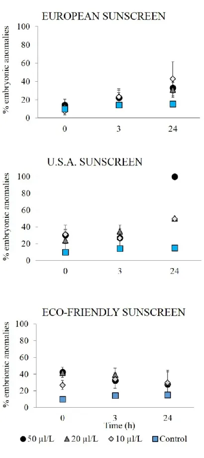
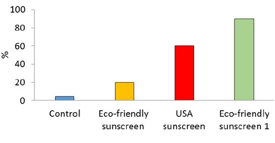
The percentage of anomalous embryos increased significantly over time after addition of the three different concentrations of European sunscreen (P < 0.001, Figure 4. 1). The percentage of abnormal embryos also increased after the addition of the different concentrations of USA sunscreen but the increase was more evident (up to 100 %) when the highest concentration (50μL L-1; P = 0.0001) was inoculated
(Figures 4. 1 and 4. 2). The different concentrations of the eco-friendly sunscreen did not cause a significant increase in the percentage of embryonic anomalies over time but a significant difference with the control was observed especially after sunscreen addition (at t0, P < 0.001, Figure 4. 1).
Figure 4. 1. Percentages of P. lividus anomalous embryos after exposure to different sunscreen products and at a different concentration over time. ± = SD.
Figure 4. 2. Unexposed embryos before the experiment (A, healthy unfertilized egg cells), fertilized egg cells at the start of the experiment (B, where the elevated fertilization layer indicates a successful fertilization (2) and sperms (1) are visible), after 70 min (C) and 2h (D) from the beginning of the experiment. Main anomalies found in early embryos after fertilization (E-H) and anomalies found in early embryos after incubation with sunscreens represented by asymmetric division into blastomeres (E-G) and undeveloped embryos (F).
In all the three treatments, significant differences in the percentages of anomalous larvae were observed after addition of each concentration of sunscreen compared to the control (P < 0.001, Figure 4. 3). We also observed that the addition of all sunscreens, at the different concentrations, determined an immediate increase in the percentage of anomalous larvae already at the beginning of the experiment (t0,
Figure 4. 3). The percentage of abnormal larvae remained constant over time in the systems treated with all concentrations of the European and eco-friendly
sunscreens, whereas in the system added with the highest concentration of U.S.A. sunscreen such a percentage increased up to 100% (P < 0.001). The different classes of anomalies found after addition of U.S.A. sunscreen (50 µlL-1) are showed in
Figure 4. 4 B-F.
Figure 4. 3. Percentages of P. lividus anomalous larvae after exposure to different sunscreen products and at a different concentration over time. ± = SD.
Figure 4. 4. Main anomalies found in P. lividus larvae after different exposure time. A: unexposed plutei, control; B: pluteus showing crossed skeletal tips at the hood apex; C, D: pluteus with joined anterior arms; E, F: incomplete or absent skeletal roots.
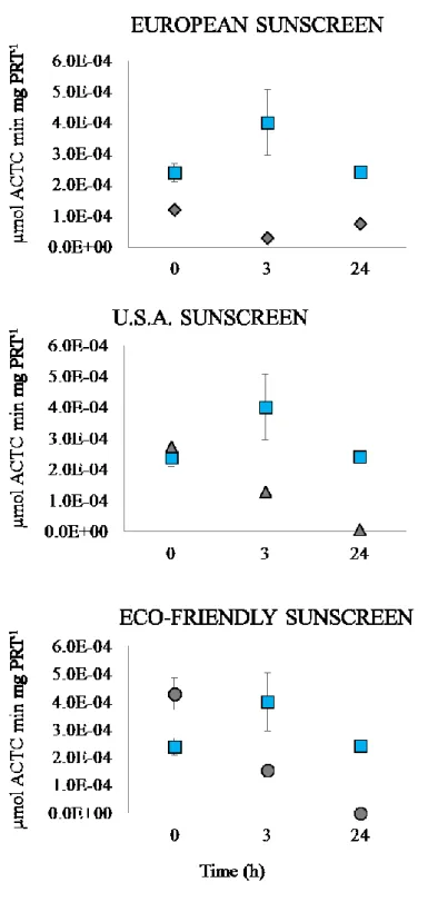
1. 4. 2 AChE activity
The exposure of the embryos to the different sunscreens (at the concentration of 50 µL L-1) caused a general decrease over time in AChE activity in all the treatments
(Figure 4. 5). AChE activity in the controls ranged from 2.40 to 4.01 x 10⁻⁴ μmoli
per minute per mg of protein (at the beginning of the experiment and after 3 h, respectively). European sunscreen determined immediately a significant decrease in AChE activity (at t0, p < 0.001), which after 3 h and 24 h continued to decrease
down to 7.46 x 10⁻⁵ μmoli per minute per mg of protein respectively. In the
treatment with USA sunscreen, AChE activity decreased significantly over time (p < 0.001) down to 7.01 x 10⁻⁶ μmoli per minute per mg of protein after 24h, whereas
Eco-friendly sunscreen determined an immediate increase of the enzymatic activity (4.30 x 10⁻⁴ μmoli per minute per mg of protein), which then dropped to zero after
Figure 4. 5. AChE activity in control (square symbols) and exposed larvae of P. lividus. Sunscreens concentration = 50 µL/L; X-axis = sampling times; Y-axis = AChE Units (AChE U), where 1 AChE U= 1 micromole of ACh hydrolyzed/min/mg protein. The square dots represent control samples. ± = SD.
1. 4. 3 Impact of sunscreens on Anemonia viridis 1. 4. 3. 1 Changes in biomass
The biomass of Anemonia viridis exposed to three different treatments did not show a significant difference compared to controls.
The general trend of the control samples was towards the loss of time-progressive biomass. The Anemonia viridis exposed at the European sunscreen showed a slight weight loss in the first exposure time (t0 and t2) and then remained constant during
the exposure, especially in recovery times (Fig. 4. 6). As opposed, in the U.S.A. and eco-friendly sunscreen, the weight of exposed organisms remains constant throughout the experiment but shows a slight decrease during the recovery time.
Figure4. 6. The weight variation (g) of Anemonia viridis in the control (square symbol) and treated. ± = SD.
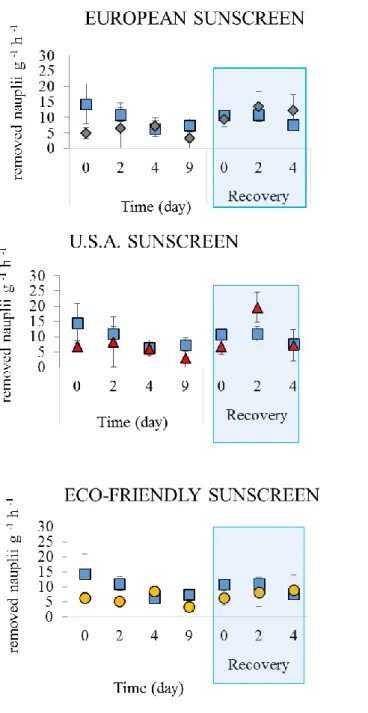
1. 4. 3. 2 Feeding rate analysis
The specimens exposed to the three treatments at a concentration of 50 μl L-1
showed the same pattern of the feeding rate (Fig. 4. 7). During the period of exposure, the number of nauplii removed from the water was similar in the threats and in the control. Also during the recovery phase, in general, we did not observe
significant differences in the rate of feeding between the organisms of A. viridis exposed to the three treatments and the control (Figure 4. 7).
Figure 4. 7. Removed nauplii of Artemia salina (g -1 h-1) in the control (square
1. 4. 3. 3 Amount of released zooxanthellae
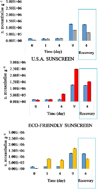
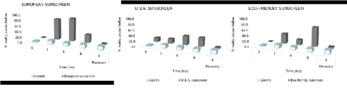
Eco-friendly and, especially USA sunscreen, determined a higher release of symbiotic organisms than in the control during the exposure phase, already after 1-4 h from the beginning of the experiment (p< 0.001). Such a release decreased during the recovery phase and particularly in the system added with Eco-friendly sunscreen, we observed a reduction of the fraction of released zooxanthellae compared to the control. After addition of the European sunscreen, a number of released symbionts was always lower compared to the control both during the exposure and recovery time.
The analysis of the health status of zooxanthellae showed that the fraction of healthy zooxanthellae, significantly increased in all treats (Figure 4. 10). In particular, the U.S.A. sunscreen caused an immediate increase in the percentage of healthy zooxanthellae already immediately after addition of the sunscreen (Figure 4. 10). In the recovery phase, all the treatments determined a decrease of healthy symbiotic algae when compared to the control.
Figure 4. 9. A number of zooxanthellae released into the water boundary in experimental systems with Anemonia viridis treated and untreated (blue columns) at different sampling times. ± = Standard Error.
Figure 4. 10. Percentage of healthy zooxanthellae released into seawater surrounding Anemonia viridis in treated and untreated (blue columns) at different sampling times.
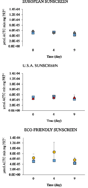
1. 4. 3. 5 AChE activity
The exposure to the European and U.S.A. sunscreens did not determine any effect on the activity of AChE, which remained rather constant over time and non-significantly different from controls (Fig 4. 11).
Figure 4. 11. AChE activity in control (square symbols) and Anemonia viridis. Sunscreens concentration = 50 µL/L; X-axis = sampling times; Y-axis = AChE Units (AChE U), where 1 AChE U= 1 micromole of ACh hydrolyzed/min/mg protein. The square dots represent control samples. ± = SD.
1. 5 DISCUSSION
1. 5. 1 Effect of sunscreen products on P. lividus
In the last decade, a number of studies have revealed that UV filters and preservatives can have a variety of negative effects on different marine organisms (Danovaro et al. 2008; Fent et al., 2010; Kim et al., 2014; Paredes et al., 2014; Maipas et al., 2015; Tovar-Sanchez et al., 2013). In particular, such effects have been investigated in coral reef ecosystems where sunscreens are generally released by tourists, including scuba divers (Ferrigno et al., 2016).
Here, we investigated the effects of three different sunscreen brands on the embryo and larval development of Paracentrotus lividus, a species widely diffused in Mediterranean and Atlantic coastal ecosystems (Verlaque and Boudouresque, 2001). Such ecosystems can be strongly affected by human activities and pollution (Mesaric et al., 2015) and recent studies have provided evidence that sunscreen products are a significant source of organic and inorganic chemicals reaching coastal ecosystems with potential ecological consequences on marine life (Tovar-Sanchez et al. 2013).
Since P. lividus is a very sensitive for a great variety of pollutants particularly during the first stages of development (Pesando et al., 2003, Carballeira et al., 2012) it is widely used as a model organism to evaluate the effects of chemicals, including components of cosmetic products (Mesaric et al., 2015; Gambardella et al., 2013; Manzo et al., 2013; Falugi et al., 2008; Pardes et al., 2014). Such information is very useful to allow comparisons with results provided in the present studies.
Our findings indicate that commonly used sunscreens can affect the embryo and larval development of Paracentrotus lividus depending on the product and concentration tested. In particular, we found that USA sunscreen at the highest concentration (50ulL-1) caused anomalies in all embryos investigated after 24h of
treatment, hampering the embryo development after the gastrula stage. Conversely, Eco-friendly sunscreen after 24 h of treatment produced a fraction of abnormal embryos similar to that found in the control suggesting a minor effect on their development.
Even USA sunscreen, at 10 uL-1 and 20ulL-1 concentrations, and European
sunscreen, at all concentrations, determined a significant effect on embryo development (after 24 of exposure, ca. 30-40%) but less severe than that induced by USA sunscreen at the highest concentration.
The embryonic abnormalities observed in the different treats were represented by defective gastrulae, with typical signs of asymmetrical migration of primary mesenchyme cells and non-developed embryos, which were especially found in the systems added with USA sunscreen.
The negative effects of the sunscreens tested were even more evident in the development phase of the larvae. Indeed, all the sunscreen products caused an immediate negative effect on the larval development, with the highest percentage of abnormalities (100%) already in the systems added with 20ulL-1 of U.S.A.
sunscreen. The fraction of abnormal larvae was high also when they were exposed to European and Eco-friendly sunscreens (50-60%) and remained rather constant over time. The anomalies observed included crossed and/or separated tips at the
hood apex, fused anterior arms, and incomplete or absent skeletal rod. The categories of the anomalies identified in both embryos and larvae were similar for all the systems exposed to sunscreens. These anomalies were previously identified also in other studies in which P. lividus larvae and gametes were exposed to nanoparticles (Gambardella et al., 2015; Mesaric et al., 2015; Gambardella et al. 2013; Manzo et al., 2013), organophosphate pesticides (Aluigi et al., 2010) and mixtures of organic wastes and contaminants of different nature (Carballeira et al. 2012). These results suggest that sunscreen products act as a wide category of pollutants that have been reported to affect the sea urchin skeleton apparatus, by modifying the location of the skeletal rods or primary mesenchymal cell migration, or by inactivating the gene regulatory system underlying the development of the embryonic skeleton (Peterson & McClay, 2003).
Despite the typologies of anomalies induced by the different sunscreens were similar, we observed that their quantitative relevance changed depending on the sunscreen product and in some cases, on the concentration tested. In particular, high concentrations of USA sunscreen determined the most severe effects on embryos and larvae since ca. 80% of them showed the block or regression of the development or death (particularly referred to larvae). Based on the criteria previously established for embryonic and larval alterations (Gambardella et al. 2013, Carballeira et al. 2012, Guillou et al., 2000), the effects due to USA sunscreen can be referred to a toxicity level 3, which represents the most severe toxicity for contaminants (Carballeira et al. 2012). The reason why such a sunscreen had a major negative effect already in the first stage of development of P. lividus can be explained by the presence of different ingredients contained within USA sunscreens
such as benzophenone 3 and homosalate, which have been reported to be highly toxic to marine organisms (Krause et al., 2012).
Despite the embryonic development of P. lividus, in terms of percentage of anomalous zygotes, was more affected by European sunscreen than Eco-friendly sunscreen, the opposite effect was observed for larval development. However, based on the morphological alterations observed, European sunscreen resulted in a higher toxicity level (resulting on average in ca. 30% of embryos and larvae with the block of their development) compared to Eco-friendly sunscreen (on average, only ca. 4% of embryos and larvae with the block of their development).
Since previous studies have reported that, being sea urchin larvae very plastic, their anomalies can be also reversible. Therefore, we suggest that the anomalous plutei exposed to Eco-friendly sunscreen could even survive and transform in healthy sea urchins. The higher toxicity of the European sunscreen than that of Eco-friendly, particularly on early stages of development of P. lividus induced by European sunscreen can be due to the presence of octocrylene, TiO2 (nanoparticles) and
preservatives such as benzyl benzoate in its composition (Petra et al., 2006). The delay of development in embryos of Paracentrotus appears to be primarily a consequence of changes in AChE activity, which is responsible for regulating neurotransmission and other relevant biological processes, including the correct cell migration during gastrulation (Drews, 1975, Falugi, 2008, Gambardella et al., 2015). It has been also reported that changes in AChE activity due to the exposure to cholinesterase inhibitors (i.e., neurotoxic pesticides) during the first life stages of P. lividus can lead to cytoskeletal alterations (Aluigi et al. 2008).
Since AChE activity is known to rapidly respond to a wide range of chemical and environmental stress (Gambardella et al., 2015; Falugi et al., 2012; Gambardella et al., 2013; Pesando et al., 2003) we analyzed the rates of AChE activity in the larvae of P. lividus exposed to sunscreens to investigate their neurotoxic effects. The analysis of the AChE activity revealed that sunscreens here tested, altered such an enzymatic activity compared to the control, determining its general decrease over time. These findings are in line with other studies where the exposure of P. lividus larvae to different micropollutants were shown to inhibit AChE activity (Gambardella et al., 2015; Mesaric et al., 2015; Aluigi et al., 2010; Pesando et al., 2003). The inhibition of AChE activity could be caused by the irreversible or reversible binding of the sunscreen ingredients to the enzyme's catalytic sites and to be involved in the skeletogenic aberrations (Falugi et al., 2008, Gambardella et al. 2012). Despite AChE activity decreased over time, also when sea urchin larvae were exposed to Eco-friendly sunscreen, at the stage of 4-arms pluteus we observed a significant increase compared with the control was observed. Previous studies have suggested that an increase in AChE activity in P. lividus might be the result of differential locomotion and feeding patterns to enhance its survival and fitness (Jennings et al. 2007, McEdward and Miner, 2007).
Overall, our findings suggest that commonly used sunscreens particularly containing chemical filters (i.e., octocrylene, homosalate and benzophenone-3) and preservative (methylisothiazolinone, benzyl benzoate) can affect the embryo-larval development of P. lividus, reducing the ability of larvae to survive, thus potentially affecting the abundances/biomasses of sea urchins and the trophic webs of coastal ecosystems. In addition, data reported here reveal that the Eco-friendly sunscreen,
whose ingredients have been defined compatible with marine life (especially of tropical ecosystems), impacted to a lesser extent the embryos and larvae of P. lividus, although stress signals due the exposure to high concentrations of such a product were still evident. Therefore, it is strongly advisable to develop eco-compatible sunscreen products, specifically tested a priori even on the early developmental stages of marine organisms, which might be more sensible than adults to these contaminants.
1. 5. 2 Effect on sunscreen products on Anemonia viridis
Sea anemones, as well as other Cnidarians, are models commonly used for studying the impact of contaminants on marine life (Venn et al., 2008). Several species of anemones contain symbiotic dinoflagellates (Symbiodinium sp.) in their tentacles known as zooxanthellae (Caparkaya et al., 2010), which are highly susceptible to a wide range of stress (Merle et al., 2007).
The experiments here conducted on Anemonia viridis exposed at 50 μL L-1 of
different sunscreen products revealed that these organisms, respond with behavioral alterations in presence of sunscreens. Indeed, already after 2 days of the beginning of the exposure (especially the anemones exposed to USA sunscreen) retracted the tentacles, contracted their body and released a large amount of mucus suggesting the stress of the animals. However, during the experiment with the different sunscreens, we did not observe a significant change in biomass, feeding rates and AChE activity in the anemones over time, as well as during the recovery phase. Conversely, the rate of release of zooxanthellae in seawater increased in all systems during the exposure at the different sunscreens. This was most evident in the systems treated with USA sunscreens where after 9 days the number of
zooxanthellae released was 19 fold higher than at the start of the experiment. We observed that Eco-friendly sunscreen determined a more important effect in the release of symbionts than European sunscreen. However, after 9 days of exposure to Eco-friendly sunscreen, we observed that most of the released zooxanthellae were healthy (ca. 70%), whereas only ca. 30% of these was healthy in the systems incubated with European sunscreen. Previous studies revealed that the release of healthy zooxanthellae by their host organism can be a reversible process (Leichter et al., 1999), suggesting a potential recovery of the organism. The results obtained during the recovery phase showed a decrease of the release of zooxanthellae in seawater in both the systems added with European and Eco-friendly sunscreen. Conversely, anemones exposed to USA sunscreen continued to release their symbionts, indicating the impossibility to recover. Indeed, during the recovery phase anemones exposed to USA sunscreen died. We hypothesize that the death of these organisms was caused by the change in seawater conditions to which they were relatively adapted.
Overall, we can suggest that A. viridis appeared to be rather resistant to sunscreens tested, except for USA sunscreen that caused the death of the organisms when the pre-impact conditions were restored. Conversely, European and Eco-friendly sunscreens, even if promoted the release of their symbionts did not affect particularly the health of anemones.
1. 6 CONCLUSION
All these results indicate that sunscreens cause damage to early stages of development of Paracentrotus lividus, resulting in decreased reproductive capacity of the sea urchin, due to the alteration in both embryo development and larvae structure. The anomalies identified in this study are similar to those identified in numerous investigations in response to exposure of sea urchin gametes to neurotoxic pesticides and nanoparticles. This may mean that the mechanism of action of these pollutants on the stages of early development of sea urchin is the same. Indeed, the occurrence of abnormalities in the sea urchin development is in accordance with the decrease in the activity of acetylcholinesterase, already reported in several studies.
Otherwise, Anemonia viridis seems to resist better to this type of stress, not showing effects in terms of change in biomass, the rate of feeding and activity of acetylcholinesterase except for their symbiotic organisms.
We can suggest that the different effect of sunscreens on P. lividus and A. viridis is due to the different sensitivity of the organisms considered and to the different life cycle considered.
Since, we observed that the sunscreens defined eco-friendly produced a relatively slight impact on larvae of P. lividus, this suggests the need to test the products on different model marine organisms, before placing them on the market, to develop cosmetic products that effectively are compatible with marine life. Finally, this study provides new evidence of the direct effects of sunscreens on marine environments and paves the way for further research on different marine organism
models, both on different trophic levels and different stage of developments, to understand how sunscreens can affect the health of marine organisms and the balance of the environment in which they live.
1. 7 REFERENCES
Aluigi, M. G., Falugi, C., Mugno, M. G., Privitera, D. & Chiantore, M. Dose-dependent effects of chlorpyriphos, an organophosphate pesticide, on metamorphosis of the sea urchin, Paracentrotus lividus. Ecotoxicology 19, 520– 529 (2010).
Amemiya, S. Complete regulation of development throughout metamorphosis of sea urchin embryos devoid of macromeres. Development Growth and Differentiation. 38, 465–476 (1996).
Amemiya, S., Yonemura, S., Kinoshita, S., Shiroya, T. Biphasic stage sensitivity to UV suppression of gastrulation in sea urchin embryos. Cell Differentiation 18, 45-49 (1986).
Anderson, M. J. Permutational multivariate analysis of variance.Department of Statistics, University of Auckland, Auckland (2005).
Baccetti, B., Burrini, A. G., Collodel, G., Falugi, C., Moretti, E., Piomboni, P. Localization of two classes of acetylcholine receptor-like molecules in sperms of different animal species. Zygote 3, 207–217 (1995).