Autophagy: A New Mechanism
of Prosurvival and Drug
Resistance in Multiple Myeloma
V. Desantis*, I. Saltarella*, A. Lamanuzzi*,
M.A. Mariggiò†, V. Racanelli*, Angelo Vacca*and M.A. Frassanito†
*
Department of Biomedical Sciences and Human Oncology, Unit of Internal Medicine and Clinical Oncology, University of Bari Aldo Moro Medical School, Bari, Italy;†Department of Biomedical Sciences and Human Oncology, Unit of General Pathology, University of Bari Aldo Moro Medical School, Bari, Italy
Abstract
Autophagy is an intracellular self-degradative process that balances cell energy source and regulates tissue homeostasis. In physiological condition, autophagy funnels cytoplasmic constituents to autophagolysosomes for degradation and is an alternative way for cell-death behavior. Here, we inspected autophagy as a prosurvival mechanism essential for drug resistance in multiple myeloma (MM). Accordingly, autophagy inhibitors used in association to conventional anti-MM drugs might enforce the effect against resistant MM plasma cells and render autophagy a new therapeutic target.
Translational Oncology (2018)11, 1350–1357
Introduction
Autophagy governs cell self-clearance by which unwanted cytoplasmic content (amino acids, fatty acids, and nucleotides) is degraded. Unfavorable conditions including nutrients deprivation, hypoxia, DNA damage, or production of reactive oxygen species (ROS) trigger autophagy to overcome energy deficiency and remove exceeded materials, hence maintaining cell homeostasis and survival. Although autophagy has been first described in yeasts[1], further studies have confirmed its pivotal role in humans for the regulation of both physiological and pathological processes.
Three types of autophagy coexist in most cells: microautophagy, chaperone-mediated autophagy (CMA), and macroautophagy, usually named “autophagy”. Microautophagy is a nonselective lysosomal degradation pathway that involves cellular components [2]. Vesicles formed by direct membrane invagination[3]are able to transfer cellular components into the lysosomal lumen that induces the degradation of soluble cytosolic constituents or of entire incorporated organelles, i.e., peroxisomes. CMA contributes to the maintenance of proteostasis and adaptation of cells to stress[4]. The vesicles are not needed. Indeed, substrates are selectively identified and delivered by cytosolic hsc70/ co-chaperones to the lysosomal surface[5], where they are internalized through a membrane translocation complex formed by multimerization of LAMP‐2A (lysosome-associated membrane protein type 2A) that is the CMA substrate-chaperone receptor [6]. The multimerization induces the substrate translocation and subsequent degradation. The
levels of CMA-active lysosomes containing both hsc70 and CMA substrates increase in response to the stressors. These (i.e., starvation, oxidative stress) upregulate CMA[7], but very low levels of CMA are also detectable at basal conditions in most cells[8].
Autophagy is a complex process regulated by different multistep signaling pathways [9]. It ensures the turnover of bulk cytoplasmic, damaged and superfluous organelles (i.e., mitochondria and peroxisomes), and invasive microbes. These agents are sequestrated by an expanding double-membrane vesicle, so-called “autophagosome” and further degraded into the lytic compartment of lysosomes [10]. After degradation, products are released into the cytosol [11] to promote generation of new energy and to support cell maintenance.
Autophagy is slightly activated at basal conditions in a large number of human cells to maintain the physiological homeostasis and/or to promote cell survival during stress conditions [12]. Mutations of autophagy genes and/or alterations of its regulation mechanism prompt
www.transonc.com
Address all correspondence to: Professor Angelo Vacca, MD, PhD, Department of Biomedical Sciences and Human Oncology, Unit of Internal Medicine and Clinical Oncology, University of Bari Medical School Aldo Moro, Policlinico-Piazza Giulio Cesare, 11 I-70124, Bari, Italy. E-mail:; [email protected]
Received 9 July 2018; Revised 24 August 2018; Accepted 28 August 2018
© 2018 Published by Elsevier Inc. on behalf of Neoplasia Press, Inc. This is an open access article under the CC BY-NC-ND license (http://creativecommons.org/licenses/by-nc-nd/4.0/). 1936-5233
the onset and/or progression of different human disorders and diseases, including cancer.
In this review, we will focus on autophagy as a tumor prosurvival mechanism especially in multiple myeloma (MM) in which it is closely connected to control progression and drug resistance (DR). The Autophagy Pathway and Regulation
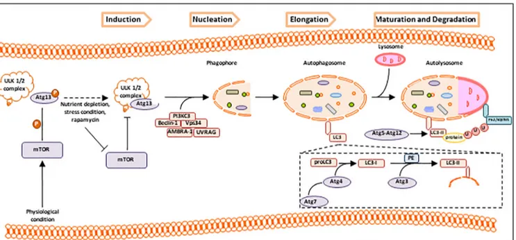
Several key markers of autophagy have been identified. The expression of AuTophaGy-related genes (ATGs) and of their Atg proteins is constant and needed for the autophagy regulation. To date, 30 ATG genes have been identified in yeast Saccharomyces cerevisiae[13], and their homologues have been well characterized in higher eukaryotes. The function and the involvement in the autophagosome formation of Atg proteins have been widely studied in association to each step of autophagy (Figure 1)[14,15].
As depicted in Figure 1, autophagy develops by different steps: induction, nucleation, elongation, maturation, and degradation of specific cytosolic components. The mammalian target of rapamycin (mTOR) pathway is a serine/threonine protein kinase highly conserved in eukaryotes [16] and is the most important negative regulator of autophagy[17]. It interacts with several proteins to form two distinct complexes, mTOR complex 1 (mTORC1) and 2 (mTORC2), that show different sensitivities to rapamycin as well as to upstream inputs and downstream outputs[18,19]. mTOR controls cell growth by activating anabolic processes (i.e., protein synthesis, transcription, and ribosome biogenesis) and by inhibiting catabolic processes (i.e., mRNA degradation and autophagy activation) [20]. Its activity is negatively regulated by multiple stress signals including hypoxia, oxidative stress,
DNA damage, and lack of nutrients, which are all inducers of autophagy
[21]. Specifically, mTORC1 is sensitive to the lack of nutrients that leads to the mTOR inactivation [22]that induces autophagy. In the stress condition, mTOR does not phosphorylate Atg13. In this form, Atg13 binds the Unc-51-like kinase-1/2 (ULK1/2) complex and stimulates its catalytic activity that results in the induction of the phagophore formation [23,24]. ULK1/2-Atg13 recruits a second kinase complex
[25] composed by Beclin-1, PI3KC3 (phosphatidylinositol-3-kinase class III), Vps34 (vacuor protein sorting-34), AMBRA-1 (autophagy and Beclin-1 regulator-1), and UVRAG (UV radiation resistance-associated gene protein) and promotes the phagophore nucleation[26].
Beclin-1 is the mammalian orthologue of yeast Atg6 and a Bcl-2-homology (BH)-3 domain protein[27]; it acts as a master regulator of autophagy depending on the specific Beclin-1 binding proteins, i.e., autophagic inducers and/or inhibitors[28](see below).
Elongation of the phagophore requires two ubiquitin-like systems, namely, the light chain3 (LC3) and the cargo receptor p62/sequestome1 (p62/SQSTM1). The LC3 activity is based on processing of microtubule-associated protein LC3 usually expressed as full length in the cytosol. Upon autophagy stimuli, nascent LC3 (pro-LC3) is proteolitically cleaved by Atg4, a cysteine protease, which generates LC3-I, a soluble cytosolic form. Atg4 is activated by Atg7, an E1-like enzyme, in an ATP-dependent manner, and induces the transfer of LC3-I to Atg3, an E2-like carrier protein[29]. At this step, activated LC3-I is conjugated to phosphatidylethanolamine (PE) and generates LC3-II that is the autophagic vesicle-associated form or lipidated form
[30]. The conversion of LC3-I to LC3-II indicates the autophagosome maturation. LC3-II is found on both internal and external autophagosome
Figure 1. Autophagy pathway. Physiologically, activated mTOR phosphorylates Atg13 which, after dissociation by ULK1/2 complex, inhibits autophagy. On the contrary, in conditions of higher nutrients depletion or stress, or in the presence of rapamycin (inhibitor of mTOR pathway), autophagy is activated. When mTOR is inhibited, dephosphorylated Atg13 is activated and reassociates to ULK1/2 complex, inducing the phagophore formation. ULK1/2-Atg13 recruits PI3KC3 complex with Beclin-1, Vps34, AMBRA-1, and UVRAG, resulting in the initial steps of phagophore nucleation. Elongation process begins with the recruitment of cytosolic nascent pro-LC3 that is cleaved by Atg-7-activated Atg4 and transformed in the soluble cytosolic form LC3-I. In the next step, Atg4 transfers LC3-I to Atg3, triggering the lipid conjugation to PE (phosphatidylethanolamine), and converting the soluble form LC3-I into autophagic vesicle-associated form LC3-II. LC3-II is conjugated to the autophagosome membrane by mean Atg5 and Atg12 and regulates both the fusion of autophagosome to lysosome membranes and the degradation of cargo binding ubiquitinated proteins trapped by p62/SQSTM1.
membrane, and its recruitment and integration depend on Atg5-Atg12 interactions. LC3-II acts as a regulator of the fusion of autophagosome and lysosome membranes and as selector of the cargo for the degradation process[31]. The second system p62/SQSTM1
[32]is an autophagy marker because its degradation follows the increase of autophagic flux. It promotes the turnover of ubiquitinated proteins in autolysosome membrane by association of protein ubiquitin-binding domain with LC3-II via its LC3-interacting region[33].
The autophagy machinery is regulated at different levels [34]. The major regulator is mTOR that operates as a signaling control point downstream of the growth factor receptors, ATP levels, and insulin signaling. Following the nutrient deprivation and low levels of ATP, mTOR is repressed and activates adenosine 5′-monophosphate–activated protein kinase (AMPK) [35,36]. Also, the mTOR inhibition by AMPK allows hypoxia to activate autophagy through hypoxia-inducible factor (HIF)–dependent and –independent effects [36,37]. Hypoxia (i.e., oxygen unavailability to accept free electrons from the respiratory chain) prompts endoplasmic reticulum (ER) stress through unfolded protein response, reduces mitochondria function in oxidative phos-phorylation, and induces autophagy that eliminates the ER compacted portions and reduces the mitochondrial mass [37]. This adaptive response to hypoxia prevents ER wasteful ATP consumption and restrains the production of ROS into mitochondria. Increased autophagy can also generate ATP from catabolism when ATP production, by oxidative phosphorylation, is limited.
Atg proteins also provide complex posttranslational modulation of autophagy[38].
Autophagy in Cancer and Cellular Stress Conditions Autophagy plays a dual role in tumor development and progression. At the early stage of tumorigenesis, it is oncosoppressive in that it prevents tumor initiation by suppressing chronic tissue damage, inflammation, and genome instability [39,40]. In contrast, once tumor is growing, autophagy promotes its growth, metabolism, progression, and DR because it allows cancer cells to overcome intracellular and environmental stress[41].
The role of autophagy in cancer has been proved by using murine models knockout for essential autophagic genes. Beclin-1 is involved in the initiation and formation of phagosome as shown in ovarian, prostate, and breast cancer [42,43]. Its levels are decreased in human breast carcinoma, and this inhibits tumorigenesis [44]. Inoculation of immortalized breast epithelial cells with the allelic loss of Beclin-1 gene into NOD/SCID mice induces genomic instability in response to a metabolic stress and rapidly promotes breast tumorigenesis [45]. Transfer of Beclin-1 in autophagy-defective human MCF7 breast carcinoma cell line promotes autophagy and supports tumor progression. Several proteins interact with Beclin-1 and regulate autophagy, inducing antiproliferative or tumor suppressor effects. Functional deficiency in mouse embryos of AMBRA1[46], a positive regulator of Beclin-1–dependent autophagy, leads to neural tube defects associated with autophagy impairment and consequent accumulation of ubiqui-tinated proteins, unbalanced cell proliferation, and excessive apoptotic cell death. BIF-1 (endophilin B1)[47]interacts with Beclin-1 through the UVRAG [48] acting as a positive mediator of the PI3KC3. In response to nutrients deprivation, BIF-1 colocalizes with Atg5 and LC3 on autophagosomes and joins the UVRAG-Beclin 1 complex that activates autophagy and induces tumor suppression[47].
The master tumor suppressor genes p53[45–50]plays a dual role in the regulation of autophagic machinery. Multiple p53 target genes
stimulate autophagy, which often results in the downregulation of the central negative regulator mTOR[51]. One of the mechanisms by which p53 downregulates mTOR is the activation of AMPK (positive regulator of autophagy) through the transcriptional regulation of Sestrins 1 and 2
[52]. p53 also transactivates several genes, includingβ1 and β2 subunits of AMPK, phosphatase and tensin homolog (PTEN), and insulin-like growth factor binding protein 3. AMPK β1 and β2 subunits are upregulated under stress conditions and antagonize the autophagy-suppressive functions of mTOR in a p53-dependent manner [53]. Another mechanism whereby p53 induces autophagy is the upregulation of the tumor suppressor protein death-associated protein kinase 1[54]. This induces autophagy by binding to and then suppressing the MAP1-B, an antiautophagic factor that interacts with LC3[55]. In contrast, the cytoplasmic p53 inhibits autophagy through a transcription-independent manner [56]. p53 knockout, by RNA interference or pharmacological inhibition, induces an increase of basal autophagy through AMPK-dependent inhibition of mTOR. This is observed in G1 and S cell cycle phases, suggesting a cell cycle–dependent regulation of autophagy by p53[57].
In association to p53, the activated RAS plays an important role into modulation of autophagy in cancer progression and invasion. Kinsey et al.[58]demonstrated that, in human pancreatic cells, activation of K-RAS and loss of p53 upregulate Placenta-specific gene 8 that induces autophagosome-lysosome fusion. Instead, in breast cancer progression, RAS and p53 can regulate heat shock transcription factor 1, inducing autophagy by binding Atg7[59].
Literature data also demonstrate that HIF-1α interferes with cancer metabolism and activates the autophagic machinery [60]. HIF-1α
participates to the stabilization of p53 and activation of H-RAS interacting with the autophagy regulation. Lili Song et al. [61]
demonstrated that, in radioresistant cervical cancer, upregulated miRNA-21 is associated to HIF-1α and p-Akt overexpression and to decreased levels of PTEN. miRNA-21 inhibition increases PTEN levels and decreases p-Akt and HIF-1α, suggesting a HIF-1α–miRNA-21 positive feedback loop through the PTEN/Akt/HIF-1α pathway. This may act on autophagy regulation via Akt-mTOR signaling pathway.
Thus, autophagy, as a tumor promotor, is closely linked to the hypoxic conditions. Exposition of human tumor cell lines to hypoxia or metabolic stress activates autophagy in vitro [62]. In these conditions, HIF-1α triggers autophagy, leading to cell survival by involving unfolded protein response [63]. Hypoxia increases the transcription of the MAP1LC3B that during autophagy is conjugated to the lipid PE (MAP1LC3B-II) and inserted into membranes of the growing autophagic vesicles [64]. Consequently, autophagosome formation and lipidation are also dependent on Atg5 through the transcription factors ATF4 and CHOP, which are regulated by PKR-like ER kinase. Regulation of autophagy by PKR-PKR-like ER kinase is important to mediate hypoxia tolerance and to target this pathway through genetic or pharmacological approaches in tumor hypoxia and irradiation treatment.
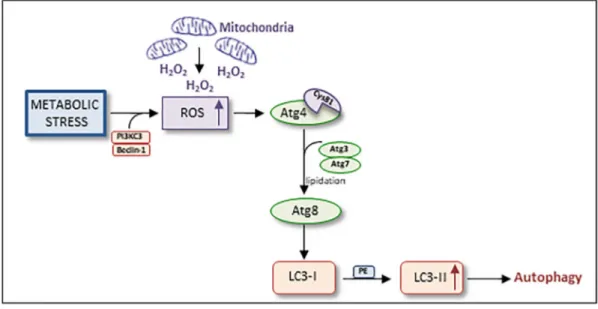
Moreover, remarkable is the role of ROS in the activation of autophagy. ROS are small and highly reactive molecules formed by incomplete one-electron reduction of oxygen and able to oxidize proteins, lipids, and DNA. Redox regulation can be involved in different signaling pathways, and the cross talk between ROS production and autophagic machinery has been demonstrated in several pathological conditions [65]. Autophagy induces the suppression of damaged organelles or protein aggregates that lead to an increase of ROS species and consequently to DNA damage[66].
Aberrant ROS levels can be associated to cancer initiation. ROS production is mainly located in the mitochondria: in this context, the autophagy is the most important process responsible for the damaged mitochondria degradation and for their turnover. Scherz-Shouval et al.[67]have demonstrated that stress conditions and amino acids deprivation induce the formation of PI3KC3/Beclin-1 complex that stimulates the mitochondria to accumulate H2O2, subsequently secreted into the cytosol. ROS prompts the oxidation of Atg4 Cys81 residue in the catalytic site, lipidating Atg8 ubiquitin-like protein with the cleavage of c-terminus residue in the presence of Atg3 and Atg7 proteins. Lipidation of Atg8 is an essential prerequisite for the conjugation of PE to LC3-I, forming LC3-II and inducing the autophagolysosome formation (Figure 2). In this condition, mitochondria act as one of the major sources of signal to induce autophagy and autophagosome biogenesis.
Collaborations between autophagy and different tumor markers have a strong impact on context-specific ways in cancer and are the basis for further investigations to provide novel pharmacological treatments. Autophagy and Apoptosis: Two Closely Connected Processes
Apoptosis (programmed cell death) is a cell suicide mechanism responsible for the removal of damaged cells and/or unnecessary nonfunctioning cells; hence, it acts for maintenance of physiological homeostasis. It can be induced by intracellular input (i.e., calcium accumulation, oxidative damage, and hypoxia) or by extracellular messages such as pathogenic bacteria, toxins, and proapoptotic molecules[68]. Deregulation of apoptosis leads to accumulation of cellular damages and contributes to carcinogenesis and failure of anticancer treatment. Mutations or aberrant expression and/or function of death receptors/ligands and/or other proteins involved in apoptotic pathways have been described in several tumors[69]. In this context, therapeutic strategies that restore the apoptotic process may serve the antitumor drug activity[70].
As autophagy is a process by which tumor cells adapt their survival in response to microenvironment critical conditions, autophagy and
apoptosis can act as mutually exclusive and simultaneously cross-linked processes[71]. Although they are two distinct biochemical processes, they form a network in terms of regulation: both pathways are necessary for the maintenance of cell homeostasis and for the prevention of tumorigenesis[72]. In cancer cells, metabolic or pharmacological stress activates autophagy, triggering cell proliferation and survival. Inhibition of autophagy prompts apoptosis in stressed cells and causes tumor regression. Finally, persistent and sustained cell stress turns the physiological protective effect of autophagy into autophagic cell death
[73]. The cross-regulation of these opposite effects, namely, autophagic cell survival and cell death, relies on a network of signal transducers involved in autophagy as well as in apoptosis[74].
Since 1999, Liang et al.[75]identified Beclin-1 as the link between autophagy and tumorigenesis: ectopic expression of Beclin-1 activates tumorigenesis in vivo. Further studies show that Beclin-1 interacts with the antiapoptotic Bcl-2, Bcl-xL, and Mcl-1 through its BH3 domain inhibiting autophagy[76]. During starvation or other stress conditions, several mechanisms (i.e., JNK-mediated phosphorylation of Bcl-2/Bcl-xL, DAPK-mediated phosphorylation of the BH3 domain of Beclin-1) destroy the Bcl-2/Bcl-xL–Beclin-1 complex and activate autophagy via the interaction of Beclin-1 with Vsp34[28]. Pattingre et al.[77]suggested that Bcl-2/Beclin-1 complex acts as a detector able to maintain autophagy levels within the homeostatic range.
Cellular FADD-like IL-1β–converting enzyme inhibitory protein (FLIP) is involved in the inhibition of both processes. Specifically, FLIP inhibits apoptosis by the interaction with Fas-associated death-domain– containing protein, preventing the activation of the caspase cascade. In addition, FLIP attenuates autophagy by blocking the lipidation of LC3 on phagosome membrane[78].
In relation to Atg proteins, Yousefi et al.[79]showed their important role in the apoptosis pathway. Atg5 is a substrate of calpains that migrates into the mitochondria and induces the release of cytochrome c. Atg12 is required for the activation of caspases by inhibition of Bcl-2 and Mcl-1, activating apoptosis[80].
Inhibition of autophagy leads to an increase of apoptosis, highlighting the prosurvival role of the autophagy[81]. Fimia et al.
Figure 2. Autophagy machinery and cellular stress. Metabolic stress induces the formation of PI3KC3/Beclin-1 complex which leads to the accumulation of H2O2 previously created into the mitochondria. ROS directly oxidizes Cys81 residue of Atg4 catalytic site that, in the presence of Atg3 and Atg7, directly cleaves the c-terminus of Atg8. Lipidation of Atg8 is essential to the conjugation of LC3-I to PE, forming LC3-II and inducing autophagolysosome formation.
[81]proved that PI3Ks class I and III pathways play an important and opposite role in the control of autophagy: the first one stimulates a repressive effect promoting cell growth and tumor development, while the second is essential for autophagy induction.
Moreover, the RAS and Akt oncoproteins are involved in this network, inhibiting the autophagy machinery [81,82]. Oncogenic RAS supports tumor cell proliferation and maintains metabolic cancer activities[83]. Human cancer cell lines with ectopical expression of oncogenic H-RAS and endogenous K-RAS mutations show activa-tion of autophagy following extracellular matrix detachment. These data suggest a mechanism by which autophagy may promote RAS-driven tumor growth in specific metabolic contexts.
Autophagy as Prosurvival Machinery in MM
In cancer cells, metabolic stress induces autophagy as a cellular alternative source of energy and metabolites [84], enhancing an adaptive cell response to cancer therapies [85]. In hematological malignancies, autophagy plays an essential role to attenuate drug-induced cell death through a chemoresistance [86]. MM is a hematological malignancy characterized by the expansion of monoclonal plasma cells (MM cells) in the bone marrow. MM cells produce large amounts of monoclonal immunoglobulins (Igs) that result in a localization of potentially toxic unfolded or misfolded proteins to ER. Thus, because of the high proliferative rate and the Igs synthesis, autophagy is essential for MM cells survival in order to degrade the excess of protein aggregates [87]. Inhibition of autophagy by Beclin-1 knockdown or treatment with autophagy inhibitors, i.e., 3-methyladenine (3-MA) and chloroquine, induces MM cells apoptosis [88,89] and blocks autophagosome formation. Recently, Wang et al.[90]demonstrated that elaiophylin, a potent inhibitor of late stage of autophagy, has an anti–MM cell activity via inhibition of the autophagic flux and persistent activation of ER stress– mediated apoptosis. On the other hand, basal autophagy is tightly regulated to avoid autophagic cell death. Lamy et al.[91]identified the
heterodimeric protease caspase-10/FLIPLas a prosurvival factor restraining basal autophagy by cleaving the Bcl-2-interacting protein Bcl-2-associated transcription factor 1. Inhibition or knockdown of caspase-10 stabilizes Bcl-2-associated transcription factor 1 that displaces Bcl-2 from Beclin-1, leading to excessive autophagy and thus to MM cell death.
Literature data focus on autophagy as an MM prosurvival mechanism able to exert a protective effect under drug treatment in that drug-resistant MM cells can escape the toxic effect of drugs via autophagy
[92]. Proteasome inhibitors, such as bortezomib and carfilzomib, are the first-line drug for MM treatment in both newly diagnosed and relapsed patients. These drugs initially exert anticancer properties, but frequently, patients become resistant. Bortezomib-resistant MM cells show high levels of AMPK and of autophagosome formation compared to bortezomib-sensitive cells. Reduction of AMPK activity compro-mises autophagosome formation[93]. Riz et al.[94]demonstrated that the transcription factor Kruppel-like factor 4 contributes to carfilzomib resistance in MM cells by binding to the promoter regions of SQSTM1 gene encoding p62. Hoang et al.[95]showed that autophagy is further enhanced by exposure of MM cells to ER stress inducers and mTOR inhibitors. Since mTOR is a well-known inhibitor of autophagy, treatment of MM cells with a pharmacological inhibitor 3-MA induces autophagic cell death in a dose-dependent manner. Co-treatment of MM cells with bortezomib and autophagy inhibitor results in a synergistic cytotoxic effect[95].
Downregulation of PI3K/Akt/mTOR signaling pathway positively correlates with cell autophagy activation mediated by ER stress[96]. Fu et al.[97]demonstrated that, in a large cohort of MM patients divided into resistant and sensitive basing on the chemotherapy efficacy, ER stress promotes autophagy and apoptosis and blocks proliferation through inhibition of PI3K/Akt/mTOR signaling. ER stress is also able to revert to DR via PI3K/Akt/mTOR pathway.
Intracellular nicotinamide adenine nucleotide plays an important role in the regulation of several cellular processes. It is highly
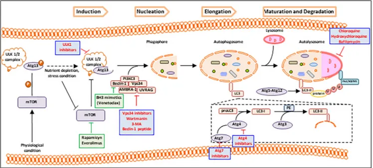
Figure 3. Autophagy targeted therapy. Pharmacological targets are involved at different levels in autophagy process to exert a positive (green plots) or negative (red plots) regulation. Rapamicyn, everolimus, and BH3 mimetics (venetoclax) positively regulate autophagy at mTOR and Beclin-1 level; on the contrary wortmanin, Beclin-1 peptide, 3-MA, chloroquine, hydroxychloroquine, bafilomycin, ULK1, Vps34, Atg7, and Atg4 inhibitors negatively regulate the autophagy machinery.
expressed in MM cells and mediates cell growth and DR[98]. Cea et al. demonstrated that the inhibition of Nampt, a rate-limiting enzyme involved in nicotinamide adenine nucleotide synthesis, induces a relevant cytotoxic activity against MM cells resistant to conventional anti-MM drugs in vitro and in vivo and overcomes the protective effect of interleukin-6, IGF-1, and bone marrow stromal cells. The cytotoxic effect of the Nampt inhibitor FK866 is due to the activation of autophagy through the inhibition of mTORC1/Akt and of ERK 1/2 pathways[98].
Activation of prosurvival autophagy can also be induced in response to multiple stressors, i.e., oxygen/nutrient deprivation, extracellular matrix degradation, and inflammation, in the tumor microenviron-ment. Recently, Frassanito et al.[99]identified a mechanism able to mediate the cross talk between MM cells and cancer-associated fibroblasts (CAFs) in MM DR. CAFs are a very important cell within the bone marrow stroma and promote cancer initiation, progression, and DR [100,101]. Co-cultures of MM cells with MM CAFs are in vitro resistant to bortezomib, implying that MM CAFs prevent bortezomib-induced apoptosis. Authors demonstrated that bortezomib treatment activates autophagy in MM CAFs through inhibition of mTOR, induction of LC3-II, and activation of TGF-β pathway[98]. Inhibition of autophagy by small-interfering RNA knockdown of Atg7 or treatment with 3-MA or TGF-β inhibitor restores the susceptibility to bortezomib in bortezomib-resistant CAFs and produces cytotoxicity in MM cells co-cultured with CAFs.
Conclusions
Autophagy is a crucial mechanism involved in tumor progression. Of note, in MM, autophagy is a prosurvival mechanism by which tumor cells overcome excessive accumulation of toxic misfolded Igs. Furthermore, autophagy fosters resistance of MM cells to proteasome inhibitors. Proteasome inhibition induces the accumulation of damaged proteins in the intracellular environment, causing ER overload and stress. Based on the tight connection among autophagy, cell stress, and apoptosis, proteasome inhibition also activates autophagy, inducing DR. Therefore, targeting autophagy to prompt MM cells death and to increase drug sensitivity may be envisaged as a novel therapeutic strategy. Specifically, because of the dual role of autophagy as mechanism of prosurvival or cell death, targeting autophagy may focus on activation (ie, rapamicyn, everolimus, BH3 mimetics) or on inhibition of autophagy (Figure 3).
Conflict of Interest Statement
The authors declare that they have no competing interests. Acknowledgements
This work was supported by the Associazione Italiana per la Ricerca sul Cancro (AIRC, Milan, Italy) through an Investigator Grant (no. 20441) to V. R. and Special Program Molecular Clinical Oncology 5 per 1000 (no. 9965) to A. V.
References
[1] Suzuki K and Ohsumi Y (2007). Molecular machinery of autophagosome formation in yeast, Saccharomyces cerevisiae. FEBS Lett581(11), 2156–2161.
[2] Mizushima N (2007). Autophagy: process and function. Genes Dev21(22), 2861–2873.
[3] Kunz JB, Schwarz H, and Mayer A (2004). Determination of four sequential stages during microautophagy in vitro. J Biol Chem279(11), 9987–9996.
[4] Jardon MA, Rothe K, Bortnik S, Vezenkov L, Jiang X, Young RN, Lum JJ, and Gorski SM (2013). Autophagy: from structure to metabolism to therapeutic regulation. Autophagy9(12), 2180–2182.
[5] Kaushik S and Cuervo AM (2012). Chaperone-mediated autophagy: a unique way to enter the lysosome world. Trends Cell Biol22(8), 407–417.
[6] Arias E (2017). Methods to study chaperone-mediated autophagy. Methods Enzymol588, 283–305.
[7] Cuervo AM, Dice JF, and Knecht E (1997). A population of rat liver lysosomes responsible for the selective uptake and degradation of cytosolic proteins. J Biol Chem272, 5606–5615.
[8] Schneider JL and Cuervo AM (2014). Autophagy and human disease: emerging themes. Curr Opin Genet Dev26, 16–23.
[9] Parzych KR and Klionsky DJ (2014). An overview of autophagy: morphology, mechanism, and regulation. Antioxid Redox Signal20(3), 460–473.
[10] Feng Y, He D, Yao Z, and Klionsky DJ (2014). The machinery of macroautophagy. Cell Res24(1), 24–41.
[11] Klionsky DJ (2005). The molecular machinery of autophagy: unanswered questions. J Cell Sci118(Pt 1), 7–18.
[12] Vakifahmetoglu-Norberg H, Xia HG, and Yuan J (2015). Pharmacologic agents targeting autophagy. J Clin Invest125(1), 5–13.
[13] Kenific CM and Debnath J (2015). Cellular and metabolic functions for autophagy in cancer cells. Trends Cell Biol25(1), 37–45.
[14] Feng Y, He D, Yao Z, and Klionsky DJ (2014). The machinery of macroautophagy. Cell Res24(1), 24–41.
[15] Mizushima N, Yoshimori T, and Ohsumi Y (2011). The role of Atg proteins in autophagosome formation. Annu Rev Cell Dev Biol27, 107–132.
[16] Rowinsky EK (2004). Targeting the molecular target of rapamycin (mTOR). Curr Opin Oncol16(6), 564–575.
[17] Jung CH, Ro SH, Cao J, Otto NM, and Kim DH (2010). mTOR regulation of autophagy. FEBS Lett584(7), 1287–1295.
[18] Sarbassov DD, Ali SM, and Sabatini DM (2005). Growing roles for the mTOR pathway. Curr Opin Cell Biol17(6), 596–603.
[19] Laplante M and Sabatini DM (2012). mTOR signaling in growth control and disease. Cell149(2), 274–293.
[20] Díaz-Troya S, Pérez-Pérez ME, Florencio FJ, and Crespo JL (2008). The role of TOR in autophagy regulation from yeast to plants and mammals. Autophagy4 (7), 851–865.
[21] Kwon G, Marshall CA, Pappan KL, Remedi MS, and McDaniel ML (2004). Signaling elements involved in the metabolic regulation of mTOR by nutrients, incretins, and growth factors in islets. Diabetes53, S225–S232.
[22] Wullschleger S, Loewith R, and Hall MN (2006). TOR signaling in growth and metabolism. Cell124(3), 471–484.
[23] Jung CH, Jun CB, Ro SH, Kim YM, Otto NM, Cao J, Kundu M, and Kim DH (2009). ULK-Atg13-FIP200 complexes mediate mTOR signaling to the autophagy machinery. Mol Biol Cell20(7), 1992–2003.
[24] Lozy F and Karantza V (2012). Autophagy and cancer cell metabolism. Semin Cell Dev Biol23(4), 395–401.
[25] Zachari M and Ganley IG (2017). The mammalian ULK1 complex and autophagy initiation. Essays Biochem61(6), 585–596.
[26] Simonsen A and Tooze SA (2009). Coordination of membrane events during autophagy by multiple class III PI3-kinase complexes. J Cell Biol186(6), 773–782.
[27] Oberstein A, Jeffrey PD, and Shi Y (2007). Crystal structure of the Bcl-XL-Beclin 1 peptide complex: Beclin 1 is a novel BH3-only protein. J Biol Chem282, 13123–13132.
[28] Kang R, Zeh HJ, Lotze MT, and Tang D (2011). The Beclin 1 network regulates autophagy and apoptosis. Cell Death Differ18(4), 571–580.
[29] Parzych KR and Klionsky DJ (2014). An overview of autophagy: morphology, mechanism, and regulation. Antioxid Redox Signal20(3), 460–473.
[30] Glick D, Barth S, and Macleod KF (2010). Autophagy: cellular and molecular mechanisms. J Pathol221(1), 3–12.
[31] Barth S, Glick D, and Macleod KF (2010). Autophagy: assays and artifacts. J Pathol221(2), 117–124.
[32] Puissant A, Fenouille N, and Auberger P (2012). When autophagy meets cancer through p62/SQSTM1. Am J Cancer Res2(4), 397–413.
[33] Pankiv S, Clausen TH, Lamark T, Brech A, Bruun JA, Outzen H, Øvervatn A, Bjørkøy G, and Johansen T (2007). p62/SQSTM1 binds directly to Atg8/LC3 to facilitate degradation of ubiquitinated protein aggregates by autophagy. J Biol Chem282, 24121–24145.
[34] Galluzzi L, Pietrocola F, Bravo-San Pedro JM, Amaravadi RK, Baehrecke EH, Cecconi F, Codogno P, Debnath J, Gewirtz DA, and Karantza V, et al (2015).
Autophagy in malignant transformation and cancer progression. EMBO J34(7), 856–880.
[35] Mihaylova MM and Shaw RJ (2011). The AMPK signalling pathway coordinates cell growth, autophagy and metabolism. Nat Cell Biol13(9), 1016–1023.
[36] Shaw RJ (2009). LKB1 and AMP-activated kinase control of mTOR signalling and growth. Acta Physiol196, 65–80.
[37] Semenza G (2009). HIF-1: upstream and downstream of cancer metabolism. Curr Opin Genet Dev20(1), 51–56.
[38] Boya P, Reggiori F, and Codogno P (2013). Emerging regulation and functions of autophagy. Nat Cell Biol15(7), 713–720.
[39] Marx J (2006). Autophagy: is it cancer's friend or foe? Science 312(5777), 1160–1161.
[40] White E (2012). Deconvoluting the context-dependent role for autophagy in cancer. Nat Rev Cancer12(6), 401–410.
[41] Kroemer G, Mariño G, and Levine B (2012). Autophagy and the integrated stress response. Mol Cell40(2), 280–293.
[42] Xu Z, Jiang H, Zhu Y, Wang H, Jiang J, Chen L, Xu W, Hu T, and Cho CH (2017). Cryptotanshinone induces ROS-dependent autophagy in multidrug-resistant colon cancer cells. Chem Biol Interact273, 48–55.
[43] Pirtoli L, Cevenini G, Tini P, Vannini M, Oliveri G, Marsili S, Mourmouras V, Rubino G, and Miracco C (2009). The prognostic role of Beclin 1 protein expression in high-grade gliomas. Autophagy5(7), 930–936.
[44] Liang XH, Jackson S, Seaman M, Brown K, Kempkes B, Hibshoosh H, and Levine B (1999). Induction of autophagy and inhibition of tumorigenesis by beclin 1. Nature402(6762), 672–676.
[45] Karantza-Wadsworth V, Patel S, Kravchuk O, Chen G, Mathew R, Jin S, and White E (2007). Autophagy mitigates metabolic stress and genome damage in mammary tumorigenesis. Genes Dev21(13), 1621–1635.
[46] Fimia GM, Stoykova A, Romagnoli A, Giunta L, Di Bartolomeo S, Nardacci R, Corazzari M, Fuoco C, and Ucar A, et al (2007). Ambra1 regulates autophagy and development of the nervous system. Nature 447(7148), 1121–1125.
[47] Takahashi Y, Coppola D, Matsushita N, Cualing HD, Sun M, Sato Y, Liang C, Jung JU, Cheng JQ, and Mulé JJ, et al (2007). Bif-1 interacts with Beclin 1 through UVRAG and regulates autophagy and tumorigenesis. Nat Cell Biol9 (10), 1142–1151.
[48] Liang C, Feng P, Ku B, Dotan I, Canaani D, Oh BH, and Jung JU (2006). Autophagic and tumour suppressor activity of a novel Beclin1-binding protein UVRAG. Nat Cell Biol8(7), 688–699.
[49] Zhang X, Cheng Q, Yin H, and Yang G (2017). Regulation of autophagy and EMT by the interplay between p53 and RAS during cancer progression. Int J Oncol51(1), 18–24.
[50] Tang J, Di J, Cao H, Bai J, and Zheng J (2015). p53-mediated autophagic regulation: a prospective strategy for cancer therapy. Cancer Lett 363(2), 101–107.
[51] Mizushima N, Yoshimori T, and Ohsumi Y (2011). The role of Atg proteins in autophagosome formation. Annu Rev Cell Dev Biol27, 107–132.
[52] Budanov AV and Karin M (2008). p53 target genes sestrin1 and sestrin2 connect genotoxic stress and mTOR signaling. Cell134(3), 451–460.
[53] Feng Z (2010). p53 regulation of the IGF-1/AKT/mTOR pathways and the endosomal compartment. Cold Spring Harb Perspect Biol2(2), a001057.
[54] Gozuacik D and Kimchi A (2006). DAPk protein family and cancer. Autophagy 2(2), 74–79.
[55] Harrison B, Kraus M, Burch L, Stevens C, Craig A, Gordon-Weeks P, and Hupp TR (2008). DAPK-1 binding to a linear peptide motif in MAP1B stimulates autophagy and membrane blebbing. J Biol Chem283(15), 9999–10014.
[56] Tasdemir E, Maiuri MC, Galluzzi L, Vitale I, Djavaheri-Mergny M, D'Amelio M, Criollo A, Morselli E, Zhu C, and Harper F, et al (2008). Regulation of autophagy by cytoplasmic p53. Nat Cell Biol10(6), 676–687.
[57] Tasdemir E, Maiuri MC, Orhon I, Kepp O, Morselli E, Criollo A, and Kroemer G (2008). p53 represses autophagy in a cell cycle–dependent fashion. Cell Cycle 7(19), 3006–3011.
[58] Kinsey C, Balakrishnan V, O'Dell MR, Huang JL, Newman L, Whitney-Miller CL, Hezel AF, and Land H (2014). Plac8 links oncogenic mutations to regulation of autophagy and is critical to pancreatic cancer progression. Cell Rep 7(4), 1143–1155.
[59] Desai S, Liu Z, Yao J, Patel N, Chen J, Wu Y, Ahn EE, Fodstad O, Tan M, and Hu YL, et al (2013). Heat shock factor 1 (HSF1) controls chemoresistance and autophagy through transcriptional regulation of autophagy-related protein 7 (ATG7). J Biol Chem288(13), 9165–9176.
[60] Hu YL, Jahangiri A, De Lay M, and Aghi MK (2012). Hypoxia-induced tumor cell autophagy mediates resistance to anti-angiogenic therapy. Autophagy8(6), 979–981.
[61] Song L, Liu S, Zhang L, Yao H, Gao F, Xu D, and Li Q (2016). MiR-21 modulates radiosensitivity of cervical cancer through inhibiting autophagy via the PTEN/Akt/HIF-1α feedback loop and the Akt-mTOR signaling pathway. Tumour Biol37(9), 12161–12168.
[62] Jin S and White E (2008). Tumor suppression by autophagy through the management of metabolic stress. Autophagy4(5), 563–566.
[63] Rouschop KM, van den Beucken T, Dubois L, Niessen H, Bussink J, Savelkouls K, Keulers T, Mujcic H, Landuyt W, and Voncken JW, et al (2010). The unfolded protein response protects human tumor cells during hypoxia through regulation of the autophagy genes MAP1LC3B and ATG5. J Clin Invest120(1), 127–141.
[64] Mizushima N, Ohsumi Y, and Yoshimori T (2002). Autophagosome formation in mammalian cells. Cell Struct Funct27(6), 421–429.
[65] Scherz-Shouval R and Elazar Z (2011). Regulation of autophagy by ROS: physiology and pathology. Trends Biochem Sci36(1), 30–38.
[66] Kongara S and Karantza V (2012). The interplay between autophagy and ROS in tumorigenesis. Front Oncol2, 1–13.
[67] Scherz-Shouval R, Shvets E, Fass E, Shorer H, Gil L, and Elazar Z (2007). Reactive oxygen species are essential for autophagy and specifically regulate the activity of Atg4. EMBO J26(7), 1749–1760.
[68] Taylor RC, Cullen SP, and Martin SJ (2008). Apoptosis: controlled demolition at the cellular level. Nat Rev Mol Cell Biol9(3), 231–241.
[69] Cotter TG (2009). Apoptosis and cancer: the genesis of a research field. Nat Rev Cancer9(7), 501–507.
[70] Dalby KN, Tekedereli I, Lopez-Berestein G, and Ozpolat B (2010). Targeting the prodeath and prosurvival functions of autophagy as novel therapeutic strategies in cancer. Autophagy6(3), 322–329.
[71] Gordy C and He YW (2012). The crosstalk between autophagy and apoptosis: where does this lead? Protein Cell3(1), 17–27.
[72] Maiuri MC, Zalckvar E, Kimchi A, and Kroemer G (2007). Self-eating and self-killing: crosstalk between autophagy and apoptosis. Nat Rev Mol Cell Biol8(9), 741–752.
[73] Giansanti V, Torriglia A, and Scovassi AI (2011). Conversation between apoptosis and autophagy: "Is it your turn or mine?". Apoptosis16(4), 321–333.
[74] Galluzzi L, Vicencio JM, Kepp O, Tasdemir E, Maiuri MC, and Kroemer G (2008). To die or not to die: that is the autophagic question. Curr Mol Med8(2), 78–91.
[75] Liang XH, Jackson S, Seaman M, Brown K, Kempkes B, Hibshoosh H, and Levine B (1999). Induction of autophagy and inhibition of tumorigenesis by beclin 1. Nature402(6762), 672–676.
[76] Maejima Y, Kyoi S, Zhai P, Liu T, Li H, Ivessa A, Sciarretta S, Del Re DP, Zablocki DK, and Hsu CP, et al (2013). Mst1 inhibits autophagy by promoting the interaction between Beclin1 and Bcl-2. Nat Med19(11), 1478–1488.
[77] Pattingre S, Tassa A, Qu X, Garuti R, Liang XH, Mizushima N, Packer M, Schneider MD, and Levine B (2005). Bcl-2 antiapoptotic proteins inhibit Beclin 1-dependent autophagy. Cell122(6), 927–939.
[78] Lee JS, Li Q, Lee JY, Lee SH, Jeong JH, Lee HR, Chang H, Zhou FC, Gao SJ, and Liang C, et al (2009). FLIP-mediated autophagy regulation in cell death control. Nat Cell Biol11(11), 1355–1362.
[79] Yousefi S, Perozzo R, Schmid I, Ziemiecki A, Schaffner T, Scapozza L, Brunner T, and Simon HU (2006). Calpain-mediated cleavage of Atg5 switches autophagy to apoptosis. Nat Cell Biol8(10), 1124–1132.
[80] Rubinstein AD, Eisenstein M, Ber Y, Bialik S, and Kimchi A (2011). The autophagy protein Atg12 associates with antiapoptotic Bcl-2 family members to promote mitochondrial apoptosis. Mol Cell44(5), 698–709.
[81] Fimia GM and Piacentini M (2010). Regulation of autophagy in mammals and its interplay with apoptosis. Cell Mol Life Sci67(10), 1581–1588.
[82] Kenific CM and Debnath J (2015). Cellular and metabolic functions for autophagy in cancer cells. Trends Cell Biol25(1), 37–45.
[83] Lock R, Roy S, Kenific CM, Su JS, Salas E, Ronen SM, and Debnath J (2011). Autophagy facilitates glycolysis during Ras-mediated oncogenic transformation. Mol Biol Cell22(2), 165–178.
[84] Rosenfeldt MT and Ryan KM (2009). The role of autophagy in tumour development and cancer therapy. Expert Rev Mol Med11, e36.
[85] Chen N and Karantza-Wadsworth V (2009). Role and regulation of autophagy in cancer. Biochim Biophys Acta1793(9), 1516–1523.
[86] Dong Z, Liang S, Hu J, Jin W, Zhan Q, and Zhao K (2016). Autophagy as a target for hematological malignancy therapy. Blood Rev30(5), 369–380.
[87] Milan E, Fabbri M, and Cenci S (2016). Autophagy in plasma cell ontogeny and malignancy. J Clin Immunol1, 18–24.
[88] Hoang B, Benavides A, Shi Y, Frost P, and Lichtenstein A (2009). Effect of autophagy on multiple myeloma cell viability. Mol Cancer Ther8(7), 1974–1984.
[89] Caro LH, Plomp PJ, Wolvetang EJ, Kerkhof C, and Meijer AJ (1988). 3-Methyladenine, an inhibitor of autophagy, has multiple effects on metabolism. Eur J Biochem175(2), 325–329.
[90] Wang G, Zhou P, Chen X, Zhao L, Tan J, Yang Y, Fang Y, and Zhou J (2017). The novel autophagy inhibitor elaiophylin exerts antitumor activity against multiple myeloma with mutant TP53 in part through endoplasmic reticulum stress-induced apoptosis. Cancer Biol Ther18(8), 584–595.
[91] Lamy L, Ngo VN, Emre NC, Shaffer AL, Yang Y, Tian E, Nair V, Kruhlak MJ, Zingone A, and Landgren O, et al (2013). Control of autophagic cell death by caspase-10 in multiple myeloma. Cancer Cell23(4), 435–449.
[92] Yun Z, Zhichao J, Hao Y, Ou J, Ran Y, Wen D, and Qun S (2017). Targeting autophagy in multiple myeloma. Leuk Res59, 97–104.
[93] Jaganathan S, Malek E, Vallabhapurapu S, Vallabhapurapu S, and Driscoll JJ (2014). Bortezomib induces AMPK-dependent autophagosome formation uncoupled from apoptosis in drug resistant cells. Oncotarget5(23), 12358–12370.
[94] Riz I, Hawley TS, and Hawley RG (2015). KLF4-SQSTM1/p62-associated prosurvival autophagy contributes to carfilzomib resistance in multiple myeloma models. Oncotarget6(17), 14814–14831.
[95] Hoang B, Benavides A, Shi Y, Frost P, and Lichtenstein A (2009). Effect of autophagy on multiple myeloma cell viability. Mol Cancer Ther8(7), 1974–1984.
[96] Fu YF, Liu X, Gao M, Zhang YN, and Liu J (2017). Endoplasmic reticulum stress induces autophagy and apoptosis while inhibiting proliferation and drug resistance in multiple myeloma through the PI3K/Akt/mTOR signaling pathway. Oncotarget8(37), 61093–61106.
[97] Frassanito MA, De Veirman K, Desantis V, Di Marzo L, Vergara D, Ruggieri S, Annese T, Nico B, Menu E, and Catacchio I, et al (2016). Halting pro-survival autophagy by TGFβ inhibition in bone marrow fibroblasts overcomes bortezomib resistance in multiple myeloma patients. Leukemia 30(3), 640–648.
[98] Cea M, Cagnetta A, Fulciniti M, Tai YT, Hideshima T, Chauhan D, Roccaro A, Sacco A, Calimeri T, and Cottini F, et al (2012). Targeting NAD+salvage pathway induces autophagy in multiple myeloma cells via mTORC1 and extracellular signal-regulated kinase (ERK1/2) inhibition. Blood 120(17), 3519–3529.
[99] Frassanito MA, Rao L, Moschetta M, Ria R, Di Marzo L, De Luisi A, Racanelli V, Catacchio I, Berardi S, and Basile A, et al (2014). Bone marrow fibroblasts parallel multiple myeloma progression in patients and mice: in vitro and in vivo studies. Leukemia28(4), 904–916.
[100] Paraiso KH and Smalley KS (2013). Fibroblast-mediated drug resistance in cancer. Biochem Pharmacol85(8), 1033–1041.
[101] De Veirman K, Rao L, De Bruyne E, Menu E, Van Valckenborgh E, Van Riet I, Frassanito MA, Di Marzo L, Vacca A, and Vanderkerken K (2014). Cancer associated fibroblasts and tumor growth: focus on multiple myeloma. Cancers (Basel)6(3), 1363–1381.