1
UNIVERSITY OF NAPLES FEDERICO II
PH.D. PROGRAM IN
C
LINICAL AND
E
XPERIMENTAL
M
EDICINE
C
URRICULUMI
NT
RANSLATIONALP
EDIATRICS
CIENCESXXXI Cycle
(Years 2015-2018)
Chairman: Prof. Francesco Beguinot
PH.D. THESIS
T
ITLE
Diet, Microbiota and Epigenetics
as target for innovative strategies against food allergy:
deciphering the protective mechanism of butyrate as crucial human
milk effector
TUTOR
PH.D. STUDENT2
TABLE OF CONTENTS
1. INTRODUCTION
5
1.1
THE CHANGING SCENARIO OF FOOD ALLERGY5
1.2 NEW INSIGHTS IN THE PATHOGENESIS OF FOOD ALLERGY
5
1.3 NATURAL HISTORY OF FOOD ALLERGY
6
1.4 GUT MICROBIOTA FEATURES IN FOOD ALLERGY:
INVESTIGATING THE METAGENOMIC AND METABOLOMICS FEATURES
7
1.5EVIDENCE ON GUT MICROBIOTA DYSBIOSIS IN FOOD ALLERGY
9
1.6 TARGETING GUT MICROBIOTA IN FOOD ALLERGY: THE IMPORTANCE OF THE DIET-GUT MICROBIOTA AXIS
10
1.7 THE ROLE OF BREASTFEEDING IN FOOD ALLERGY
15
1.8 ENGINEERING GUT MICROBIOTA WITH PROBIOTICS IN FOOD ALLERGY
16
1.9 EPIGENETICS REGULATION OF FOOD ALLERGY
21
2. AIM
24
3. MATERIALS AND METHODS
25
3.1 DONORS AND COLLECTION OF HUMAN MILK SAMPLES
25
3.2 DETERMINATION OF BUTYRATE CONCENTRATION IN HUMAN MILK SAMPLE
25
3.3 PERIPHERAL BLOOD MONONUCLEAR CELLS STIMULATION AND MEASUREMENT OF IL-4, IL-5, IL-13, IL-10, AND IFN-γ CULTURE
3 MEDIA CONCENTRATION
3.4 DNA AND RNA EXTRACTION FROM CD4+ T-CELLS
27
3.5 DNA METHYLATION ANALYSIS
27
3.6 HUMAN ENTEROCYTES CELL LINE 28
3.7 IMMUNE AND NON-IMMUNE BIOMARKERS ANALYSIS ON HUMAN ENTEROCYTES
28
3.8 HDAC ACTIVITY ASSAY
30
3.9 FOOD ALLERGY ANIMAL MODEL
30
3.10 FOOD ALLERGEN SENSITIZATION AND CHALLENGE
31
3.11 ELISAs
32
3.12 SPLEEN CULTURE AND CYTOKINE MEASUREMENT
32
3.13 PREPARATION OF ISOLATED MITOCHONDRIA AND POLAROGRAPHIC MEASUREMENT OF RESPIRATION
32
3.14 DETERMINATION OF MITOCHONDRIAL ENZYMATIC ACTIVITIES AND H2O2 RELEASE
33
3.15 STATISTICAL ANALYSIS
34
4. RESULTS
35
4.1 BUTYRATE CONCENTRATION IN HUMAN MILK
35
4.2 EFFECTS OF BUTYRATE ON CD4+ T CELLS
35
4.3. EFFECTS OF BUTYRATE ON HUMAN ENTEROCYTES
36
4
4.5 EFFECTS OF BUTYRATE ON HEPATIC
MITOCHONDRIAL OXIDATIVE CAPACITY AND HEPATIC
OXIDATIVE STRESS
37
5. DISCUSSION
38
6. CONCLUSIONS
43
7. REFERENCES
44
8. TABLES and FIGURES
68
5
1. Introduction
1.1 The changing scenario of food allergy
Food allergy (FA) is an adverse health effect arising from a specific immune response that occurs reproducibly on exposure to a given food (1). It is one of the most common allergic disorders in the pediatric age, and it has been recognized as a global health problem,
particularly in industrialized countries. Studies have suggested that the epidemiology of FA has changed during the last two decades, with a dramatic rise in the prevalence, severity of clinical manifestations, and risk of persistence into later ages, leading to an increase in
hospital admissions, medical visits, treatments, burden of care on families and to an important economic impact, with significant direct costs for the families and healthcare system (2-4). In Europe, around 7 million of people suffer from challenge-proven FA. If this prevalence is projected onto the world’s population of 7 billion, it translates into 63 million-1.16 billion of
potential food-allergic people, with a greater incidence in children (5-8%) than in adults (1-2%) (5). Furthermore, about 8% of people suffering from FA are exposed to the risk of potentially life-threatening allergic reaction resulting in death, mainly amongst children aged 0–14 years. According to the most recent epidemiological data, time trend analysis showed up to 7-fold increase in hospital admissions for food severe allergic reactions in children in the UK, USA, Italy and Australia over the last 10 years (6-10). Although more than 170 foods have been identified as triggers of FA, there is rather short list of foods account for most of the more serious disease burden, namely peanut, tree nuts, fish, shellfish, egg, milk, wheat, soy, and seeds, with national and geographical variations concerning the most common FA (1,11-16).
1.2 New insights in the pathogenesis of food allergy
FA derives from a breakdown of immune tolerance (17). Induction and maintenance of tolerance to food antigens requires active generation of food antigens specific regulatory T
6 cells (Tregs), which are influenced by the resident microbiome (18,19). Current knowledge suggests that the epidemiology of FA might be influenced by genetics and
genome-environment interactions leading to immune-system dysfunction, mediated at least in part by epigenetic mechanisms (20,21). Many factors have been postulated to contribute to the onset of FA. Among the multiple immutable risk factors that could influence FA onset, there are the male sex, ethnicity (increased risk among Asian and black children compared with white children), and genetics (familial associations, HLA, and specific genes) (22-26). In addition, there are other risk factors that can be potentially addressed to reduce/prevent FA. These factors are related (mode of delivery, breast milk, use of acid-suppressive medications or antibiotics, use of antiseptic agents, rural environment, junk food-based and/or low fibers/high fat diet, consumption of unpasteurized milk or fermented foods, exposure to pets) or unrelated (comorbid atopic dermatitis, vitamin D insufficiency, reduced consumption of omega-3-polyunsaturated fatty acids or antioxidants, timing and route of exposure to foods) to an influence on gut microbiota development and function (27-30).
1.3 Natural history of food allergy
Many subjects with FA naturally outgrow it over time; however, the natural course highly depends on the causative allergen. Cow’s milk allergy (CMA), hen’s egg and wheat allergy
approximately resolve in 50% of children by the age of 5-10 years. Other FAs (including peanut, tree nut, fish) have low rates of resolution or are considered persistent (31). In addition, many forms of FA, including CMA, may be associated with later development of other allergic diseases such as asthma, oculorhinitis, urticaria, and atopic dermatitis (the so called “Atopic March”) (32), as well as other diseases such as functional gastrointestinal
disorders (FGIDs) (33), inflammatory bowel diseases (IBD) (34), and psychiatric disorders, such as attention deficit hyperactivity disorder (ADHD), autistic spectrum disorders (ASD)
7
and obsessive-compulsive disorder (OCD) (35). The pathogenesis of these events is still largely unknown, but increasing evidence suggest that perturbation of gut microbiota, leading to alterations in immune system and gut-brain axis, could influence the occurrence of FA and FA-related conditions later in the life (Figure 1).
1.4 Gut microbiota features in food allergy: investigating the metagenomic and metabolomics features
The knowledge and awareness of the role the gut microbiota and metabolites in the balance between health and disease is rapidly increasing. This is mainly due to the advance in
technology and the availability we currently have of high sensitivity means to study microbial communities in any type of ecosystem. It is important for the clinicians and researchers dedicated to the FA field to know potential and limits of these technologies to better understand the value and significance of the findings reported in literature. Thanks to the power of genome DNA sequencing, we have learned much about the composition of gut microbial communities. In addition, the potential of transcriptomics, proteomics and metabolomics are enlarging our understanding of the gut microbiota role in human health. Until the 1990s, knowledge of the gut microbiota was limited because the only technique used to study and characterize the composition of gut microbiota was bacteriological culture. Since the 1990s, there were advances in culture-independent techniques. These new
techniques are fast, facilitate high throughput, identify organisms that are uncultured to date and enable enumeration of organisms present in the gut microbiota. In the last decade, the composition of the gut microbiota was described by next generation sequencing of 16S ribosomal RNA genes. Lately, it is widening the amount of information that can be retrieved by studying metagenomes from human samples, with the capability to infer the abundance of genes and potential metabolic pathways that characterize a microbial community. It is
8 potential functions in a given system. Such methodological background is fundamental to investigate associations between microbiota structure and health as well as other
environmental factors (36) and also to observe the changes of the gut microbiota in response to disease or perturbations in diet or lifestyle. An advanced technique to investigate gut microbiota at deep level is shotgun sequencing that represents a massive parallel sequencing of the whole genome. This is done by massive parallel sequencing of the mixed DNA sample. Shotgun sequencing involves random fragmentation of DNA, sequencing of DNA fragments and reconstruction of overlapping sequences to assemble them into a continuous sequence (37). Metabolomics represents one of the meta-omic approach to study gut microbiota function. Metabolomics uses high throughput techniques to characterize and quantify small molecules in several biological samples such as feces, urine, plasma, serum, saliva (38). The use of metabolomics is considered a powerful top-down systems biology approach, and it is essential to reveal the genetic-environment-health relationship, as well as the clinical
biomarkers of diseases (39). Currently, the rapid development of several analytical platform, including Gas Chromatography Mass Spectrometry (GC- MS), liquid chromatography (LC), high pressure LC (HPLC), ultra pressure LC (UPLC), Fourier transform infrared spectroscopy (FTIR), ion cyclotrone resonance-FT (ICR-FT), capillary electrophoresis (CE) coupled to mass spectrometry (MS), and nuclear and proton nuclear magnetic resonance spectroscopy (NMR-1H-NMR), allowed to separate, detect, characterize and quantify metabolites and their metabolic pathways (40). What is needed is a transition from descriptive research to
understanding the ways the microbiome interacts with the host and plays a role in health and disease. In this frame, controlled clinical interventions are of utmost importance to establish microbiota causative involvement and are the basis to implement approaches of personalized medicine (41,42). The study of the relationship between microbiota and FA may start from association and be translated to causation and clinical practice with appropriate advances in
9 knowledge. An initial wide screening of microbial diversity in gut microbiota of patients with a sure diagnosis of FA, including a well-matched control population, may identify useful signatures in the microbiota that are specific for certain types of FA (43,44). If the wide screening included cohorts of patients with different dietary style or ethnicity, the common microbial signatures would be even stronger and provide a solid indication of the microbial biomarkers of FA. A further mapping of the genomic features associated to FA maybe inferred by metagenomics and metabolomics, which may inform on the functional microbial signatures that can be recognized in FA patients.
Biomarkers strains or defined microbial systems may be tested in gnotobiotic or humanized animal models to observe the development of the disease, and beneficial vs detrimental microbial metabolites can be recognized and used as final target of microbiome-targeted personalized interventions. The identification of bacterial metabolites, that affect positively the immune tolerance network, may be an interesting strategy against FA using a post-biotic approach.
1.5 Evidence on gut microbiota dysbiosis in food allergy
There is mounting evidence that the alterations of gut microbiota composition (dysbiosis) early in life play a key role in early host immunological development and represents a critical factor underlying FA and occurrence of other allergic and not allergic diseases later in the life (28,30,45,46). Many epidemiologic data suggest a link between environmental factors able to influence gut microbiota composition and function, and the occurrence of FA (Figure 2). But these data only support the notion that several factors potentially influencing gut microbiota may be key risk modifiers for the development of FA. Unfortunately, data characterizing the microbiota of patients with FA are still preliminary. Table 1 summarizes main evidences on FA-associated gut microbiota features. Heterogeneity in study design, including sampling
10
time points, methods used to characterize the gut microbiota, and different allergic phenotypes under study, make it difficult to establish a causal relation between specific bacterial taxa and development of FA. Despite these limitations at least 4 relevant observations on FA-associated gut microbiota can be raised:
- Dysbiosis precedes the FA onset;
- Microbial community structure early in life, in particular in the first 6 months of life, is more relevant in FA development;
- No specific bacterial taxa could be consistently associated with FA onset, with a broad range of microbes that could have positive or negative influence on tolerogenic
mechanisms;
- Dysbiosis could influence not only the occurrence, but also the disease course of FA (46).
1.6 Targeting gut microbiota in food allergy: the importance of the diet-gut microbiota axis
Advances in metagenomics and metabolomics implicate diet and gut microbiota (the diet-gut microbota axis) as key modulators of the maturation of the immune system. Findings from a recent systematic review further support the relationship between maternal diet during pregnancy and lactation and allergic sensitization to food during childhood (47). Diet during the first 1000 days of life, from conception up to the first 24 months of age, may influence the risk of developing FA (48-50). A recent study examining the influence of dietary patterns on the development of FA at the age of 24 months suggests that a healthy child diet with high levels of fruits, vegetables, and home-made foods is associated with less FA (51).
11 The role of maternal and infant diet in the development of food allergy has been a major focus of research throughout this period. Allergen exposure can potentially occur in utero could impact on food allergy development. Early introduction of peanut has been shown to be protective against the development of peanut allergy in high-risk children with the body of evidence suggesting the same is true for egg allergy (52). One cohort reported that the delayed introduction of rice/wheat cereal (>6 months of age) was associated with a lower risk of food allergy (53). In addition to allergenic food introduction, postnatal vitamin D status and prebiotic and/or probiotic supplementation has been suggested to be associated with the development of food allergy. Zhang et al. (54) recently performed a meta-analysis of randomized controlled trials about probiotic supplementation during pregnancy and/ or infancy and their effects on atopy in children. They found that administering probiotics prenatally to pregnant mothers and postnatally to the child both could reduce the risk of food sensitization. Based on a review of the literature, the World Allergy Organisation guideline panel suggests using prebiotic supplementation in infants who are not exclusively breastfed, but not for exclusively breastfed infants (55).
Several studies reported that nutrients impact the gut microbiota and the bacterial metabolites production (56,57). The Mediterranean diet (MD) is highly regarded as a healthy balanced diet. It is characterized by high consumption of assorted fruits, vegetables, cereals, legumes, olive oil, and nuts; moderate consumption of fish, poultry, and red wine; and a lower intake of dairy products, red meat, processed meat and sweets. It has been demonstrated that adherence to MD during pregnancy and early life has a protective effect on allergic disease in children (58). These effects could derive from the high intake of non-digestible dietary carbohydrates (NDC), the beneficial fatty acid profile that is rich in omega-3, the high levels of polyphenols and other antioxidants (59). Non-digestible dietary carbohydrates represent the primary nutrient source for the gut bacteria and their fermentation leads to the production of short
12 chain fatty acids (SCFA) (60). It has been demonstrated that reduced availability of NDC lowered the concentration of fiber-degrading bacteria and increased mucin degrading bacteria (61). De Filippis et al. observed a significant association between degree of adherence to the MD and increased levels of SCFAs, Prevotella bacteria, and other Firmicutes (62).
The immunomodulatory mechanisms stimulated by SCFAs represent one of the strongest connections between diet, gut microbiota, and allergic diseases (63). SCFAs are 2-carbon to 5-carbon weak acids, including acetate, propionate, butyrate and valerate (64).
SCFAs-producing bacteria represent a functional group, including Bacteroidetes phylum that are good producers of acetate and propionate, whereas Faecalibacterium prausnitzii, which belongs to the Clostridium leptum cluster (or clostridial cluster IV), and Eubacterium rectale/Roseburia spp., which belong to the Clostridium coccoides cluster (or clostridial cluster XIVa) of Firmicutes bacteria are efficient butyrate producers (65). SCFAs are major energy source for colonocytes and influence gene expression necessary for the expression of epithelial barrier-forming molecules and mucin production defense functions and regulation of immune cells, such as macrophages, neutrophils, DCs, T and B-cells (66-72). They are absorbed by colonocytes and other cells via transporters (SLC16a1 and SLC5a8), via simple diffusion or through G-protein coupled receptors (GPCRs), such as GPR43, GPR41, GPR109A and Olfr78 (73-76). GPR43 and GPR41 are highly expressed by intestinal epithelial cells (77). Neutrophils, macrophages and DCs, express GPR43 and GPR109A, but T- and B-cells do not express these SCFA receptors (78-82).
Among SCFAs, butyrate exerts a pivotal role in immune tolerance induction. It is able to regulate DCs, reducing pro-inflammatory cytokines and chemokines production and enhancing retinoic acid (RA) expression and subsequent generation of RA-regulated tolerogenic DCs (83).
13 It has been found that SCFA individually or in combination (SCFA mix) are able to increase colonic Treg frequency and number and that this effect coincide with increased luminal SCFA. Furthermore, SCFA are able to increase also CD4+ T cell frequency and number but did not alter colonic Th1 or Th17 cell numbers (84). In vitro treatment of colonic Tregs from
germ free mice with propionate significantly increased FoxP3 and IL-10 expression, a key
cytokine in Treg-mediated suppression suggesting that SCFA specifically induce FoxP3+ IL-10-producing Tregs (85). In a mouse model, it has been demonstrate that butyrate, facilitated generation of colonic Treg cells, acting as to HDAC-inhibitory and enhance acetylation of the Foxp3 locus, induced a decrease of proinflammatory cytokine expression within DCs to stimulate colonic Tregs (86).
The mechanisms are multiple and involve a strong epigenetic regulation of gene expression through the inhibition of histone deacetylase (HDAC). Butyrate promoting B-cell
differentiation and increasing IgA and IgG production through their HDAC inhibitory activity (87). It has been demonstrated that butyrate suppresses CD4+ T-cell proliferation and increase colonic FoxP3+ Tregs, potentially through their HDAC inhibitor activity (88-90). The
inhibition of HDAC 9 and 6 increases FoxP3 gene expression, as well as the production and suppressive function of Tregs (91).
Butyrate deficiency has been observed in allergic patients (92). Bacteria-produced SCFAs have been implicated in the regulation of both the proportions and functional capabilities of Tregs, which, in some studies, has been specifically attributed to butyrate production by spore-forming Clostridiales. It has been observed an enrichment of taxa from the Clostridia class and Firmicutes phylum in human subjects with faster CMA resolution (92). Altogether these evidences suggest the potential of a “post-biotic” approach, based on the use of butyrate against FA. In this light, data from our laboratory showed that oral butyrate induces a
14 temperature decrease, intestinal permeability increase, anti-β lactoglobulin (BLG) IgE, IL-4 and IL-10 production in a murine model of CMA, suggesting a protective role of butyrate against FA (93). We evaluated the direct effects of butyrate on peripheral blood mononuclear cells (PBMCs) from children affected by challenge-proven IgE-mediated CMA. PBMCs were stimulated with BLG in the presence or absence of butyrate. Preliminary results showed that butyrate stimulates IL-10 and IFN-γ production and decreases DNA methylation rate of these two cytokines. Same effective butyrate dose induces FoxP3 promoter region demethylation and HDAC6/HDAC9 expression down-regulation (93,94).
Additional potential mechanisms by which diet could exert pro-tolerogenic effects in the gut are related to the production of immunoregulatory metabolites, which interact with the host immune cells to promote non-responsiveness to innocuous luminal antigens (95). Tryptophan is essential amino acid which cannot be synthesized independently by humans, thus, need to be ingested with the diet. A part of tryptophan is utilized to synthesize protein, the other portion is catabolized to produce variety bioactive compounds, such as kynurenine, serotonin, melatonin. Tryptophan absorbed by intestinal epithelial cells directly activates the mTOR pathway by intracellular tryptophan receptors through a PI3K/AKT-independent mechanism (96,97). As we known, mTOR plays an important role in connecting metabolism and immune system. During an inflammatory process, tryptophan is metabolized through the kynurenine (Kyn) pathway. Kyn is an active metabolite and its biological activity is mediated by aryl hydrocarbon receptor (AhR) (98). The bond of Kyn to AhR receptor (99) lead to the inhibition of dendritic cells (DCs) maturation (100) and the proliferation of Th17 cells and Treg, increasing IL-22 and IL-10 production (101,102). Indole, indole 3-propionic acid (IPA) and indole-3-aldehyde (I3A) are produced by catabolism of tryptophan through intestinal commensal bacteria. A study demonstrated that Peptostreptococcus anaerobius CC14N and three strains of Clostridium cadaveris utilizing tryptophan to produce IPA. Another study
15 suggested that IPA could promote the intestinal barrier function via PXR and TLR4 pathway. Tryptophan can be also catabolized by lactobacilli to I3A. This metabolite protects gut mucosa against inflammation through AhR recognition (103).
It has been demonstrated that indole-3-carbinole (I3C), an AhR ligand, was able to boost oral tolerance in ovalbumin (OVA)-sensitized mouse model. Mice fed I3C showed lower titers of anti-OVA IgG1 antibodies and higher number of CD103+MHC-II+ tolerogenic DCs compared to normal chow-fed control mice (104).
1.7 The role of breastfeeding in food allergy
Breastfeeding has important health benefits for infants and represents the gold standard of nutrition for infants (105). However, the role of human milk (HM) in the primary prevention of FA (10) are still undefined. One birth cohort from Taiwan reported exclusive or partial breastfeeding for 6 months was associated with reduced milk sensitization at ages 1 and 1.5 years compared with formula feeding (106). An influence of breast milk on the development of disease in offspring may also be explained by the composition of breast milk. Mothers’ diet during lactation can alter the composition of breast milk, which might have an impact on allergic outcomes. A small cohort study (FARMFLORA) in Sweden showed mothers
consuming more margarine, margarine and oils and low-fat milk during pregnancy and were more likely to have pediatrician-confirmed allergic children (107). HM is a complex living nutritional fluid that contains antibodies, enzymes, and hormones, all of which have health benefits (108). It has been demonstrated a pivotal role of breast-milk antibodies for the development immature immune system of a newborn (109). Breast-milk antibodies that transfer in the newborn’s gut can help to determine the composition of microbes that colonize the intestine, and thereby prevent excessive immune responses to non-pathogenic microbes (109,110). Yet despite this maternally provided immune protection, an infant still faces an
16 extremely sensitive period in early life when the progressive microbial colonization of
internal and external body surfaces occurs concurrently with the development and maturation of the immune system (111). In addition to passive immunity (from bioactive components such as secretory IgA and IgG), human milk also contains factors that actively stimulate the infant immune system (112). Accumulated data suggests that a wide range of bioactive factors: such as proteins, polyunsaturated fatty acids, oligosaccharides, microbial content, metabolites, lipids and micronutrients present in HM can influence the infant’s gut immune maturation (113). Milk lipids is the main source to supply energy for newborns such that it comprises ~50% of the energy that they need (114). Milk lipids provide essential nutrients, which are needed, as structural elements, for all cell membranes and, as integral constituents of neural tissues, for rapidly growing infants (115-117). Among fatty acids, human milk has been examined as a potential source of the major SCFAs butyrate for neonates, with a
different content of SCFAs than infant formula (118). However, butyrate levels in HM are not well established.
1.8 Engineering gut microbiota with probiotics in food allergy
Immune tolerance is a major therapeutic target in FA. Evidence support the concept that probiotics, defined as live microorganisms which wheningested in adequate amounts confer a beneficial effect on the host (119), could act at different level on immune tolerance network in FA: modulating gut microbiota composition and function (increased production of
butyrate) (43); interacting with enterocytes with subsequent modulation of non-immune (gut permeability and mucus thickness) (120-123) and immune tolerogenic mechanisms
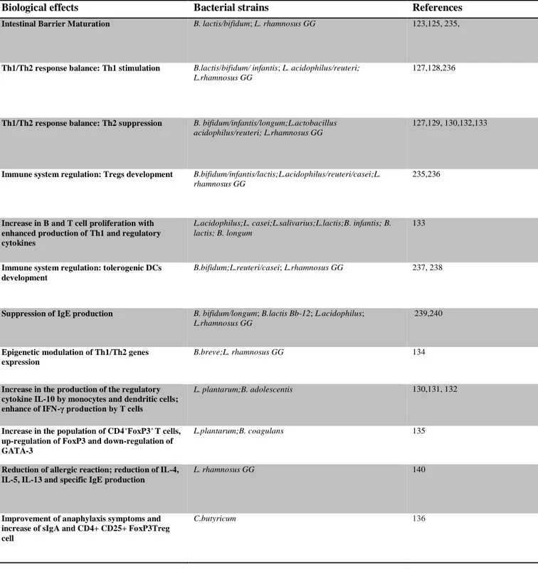
(stimulation of sIgA and -defensins production) (124); modulation of cytokines response by immune cells (125-128). Main pre-clinical evidence on the probiotics activity against FA are summarized in Table 2. In the last decades, a number of experimental investigations have
17 been developed in order to characterize those organisms could be used to modulate the gut microbiota for FA. Stimulation of human peripheral blood mononuclear cells (PBMCs) with selected probiotic strains is a commonly used experimental tool for the investigation of the effect of these microorganisms on immune cells. The incubation of PBMCs with
lactic acid bacteria (LAB) strains (such as L.plantarum and B.adolescentis) resulted in an increased production of the regulatory cytokine IL-10 by monocytes and dendritic cells, and to an enhance of IFN-γ production by T cells (129-131). The addition of probiotics mixture (L.acidophilus W55, L.casei W56, L.salivarius W57, L.lactis W58, B infantis W52, B.lactis W18 and B.longum W51) to PBMCs from children with FA resulted in increased T cell proliferation with enhanced production of Th1 and regulatory cytokines. An increase in T and B cells proliferation and a reduction in IgE production were also observed in PBMCs from children with FA treated for 3 months with the same probiotic mixture (132). Using a 3D co-culture model of intestinal epithelial cells and PBMCs as an in vitro model of the intestinal mucosal immune system, Ghadimi et al demonstrated that the probiotics B.breve and
L.rhamnosus GG (LGG) inhibit activation of inflammatory IL-23 and IL-17 cytokines,
thereby reducing histone acetylation and simultaneously enhancing DNA methylation (133). The limitation of studying the effect of probiotics in vitro lies in the extrapolation of the results to in vivo benefits. For that reason, another commonly used experimental tool in this area is based on the use of animal model of FA. Differential effects in relation to molecular action of oral administration of three LAB strains (B. coagulans 09.712, L.plantarum 08.923 and B.infantis 11.322) in alleviating Th2-driven intestinal inflammation and other symptoms associated with food-induced anaphylaxis were demonstrated in a murine model induced by a major shrimp allergen, ST. In particular, oral supplementation with B.coagulans 09.712 and
L.plantarum 08.923 significantly ameliorates anaphylaxis symptoms and increases the
18
FoxP3 up-regulation and GATA-3 down-regulation (134). Zang et al investigated the
preventive and therapeutic effects of oral C.butyricum on anaphylactic symptoms in FA mice model induced by a β -lactoglobulin (BLG) as allergen, a well-established model of CMA.
The authors observed that the oral treatment with C.butyricum significantly ameliorated anaphylaxis symptoms and increased sIgA and CD4+ CD25+ FoxP3+Treg cell in spleen from BLG-sensitized mice (135). Neonatal monocolonization of germ-free mice by L.casei BL23 modulated the allergic sensitization to cow’s milk proteins, developed higher IgG responses
against caseins, elicited by L.casei that was able to hydrolyze insoluble caseins into soluble immunogenic peptides (136). Using OVA-sensitized murine model, it was demonstrated that oral administration of B.infantis ameliorated allergic symptoms, reducing OVA specific-IgE, and -IgG1 levels in the serum, and Th2 cytokines release in spleen. Moreover, gut microbiota analysis showed that the probiotics-mediated protection was conferred by up-regulation of the relative abundance of Coprococcus and Rikenella at genus level (137). Similar results was obtained by others that observed a decrease of concentrations of IgE, IL-4 and IL-13 following administration of B.infantis CGMCC313-2 in BLG-sensitized mice (138). Oral administration of VSL#3 (a mixture of Streptococcus thermophilus BT01, B. breve
BB02, B.longum BL03, B.infantis BI04, L.acidophilus BA05, L.plantarum BP06, L.paracasei BP07, L.delbrueckii subsp. bulgaricus BD08) to sensitized mice significantly reduces Th2 immune responses and protects against anaphylactic reactions in a mouse model of FA. Also the incubation of mouse spleen cells from sensitized mice with probiotic mixture reduced allergen-stimulated IL-5 and IL-13 production and increased of IFN-γ and IL-10 production (139). An immunoregulatory action by LGG has been demonstrated in a murine model of CMA. LGG administration was able to suppress of Th2 responses such as reduced
hypersensitivity scores and lowered serum CMP-specific IgG1 while promoting Th1 responses by causing elevated IFN-γ and CMP-specific IgG2a levels (140). Similar results
19
have been reported by our group in a BLG-sensitized mice model, in which we found that the administration of LGG added to the extensively hydrolyzed casein formula (EHCF) elicited a significantly reduction of allergic reaction, and of IL-4, IL-5, IL-13 and specific IgE
production (141). Clinical studies have investigated the efficacy of selected probiotic strains against FA. The effect appears strain-specific and more evident in the pediatric age. In a randomized double-blind placebo-controlled trial, it has been demonstrated that the
administration of L.casei CRL431 and B.lactis BB12 added to hypoallergenic formula for
12 months did not affect the acquisition of immune tolerance to cow’s milk proteins in infants with CMA (142). Whereas, using a similar study design we have demonstrated that the addition of the probiotic LGG to EHCF is able to accelerate immune tolerance acquisition in infants with CMA. Infants (aged 1–12 months), consecutively referred for suspected CMA (IgE- or non-IgE-mediated), but still receiving cow’s milk proteins, were invited to participate in the study. Subjects were randomly allocated to one of the two groups of dietary
interventions: control group, received an EHCF; and active group, received an EHCF containing LGG (at least 1.4×10⁷ CFU/100 mL). After 12 months, the double-blind
placebo-controlled food challenge was negative in 15 of 28 control infants (53.6%) and in 22 of 27 infants receiving EHCF with LGG (81.5%, p = 0.027) (143). The results were confirmed in a subsequent trial, when the effect of 5 different dietary strategies was investigated: EHCF,
EHCF + LGG, partially hydrolyzed rice formula, soy formula or amino acid-based formula, in
children affected by IgE- or non-IgE-mediated CMA. After the treatment period of 12
months, the proportion of children acquiring immune tolerance to cow’s milk proteins was
significantly higher in the group receiving EHCF+ LGG (78.9%) than in other groups (144).
At the 3-year follow-up of another pediatric cohort, a further confirmation of a greater rate of
resolution of IgE-mediated CMA as well as a lower incidence of other atopic manifestations
20
part by a modulation elicited by selected LGG components on immune functions through
different pathways including enterocytes, monocytes, mast cells, DCs and Tregs (146-149), and by an expansion of butyrate-producing gut microbiota (43). Accordingly, studies in
infants with eczema and/or CMA who received EHCF supplemented with LGG showed benefits in decreasing gastrointestinal symptoms and inflammation (107,150). Probiotics has been also proposed to reinforce the effectiveness of immunotherapy (151). Oral food
immunotherapy (OIT) is currently the most investigated approach for persistent FA and it is based on the concept that repeated oral/intestinal exposures to antigens normally leads to tolerance. Tang et al. performed a randomized double-blind placebo-controlled trial with the probiotic L.rhamnosus CGMCC 1.3724 and peanut OIT (PPOIT) in 62 children with peanut allergy. Subjects received a fixed dose of probiotic (or placebo) together with peanut OIT (or placebo) once daily for a total of 18 months. Sustained unresponsiveness (SU), determined by DBPCFC conducted 2 to 5 weeks after discontinuation of treatment, was achieved in 82.1% of patients receiving PPOIT compared with 3.6% of those receiving placebo, the highest rate of SU reported for any food immunotherapy treatment evaluated in a randomized controlled study to date. PPOIT also induced high rates of desensitization (90%) and was associated with reduced peanut skin test reactivity, decreased specific IgE, and increased peanut-specific IgG4 levels. PPOIT was well tolerated with no participants withdrawing because of adverse reactions (6 participants withdrew for reasons unrelated to PPOIT treatment); this is in stark contrast to OIT whereby 10% to 30% of participants withdraw because of adverse reactions. At approximately 4 years after the study ended, 67% were still consuming peanut, and only 58% of the 12 participants who stopped peanut ingestion for 8 weeks demonstrated sustained unresponsiveness. Importantly, no OIT control group was evaluated to determine if the probiotic itself had any effect on SU (152). Further studies comparing peanut OIT with probiotic with peanut OIT with placebo will be required to evaluate this strategy further.
21 1.9 Epigenetics regulation of food allergy
Epigenetic modifications are biochemical changes of the chromatin, in other words, DNA or histones, that are functionally relevant, but do not affect the nucleotide sequence of the genome. DNA methylation, a covalent addition of a methyl group, occurs at the cytosine nucleotide belonging to CpG dinucleotide (called ‘CpG site’), which is a DNA sequence where a cytosine nucleotide (C) is directly followed by a guanine nucleotide (G). CpG sites frequently cluster to form ‘CpG islands’, typically located in the elements of a gene
regulatory element with impact on its transcription, for example, promoters or enhancers (153,154). High DNA methylation levels in the CpG island of a promoter are usually associated with lower gene expression up to full gene silencing. The reaction of DNA methylation is catalyzed by DNA methyltransferases (DNMTs), including DNMT1 and DNMT3A and DNMT3B (156-159). The best-characterized post-translational histone modifications include phosphorylation, ubiquitination, acetylation and methylation, the last two of which are the most extensively studied (160,161). Histone acetylation occurs at the lysine residues and it is catalyzed by histone acetyltransferases, while the opposite reaction by histone deacetylases (HDACs). Histone acetylation independent of the position of the lysine amino acid generally correlates with potentially active genes or gene regulatory elements (162,163). DNA methylation and histone modifications mutually interact (164). MicroRNAs (miRNAs) represent post-transcriptional control elements, important epigenetic regulators of gene expression (165;166). These approximately 22-nt noncoding RNA molecules are highly abundant, with more than 2500 mature miRNA molecules characterized in humans. To exert its function, mature miRNAs become incorporated into the RNA-induced silencing complex. The RNA-induced silencing complex is in turn guided by miRNAs to specifically target mRNAs. This leads to the cleavage or degradation of the bound mRNA molecule or suppression of its translation by reducing the speed of the ribosomal machinery (165-170).
22 Considering their biological importance, miRNAs have been involved in multiple human pathologies (167). These include also allergic diseases, in which the role of miRNAs has been rather extensively studied (171-175). It is also worth mentioning that the mechanism of RNA-mediated silencing of gene expression has been utilized in biomedical research as a powerful laboratory tool (176) and in therapeutic applications as one of the possible antisense
approaches (177). Connections between miRNAs and DNA methylation/histone
modifications have also been described (165,167,169,170, 178). Several studies provide direct evidence linking epigenetics and FA (179-184). Martino et al. investigated whether variation in DNA methylation underscores the suboptimal neonatal CD4+ T-cell gene expression associated with the development of FA (179), including impaired T-cell expansion and
reduced IFN- γ production (185-188). In a follow-up study, it was examined the genome-wide DNA methylation profiles in CD4+ T-cells from 12 children with FA and from 12
non-allergic controls at birth and again at 12 months. The authors found that the dysregulation of DNA methylation at MAPK signaling-associated genes during early CD4+ T-cell
development may contribute to T lymphocyte responses in early childhood associated with the development of FA (179). Further linking epigenetics and FA, the first genome-wide association study (GWAS) of FA in 2759 US participants revealed the important role of differential DNA methylation in mediating identified genetic risk factors for peanut allergies. (189). Syed et al demonstrated that subjects who acquired immune tolerance to peanuts after 3 months of immunotherapy had higher numbers of Tregs with higher levels of FoxP3
demethylation, compared to non-tolerant and healthy subjects (180). Recent study
demonstrated that naive CD4+T cells from children with FA exhibit an intrinsic molecular defect during the early state of priming and depressed capacity for proliferation, which is related to epigenetic changes in metabolic (RPTOR, PIK3D, MAPK1, FOXO1) and
23 who fail to resolve FA in later childhood exhibit cumulative increases in epigenetic disruption at T cell activation genes and a decrease of CD4+ T cells proliferative responses compared to children who resolved FA (190). Preliminary cross-sectional studies have suggested that Th1/Th2 cytokine genes DNA methylation pattern and selected miRNAs expression change significantly during CMA disease course (182-184). In particular, we demonstrated a potential role of miR-193a-5p in regulation of Th2 response in children with IgE-mediated CMA (184). Different demethylation rates in Treg-specific-demethylation region (TSDR) of FoxP3 have been also demonstrated comparing CMA children with active disease with those with recent evidence of immune tolerance acquisition (183). Dietary factors exert a pivotal role in the regulation of epigenetic mechanisms (191). Previous observations suggest that formula choice for CMA treatment could influence these mechanisms. Specifically, we observed a significant difference in DNA demethylation rate in TSDR of FoxP3, and in the promoter region of T helper (Th)1/Th2 cytokine genes in children who acquired immune tolerance after treatment with extensively hydrolyzed casein formula containing the probiotic
Lactobacillus rhamnosus GG (EHCF+LGG) compared to subjects who received other
formulas (182-184).
The study of epigenetics in FA is a promising avenue that may lead to a better understanding of the mechanisms underlying FA etiology. It has been shown that both genetics and
environmental factors can alter epigenetic profile (192-193). Thus, epigenetics may be the missing piece to understanding environmental–genetic interactions and FA risk.
24
2. Aim
The aim of our study was to evaluate butyrate concentrations in HM and to see whether these butyrate concentrations can exert protective actions against FA, exploring several immune and non-immune tolerogenic mechanisms in different experimental models:
1. CD4+ T cells from healthy controls and children with IgE-mediated FA 2. human enterocytes cell lines, HT-29 and Caco-2
25
3. Materials and Methods
3.1 Donors and collection of human milk samples
Mothers who participated in the study were enrolled after full-term, singleton births, with all mothers intending to breastfeed infants for at least 75% of feedings for 3 months from the Villa Betania Evangelic Hospital (Naples, Italy) in accordance with the Research Ethics Committee of the University of Naples “Federico II”. All donors were healthy, aged 21-42 years and HM samples were donated at 3 days (colostrum) and during the first 5 months post-partum. Written informed consent was obtained from all participants. HM samples were collected by either manual or electric breast pump expression into 2 mL a sterile milk tubes. Samples were immediately frozen and then stored at −80°C. For each enrolled subject, anamnestic, demographic, clinical and laboratory data were recorded in a data collection sheet. 3-days dietary diary before the collection of HM samples was obtained. The sampling procedures applied ensured that breastfeeding had been well established and that the baby was thriving.
3.2 Determination of butyrate concentration in human milk samples
Butyrate extraction from HM was performed as previously described (Clinica Chimica Acta, 78 (1977) 243-250 Pretreatment methods prior to gaschromatographic analysis of volatile fatty acids from faecal samples J.B.Zijlstra, J.Beukema, B.G.Wolthers, B.M.Byrne, A.Groen, J.D.Anker). 0.5 ml of HM were acidified with 20 μl of 85% (w/v) ortophosphoric acid and 0.5 ml of ethyl acetate, mixed, centrifuged (12,000 x g) for 1 h and extracted in duplicate. About 0.5 ml of extract containing 3mmol/L of 2-ethylbutyric acid (internal standard) was transferred into a 2 ml glass vial and loaded onto an Agilent Technologies (Santa Clara, CA, USA) 7890 gas chromatograph (GC) system with automatic loader/injector. The GC column was an Agilent J&W DB-FFAP (Agilent Technologies) of 30 m, internal diameter 0.25 mm
26 and film thickness 0.25 μm. The GC was programmed to achieve the following run
parameters: initial temperature 90°C, hold 0.5 min, ramp of 20°C min−1 up to a final temperature of 190°C, total run time 8.0 min, gas flow 7.7 ml min−1 split less to maintain 3.26 p.s.i. column head pressure, septum purge 2.0 ml min−1. Detection was achieved using a flame ionization detector. Peaks were identified using a mixed external standard and
quantified by peak height/internal standard ratio.
3.3 Peripheral blood mononuclear cells stimulation and measurement of 4, 5, IL-13, IL-10, and IFN-γ culture media concentration
Two healthy children (Caucasian male, age 24 months with negative clinical history for any allergic conditions and not at risk for atopic disorders), referred to the Pediatric Department of the University of Naples “Federico II” because of minimal surgical procedure, and six
children with challenge-proven FA (2 cow’s milk allergy, 2 peanut allergy, 2 egg allergy; all Caucasian male, age 24 months) were recruited. Patients and control subjects donated a venous blood samples in heparin tubes (8 ml), after written informed consents. Peripheral blood mononuclear cells (PBMCs) were isolated using the Ficoll-Paque (Sigma-Aldrich, St. Louis, MO, USA) method, as described previously (183). PBMCs cells were stimulated with beta-lactoglobulin (BLG;100μg/ml), peanut extract (PN;100μg/ml) or ovalbumin
(OVA;100μg/ml) in the presence or absence sodium butyrate (Sigma-Aldrich, Darmstadt, Germany) at dose of 0.75mM for 24h. After stimulation, the supernatants were collected and the cells were washed and harvested for CD4+T cells isolation. The concentrations of IL-4 and IL-10 were measured in supernatants with a Human IL4/IL10 Enzyme immunoassay kit (Boster Biological Technology, Ltd., Fremont, CA, USA) according to the type of stimulation and stimulant. Human IL-5, IL-13 and IFN-γ ELISA, High Sensitivity (BioVendor, Brno, Czech Republic) were used to detect the IL-5, IL-13 and IFN-γ concentrations. Absorbance
27 was read at 450 nm. The minimum detection concentrations were 15.6 pg/ml for IL-4, 7.8 pg/ml for IL-5, and IL-10, 1.6 pg/ml for IL-13, and 0.78 pg/ml for IFN-γ.
3.4 DNA and RNA extraction from CD4+ T-cells
CD4+ T-cells were obtained by negative selection using the CD4+ T-Cell Isolation Kit II (Miltenyi Biotec, Bergisch Gladbach, Germany) from stimulated PBMCs. Non-target cells were labeled with a cocktail of biotin-conjugated monoclonal antibodies (MicroBead
Cocktail, Miltenyic Biotec) and the magnetically labeled non-target T cells were retaining on a column in the magnetic field of a separator (Miltenyi Biotec). This protocol produces >95% pure CD4+ T cells, as tested by fluorescence-activated cell sorting analysis. Cells were resuspended at 2x10⁶ cells/ml in RPMI-1640 cultur medium (Gibco) supplemented with 10% fetal bovine serum, penicillin/streptomycin (1%) (Lonza), L-glutamine (1%), sodium pyruvate (1%) (Lonza) and NEAA (1%) (Lonza). Cells were cultured at 37°C in complete medium at concentrations of 2x10⁵cells/ml in 24-well plate (Nunc, Roskilde, Denmark). CD4+ T-cells obtained were processed for DNA and RNA extraction. All experiments were performed in triplicate and repeated twice.
3.5 DNA methylation analysis
One microgram of DNA, extracted from CD4+T cells, was modified with sodium bisulfite to convert all unmethylated, but not methylated-cytosines to uracil. Bisulfite conversion was carried out using the EZ DNA Methylation Gold Kit (ZYMO Research Co., Orange, CA, USA), according to the manufacturer’s instructions. The converted DNA was stored at −20°C until used. Fully methylated and fully unmethylated DNA (Merck Millipore, Darmstadt, Germany) were used as controls for the optimization of the assay conditions and to calculate the percent of methylation (0% to 100%). The primers used for DNA methylation analysis of
28 IL-4, IL-5, IL-13, IL-10, IFN-gamma and FoxP3 TSDR are reported elsewhere (182,183). High-resolution melting real-time PCR for methylation analysis was performed as described previously (182,183). The results of methylation analysis were verified by direct sequencing (using the Sanger method modified as follows: ddNTPs labeled with four different
fluorophores) and analyzed by capillary electrophoresis (the analytical specificity and
sensitivity of the test was >99 %). Real-time PCR was performed with the LightCycler® 480 instrument (Roche Applied Science, Penzberg, Germany) using 96-well plates (Roche Applied Science).
3.6 Human enterocytes cell line
Human enterocytes cell line Caco-2 and HT-29 were used (American Type Culture
Collection, Teddington, Middlesex TW11 0LY, UK). Caco-2 cells were grown to confluence in Dulbecco’s modified Eagle’s medium (DMEM; Gibco, Berlin, Germany) supplemented with 10% fetal calf serum (Lonza, Visp, Switzerland), 1% L-glutamine (Lonza), 1%
nonessential amino acids, and 1% penicillin/streptomycin (Lonza). HT-29 cells were grown to confluence in RPMI medium 1640 (Gibco, Berlin, Germany) supplemented with 10% fetal calf serum (Lonza, Visp, Switzerland) 1% L-glutamine (Lonza) and 1%
penicillin/streptomycin (Lonza). Cells were cultured at 37°C in a water-saturated atmosphere consisting of 95% air and 5% CO2, and the medium was changed every 2 days. All
experiments were performed in triplicate and repeated twice.
3.7 Immune and non-immune biomarkers analysis on human enterocytes
At full confluence (15 days), when a human enterocytes monolayer was obtained from Caco-2 and HT-29 in six-well plates (Falcon, Heidelberg, Germany), cells were stimulated for 48h with or without sodium butyrate at different doses (0.1; 0.5; 0.75; 1; 2 mM). Afterward, the
29 supernatants were harvested and stored at −80°C for further use. Cells were used for RNA extraction to perform real-time PCR experiments. The concentration of β-defensin 3 (HBD-3) in the supernatants was measured using a commercially available ELISA kit specific for human HBD-3 (MyBioSource, San Diego, CA, USA) with a detection limit of 11.3 pg/ml. The ELISA was conducted according to the manufacturer’s recommendations. For ZO-1, Occludin, Muc2 and Muc5AC and FoxP3 expression analysis, total cellular RNA was extracted from cells with TRIzol reagent (Gibco BRL, Paisley, Scotland). RNA (1 μg) was reverse transcribed at 37°C in cDNA with a High-Capacity RNA-to-cDNA™ Kit (Life Technologies, Waltham, MA, USA) according to the manufacturer’s instructions.
Complementary DNA (cDNA) was stored at −20°C until use. Quantitative real-time PCR (qRT-PCR) analysis was performed with the TaqMan miRNA assay kit and the TaqMan gene expression assay kit, respectively (both from Applied Biosystems, Grand Island, NY, USA) according to the manufacturer’s instructions. Samples were run in duplicate at 95°C for 15 seconds and 60°C for 1 minute using an ABI Prism 7900 HT Sequence Detection System (Applied Biosystems). After a hot start, the amplification protocol was 40 cycles of 30 sec of denaturation at 95°C, 30 sec of annealing at 60°C, and 1 min of elongation at 72°C. Data analysis was performed with the comparative threshold cycle (CT) method. We used the GAPDH gene to normalize the level of mRNA expression.
For measurement of the mucus thickness, human enterocytes cell lines were seeded onto polycarbonate membranes (0.4 μm pore size) from TranswellTM (Corning Inc., New York, US) at 37°C in an atmosphere with 95% humidity and 5% CO2. At full confluence (14 days), the cells were stimulated for 24, 48 and 72 h with or without butyrate. A pair of membranes cultured under the same conditions were embedded immediately in an optimal cutting temperature compound (BioOptica, Milan, Italy) to avoid mucus damage during processing. Five-micrometre frozen sections of each sample were cut, and after rinsing in 3% acetic acid
30 (Merck, Germany), the cells were stained with 1% Alcian Blue (Merck, Germany) and
oxidized in 1% periodic acid (Merck, Germany). Finally, sections were counterstained with Mayer's haematoxylin and mounted with Aquamount (BDH, Poole, UK). Measurement of the mucus thickness was performed in the middle of the membrane using the ruler tool provided by the Zeiss Axio Observer/ApoTome.
3.8 HDAC activity assay
To assess HDAC activity, HT-29 cells were treated with 10 nmol/L trichostatin A (TSA, Sigma-Aldrich) and with butyrate (0.1; 0.5; 0.75; 1; 2 mM) for 48 hours. HT-29 cells nuclear extracts (3x10⁷ cells/well in 10 mL of culture medium) were prepared by using the Nuclei EZ
Prep kit (Sigma-Aldrich) and quantified by with the Micro BCA Protein Assay Kit. HDAC activity was measured with the EpiQuik HDAC Activity/Inhibition Assay Kit (Epigentek, Farmingdale, NY), according to the manufacturer’s instructions.
3.9 Food allergy animal model
For all experiments three week-old female C3H/HeJ mice were purchased from Charles River Laboratories (Calco, Lecco, Italy). Mice were housed in the animal facility under a 12L:12D lighting cycle, 20-24ºC range of ambient temperature and 40-70% of relative humidity. The mice were acclimated to their environment for 1 week before experiments. Each experiment was littermate controlled and mice were cohoused throughout manipulations. All procedures involving the animals were carried out in accordance with the Institutional Guidelines and complied with the Italian D.L. no.116 of January 27, 1992 of the Italian Ministry of Health and associated guidelines in the European Communities Council Directive of November 24, 1986 (86/609/ECC) and they were approved by the Institutional Committee on the Ethics of
31 Animal Experiments (CSV) of the University of Naples “Federico II” and by the Ministero della Salute (protocol no.2012-0024683).
3.10 Food allergen sensitization and challenge
The experimental design is reported in Figure 4. Briefly, two weeks prior to sensitization, mice were given 30 mg/kg/day of butyrate by oral gavage and continued during the whole study. The control animal received only PBS. After 14 days, mice were sensitized orally using a blunt needle on day 0, 7, 14, 21, 28 with 20 mg of β-lactoglobulin (BLG) (Sigma-Aldrich, Steinheim, Germany) or 1 mg of ovalbumin (OVA) (Sigma-Aldrich, Steinheim, Germany) or 12 mg of peanut extract (kindly provided by Prof. Nagler) mixed with 10 µg cholera toxin (CT) as adjuvant. The control mice receive CT only.
One week after the final sensitization, two doses of 50 mg BLG or 5 mg OVA or 36 mg PN each were administered via intragastric gavage 30 minutes apart. Core body temperature was measured prior to allergen challenge and every 5 minutes after the first challenge until at least 30 minutes after the second challenge using a rectal probe (Mitutoyo, Lainate, Italy).
Anaphylaxis symptoms were scored by an investigator blind to the study group assignment 1 hour after oral challenge, as previously described [16]: 0 = no symptom; 1 = scratching and rubbing around the nose and head; 2 = reduced activity; 3 = activity after prodding and puffiness around the eyes and mouth; 4 = no activity after prodding, labored respiration, and cyanosis around the mouth and the tail; and 5 = death. Serum was collected 1 hour after the second challenge to measure mMCP-1 levels. Spleen and serum were collected 24 hours after challenge for splenocyte culture and antibody measurements.
32 3.11 ELISAs
mMCP-1 was quantified in serum collected 1 hour after challenge according to the manufacturer’s protocol (eBioscience). BLG/PN-specific ELISAs were performed using protocols modified from ref X. Briefly, plates were coated overnight at 4˚C with 100µg/mL BLG/PN in 100mM carbonate-bicarbonate buffer (pH 9.6). Plates were blocked for 2 hours at room temperature with 3% BSA. Samples were added in 1% BSA and incubated overnight at 4˚C. Assays were standardized with BLG/PN-specific antibodies (IgE) purified from mice immunized with BLG+alum or PN+alum on a CNBr-Sepharose affinity column(X). Secondary antibody (biotin-anti-IgE, BD Pharmingen) was added at a 1:500 dilution overnight at 4˚C. On the third day, the plate was incubated with streptavidin-HRP
(ThermoFisher) for 1 hour at room temperature and developed with TMB (Sigma) and 15 min were allowed for the development of colorimetric reactions. Absorbance were read at a wavelength of 450 nm in a microplate reader. OVA-specific IgE were measured using commercially ELISA kit (eBioscience).
3.12 Spleen culture and cytokine measurement
Single-cell suspensions were prepared from spleens harvested 24 h after challenge. Cells were plated at 2×10⁵cells per well with media alone, 1 μg/mL anti-CD3 (clone 2C11), or 200 μg/mL BLG/OVA/PN and incubated at 37 °C for 72 h. After 72 h, plates were frozen at −20 °C. IL-4, IL-13, IL-10, and INF-γ concentrations in supernatants were measured using commercially ELISA kit (eBioscience).
3.13 Preparation of isolated mitochondria and polarographic measurement of respiration
33 After removal, the livers were finely minced and washed in a medium containing 220 mM-mannitol, 70 mM sucrose, 20 mM -N’-(2-hydroxyethyl)piperazine-N-2-ethanesulfonic acid (HEPES) (pH 7.4), 1 mM-EDTA, and 0•1 % (w/v) fatty-acid-free bovine serum albumin (BSA). Tissue fragments were homogenised with the above medium (1:4, w/v) in a Potter Elvehjem homogeniser (Heidolph, Kelheim, Germany) set at 500 rpm (4 strokes/min). The homogenate was centrifuged at 1000 gav for 10 min and the resulting supernatant fraction was again centrifuged at 3000 gav for 10 min. The mitochondrial pellet was washed twice and finally re-suspended in a medium containing 80 mM-KCl, 50 mM-HEPES (pH 7.0), 5 mM KH2PO4, and 0.1% (w/v) fatty-acid-free BSA. The protein content of the mitochondrial suspension was determined by the method of Hartree (1972) using BSA as the protein standard. Isolated mitochondria were then used for the determination of respiratory
parameters. Mitochondrial O2 consumption was estimated by a Clark type electrode (Yellow Springs Instruments, Yellow Springs, OH, USA), maintained in a water-jacketed chamber at 30°C. Hepatic mitochondria (0.5 mg protein) were incubated in a medium (3 ml) containing 80 mM KCl, 50 mM HEPES, 1 mM EGTA, 5 mM KH2PO4 (pH 7.0), and 0.1% (w/v) fatty-acid-free BSA. The substrates used for liver respiration were 10 mM succinate + 3.75 μM-rotenone or 40 μM-palmitoyl-carnitine + 2.5 mM-malate for the determination of fatty acid oxidation rate. State 3 measurements were performed in the presence of 0.6 mM ADP. State 4 respiration was measured in the absence of ADP in isolated mitochondria. The ratio between state 3 and 4, called the respiratory control ratio, was calculated according to Estabrook (1967).
3.14 Determination of mitochondrial enzymatic activities and H2O2 release
Total activity of the Carnitine Palmitoyl-CoA Transferase (CPT) was followed
34 (DTNB) and as substrate palmitoyl CoA. The medium consisted of 50 mM KCl, 10 mM Hepes (pH 7.4), 0.025% Triton X-100, 0.3 mM DTNB, and 10-100 pg of mitochondrial protein in a final volume of 1.0 ml. Two cuvettes were used, both containing the same
medium as that used in the mitochondrial samples. In addition, the sample cuvette contained 1 mM L-carnitine. The reaction was started by simultaneous addition of acyl-CoA to both cuvettes, and the change in absorbance difference between the two cuvettes was followed at 412 nm. Enzyme activity was calculated from E412 = 13,6OO/(M X cm). The temperature was thermostated to 25°C. (Alexson 1988). Mitochondria (40–60 μg) were solubilized in 1% Triton X-100. Aconitase specific activity was measured in a medium containing 30 mmol/l sodium citrate, 0.6 mmol/l MnCl2, 0.2 mmol/l NADP, 50 mmol/l Tris-HCl, pH 7.4, and 2
units isocitrate dehydrogenase, and the formation of NADPH was followed
spectrophotometrically (340 nm) at 25°C. (Hausladen, 1996). The rate of mitochondrial H2O2
release was measured at 30°C following the linear increase in fluorescence (exicitation at 320nm, emission at 400 nm) due to oxidation of homovanillic acid by H2O2 in the presence of
horseradish peroxidase in a computer controlled Jasco fluorometer equipped with a thermostatically controlled cell-holder. The reaction mixture consisted of 0.4 mg of
mitochondrial proteins and succinate (0.6M) added at the end to stat the reaction in the same incubation buffer used for measurements of O2 consumption. Known concentrations of H2O2
were used to establish the standard concentration curve (Barja, 1998). 3.15 Statistical Analysis
The Kolmogorov-Smirnov test was used to determine whether variables were normally distributed. To evaluate the differences among continuous variables, the independent
sample t-test was performed.. The level of significance for all statistical tests was 2-sided, p < 0.05. All analyses were conducted by a statistician, using SPSS version 19.0 for Windows (SPSS Inc., Chicago, IL, USA) and Graph Pad Prism 5.0.
35
4. Results
4.1 Butyrate concentration in human milk
100 healthy lactating women were consented to participate in the study and provided HM samples during the first 5 months of lactation. Butyrate concentrations in colostrum and in mature HM are reported in Figure 5. Butyrate concentration resulted significantly higher in mature HM than in colostrum starting from the 0.5 to 0.75 mM in the 2 ° month of lactation, and remain stable until the 5° month of lactation. This result was in line with previous study in which HM has been examined as a potential source of butyrate for neonates (194). Median butyrate concentration in mature HM was 0.75 mM. This means that a breastfed infant could receive a daily dose of butyrate of about 30 mg/Kg body weight.
4.2 Effects of butyrate on CD4+ T cells
To investigate immunological effects of butyrate, we performed time-course and dose
response experiments on PBMCs from healthy children. Butyrate was able to stimulate IL-10 production, with a maximal effect at 0.75 mM after 24 hours of incubation. No modulation was observed for IL-4, IL-5, IL-13 and IFN-γ production (data not shown). PBMCsfrom IgE-mediated FA patients, stimulated with BLG, OVA, PN in absence of butyrate, resulted in a significant increase in Th2 cytokines production: IL-4, IL-5 and IL-13. Co-incubating the cells with 0.75 mM, butyrate induced a significant inhibition of these effects (Figure 6). This effect was independent on a methylation of the promoter region of the IL-4, IL-5 and IL-13 genes. Instead, butyrate stimulated the tolerogenic cytokines IL-10 and IFN-γ production through a demethylation of respective genes at 0.75 mM dose. This effect paralleled with a modulation of TSDR FoxP3 demethylation and its expression (Figure 6).
36 4.3. Effects of butyrate on human enterocytes
The direct regulatory action elicited by butyrate on human enterocytes in the regulation of non-immune tolerogenic mechanisms is depicted in Figure 5. Butyrate stimulated the expression of tight junction (TJ) proteins, ZO-1 and occludin in Caco-2 cells. The maximal effective dose was 0.75 mM. The same butyrate dose was also able to up-regulate Muc2 and Muc5AC expression, in HT-29 and Caco-2 cells, respectively (Figure 7). Accordingly, 0.75 mM butyrate was able to increase the enterocyte mucus layer thickness (from 0.0 to 5±2 µm, NT vs 0.75 mM, p<0.05). We also investigated the butyrate effect on the innate immune peptide synthesis, HBD-3, that is involved in Foxp3+ T cells induction (195). As shown in Figure 7, butyrate elicited a significant effect on HBD-3 synthesis by human enterocytes with maximum effective dose of 0.75 mM.
To determine whether butyrate acts as an HDAC inhibitor in HT-29 cells, we analyzed the HDAC activity in comparison with TSA, a well-established HDAC inhibitor. We observed that HDAC activity was reduced in a dose-dependent manner in HT-29 cells treated with butyrate (Figure 7).
4.4 Effects of butyrate in FA animal model
BLG-, OVA- and PN- sensitized mice showed a significantly higher anaphylactic symptoms score and body temperature decrease, compared to control animals. Exposing the animal to oral butyrate caused a significant inhibition of these parameters of allergic response (Figure 8).
To determine whether the butyrate administration was effective in reducing local mucosal mast cell degranulation in sensitized mice, mMCP-1 serum concentrations were measured after oral challenge. Serum mMCP-1 concentration was significantly increased in BLG-,
37 OVA- and PN- sensitized mice compared to the control animals. Whereas, oral butyrate caused a significant reduction in mMCP-1 concentration compared to control mice. According with these data, butyrate administration also caused a reduction in specific IgE concentrations compared to control group (Figure 8).
To study the mechanism underlying the effect of butyrate in reducing allergic response, splenocytes cytokines production was investigated. BLG-, OVA- and PN- sensitized mice showed a significant increase in Th2 cytokines production (IL-4 and IL-13) and a significant decrease in Th1 cytokines production (IL-10 and IFN-gamma) compared to control animals. These effects were significantly inhibited by butyrate administration (Figure 8).
4.5 Effects of butyrate on hepatic mitochondrial oxidative capacity and hepatic oxidative stress
Pivotal role for hepatic mitochondrial dysfunction, and consequent excessive generation of ROS, in a murine model of FA has been recently demonstrated (196). H2O2 yield and
ROS-induced damage on aconitase activity were measured in hepatic mitochondria. The increased ROS production was proven by the higher mitochondrial H2O2 yield and the lower basal/total
aconitase ratio in all sensitized animals when compared to the control group. Butyrate administration efficiently modulated the oxidative stress as demonstrated by the lower H2O2
release and by the reactivation of the aconitase enzyme in all treated groups when compared to control animals (Figure 9).
38
5. Discussion
The results of our study demonstrate that HM butyrate stimulates a wide range of immune and non-immune tolerogenic mechanisms able to protect against FA. Immune tolerance is a state of active non-responsiveness to ingested soluble antigens mediated by gut-associated
intestinal lymphoid tissue. Inducible FoxP3+ CD4+ Treg cells are central to the maintenance of immune homeostasis and tolerance throughout the body, particularly in the gut. Other
evidence suggests also a role for the complex interaction between gut microbiota and immune and non-immune cells in shaping immune tolerance. The presence of both diet-and microbe-induced populations of Treg cells is required for oral tolerance to food antigens (197). In our study, we observed that butyrate is able to modulate these mechanisms of oral tolerance. We evaluated the direct effects of butyrate on CD4+ T cells isolated from PBMCs from
children affected by challenge-proven IgE-mediated FA. CD4+ T cells were stimulated in
vitro with different allergens, BLG, PN and OVA, in the presence or absence of butyrate.
Butyrate stimulated, in a dose-dependent manner, tolerogenic cytokines, such as IL-10 production, through a demethylation of respective gene. We found also that butyrate induced a FoxP3 demethylation and a concomitantly increase of its expression. The mechanisms of action of butyrate are multiple, many of these involve an epigenetic regulation of gene expression through the inhibition of histone deacetylase (HDAC) (198). It has been demonstrated that the inhibition of HDAC 6 and 9 is responsible for the increase in FoxP3 gene expression and increase of Tregs (199). Acetylation of Foxp3 is an important
posttranslational mechanism that affects Foxp3 abundance, because it protects Foxp3
proteasomal degradation (200). This is a signature mechanism of action of several HDACs in Tregs (201-205).
We also explore the direct effects of butyrate on human enterocytes. Our data suggest that SCFA butyrate contribute to mucosal homeostasis through the induction of Treg cells and the
39 regulation of epithelial barrier integrity. Loss of epithelial integrity in the gut increases
antigen uptake and promotes secretion of epithelial-derived cytokines IL-33, thymic stromal lymphopoietin, and IL-25 (206). These cytokines promote Th2-type allergic response by activation of ILC2s, mast cells, basophils, and DCs (207). Activation of ILC2s stimulates production of IL-4–, IL-5–, and IL-13–promoting Th2-type allergic responses (208). Overall, the state of the epithelial barrier is thought to be important for sensitization to food antigens. The positive modulation of gut mucosa integrity by butyrate is supported by the up-regulated expression of TJ proteins, which in turn are involved in the tuning of epithelial permeability. These results are in line with previous observations demonstrating that butyrate is able to maintain epithelium barrier integrity through an increase of ZO-1 and occludin expression (209).
A significant increase of Muc2, Muc5AC expression and mucus layer thickness was also observed after stimulation of human enterocytes with butyrate. Similarly, Gaudier et al. demonstrated that butyrate differently stimulates the expression of various mucin genes in the colon, with maximum effects on Muc2 expression (210). The mucus layer, covering the gastrointestinal mucosa, is considered as the first line of defense against mechanical,
chemical, or microbiological aggressions arising from the luminal contents (211). The mucus layer does not merely form a nonspecific physical barrier, but also constrains the
immunogenicity of gut antigens by delivering tolerogenic signals (212). In the colon, the mucus layer is directly in contact with the butyrate produced by gut microbiota, representing the major energy source for colonocytes (213).
Moreover, butyrate stimulates the HBD-3 synthesis, an innate immune peptide, involved in induction of FoxP3+ T cells (195).
To confirm the butyrate immune and non immune effects demonstrated in vitro, we performed an in vivo model of FA. In a murine model of FA, the results of anaphylactic