Claudia Loganes, Elisa Marini, Samuele Naviglio, Tarcisio Not, Annalisa Marcuzzi, Department of Medicine, Surgery, and Health Sciences, University of Trieste, Strada di Fiume, 447 Trieste, Italy
Erica Valencic, Alessia Pin, Stefano Martelossi, Luigina De Leo, Tarcisio Not, Lorenzo Monasta, Alberto Tommasini, Institute for Maternal and Child Health - IRCCS “Burlo Garofolo”, I-34137 Trieste, Italy
Author contributions: Loganes C, Valencic E and Tommasini A designed the experimental plan and conceived and designed the research; Pin A, Marini E, De Leo L performed all experiments; Loganes C, Monasta L performed the statistical analyses; Martelossi S, Naviglio S and Not T enrolled the patients and provided patient samples; Loganes C, Tommasini A, Marcuzzi A wrote the paper; Martelossi S and Marcuzzi A revised the manuscript critically for important intellectual content; all authors read and approved the final version of this manuscript.
Supported by Institute for Maternal and Child Health - IRCCS “Burlo Garofolo”, No. RC 03/09.
Institutional review board statement: The study was reviewed and approved by the Institutional Review Board of the Institute for Maternal and Child Health - IRCCS “Burlo Garofolo”, Trieste, Italy (RC 03/2009).
Conflict-of-interest statement: All the authors declare no conflict of interest.
Data sharing statement: Dataset available from the corresponding author at [email protected].
Open-Access: This article is an open-access article which was selected by an in-house editor and fully peer-reviewed by external reviewers. It is distributed in accordance with the Creative Commons Attribution Non Commercial (CC BY-NC 4.0) license, which permits others to distribute, remix, adapt, build upon this work non-commercially, and license their derivative works on different terms, provided the original work is properly cited and the use is non-commercial. See: http://creativecommons.org/ licenses/by-nc/4.0/
Manuscript source: Invited manuscript
Correspondence to: Alberto Tommasini, MD, PhD, Institute for Maternal and Child Health - IRCCS “Burlo Garofolo”, Via dell’Istria 65/1, I-34137 Trieste,
Italy. [email protected] Telephone: +39-40-3785422
Received: June 28, 2016
Peer-review started: June 29, 2016 First decision: July 29, 2016 Revised: August 12, 2016 Accepted: September 6, 2016 Article in press: September 6, 2016 Published online: November 28, 2016
Abstract
AIMTo evaluate how mucosal bacteria impact on the spontaneous and muramyl dipeptide (MDP)-induced inflammation in Crohn’s disease (CD) and ulcerative colitis (UC).
METHODS
Colonic mucosal biopsies were collected from children with active or remissive CD, UC and controls. Two tissue samples were taken from inflamed mucosal segments (in patients with active disease) or from non-inflamed mucosa [in patients in remission or in healthy controls (HC)]. Experiments were performed in the presence or absence of antibiotics, to assess whether the disease-associated microbiota can modulate the cytokine response
ex vivo
. For this purpose, each specimen was half-cut to compare spontaneous and MDP-induced inflammation in the presence of live bacteria (LB) or antibiotics. After 24 h of culture, an array of 17 cytokines was assessed in supernatants. Statistical analyses were performed to find significantORIGINAL ARTICLE
Ex vivo
response to mucosal bacteria and muramyl
dipeptide in inflammatory bowel disease
Basic Study
Claudia Loganes, Erica Valencic, Alessia Pin, Elisa Marini, Stefano Martelossi, Samuele Naviglio, Luigina De
Leo, Tarcisio Not, Lorenzo Monasta, Alberto Tommasini, Annalisa Marcuzzi
differences in single cytokines or in patterns of cytokine response in the different groups.
RESULTS
We demonstrated that subjects with CD display a spontaneous production of inflammatory cytokines including granulocyte-colony stimulating factor (G-CSF), interleukin (IL) 6, IL8, IL10 and IL12, that was not significantly influenced by the addition of antibiotics. UC specimens also displayed a trend of increased spontaneous secretion of several cytokines, which however was not significant due to broader variability among patients. After the addition of antibiotics, spontaneous IL8 secretion was significantly higher in UC than in controls. In HC, a trend towards the weakening of spontaneous IL8 production was observed in the presence of live mucosal bacteria with respect to the presence of antibiotics. In contrast, in the presence of LB UC showed an increasing trend of spontaneous IL8 production, while MDP stimulation resulted in lower IL8 production in the presence of antibiotics. We also showed that subjects with CD seem to have a lowered production of IL8 in response to MDP in the presence of LB. Only with the addition of antibiotics, likely reducing the contribution of LB, multivariate statistical analysis could identify the combination of measures of G-CSF, tumor necrosis factor alpha, IL4 and IL17 as a good discriminator between CD and UC.
CONCLUSION
We showed that the presence of LB or antibiotics can significantly influence the inflammatory response
ex
vivo
in inflammatory bowel diseases.Key words: Gut-microbiota; Crohn’s disease; Ulcerative colitis; Cytokines; Inflammation
© The Author(s) 2016. Published by Baishideng Publishing
Group Inc. All rights reserved.
Core tip: Even though previous studies have already considered cytokine secretions as marker of an infla-mmatory condition and mucosal imbalance in Crohn’s disease (CD) subjects, they did not take into account the autochthonous colonization of the intestinal mucosa by the disease-associated microbiota. In this work we investigated whether the microbiota can modulate the
ex vivo
cytokine response, in the presence or absence of antibiotics, or if a selection of cytokines could dis-criminate different inflammatory bowel diseases types. Through a multivariate logistic model we identified, only in specimens treated with antibiotics, a specific cytokine profile able to discriminate CD from ulcerative colitis.Loganes C, Valencic E, Pin A, Marini E, Martelossi S, Naviglio S, De Leo L, Not T, Monasta L, Tommasini A, Marcuzzi A.
Ex vivo response to mucosal bacteria and muramyl dipeptide
in inflammatory bowel disease. World J Gastroenterol 2016; 22(44): 9734-9743 Available from: URL: http://www.wjgnet.
com/1007-9327/full/v22/i44/9734.htm DOI: http://dx.doi. org/10.3748/wjg.v22.i44.9734
INTRODUCTION
Inflammatory bowel diseases (IBD) are intestinal disorders characterized by a complex inflammatory process that may involve any part of the digestive tract. IBD include mainly Crohn’s disease (CD) and ulcerative colitis (UC), which are characterized by a different spectrum of clinical and pathological features[1,2].
In CD, the inflammation is extended through the whole intestinal wall of the gut with a segmental distribution, while in UC the inflammatory process is continuous and predominantly confined to the colonic mucosa[2].
IBD are characterized by an aberrant activation of the mucosal immune system with reaction to luminal content in genetically susceptible subjects[3], resulting
in a chronic inflammatory state with dysregulation of several cytokines[4-6].
Cytokines are important mediators in the gut-associated immune system, both in physiological conditions and in disease[7]. In normal conditions, the
mucosal immune activation reflects an active balance between tolerance of luminal content and response to commensal and pathogenic bacteria. Indeed, this basal activation is associated with a predominance of cytokines such as interleukin (IL) 10 and transforming growth factor beta and the production of antimicrobial peptides that can contribute to the shaping of the “healthy” microbiota. In pathological conditions, other cytokines and mediators are produced, which can, at the same time, lead to tissue injury and altered regulation of the microbiota and dysbiosis[4,8-10].
Chronic tissue damage results in clinical intestinal symptoms in IBD patients (abdominal cramping and pain, diarrhea, constipation and bowel obstruction, nausea and vomiting), even though extra-intestinal manifestations (weight loss, fever, asthenia and arthralgias) and immune disorders are also associated with this pathologies[11].
Despite the large number of studies and clinical analyses, several issues remain to be solved regard-ing the immune pathogenesis of IBD. In particular, the therapy of IBD is still based mainly on anti-infla-mmatory and immunosuppressive therapies, while mounting evidence supports the idea that defective immunity may underlie IBD pathogenesis, especially in cases with an early onset in childhood[12]. In fact,
excessive inflammation in IBD could either result from a hyper-response of the immune system or it may just represent a compensatory response to luminal contest in the background of defective immunity[13].
Indeed, CD has been alternatively classified as an autoinflammatory disorder or a disease with primary immune deficiency[14].
On the one hand, some studies suggested that CD is an autoinflammatory disease, characterized by an increase in the inflammatory response due to nucleotide-binding oligomerization domain-containing protein 2 (NOD2) variants, even though data su-pporting this hypothesis are controversial. NOD2 is an intracellular receptor involved in gastrointestinal immunity; it detects bacterial components and has been identified as the first susceptibility gene for CD[15-17]. The binding of muramyl dipeptide (MDP) to
NOD2 leads to the downstream activation of mitogen-activated protein kinases and nuclear factor-kappa beta (NF-κB) signaling, which in turn stimulates the transcription of several immune response genes. However, early studies have shown the association of the most common mutation 3020insC with a reduced activation of NF-κB[18]. In contrast, later investigations
proposed that the pro-inflammatory action is caused by a dysregulated autophagy[19].
On the other hand, several studies have found evidence that CD patients present immune response defects. In fact, a decreased bacterial clearance by the phagocytic system has been observed in peripheral blood cells from patients with CD; furthermore, NOD2-/- mice exhibit a reduced production of anti-microbial peptides by Paneth cells that leads to an imbalance in the microbiota in the presence of certain microbial pathogens, and intestinal inflammation can be prevented in these models by the forced expression of defensins in the intestinal cells[20].
For these reasons, we wanted to describe what happens in the disease itself, not by using simple animal models, but by studying the relationships between the mucosal immune system, influenced by its multigenic background, and the intestinal microbiota colonizing the diseased epithelium.
In these conditions, we tried to understand how the inflammatory response to stimuli such as MDP is influenced ex vivo by the presence of live or antibiotic-inactivated bacteria.
We performed an ex vivo culture of colonic explants to investigate the mucosal response both in spon-taneous conditions and after the stimulation with MDP.
MATERIALS AND METHODS
Patients and gut biopsies collection
Forty-eight children with IBD and eight control subjects were enrolled. Participation in the study was proposed to all patients with IBD or their guardians when the performance of a colonoscopy was needed for clinical assessment, required for the diagnosis or the follow-up of their intestinal disease.
Patients with IBD were divided into four groups based on the type of intestinal disorder and on endo-scopic mucosal disease activity: active Crohn’s disease (CD, n = 23), remissive Crohn’s disease (rCD, n = 3), active ulcerative colitis (UC, n = 15) and remissive ulcerative colitis (rUC, n = 7). Disease activity scores,
namely the Pediatric Crohn’s Disease Activity Index and the Pediatric Ulcerative Colitis Activity Index were also calculated. Any other recent acute disease or chronic inflammatory conditions are reported in these patients.
Control subjects were selected among children who underwent colonoscopy as part of their clinical investigations for various reasons not related to inflammatory enteropathies, such as routine evaluation of juvenile intestinal polyps, autoimmune gastritis, unexplained abdominal symptoms and weight loss. They had evidence of normal, non-inflamed mucosa and were thus considered healthy controls (HC).
Colonic mucosal biopsies were taken during colono-scopy. For each subject, two specimens were collected from the same mucosal area, in addition to the biopsies obtained for clinical purposes. In patients with active disease, two inflamed biopsies were collected, while in patients with inactive disease or HC two biopsies were taken from non-inflamed areas.
All subjects were recruited from the Gastro-enterology and Clinical Nutrition Unit, Institute for Maternal and Child Health - IRCCS “Burlo Garofolo”, Trieste, Italy and written informed consent was obtained from patients and donors (or their guardians) according to a protocol approved by the Independent Ethical Board at the Institute (n.185/08, 19/08/2008).
Ex vivo specimen culture
Immediately after collection, biopsies were cut in half following the intestinal layers’ orientation and split in two tubes containing a physiological saline solution. Once they reached the laboratory, the specimens were transferred to sterile tubes with culture medium (X-VIVO, Lonza, Verviers, Belgium) supplemented with 10% human AB serum (Sigma-Aldrich, Milano, Italy) and 2 mmol/L L-glutamine (EuroClone, Milano, Italy). The intestinal tissue was exposed to 250 ng/mL MDP (N-Acetylmuramyl-L-alanyl-D-isoglutamine hydrate, Sigma-Aldrich) or medium alone, in the presence or absence of antibiotics: 100 U/mL penicillin (EuroClone) and 0.1 mg/mL streptomycin (EuroClone).
After 24 h culture at 37 ℃ in a humidified atmo-sphere (95% air and 5% CO2), supernatants were
collected and stored at -20 ℃ for cytokine analysis.
Cytokine quantifications
The cytokines and the chemokines released in the culture medium were measured using the Bio-Plex Pro® Human Cytokine 17-plex Assay (BioRad, Hemel
Hempstead, United Kingdom), according to the manufacturer’s instructions.
Analytes examined by this method were IL1β, IL2, IL4, IL5, IL6, IL7, IL8, IL10, IL12 (p70), IL13, IL17, granulocyte-colony stimulating factor (G-CSF), GM-CSF, IFNγ, MCP1 (MCAF), MIP1β, and TNFα.
Samples were analyzed with the Bio-Plex® 200
RESULTS
Experimental design
Clinical characteristics of all patients included in the study, obtained from the medical sheets and from the colonoscopy records, are reported in Supplementary Table 1 and Supplementary Table 2 and summarized in Table 1.
Ex vivo culture of biopsies is thought to
repro-duce both the immunological and the microbiologic background of the disease. However, to isolate the contribution of live bacteria (LB) to inflammation, biopsies were stimulated in absence or in presence of antibiotics. Thus, for each sample we examined four conditions: basal with LB or with antibiotics (PS), and MDP-stimulated with (PS + MDP) or without antibiotics (LB + MDP).
After a 24 h culture, the media were harvested to proceed with the quantification of 17 analytes, including cytokines, chemokines and growth factors (Figure 1).
The levels of three cytokines (IL5, IL7 and IL13) were under the lower limit of detection in all evaluated conditions and were thus excluded.
Basal cytokine network in IBD
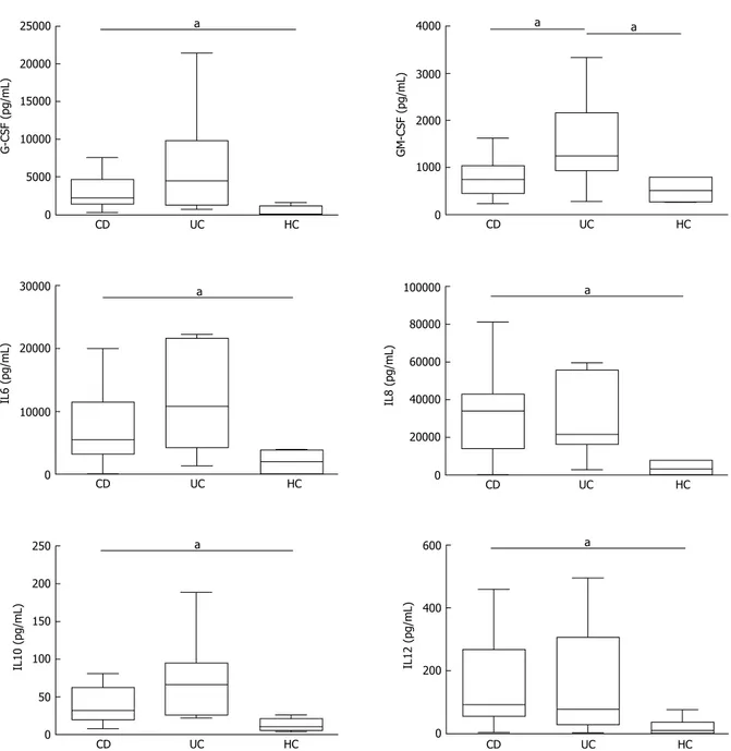
In basal conditions with live mucosal bacteria, five cytokines had levels significantly higher in CD than in controls (HC) (CD vs HC: G-CSF, P = 0.024, IL6 P = 0.037, IL8 P = 0.033, IL10 P = 0.049, IL12, P = 0.028); levels of most cytokines tended to be even higher in UC, but because of the high variability among patients, differences did not reach significance, except for GM-CSF, whose levels were higher in UC compared with both HC (P = 0.031) and CD (P = 0.034) (Figure 2).
Levels of IL1β, IL2, IL4, IL17, IFNγ, TNFα, MCP1 and MIP1β did not differ significantly between the three studied groups, even if median levels of several cytokines such as IL2, IL4 and TNFα tended to be higher in UC than in HC. In general, HC showed a narrower variability compared to patients (Supplementary Figure 1).
Median levels of all cytokines in the different groups and conditions are rendered in Figure 3 with a color analog scale (CAS): for each cytokine, values 0-24% of the higher value are displayed in green, 25%-49% in orange, 50%-74% in red and above 75% in dark red.
In specimens from patients with endoscopically inactive disease, almost all cytokines were detected, as expected, at lower levels than in active disease. The decrease is significant in UC for GM-CSF (P < 0.01), IL6 (P < 0.05), IL10 (P < 0.05) and TNFα (P < 0.05) (Data not shown).
The addition of antibiotics in the bioptic culture to inhibit mucosal bacteria did not lead to relevant changes in the secretion pattern (Supplementary Figure 2), with the exception of IL8 in UC samples. software, which returned data as Median Fluorescence
Intensity and concentration (pg/mL).
Statistical analysis
All statistical analyses were carried out using Stata/IC 14.1 (StataCorp LP, College Station, United States) and GraphPad Prism software version 5 (GraphPad, San Diego, United States). Values assumed by cytokines were described as medians and interquartile ranges and represented with box plots using Tukey’s whiskers. Statistical significance was set as P < 0.05 (a), P < 0.01 (b) and P < 0.001 (c).
Associations between cytokines and specific condi-tions (HC vs IBD, and CD vs UC) were analyzed firstly by univariate logistic regression and then by multivariate logistic regression. For the multiple comparisons carried out by univariate logistic regression, considering the relatively small sample size, which implied having relatively large P values, we decided not to apply any correction to the P value for statistical significance: we realize that we carried out independent univa-riate logistic regressions on different outcomes and separately for 17 cytokines, chemokines and growth factors. However, this just is considered as a preliminary step for the selection of cytokines to be included in the multivariate analysis: significant variables (P < 0.05 in the univariate analysis) were entered into multivariate analysis. Multivariate logistic regression analysis was employed to determine cytokine profiles associated with the different forms of IBD. Receiver operating characteristic (ROC) curves were constructed to evaluate the performance of the multivariate models. The area under the curve (AUC), sensitivity and specificity were also calculated. For each model, we identified the optimal cut-off by maximizing the sum of sensitivity and specificity.
Table 1 Characteristics of patients included in the study Patients
(n = 56) (mean ± SD)Age (yr) Male/female remissionActive/
IBD (n = 48) CD (n = 26) 11 ± 3.08 16/10 23/3 UC (n = 22) 15 ± 3.88 12/10 15/7 CONTROLS
(n = 8)
HC (n = 8) 10 ± 4.60 4/4
-IBD: Inflammatory bowel disease; CD: Crohn’s disease; UC: Ulcerative colitis; HC: Healthy controls.
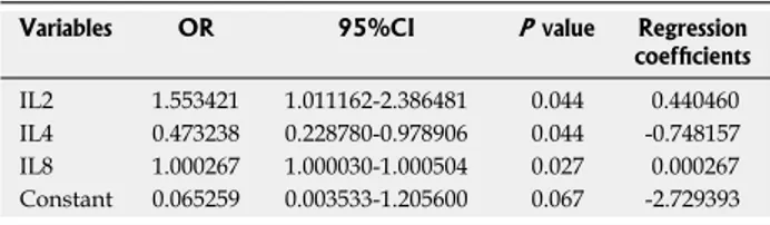
Table 2 Multivariate logistic model for inflammatory bowel disease patients vs healthy controls in the presence of antibiotics and without muramyl dipeptide stimulation Variables OR 95%CI P value Regression
coefficients
IL2 1.553421 1.011162-2.386481 0.044 0.440460 IL4 0.473238 0.228780-0.978906 0.044 -0.748157 IL8 1.000267 1.000030-1.000504 0.027 0.000267 Constant 0.065259 0.003533-1.205600 0.067 -2.729393
The trend of IL8 increase after antibiotics in UC is so clear that the difference compared with HC becomes significant (Figure 4).
Cytokine network induced by MDP
MDP was used to investigate the function of the NOD2-pathway ex vivo in CD compared with UC and HC.
In general, the response to MDP appeared to be higher in UC than in CD (see the CAS in Figure 3). After stimulation with MDP in the presence of LB, even
though the differences are not significant, there is a clear trend of increased secretion of IL2, IL6, IL8, IL17 and IFNγ in UC, while no change was observed in CD. Indeed, even if in a smaller scale, HC tended to secrete reduced amounts of IL2, IL6 and IFNγ (Figure 5).
However, after the addition of antibiotics, this pattern was subverted and MDP stimulation failed to produce an evident increase in cytokine secretion in any group and it was even possible to note a reduced trend of secretion for IL8 in UC (Figure 6).
No variation was noted in the remaining cytokines, whose values are reported in supplementary Tables 3 and 4.
Multivariate logistic regression and ROC analysis
to identify profiles of pro-inflammatory cytokines
associated with IBD diagnosis
Multivariate logistic regression were carried out to assess if any cytokine combination was associated with IBD related outcomes.
Only experiments in the presence of antibiotics provided profiles significantly associated with IBD. In particular, without MDP stimulation, IL2, IL4 and IL8 were simultaneously associated with IBD in a multivariate analysis (Table 2). ROC analysis (Figure 7A) shows that the AUC was 0.98 (95%CI: 0.93-1.00), and we could have a sensitivity of 100% with a specificity of 71%.
After MDP stimulation another set of cytokines (MCP1, IL8 and IL10) was significantly associated with IBD (Table 3). The AUC for this model was 0.91 (95%CI: 0.79-1.00) (Figure 7B). Keeping sensitivity
Two colonic biopsies from the same intestinal region
Cut of each biopsy in half following the intestinal
layers’ orientation
24 h culture at 37 ℃ of mucosal speciments
with different stimuli
No stimulus (LB) + Antibiotics (PS) + MDP (LB + MDP) + Antibiotics + MDP (PS + MDP) Collection of culture supernatants and
cytokine quantifications
Figure 1 Schematic culture protocol of colonic biopsies derived from colonoscopy. LB: Live Bacteria; PS: Penicillin-streptomicin; MDP: Muramyl dipeptide.
Table 3 Multivariate logistic model for inflammatory bowel disease patients vs healthy controls in the presence of antibiotics and muramyl dipeptide stimulation
Variables OR 95%CI P value Regression coefficients
MCP1 0.997248 0.995067-0.999434 0.014 -0.0027555 IL8 1.000094 1.000019-1.000170 0.014 0.0000944 IL10 1.098966 1.009008-1.196944 0.030 0.0943694 Constant 0.529026 0.108222-2.586054 0.432 -0.636717
Table 4 Multivariate logistic model for Crohn’s disease vs ulcerative colitis in the presence of antibiotics and muramyl dipeptide stimulation
Variables OR 95%CI P value Regression coefficients G-CSF 0.999703 0.999484-0.999921 0.008 -0.0002975 TNFα 0.990492 0.981420-0.999649 0.042 -0.0095533 IL4 1.155281 1.012918-1.317651 0.031 0.1443433 IL17 1.009361 1.000308-1.018497 0.043 0.0093177 Constant 1.043247 0.333344-3.264992 0.924 0.042338
for the detection of IBD at 94%, we obtained a specificity of 75%, which would permit to identify 45 IBD patients out of 48, with 2 false positives out of 8 HC.
After MDP stimulation it was also possible to discri-minate between CD and UC based on the measure of G-CSF, TNFα, IL4 and IL17 (Table 4). According to the ROC analysis (Figure 7C), the AUC was 0.78 (95%CI: 0.65-0.91), and we could obtain a sensitivity on CD of 92.31% with a specificity of 50%.
DISCUSSION
It is well known that subjects with active IBD display increased secretion of several inflammatory cytokines in the intestinal mucosa. For example, aberrant secretions
of IL8 after challenge with E. coli or other intestinal bacteria were recorded by Edward and colleagues and considered as a marker of the mucosal imbalance typical of CD[21]. The authors, however, did not take
into account the autochthonous colonization of the intestinal mucosa by the disease-associated microbiota. In the present work, we compared the cytokine profile of the intestinal mucosa in the presence or absence of antibiotics, to assess whether disease-associated microbiota can modulate the cytokine response ex
vivo. We demonstrated that subjects with CD display
a spontaneous production of inflammatory cytokines besides IL8, and including G-CSF, IL6, IL10 and IL12. The addition of antibiotics to the culture produced only a slight trend towards increased spontaneous cytokine secretion, both in CD and in UC, but no
a 600 400 200 0 CD UC HC IL12 (pg/mL) a 250 200 150 100 50 0 CD UC HC IL10 (pg/mL) a 100000 80000 60000 40000 20000 0 CD UC HC IL8 (pg/mL) a 30000 20000 10000 0 CD UC HC IL6 (pg/mL) a 4000 3000 2000 1000 0 CD UC HC GM-CSF (pg/mL) a a 25000 20000 15000 10000 5000 0 CD UC HC G-CSF (pg/mL)
Figure 2 Cytokine secretion levels by colonic biopsies from inflammatory bowel diseases patients. Levels of cytokine secretion by colonic biopsies from
inflammatory bowel diseases (IBD) patients in active phase [Crohn’s disease (CD); ulcerative colitis (UC)] and healthy controls (HC) in basal condition [live bacteria (LB)]. Statistical significances are denoted using alphabetical letters (aP < 0.05).
difference was significant, except for increased IL8 secretion in UC. These data may support the idea that antibiotics can influence UC, as suggested by other authors[22]. However, whilst some protocols that include
metronidazole in vivo are shown to have protective effects on disease activity[22], other combinations had
no effect[23]. Actually, it is well known that the risk
of CD can be raised by previous use of antibiotics in
childhood, but this is not believed to occur for UC[24].
Thus, the significance of increased levels of IL8 in UC after the addition of antibiotics remains without a clear explanation.
Even if the difference is not significant, there is a trend toward higher concentrations of inflammatory cytokines in UC if compared to CD. Actually, this is not surprising if we consider that we are sampling just superficial specimens where UC is expected to be more expressed than CD, which, on the contrary, may extend the inflammatory process throughout the whole intestinal wall[25].
Of note, in HC, the IL8 secretion with or without MDP is reduced in the presence of LB if compared to antibiotics. This effect is likely due to a modulatory effect of LB on mucosal immunity. In contrast, samples from UC showed a trend of increase of cytokines such as IL6 and IL8 when stimulated with MDP in the presence of LB. However, in the presence of antibiotics, stimulation with MDP was associated with decreased HC LB PS LB + MDP PS + MDP CD LB PS LB + MDP PS + MDP UC LB PS LB + MDP PS + MDP IL1 β
IL2 IL4 IL6 IL8 IL10 IL12 IL17 TNF
α MIP1 β G-CSF GM-CSF INF γ MCP1 IL1 β
IL2 IL4 IL6 IL8 IL10 IL12 IL17 TNF
α MIP1 β G-CSF GM-CSF INF γ MCP1 IL1 β
IL2 IL4 IL6 IL8 IL10 IL12 IL17 TNF
α MIP1 β G-CSF GM-CSF INF γ MCP1
Figure 3 Color analog scale representing the median levels of all cytokines analyzed. Median levels of all cytokines in healthy controls (HC),
Crohn’s disease (CD) and ulcerative colitis (UC) groups in the four experimental conditions: live bacteria (LB), antibiotics (PS), live bacteria + Muramyl dipeptide (LB + MDP) and antibiotics + MDP (PS + MDP). For each cytokine, values 0-24% of the higher value are displayed in green, 25%-49% in orange, 50%-74% in red and above 75% in dark red. Asterisks indicate the set of cytokines whose measure allowed to discriminate CD from UC with 92% sensitivity and 50% specificity. 200000 150000 100000 50000 0 IL 8 (p g/ m L) -PS +PS -PS +PS -PS +PS CD UC HC b
Figure 4 Dosage of IL8 secretion by colonic biopsies from inflammatory bowel diseases patients. Levels of IL8 secretion by colonic biopsies from
patients with active Crohn’s disease (CD), active ulcerative colitis (UC) and from healthy controls (HC), in the presence (+PS) or absence (-PS, live bacteria) of antibiotic mixture. Statistical significances are denoted using alphabetical letters (bP < 0.01). 200000 150000 100000 50000 0 IL 8 (p g/ m L) CD UC HC -MDP +MDP -MDP -MDP +MDP +MDP
Figure 6 Representative trend of cytokine secretion after muramyl dipeptide stimulation, with antibiotics. Variation trend of IL8 secretion after muramyl
dipeptide (MDP) stimulation in patients suffering from active Crohn’s disease (CD), active ulcerative colitis (UC) and healthy controls (HC), in presence of antibiotics (+PS). No statistical significance was found.
50000 40000 30000 20000 10000 0 IL 6 (p g/ m L) CD UC HC -MDP +MDP -MDP +MDP -MDP +MDP
Figure 5 Representative trend of cytokine secretion after muramyl dipeptide stimulation, with live bacteria. Variation trend of IL6 secretion after muramyl
dipeptide (MDP) stimulation in patients suffering from active Crohn’s disease (CD), active ulcerative colitis (UC) and healthy controls (HC), in absence of antibiotics [live bacteria (LB)]. No statistical significance was found.
IL8 secretion. Thus, antibiotics alone seem to increase IL8 production in UC, either because of a loss of the protective effect of mucosal bacteria or for the release of pro-inflammatory bacterial compounds. Conversely, we observed a reduction of IL8 production in UC when MDP is used together with antibiotics. Even though there is no clear explanation for this phenomenon, we can argue that in this case antibiotics and MDP-induced peptides may synergize in controlling both protective and harmful bacteria[26].
We also showed that subjects with CD seem to have a lowered production of IL8 in response to MDP in the presence of LB. Although it is not clear whether defects in NOD2 signaling might play a role in this finding, these results support the idea that CD is not usually associated with a hyper-inflammatory response to MDP, i.e., it is not just an autoinflammatory disorder. Recent data suggest that inflammation in CD may be the result of compensatory responses to defective immunity[27].
Although it was not the primary aim of our work, we could demonstrate that culture with antibiotics is the only ex vivo condition in which it was possible to identify profiles of cytokine secretions able to discriminate between CD and UC with good sensitivity and specificity. Indeed, the combination of measures of G-CSF, TNFα, IL4 and IL17 was shown to identify
CD with respect to UC with sensitivity and specificity respectively of 92% and 50%. Rather than being used to assist diagnosis, which relies only on clinical and histological features, these results highlight once more the importance of taking into account the presence of mucosal bacteria when dealing with ex vivo analyses of IBD.
Among the limitations of this study, we need to mention the small sample size. The decision not to apply corrections to the p values for significance for multiple univariate comparisons was a consequence of this constrain and it should be taken into account.
However, despite the small sample size, valid results have been obtained with this ex vivo mucosal culture, that allowed us to discern the two IBD forms.
In conclusion, we showed that CD and UC have distinct cytokine profiles in the intestinal mucosa and that the mucosal-associated microbiota can differentially impact on the inflammatory response in the two conditions.
COMMENTS
Background
Inflammatory bowel diseases (IBD) are associated with an unbalanced crosstalk between the mucosal immune system and the luminal content. Production of cytokines and antimicrobial-peptides in gut mucosa may contribute to the shaping of an altered intestinal microbiota and, conversely, microbes can
1.00 0.75 0.50 0.25 0.00 Sensitivity 0.00 0.25 0.50 0.75 1.00 1-specificity
Area under RCC curve = 0.9063 1.00 0.75 0.50 0.25 0.00 Sensitivity 0.00 0.25 0.50 0.75 1.00 1-specificity
Area under RCC curve = 0.9788 1.00 0.75 0.50 0.25 0.00 Sensitivity 0.00 0.25 0.50 0.75 1.00 1-specificity
Area under RCC curve = 0.7815
A
B
C
Figure 7 Multivariate combinations. A: Roc curve of IL2, IL4 and IL8: the best cutoff value was for a sensitivity of 100% and specificity of 71% to discriminate IBD (UC
and CD) from healthy controls (AUC = 0.98, 95%CI: 0.93-1.00); B: Roc curve of MCP1, IL8 and IL10: the best cutoff value was for a sensitivity of 94% and specificity of 75% to discriminate IBD (UC and CD) from healthy controls (AUC = 0.91, 95%CI: 0.79-1.00); C: Roc curve of G-CSF, TNFα, IL4 and IL17: the best cutoff value was for a sensitivity of 92% and specificity of 50% to discriminate CD from UC (AUC = 0.78, 95%CI: 0.65-0.91).
translocate across the epithelium and induce an excessive inflammatory response and recruit adaptive immunity. Treatment of IBD with antibiotics, however, gave inconsistent results as different bacterial species may have protective or harmful effects.
Research frontiers
Studies on the pathogenesis of IBD should involve the specific disease-associated microbiota together with the mucosal immunity, with its genetic background. Thus, ex vivo experiments that bring together these two factors are of particular importance to unravel the pathogenesis of IBD.
Innovations and breakthroughs
By reproducing four different conditions, we could dissect the effect of disease-associated live or antibiotic-inactivated bacteria on the mucosal response to muramyl dipeptide (MDP). We showed that live bacteria affect the response to MDP in different ways in Crohn’s disease (CD) and in ulcerative colitis (UC).
Applications
Ex vivo analyses of the inflammatory response in the presence of antibiotics and soluble stimuli may allow studying the action of different antibiotics in patients with CD or UC.
Terminology
MDP represents a pro-inflammatory stimulus used to mimic the inflammatory activation.
Peer-review
Some strengths of the article include: an adequate sample to test the hypothesis, novel study looking at the autochthonous colonization of the intestinal mucosa by the disease-associated microbiota, and seemingly good methods to address the question. It also provides a novel method to identify CD and UC based off of specific cytokines markers.
REFERENCES
1 Vermeire S. Towards a novel molecular classification of IBD.
Dig Dis 2012; 30: 425-427 [PMID: 22796810 DOI: 10.1159/000 338147]
2 Vermeire S, Van Assche G, Rutgeerts P. Classification of
inflammatory bowel disease: the old and the new. Curr Opin Gastroenterol 2012; 28: 321-326 [PMID: 22647554 DOI: 10.1097/ MOG.0b013e328354be1e]
3 Nagalingam NA, Lynch SV. Role of the microbiota in inflammatory
bowel diseases. Inflamm Bowel Dis 2012; 18: 968-984 [PMID: 21936031 DOI: 10.1002/ibd.21866]
4 Sanchez-Munoz F, Dominguez-Lopez A, Yamamoto-Furusho
JK. Role of cytokines in inflammatory bowel disease. World J Gastroenterol 2008; 14: 4280-4288 [PMID: 18666314 DOI: 10.3748/wjg.14.4280]
5 Baumgart DC, Sandborn WJ. Crohn’s disease. Lancet 2012; 380: 1590-1605 [PMID: 22914295 DOI: 10.1016/S0140-6736
(12)60026-9]
6 Geremia A, Biancheri P, Allan P, Corazza GR, Di Sabatino A.
Innate and adaptive immunity in inflammatory bowel disease. Autoimmun Rev 2014; 13: 3-10 [PMID: 23774107 DOI: 10.1016/ j.autrev.2013.06.004]
7 Kleiner G, Zanin V, Monasta L, Crovella S, Caruso L, Milani D,
Marcuzzi A. Pediatric patients with inflammatory bowel disease exhibit increased serum levels of proinflammatory cytokines and chemokines, but decreased circulating levels of macrophage inhibitory protein-1β, interleukin-2 and interleukin-17. Exp Ther Med 2015; 9: 2047-2052 [PMID: 26136934 DOI: 10.3892/ etm.2015.2370]
8 Strober W, Fuss IJ. Proinflammatory cytokines in the pathogenesis
of inflammatory bowel diseases. Gastroenterology 2011; 140: 1756-1767 [PMID: 21530742 DOI: 10.1053/j.gastro.2011.02.016] 9 Manoharan I, Suryawanshi A, Hong Y, Ranganathan P,
Shanmugam A, Ahmad S, Swafford D, Manicassamy B, Ramesh G, Koni PA, Thangaraju M, Manicassamy S. Homeostatic PPARα Signaling Limits Inflammatory Responses to Commensal Microbiota in the Intestine. J Immunol 2016; 196: 4739-4749 [PMID: 27183583 DOI: 10.4049/jimmunol.1501489]
10 Al Nabhani Z, Lepage P, Mauny P, Montcuquet N, Roy M, Le
Roux K, Dussaillant M, Berrebi D, Hugot JP, Barreau F. Nod2 Deficiency Leads to a Specific and Transmissible Mucosa-associated Microbial Dysbiosis Which Is Independent of the Mucosal Barrier Defect. J Crohns Colitis 2016; Epub ahead of print [PMID: 27147452 DOI: 10.1093/ecco-jcc/jjw095]
11 de Souza HS, Fiocchi C. Immunopathogenesis of IBD: current
state of the art. Nat Rev Gastroenterol Hepatol 2016; 13: 13-27 [PMID: 26627550 DOI: 10.1038/nrgastro.2015.186]
12 Bianco AM, Zanin V, Girardelli M, Magnolato A, Martelossi
S, Tommasini A, Marcuzzi A, Crovella S. A common genetic background could explain early-onset Crohn’s disease. Med Hypotheses 2012; 78: 520-522 [PMID: 22309886 DOI: 10.1016/ j.mehy.2012.01.02]
13 Notarangelo LD, Tommasini A. Defective and excessive immunities
in pediatric diseases. Curr Pharm Des 2012; 18: 5729-5734 [PMID: 22726115 DOI: 10.2174/138161212803530943]
14 Tontini GE, Vecchi M, Pastorelli L, Neurath MF, Neumann H.
Differential diagnosis in inflammatory bowel disease colitis: state of the art and future perspectives. World J Gastroenterol 2015; 21: 21-46 [PMID: 25574078 DOI: 10.3748/wjg.v21.i1.21]
15 Hugot JP, Chamaillard M, Zouali H, Lesage S, Cézard JP,
Belaiche J, Almer S, Tysk C, O’Morain CA, Gassull M, Binder V, Finkel Y, Cortot A, Modigliani R, Laurent-Puig P, Gower-Rousseau C, Macry J, Colombel JF, Sahbatou M, Thomas G. Association of NOD2 leucine-rich repeat variants with susceptibility to Crohn’s disease. Nature 2001; 411: 599-603 [PMID: 11385576 DOI: 10.1038/35079107]
16 Ogura Y, Bonen DK, Inohara N, Nicolae DL, Chen FF, Ramos R,
Britton H, Moran T, Karaliuskas R, Duerr RH, Achkar JP, Brant SR, Bayless TM, Kirschner BS, Hanauer SB, Nuñez G, Cho JH. A frameshift mutation in NOD2 associated with susceptibility to Crohn’s disease. Nature 2001; 411: 603-606 [PMID: 11385577 DOI: 10.1038/35079114]
17 Naser SA, Arce M, Khaja A, Fernandez M, Naser N, Elwasila S,
Thanigachalam S. Role of ATG16L, NOD2 and IL23R in Crohn’s disease pathogenesis. World J Gastroenterol 2012; 18: 412-424 [PMID: 22346247 DOI: 10.3748/wjg.v18.i5.412]
18 Cho JH. The Nod2 gene in Crohn’s disease: implications for future
research into the genetics and immunology of Crohn’s disease. Inflamm Bowel Dis 2001; 7: 271-275 [PMID: 11515855 DOI: 10.1097/00054725-200108000-00014]
19 Stappenbeck TS, Rioux JD, Mizoguchi A, Saitoh T, Huett A,
Darfeuille-Michaud A, Wileman T, Mizushima N, Carding S, Akira S, Parkes M, Xavier RJ. Crohn disease: a current perspective on genetics, autophagy and immunity. Autophagy 2011; 7: 355-374 [PMID: 20729636 DOI: 10.4161/auto.7.2.13074]
20 Biswas A, Liu YJ, Hao L, Mizoguchi A, Salzman NH, Bevins
CL, Kobayashi KS. Induction and rescue of Nod2-dependent Th1-driven granulomatous inflammation of the ileum. Proc Natl Acad Sci USA 2010; 107: 14739-14744 [PMID: 20679225 DOI: 10.1073/pnas.1003363107]
21 Edwards LA, Lucas M, Edwards EA, Torrente F, Heuschkel
RB, Klein NJ, Murch SH, Bajaj-Elliott M, Phillips AD. Aberrant response to commensal Bacteroides thetaiotaomicron in Crohn’s disease: an ex vivo human organ culture study. Inflamm Bowel Dis 2011; 17: 1201-1208 [PMID: 21484962 DOI: 10.1002/ibd.21501] 22 Sato K, Chiba T, Ohkusa T. Serial changes of cytokines in active
ulcerative colitis: effects of antibiotic combination therapy. Hepatogastroenterology 2009; 56: 1016-1021 [PMID: 19760932 DOI: 10.1016/S0016-5085(09)61192-6]
23 Mantzaris GJ, Archavlis E, Christoforidis P, Kourtessas D,
Amberiadis P, Florakis N, Petraki K, Spiliadi C, Triantafyllou G. A prospective randomized controlled trial of oral ciprofloxacin in acute ulcerative colitis. Am J Gastroenterol 1997; 92: 454-456
[PMID: 9068468]
24 Ungaro R, Bernstein CN, Gearry R, Hviid A, Kolho KL, Kronman MP, Shaw S, Van Kruiningen H, Colombel JF, Atreja A. Antibiotics associated with increased risk of new-onset Crohn’s disease but not ulcerative colitis: a meta-analysis. Am J Gastroenterol 2014; 109: 1728-1738 [PMID: 25223575 DOI: 10.1038/ajg.2014.246] 25 Abraham C, Cho JH. Inflammatory bowel disease. N Engl J
Med 2009; 361: 2066-2078 [PMID: 19923578 DOI: 10.1056/
NEJMra0804647]
26 Reinoso Webb C, Koboziev I, Furr KL, Grisham MB. Protective and pro-inflammatory roles of intestinal bacteria. Pathophysiology 2016; 23: 67-80 [PMID: 26947707 DOI: 10.1016/j.pathophys.2016. 02.002]
27 Marks DJ, Rahman FZ, Sewell GW, Segal AW. Crohn’s disease: an immune deficiency state. Clin Rev Allergy Immunol 2010; 38: 20-31 [PMID: 19437144 DOI: 10.1007/s12016-009-8133-2]
P- Reviewer: Kojima N S- Editor: Qi Y L- Editor: A E- Editor: Zhang FF