Edited by: Marco Ledri, Lund University, Sweden Reviewed by: Gunther Sperk, Innsbruck Medical University, Austria Deniz Yilmazer-Hanke, University of Ulm, Germany *Correspondence: Barbara Bettegazzi [email protected] Received: 18 September 2020 Accepted: 21 December 2020 Published: 22 January 2021 Citation: Cattaneo S, Verlengia G, Marino P, Simonato M and Bettegazzi B (2021) NPY and Gene Therapy for Epilepsy: How, When,... and Y. Front. Mol. Neurosci. 13:608001. doi: 10.3389/fnmol.2020.608001
NPY and Gene Therapy for Epilepsy:
How, When,... and Y
Stefano Cattaneo
1,2, Gianluca Verlengia
2,3, Pietro Marino
3,4, Michele Simonato
1,2,3and
Barbara Bettegazzi
1,2*
1Vita-Salute San Raffaele University, Milan, Italy,2San Raffaele Scientific Institute, Milan, Italy,3Department of Neuroscience
and Rehabilitation, Section of Pharmacology, University of Ferrara, Ferrara, Italy,4Department of Medical Sciences, Section
of Pediatrics, University of Ferrara, Ferrara, Italy
Neuropeptide Y (NPY) is a neuropeptide abundantly expressed in the mammalian central
and peripheral nervous system. NPY is a pleiotropic molecule, which influences cell
proliferation, cardiovascular and metabolic function, pain and neuronal excitability. In the
central nervous system, NPY acts as a neuromodulator, affecting pathways that range
from cellular (excitability, neurogenesis) to circuit level (food intake, stress response, pain
perception). NPY has a broad repertoire of receptor subtypes, each activating specific
signaling pathways in different tissues and cellular sub-regions. In the context of epilepsy,
NPY is thought to act as an endogenous anticonvulsant that performs its action through
Y2 and Y5 receptors. In fact, its overexpression in the brain with the aid of viral vectors
can suppress seizures in animal models of epilepsy. Therefore, NPY-based gene therapy
may represent a novel approach for the treatment of epilepsy patients, particularly for
pharmaco-resistant and genetic forms of the disease. Nonetheless, considering all the
aforementioned aspects of NPY signaling, the study of possible NPY applications as a
therapeutic molecule is not devoid of critical aspects. The present review will summarize
data related to NPY biology, focusing on its anti-epileptic effects, with a critical appraisal
of key elements that could be exploited to improve the already existing NPY-based gene
therapy approaches for epilepsy.
Keywords: viral vectors, epilepsy, gene therapy, Y2 receptor, NPY
NPY DISCOVERY, EVOLUTION, AND FUNCTION
Described in 1982, neuropeptide Y (NPY) is a 36-aminoacid peptide that shares high homology
with its family members pancreatic peptide (PP) and peptide YY (PYY). The NPY ancestral
gene appeared in vertebrates, evolving from an ortholog NPY-like system that regulates energy
homeostasis in invertebrates acting on growth and reproduction (
De Jong-Brink et al., 2001;
Kooijman and Troost, 2007; Gershkovich et al., 2019
). The family of Y peptides probably originated
through a chromosome quadruplication event that took place during jawed vertebrate emergence
(
Larhammar and Salaneck, 2004
).
NPY has a widespread expression throughout the central (CNS) and peripheral nervous system
(PNS) and it is typically co-released with other neurotransmitters. An unusually broad repertoire of
receptor subtypes mediate its actions, each activating specific signaling pathways in different tissues
and cellular sub-regions (
Leblanc et al., 1987; Keast, 1991; Dumont et al., 1992; Elfvin et al., 1997;
Cerdá-Reverter and Larhammar, 2000; Wai et al., 2004
).
During evolution, the NPY-like system has increased the
complexity of its actions, with effects that in humans range from
cell proliferation to the control of energy metabolism, pain and
neuronal activity (
Kuo et al., 2007; Tilan and Kitlinska, 2016
).
NPY is involved in cardiovascular and metabolic diseases, as well
as in respiratory and neurologic disorders (
Pedrazzini et al., 2003;
Vezzani and Sperk, 2004; Atanasova and Reznikov, 2018
), acting
as a paracrine hormone in the periphery and behaving like a
neuromodulator in the CNS.
In the CNS, NPY exerts its modulatory action both at cellular
(excitability, neurogenesis) and at circuit level (food intake,
stress response, and pain perception). It is expressed in different
areas of the brain, from the neocortex to the posterior root of
spinal nerves, usually in GABAergic interneurons, but also in
long projecting catecholaminergic neurons; e.g., in the brainstem
and in certain hypothalamic nuclei (
Chronwall et al., 1985; de
Quidt and Emson, 1986; Silva et al., 2005a; Benarroch, 2009
). In
the mesial temporal lobe, NPY is widely expressed in different
subnuclei of the amygdala, where it is thought to exert a potent
anxiolytic effect (
Tasan et al., 2010; Wood et al., 2016
), and in the
hippocampus, where it displays an inhibitory action on excitatory
synaptic transmission, mostly by reducing glutamate release
(
Colmers et al., 1985; Klapstein and Colmers, 1992; Greber et al.,
1994; Mcquiston and Colmers, 1996
). It is worth noting that,
coherently with its homeostatic role, NPY projecting neurons
are also close to circumventricular organs and sensory/secretory
blood-brain interfaces (
Wagner et al., 2015
).
GENE STRUCTURE
The human NPY gene (∼8 kb) is located on chromosome
7p15 (genomic coordinates (GRCh38): 7:24,284,189-24,291,861).
Regulatory elements have been found within 530 bases from
the transcription start site and further regulatory sequences
enhancing transcription and mRNA stability may be present
up/downstream that region or even inside introns (
Waldbieser
et al., 1992; Waschek, 1995; Zhou et al., 2008
). Single nucleotide
polymorphisms (SNPs) in the coding region may increase NPY
synthesis (
Mitchell et al., 2008
). The full length mRNA is 551 bp
long (
Minth et al., 1984
). After translation in the endoplasmic
reticulum, upon signal peptide truncation, NPY is directed to the
secretory pathway.
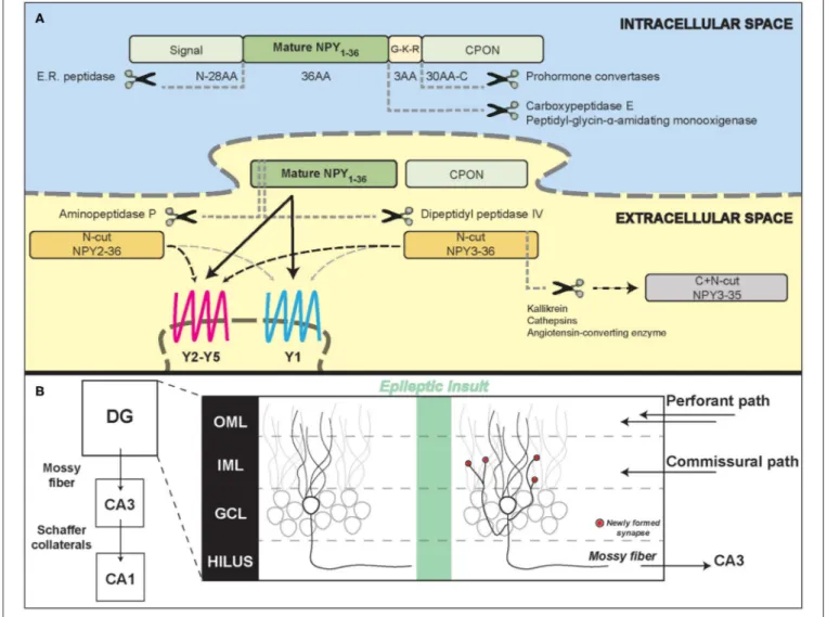
PEPTIDE TRAFFICKING, PROCESSING
AND RELEASE
While trafficking inside dense core vesicles (DCVs), the full
coding sequence of NPY, prepro-NPY, is sequentially split
into three fragments (Figure 1A): (1) an N-terminus
28-amino acid (aa) signaling peptide, (2) the mature 36 aa,
4.2 kDa, peptide (NPY
1−36), and (3) a 30-aa C-terminal
flanking peptide of neuropeptide-Y (CPON). A
glycine-lysine-arginine (G-K-R) site in proximity of the C-terminus of the
mature 36 aa peptide is crucial for CPON cleavage by
pro-hormone convertases and for the amidation of the mature
NPY, performed by carboxypeptidase E and
peptidyl-glycin-α
-amidating monooxygenase. The CPON structure is highly
conserved during evolution (
Cerdá-Reverter and Larhammar,
2000
). It has been suggested that it may play a role in epilepsy
control, but current data do not confirm this hypothesis (
Soud
et al., 2019
).
NPY and CPON containing DCVs are released upon calcium
influx. The need of a long, high frequency firing rate for
NPY release (
Lundberg et al., 1986; van den Pol, 2012
)
has been questioned by evidence that NPY is released by
hippocampal neurons even during physiological synaptic activity
(
Li et al., 2017
).
METABOLISM
Once released in the extracellular space, mature NPY can bind
to its receptors and activate signal transduction (
Walther et al.,
2011
) or be metabolized, either close or far away from its
release site, in the cerebrospinal fluid or in the blood. Proteolytic
processing can alter the NPY signaling at either the N-terminal
or C-terminal portion of the peptide and usually results in a
modification of receptor binding affinity or inactivation followed
by complete degradation, depending on a number of peptidases
with compartment-dependent concentration and activity (
Allen
et al., 1987; Wagner et al., 2015
).
The most common pathway of NPY metabolism is N-terminal
cleavage by dipeptidyl peptidase IV (DP4) which is responsible
for the formation of NPY
3−36, followed by C-terminal processing
by enzymes like kallikrein, cathepsins or angiotensin-converting
enzyme (ACE) that in turn yield inactive NPY fragments.
Aminopeptidase (AmP) instead produces NPY
2−36, catalyzing
a less efficient cleavage within the N-terminal region compared
to DP4, which results in a lower relative concentration
of this metabolite (
Abid et al., 2009
). Both NPY
3−36and
NPY
2−36display a decreased affinity for Y1 receptors, therefore
preferentially binding to other (Y2 and Y5) receptor subtypes
(
Grandt et al., 1996; Hubers et al., 2018; Yang et al., 2018
).
After inactivation, other plasmatic peptidases catalyze the
metabolism of smaller fragments, with the kidney playing a
major role in residual NPY metabolism (
Satoh et al., 1999
).
The estimated plasma half-life in human and animal studies is
between 5 and 20 min (
Pernow et al., 1986; Potter, 1987
).
NPY RECEPTORS
The NPY system is not only multi-ligand, as described above,
but also multi-receptor, and this makes it a complex target for
therapeutic applications.
In fact, five different NPY receptors are expressed in
mammals: Y1, Y2, Y4, Y5, and y6. While Y1, Y2, Y4, and Y5 are
functional in all mammals, y6 is a pseudogene in humans and
other primates and is missing also in the rat genome (
Larhammar
and Salaneck, 2004
). NPY displays an especially high affinity
for the Y1, Y2, and Y5 receptor subtypes: even if structurally
different, these three receptors can respond to the same ligands.
Y1 and Y4 form a receptor superfamily, while Y2 and Y5 have
FIGURE 1 | Neuropeptide Y processing and its potential role in the epileptic hippocampal network. (A) Schematic representation of NPY intracellular processing and extracellular metabolism. (B) Illustration of hippocampal formation rearrangements after an epileptic insult. Red dots represent synapses newly formed by the mossy fiber sprouting in the inner molecular layer that contain NPY and pre-synaptic Y2 receptors. DG, dentate gyrus; CA3/CA1, Cornu Ammonis; OML, outer molecular layer; IML, inner molecular layer; GCL, granule cell layer.
distinct, individual features (
Larhammar and Salaneck, 2004
).
NPY receptors (YRs) have different affinities for the Y family
hormone ligands, with Y4Rs binding preferably PP and Y2Rs
binding NPY and N-terminally truncated peptides with similar
affinity (
Lindner et al., 2008
). The genes encoding for NPY
receptors are located on human chromosome 4 and probably
arose by a duplication event from an ancestral
NPY/PYY-binding receptor. All NPY receptors are widely expressed in
the mammal brain, Y2 being the most abundant (
Dumont
et al., 1998
). High levels of NPY binding can be revealed in
the cortex, hippocampus, amygdala, striatum and cerebellum
(
Dumont et al., 1993
).
Specific binding to Y1 receptors can be visualized in different
layers of the cortex, in the CA1 and CA3 stratum radiatum,
oriens, in the dentate gyrus of the hippocampus in the amygdala,
striatum, cerebellum and, at lower levels, in some thalamic,
hypothalamic and brainstem nuclei (
Dumont et al., 1990, 1993;
Aicher et al., 1991; Cabrele and Beck-Sickinger, 2000; Kopp
et al., 2002
). Outside the CNS, Y1Rs are also found in the
adipose tissue and in vascular smooth muscle cells (
Castan et al.,
1993; Lindner et al., 2008
). Y1Rs are mainly localized
post-synaptically in neurons of the hippocampus (especially in CA3,
CA1 and dentate gyrus), striatum and cortex (
Wahlestedt et al.,
1986; Caberlotto, 1997; Kopp et al., 2002
), with a prominent
somatic and dendritic localization (
Kopp et al., 2002
). However,
some studies also suggest a pre-synaptic localization (
Colmers
et al., 1987, 1988; Flood and Morley, 1989; Pickel et al., 1998;
Brumovsky et al., 2002; Glass et al., 2002; Kopp et al., 2002;
Stani´c et al., 2006; Li et al., 2017
). Albeit NPY and Y1R
scarcely co-localize (
Stani´c et al., 2011
), the presence of Y1R
on the cell soma of NPY-containing hilar interneurons and
cultured hippocampal neurons is suggestive of a possible role
of these receptors in an autoinhibitory feedback (
St-Pierre et al.,
2000; Paredes et al., 2003
).
Together with Y5Rs, Y1Rs play an important role in regulating
feeding behavior and energy homeostasis (
Baldock et al., 2007;
Nguyen et al., 2012
). Y1R-mediated antidepressant and anxiolytic
effects have been described in rodents (
Wahlestedt et al.,
1993; Verma et al., 2012
), while the role in epilepsy remains
controversial (see below). The anxiolytic effect of NPY in the
basolateral amygdala has been attributed to the activation of Y1Rs
(
Sajdyk et al., 2004; Giesbrecht et al., 2010
).
Y2Rs are expressed in many brain regions, including the
hippocampus, thalamus, hypothalamus and cortex; in the
peripheral nervous system, Y2Rs are found in parasympathetic,
sympathetic and sensory neurons; finally, they are also present
in the intestine and in certain blood vessels (
Wahlestedt et al.,
1986; Stjernquist and Owman, 1990; Gehlert et al., 1992;
Dumont et al., 1993; Rettenbacher and Reubi, 2001
). In the
hippocampus, Y2 receptors are particularly enriched in the
CA1 and CA3 areas, respectively in the stratum radiatum and
in the pyramidal cell layer (
Colmers et al., 1987, 1988, 1991;
Monnet et al., 1992
). Expression of Y1 and Y2 receptors is often
complementary. For example, high levels of Y2Rs are detectable
in the stratum oriens and radiatum of CA1-CA3, where Y1
receptor levels are relatively low, while the opposite is true in
the dentate gyrus molecular layer (
Stani´c et al., 2011
). Y2Rs are
highly expressed in the terminal regions of mossy fibers and
Schaffer collaterals (
Jacques et al., 1997
), where they act
pre-synaptically by inhibiting calcium-mediated neurotransmitter
release (
Klapstein and Colmers, 1993
). While NPY and a Y2R
selective agonist inhibit evoked EPSPs on CA1 pyramidal cells,
a Y2R selective antagonist is able to block the inhibitory action of
NPY on glutamate release (
El Bahh et al., 2002
).
Y2Rs are expressed by both GABAergic and glutamatergic
terminals (
Stani´c et al., 2006, 2011
) and may therefore inhibit
the release of both neurotransmitters, in particular under chronic
epileptic conditions (
Martire et al., 1993; Greber et al., 1994;
Klapstein and Colmers, 1997; Vezzani and Sperk, 2004; Silva
et al., 2005b
). This makes Y2Rs an interesting target in epilepsy
(
Vezzani and Sperk, 2004
). Y2Rs can also be localized along the
course of axons in fiber tracts (in Schaffer collaterals, the fimbria
and the stria terminalis (
Dum et al., 2017
)). These receptors are
functionally coupled with G-protein signaling and show high
affinity for their ligand (
Dum et al., 2017
), leaving open the
possibility of a modulation through NPY volume transmission.
Y5Rs are mainly found in the hypothalamus and in the
hippocampus (in the pyramidal cell layer of the CA2 region,
with lower concentrations in the hilar region of the dentate
gyrus and in the CA3 subregion), where they participate in
the modulation of hippocampal excitability (
Gerald et al.,
1996; Dumont et al., 1998; Guo et al., 2002
). Together with
Y1Rs, Y5Rs contribute to the regulation of food intake and
energy homeostasis, but they also display anticonvulsant effects
(
Woldbye et al., 1997; Criscione et al., 1998; Nanobashvili et al.,
2004
). Y5R KO mice display a reduced NPY-mediated inhibition
of glutamatergic synaptic transmission and are therefore more
susceptible to kainate-induced seizure mortality (
Marsh et al.,
1999; Baraban, 2004
).
NPY receptors are G protein-coupled receptors (GPCRs)
with seven transmembrane domains, acting preferentially via
hetero-trimeric Gi/o proteins (
Michel et al., 1998
). They can
trigger a variety of intracellular responses, including inhibition of
adenylyl cyclase, regulation of potassium and calcium channels
and activation of the mitogen-activated protein kinase (MAPK)
cascade in some cell types (
Howell et al., 2005; Lu et al., 2010;
Thiriet et al., 2011; Shimada et al., 2012
). Binding of the ligand to
the receptor stabilizes an active receptor conformation, essential
for inducing intracellular signal transduction. NPY binding
modes vary with individual receptors, with different amino acids
impacting anchoring, affinity and binding (
Beck-Sickinger et al.,
1994; Merten et al., 2007; Walther et al., 2012; Pedragosa-Badia
et al., 2013; Kaiser et al., 2015; Yang et al., 2018
). NPY peptides
reach the receptors by lateral diffusion, after being pre-associated
with the membrane through their C-terminal domain (
Lerch
et al., 2004; Thomas et al., 2005
) that is also essential for the
binding of NPY to specific receptors, in particular Y2 (
Beck-Sickinger et al., 1994
).
NPY receptors are predominantly expressed at the cell surface
and sequence motifs essential for endoplasmic reticulum export
and delivery to the membrane have been identified, particularly
in the C-terminal portion of the protein (
Walther et al.,
2011, 2012
). Y2Rs display desensitization (
Ziffert et al., 2020a
)
but can undergo arrestin beta3-dependent and independent
internalization only when exposed to high concentrations of
agonist (
Lundell et al., 2011; Walther et al., 2011
). The low rate of
Y2R internalization may depend on the presence of a N-terminal
extracellular domain rich in acidic/anionic residues (
Parker et al.,
2001; Gicquiaux et al., 2002
).
NPY AND EPILEPSY
A consistent amount of data demonstrates the functional
involvement of the NPY system in epilepsy. This statement is
supported by two lines of evidence: (1) the epileptogenic process
and epilepsy itself modify the expression pattern of the genes
encoding NPY and its receptors; (2) acting as neuromodulators,
NPY peptides control network excitability and homeostasis.
NPY expression is increased both in rodent and human
hippocampal sections from temporal lobe epilepsy (TLE) surgical
samples (
Sperk et al., 1992; Furtinger et al., 2001
), despite the
strong loss of hilar GABAergic interneurons that physiologically
express NPY. This is because the excitatory granule cells, which
in epilepsy give rise to mossy fiber sprouting (MFS), have been
demonstrated to ectopically produce and release NPY (
Mathern
et al., 1995; McCarthy et al., 1998
). MFS, the aberrant sprouting of
granular axons that recurrently innervate granule cell dendrites
in the molecular layer generating an auto-excitatory loop
(Figure 1B), is a marker of TLE, even if its pathophysiological
role is still controversial (
Cavarsan et al., 2018
).
In patients with hippocampal sclerosis, another common
pathological trait of TLE, a shift toward higher Y2 receptor
density is observed in the CA1, CA3, in the hilar region and in
the inner molecular layer of the hippocampus (
Furtinger et al.,
2001
). This receptor up-regulation may support a persistent Y2R
signaling, because it has been recently shown that Y1, but not
Y2, receptors are rapidly internalized and recycled after binding
to their ligand (
Ziffert et al., 2020a,b
). As noted above, increased
Y2Rs signaling may imply an anti-epileptic effect (
El Bahh et al.,
2005
). In fact, Y2R knockout mice are totally insensitive to
the anti-epileptic actions of NPY, both in vitro and in vivo
(
Woldbye et al., 2005
).
As opposed to Y2 receptor up-regulation in the epileptic
hippocampus, it has been shown that Y1 receptor mRNA and
binding actually decrease in kindled rats (
Gobbi et al., 1998
) and
in intra-hippocampal kainate-treated mice (
O’Loughlin et al.,
2014
). A reduced density of Y1Rs has been also demonstrated in
human patients with hippocampal sclerosis, indicating a reduced
expression of the receptor or a loss of Y1R-expressing neurons
(
Kofler et al., 1997; Furtinger et al., 2001
). In addition, as
mentioned above, Y1Rs are rapidly internalized after binding
to NPY (
Ziffert et al., 2020a,b
). Y1R may be responsible of
unfavorable effects in epilepsy, because administration of Y1R
antagonists produces antiepileptic effects in animal models
(
Gariboldi et al., 1998; Vezzani et al., 2000
) and Y1 KO mice
display reduced mortality rate upon NPY administration (
Lin
et al., 2006
). Thus, their reduced density and signaling may be
interpreted as an antiepileptic adaptive mechanism. It cannot
be excluded, however, that this adaptive downregulation could
be linked to epilepsy-induced depressive or anxious behavior,
described in patients and in animal models (
Yilmazer-Hanke
et al., 2016; Vrinda et al., 2017; Zanirati et al., 2018
).
Similarly, the decreased density of Y5R in epilepsy models
(
Bregola et al., 2000
) may represent a maladaptive alteration
because the pharmacological activation of Y5Rs has been
reported to exert antiseizure effects (
Woldbye et al., 1997
).
Expression levels of NPY-related genes may strongly
vary across species, with rats having higher expression of
both NPY and Y2 compared to mice (
Nadler et al., 2007;
Károly et al., 2015
). Discrepancy between rodents and
humans have been also found at the electrophysiological
level. In human slices, prepared from surgically resected
hippocampi of drug-resistant patients, NPY application
reduces both lateral perforant path-evoked excitatory response
in granule cells (
Patrylo et al., 1999
) and currents evoked
by medial perforant path stimulation (
Ledri et al., 2015
).
Conversely, experiments on hippocampal slices from an animal
model of epilepsy (pilocarpine-treated rats) show that NPY
does not affect the response of granule cells to perforant
path stimulation but reversibly inhibits recurrent synaptic
transmission of mossy fibers on granule cells themselves (
Tu
et al., 2005
).
Even if the precise mechanism of action of the NPY system
on the epileptic network has not been completely clarified, a
clear effect of the neuropeptide in inhibiting epileptiform activity
on human hippocampal sections challenged with [0] Mg
2+/4-amino-piridine has been demonstrated (
Wickham et al., 2019
),
further corroborating the idea that the anti-epileptic effect is
predominantly mediated by Y2. It has been shown indeed that
the effect of NPY administration can be abolished by treatment
with a specific Y2 receptor antagonist (
Tu et al., 2005; Ledri et al.,
2015; Wickham et al., 2019
).
An epileptic insult in the brain can result in a synchronous
activation of granule cells that fail to inhibit the propagation
of excitation from the entorhinal cortex to the hippocampus.
Subsequent compensation mechanisms might arise, and it is
tempting to speculate that granule cells, with the death of their
target inhibitory neurons, sprout their axons to the molecular
layer, increasing excitability but, at the same time, producing
synapses containing both NPY and Y2R at the presynaptic level.
Within this view, NPY would act as a compensatory negative
feedback, activated upon high frequency stimulation, where
NPY is released from granular axons and reduce the overall
hyperactivity of the local neuronal network. This hypothesis
is also in line with the discrepancies that have been observed
between mice and rats, with the latter showing higher recurrent
mossy fiber sprouting and displaying higher levels of NPY and Y2
immunoreactivity coupled with a stronger inhibitory effect upon
NPY application (
Tu et al., 2005
).
Taken together, these data suggest a significant involvement
of NPY in the epileptogenic process, supporting the idea that
both pharmacological and genetic approaches targeting the NPY
system may represent effective strategies for the treatment of
epilepsy. In the frame of this article, we will focus on the latter
(gene therapies).
EXPLOITING NPY IN GENE THERAPY
In the last two decades, a great effort has been devoted to
the development of gene therapy products for life-changing
treatments in epilepsy. In that context, one of the most prominent
strategies has been the direct infusion in epileptogenic areas
of recombinant adeno-associated vectors (rAAVs) designed to
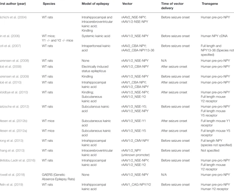
modulate the NPY system (Table 1).
Early attempts in this direction explored the anti-seizure
potential of NPY overexpression mediated by rAAV serotype 2
(rAAV2) vector injection in the hippocampus (
Richichi et al.,
2004
) or piriform cortex (
Foti et al., 2007
) in the rat kainate
model of epilepsy. Importantly,
Richichi et al. (2004)
compared
the effects of serotypes AAV2 and chimeric AAV1/2, both
vectors with the human NPY gene driven by the neuron-specific
enolase promoter (pNSE). A long-term transgene expression,
confined in hilar interneurons, was observed with AAV2, while
more widespread expression in diverse subtypes of neurons was
observed with the AAV1/2 serotype, that also conferred a more
robust protection from epileptogenesis and chronic seizures. Y1
or Y2 double knockout mice, contrary to the wild type, did
not display any protection from seizure activity upon NPY gene
therapy, indicating that activation of one (most likely Y2) or
both of these receptor subtypes was essential for the NPY effect
(
Lin et al., 2006
). More recently, the AAV1/2 expressing-NPY
vector was infused into the thalamus or somatosensory cortex
in a rat model of genetic generalized epilepsy (GAERS, Genetic
Absence Epileptic Rats from Strasbourg), resulting in a reduced
seizure activity, in particular when injected in the thalamus
(
Powell et al., 2018
).
Some concerns on the potential for translatability to human
application were raised by
Sørensen et al. (2008)
. These
TABLE 1 | Comparison of different gene therapy strategies designed to modulate the NPY system, based on the use of recombinant adeno-associated vectors.
First author (year) Species Model of epilepsy Vector Time of vector delivery
Transgene
Richichi et al. (2004) WT rats Intrahippocampal and intracerebroventricular kainic acid; Kindling
rAAV2_NSE-NPY; rAAV1/2-NSE-NPY
Before seizure onset Human pre-pro-NPY
Lin et al. (2006) WT mice;
Y1 -/- and Y2 -/- mice
Systemic kainic acid rAAV1/2_NSE-NPY Before seizure onset Human NPY cDNA
Foti et al. (2007) WT rats Intraperitoneal kainic acid
rAAV2_CBA-NPY; rAAV2_CBA-NPY13-36
Before seizure onset Full length and NPY13-36 (Species not specified)
Sørensen et al. (2008) WT rats None rAAV1/2_NSE-NPY N/A Human pre-pro-NPY
Noè et al. (2008) WT rats Electrically induced
status epilepticus
rAAV1/2_CBA-NPY After seizure onset Human pre-pro-NPY
Sørensen et al. (2009) WT rats Kindling rAAV1/2_NSE-NPY Before seizure onset Human pre-pro-NPY
Noè et al. (2010) WT rats Intrahippocampal
kainic acid
rAAV1_CBA-NPY; rAAV1/2_CBA-NPY
After seizure onset Human pre-pro-NPY
Woldbye et al. (2010) WT rats Kindling;
Subcutaneous kainic acid
rAAV1/2_NSE-NPY; rAAV1/2_NSE-Y2
After seizure onset Human pre-pro-NPY Full length mouse Y2 receptor
Gøtzsche et al. (2012) WT rats Subcutanous kainic acid
rAAV1/2_NSE-Y5; rAAV1/2_NSE-NPY
Before seizure onset Human pre-pro-NPY Full length mouse Y5 receptor
Olesen et al. (2012b) WT mice Subcutaneous kainic acid
rAAV1/2_NSE-Y1 After seizure onset Full length mouse Y1 receptor
Olesen et al. (2012a) WT mice Subcutaneous kainic acid
rAAV1/2_NSE-Y5 After seizure onset Full length mouse Y5 receptor
Dong et al. (2013) WT rats Intrahippocampal
kainic acid
rAAV1/2_CMV-NPY Before seizure onset Full length NPY (species not specified)
Zhang et al. (2013) WT rats Intracerebroventricular kainic acid
rAAV1/2_NPY (unknown promoter)
Before seizure onset Not specified
Nikitidou Ledri et al. (2016) WT rats Intrahippocampal kainic acid
rAAV1/2_NSE-NPY; rAAV1/2_NSE-Y2
Before seizure onset Human pre-pro-NPY Full length mouse Y2 receptor
Powell et al. (2018) GAERS (Genetic Absence Epilepsy Rats)
None rAAV1/2_NSE-NPY N/A Human pre-pro-NPY
Melin et al. (2019) WT rats Intrahippocampal
kainic acid
rAAV1_CAG-NPY/Y2 Before seizure onset Human pre-pro-NPY Human Y2 receptor
authors claimed an impairment of synaptic plasticity and the
attenuation of long-term potentiation of Schaffer collateral-CA1
synapses in naive rats upon unilateral vector injection in the
hippocampus, with consequent deficits of hippocampal-based
spatial discrimination learning (
Sørensen et al., 2008
). These
unexpected findings were contrasted by the authors themselves
in a following study that showed seizure protection with no
impact on working memory performance tasks in kindled rats
injected in both hippocampi with the AAV1/2-pNSE-NPY vector
(
Sørensen et al., 2009
).
In any event, the initial attempts of NPY gene therapy had
limited relevance for clinical translation: they were all carried
out before epilepsy onset, in a scenario that is obviously
non-reproducible in real patients and that did not take into account
the aberrant changes occurring during epileptogenesis, which
may significantly affect treatment effectiveness. In order to
overcome this limitation,
Noè et al. (2008)
tested the effect
of hippocampal injection of an AAV1/2 vector expressing
NPY after the establishment of epilepsy in rats and found
a decrease in seizure activity. Interestingly, this study also
demonstrated preserved levels of Y2R into the AAV-injected
hippocampus, with functional transport and high levels of
release of the recombinant NPY to nerve terminals upon
induction of neuronal depolarization. In a following report,
the same authors delivered NPY using rAAV1, and observed a
widespread transgene expression pattern throughout the injected
hippocampi and a potent effect on seizure reduction, with no
detectable evidence of immune response or cognitive impairment
(
Noè et al., 2010
).
NPY is directly involved in the regulation of brain excitability
by regulation of intracellular calcium and glutamate release,
mainly through binding to and activation of Y1, Y2, and Y5
receptors (
Berglund et al., 2003
). As described above, whereas
converging evidence supports an anti-epileptic role of Y2 (and to
a lesser extent of Y5) receptors, the involvement of Y1Rs remains
debated, with some evidence of pro-epileptic effects. Therefore, a
simple increase in NPY levels may become a double-edged sword.
These considerations prompted alternative gene therapy
strategies, oriented not only at increasing NPY secretion into the
epileptic focus but, also, at re-shaping the NPY ligand-receptor
system by the delivery of genes encoding for the different NPY
receptors. To date, the only study performed to evaluate the
effects of a brain overexpression of Y1 in an animal model of
epilepsy indicates an increased susceptibility to kainate-induced
seizures (
Olesen et al., 2012b
), consistent with the mentioned
evidence of Y1R-mediated pro-epileptic effects (
Gariboldi et al.,
1998; Benmaamar et al., 2003
). One study proved seizure
reduction through the delivery in the rat hippocampus of an AAV
pool of vectors for the concomitant expression of both Y5 and
NPY (
Gøtzsche et al., 2012
), but no protective effect was observed
with the AAV-Y5 vector alone (
Gøtzsche et al., 2012; Olesen et al.,
2012a
). More robust and promising data have been obtained
by overexpressing Y2 receptors, i.e., by seconding the adaptive
up-regulation of these receptors observed in the epileptic tissue.
Y2Rs proved to be sufficient to suppress acute seizures even
when overexpressed alone, although the therapeutic outcome
significantly increased in the case of concurrent treatment with
an NPY expressing vector (
Woldbye et al., 2010
).
Attempts of combinatorial gene delivery have been
accomplished by using two separate rAAV vectors (
Nikitidou
Ledri et al., 2016
). This procedure, however, faces some
limitations, such as an unknown transduction efficiency of the
different vectors upon brain infusion or the potential obstacles
that a heterogeneous viral pool could face in case of clinical
application. In order to solve such issues,
Melin et al. (2019)
used
an AAV1-based vector specially designed for the concurrent
expression of both NPY and Y2 from a single viral construct,
injected into both dorsal and ventral hippocampus to target
the epileptogenic focus. This dual-gene vector delivery led to
a detectable overexpression of both NPY and Y2R within the
injected hippocampi, particularly pronounced into the dorsal
CA1 and CA3 regions, and resulted in a remarkable decrease of
EEG seizure frequency and duration in the kainic acid model of
TLE (
Melin et al., 2019
).
PROBLEMS AND OPPORTUNITIES
As described above, both NPY and its receptors display a
high degree of complexity, from synthesis, processing and
compartmentalized delivery or regulated secretion, to an
intricated variety of biological effects, both at local and global
circuit level. These elements have profound implications for
gene therapy.
The majority of data reported in the literature derive from
experiments performed with viral vectors constructed to express
pre-pro-NPY (Table 1). In this context, the use of the full length
NPY sequence may be advantageous, since it allows using the
endogenous cellular machinery to process and pack the
pro-peptide into vesicles, where the mature NPY is formed and then
stored. In this way stimulus-dependent release of the peptide
(e.g., at the onset of a seizure) can be preserved. Biosynthesis
and stimulus-dependent release of mature NPY have been indeed
shown ex vivo (
Noè et al., 2008
). While all this may occur in
cells that physiologically express NPY, NPY gene delivery alone
may not be sufficient for regulated release of the mature peptide
in cells lacking/under-expressing one or more of the regulatory
elements (e.g., processing enzymes, trafficking proteins) needed
in such a complex multi-step system. One option to circumvent
this problem could be linking the NPY gene sequence to the
sequence of the laminar protein fibronectin (FIB), which induces
a constitutive secretion as opposed to a regulated secretion (
Foti
et al., 2007
). Finally, even after release, the effects of peptidases
should be taken into account to understand and modulate
NPY signaling.
The modulation of YRs expression requires an even more
finely regulated sequence of events. Functional specificity of the
NPY system depends largely on receptors. In this context, the
processes of anterograde transport, internalization, recycling or
degradation have been thoroughly characterized for only a few
NPY receptors. These considerations lead to the suggestion that,
if no specific cell targeting strategy is employed, gene
therapy-induced overexpression of NPY or NPY receptors may be more
efficient in (or even restricted to) cells that physiologically or
pathophysiologically express them. The levels of released NPY
and the coupling between ligand and receptor are also crucial
for inducing the desired effect in the right cell target. It may be
possible to obtain a certain degree of receptor selectivity by using,
for example, N-terminally truncated forms of NPY (like NPY
3−36or NPY
13−36(
Beck-Sickinger and Jung, 1995; Sajdyk et al., 2002;
Foti et al., 2007; Pedragosa-Badia et al., 2013
) that could favor
Y2Rs dependent signaling.
OUTLOOK FOR HUMAN STUDIES USING
VIRAL VECTOR-BASED STRATEGIES
Despite
this
complexity,
several
anti-epileptic
gene
therapy strategies proved successful in modulating the
inhibitory/excitatory balance within animal brain regions
involved in seizure onset by focal overexpression of NPY alone
or in combination with Y2 or Y5 receptors. Even if extended
long-time studies to exclude side effects or neuropathological
changes due to application of viral vectors still need to be
performed (optimally in non-human primates), these compelling
preclinical data may concretely prompt the design of a
first-in-human gene therapy trial in drug-resistant epileptic patients. As
an example, patients deemed suitable for surgical resection of a
clearly mapped epileptogenic region may be enrolled in a first
putative human study. This would allow to design a confined
(and presumably more effective) transgene expression within
the epileptogenic lesion only, while preserving the unaffected
brain tissue and thereby lowering the risk of unpredictable side
effects. In addition, should the treatment not prove to be effective
or well-tolerated, patients would undergo resective surgery as
originally planned.
Several issues should be taken into account in the study
design. For example, hippocampal sclerosis, if extensive, may
reduce vector diffusion and transduction efficacy, imposing
a personalization of the dose. The choice of vector would
largely depend on the strategy employed to regulate the
expression of the therapeutic gene. As described in this review,
all studies on gene therapy-mediated overexpression of NPY
and/or its receptors in epilepsy models have been performed
by using AAV vectors. However, the limited cargo capacity of
AAVs may hinder their adaptability for clinical translation, in
particular when complex regulatory mechanisms must be set
in place. In fact, it would be desirable to regulate the levels of
transgene expression in a patient-tailored manner, in response
to endogenous and/or exogenous clues. While an endogenous
control of the transgene expression system that responds to
physiological stimuli (for example, glutamate accumulation)
would be preferable, the time needed for the biosynthesis and
delivery of the therapeutic protein(s) would be too long to
arrest an ongoing seizure. Therefore, a more concrete alternative,
although not applicable to the response to individual seizures,
but rather on a general control of seizure threshold, may rely on
the administration of external factors (i.e., specific molecules).
These elements could selectively activate or inhibit transgene
expression, by acting on specific regulatory sequences delivered
along with the therapeutic gene cassette, in the same viral
vector. This option, however, would require the exploitation
of neurotropic vectors capable to host much larger exogenous
DNA cargos, for example HSV derived vectors (
Ingusci et al.,
2019a,b
).
Once the remaining gaps in knowledge and hurdles for
gene therapy will be overcome, we may finally be able to treat
epilepsy by acting on endogenous systems of neuromodulation.
In a way, this is something that we may have already done,
unconsciously and much less finely, with certain anti-epileptic
drugs (
Brill et al., 2006
).
AUTHOR CONTRIBUTIONS
All authors listed have made a substantial, direct and
intellectual contribution to the work, and approved it
for publication.
FUNDING
This work was supported by a grant from the European
Community [FP7-HEALTH project 602102 (EPITARGET)].
REFERENCES
Abid, K., Rochat, B., Lassahn, P. G., Stöcklin, R., Michalet, S., Brakch, N., et al. (2009). Kinetic study of neuropeptide Y (NPY) proteolysis in blood and identification of NPY3-35. A new peptide generated by plasma kallikrein. J. Biol. Chem. 284, 24715–24724. doi: 10.1074/jbc.M109. 035253
Aicher, S. A., Springston, M., Berger, S. B., Reis, D. J., and Wahlestedt, C. (1991). Receptor-selective analogs demonstrate NPY/PYY receptor heterogeneity in rat brain. Neurosci. Lett. 130, 32–36. doi: 10.1016/0304-3940(91) 90220-N
Allen, J., Novotny, J., Martin, J., and Heinrich, G. (1987). Molecular structure of mammalian neuropeptide Y: analysis by molecular cloning and computer-aided comparison with crystal structure of avian homologue. Proc. Natl. Acad. Sci. U. S. A. 84, 2532–2536. doi: 10.1073/pnas.84.8.2532
Atanasova, K. R., and Reznikov, L. R. (2018). Neuropeptides in asthma, chronic obstructive pulmonary disease and cystic fibrosis. Respir. Res. 19:149. doi: 10.1186/s12931-018-0846-4
Baldock, P. A., Allison, S. J., Lundberg, P., Lee, N. J., Slack, K., Lin, E. J. D., et al. (2007). Novel role of Y1 receptors in the coordinated regulation of bone and energy homeostasis. J. Biol. Chem. 282, 19092–19102. doi: 10.1074/jbc.M700644200
Baraban, S. C. (2004). Neuropeptide Y and epilepsy: recent progress, prospects and controversies. Neuropeptides 38, 261–265. doi: 10.1016/j.npep.2004.04.006 Beck-Sickinger, A. G., and Jung, G. (1995). Structure–activity relationships of
neuropeptide Y analogues with respect to Y1 and Y2 receptors. Biopolymers 37, 123–142. doi: 10.1002/bip.360370207
Beck-Sickinger, A. G., Weland, H. A., Wittneben, H., Willim, K. -D, Rudolf, K., and Jung, G. (1994). Complete L-alanine scan of neuropeptide Y reveals ligands binding to Y1 and Y2 receptors with distinguished conformations. Eur. J. Biochem. 225, 947–958. doi: 10.1111/j.1432-1033.1994.0947b.x
Benarroch, E. E. (2009). Neuropeptide Y: its multiple effects in the CNS and potential clinical significance. Neurology 72, 1016–1020. doi: 10.1212/01.wnl.0000345258.18071.54
Benmaamar, R., Pham-Lê, B. T., Marescaux, C., Pedrazzini, T., and Depaulis, A. (2003). Induced down-regulation of neuropeptide Y-Y1 receptors delays initiation of kindling. Eur. J. Neurosci. 18, 768–774. doi: 10.1046/j.1460-9568.2003.02810.x
Berglund, M. M., Hipskind, P. A., and Gehlert, D. R. (2003). Recent developments in our understanding of the physiological role of PP-fold peptide receptor subtypes. Exp. Biol. Med. 228, 217–244. doi: 10.1177/1535370203228 00301
Bregola, G., Dumont, Y., Fournier, A., Zucchini, S., Quirion, R., and Simonato, M. (2000). Decreased levels of neuropeptide Y5 receptor binding sites in two experimental models of epilepsy. Neuroscience 98, 697–703. doi: 10.1016/S0306-4522(00)00162-7
Brill, J., Lee, M., Zhao, S., Fernald, R. D., and Huguenard, J. R. (2006). Chronic valproic acid treatment triggers increased neuropeptide Y expression and signaling in rat nucleus reticularis thalami. J. Neurosci. 26, 6813–6822. doi: 10.1523/JNEUROSCI.5320-05.2006
Brumovsky, P. R., Shi, T. J., Matsuda, H., Kopp, J., Villar, M. J., and Hökfelt, T. (2002). NPY Y1 receptors are present in axonal processes of DRG neurons. Exp. Neurol. 174, 1–10. doi: 10.1006/exnr.2001.7845
Caberlotto, L. (1997). Localization of neuropeptide Y Y1 mRNA in the human brain: abundant expression in cerebral cortex and striatum. Eur. J. Neurosci. 9, 1212–1225. doi: 10.1111/j.1460-9568.1997.tb01476.x
Cabrele, C., and Beck-Sickinger, A. G. (2000). Molecular characterization of the ligand-receptor interaction of the neuropeptide Y family. J. Pept. Sci. 6, 97–122. doi: 10.1002/(SICI)1099-1387(200003)6:3<97::AID-PSC236>3. 0.CO;2-E
Castan, I., Valet, P., Larrouy, D., Voisin, T., Remaury, A., Daviaud, D., et al. (1993). Distribution of PYY receptors in human fat cells: an antilipolytic system alongside the α2-adrenergic system. Am. J. Physiol. 265(1 Pt 1), E74–E80. doi: 10.1152/ajpendo.1993.265.1.E74
Cavarsan, C. F., Malheiros, J., Hamani, C., Najm, I., and Covolan, L. (2018). Is mossy fiber sprouting a potential therapeutic target for epilepsy? Front. Neurol. 9:1023. doi: 10.3389/fneur.2018.01023
Cerdá-Reverter, J. M., and Larhammar, D. (2000). cNeuropeptide Y family of peptides: Structure, anatomical expression, function, and molecular evolution. Biochem. Cell Biol. 78, 371–392. doi: 10.1139/o00-004
Chronwall, B. M., DiMaggio, D. A., Massari, V. J., Pickel, V. M., Ruggiero, D. A., and O’donohue, T. L. (1985). The anatomy of
neuropeptide-y-containing neurons in rat brain. Neuroscience 15, 1159–1181.
doi: 10.1016/0306-4522(85)90260-X
Colmers, W. F., Klapstein, G. J., Fournier, A., St-Pierre, S., and Treherne, K. A. (1991). Presynaptic inhibition by neuropeptide Y in rat hippocampal
slice in vitro is mediated by a Y2 receptor. Br. J. Pharmacol. 102, 41–44. doi: 10.1111/j.1476-5381.1991.tb12129.x
Colmers, W. F., Lukowiak, K., and Pittman, Q. J. (1985). Neuropeptide Y reduces orthodromically evoked population spike in rat hippocampal CA1 by a possibly presynaptic mechanism. Brain Res. 346, 404–408. doi: 10.1016/0006-8993(85)90880-7
Colmers, W. F., Lukowiak, K., and Pittman, Q. J. (1987). Presynaptic action of neuropeptide Y in area CA1 of the rat hippocampal slice. J. Physiol. 383, 285–299. doi: 10.1113/jphysiol.1987.sp016409
Colmers, W. F., Lukowiak, K., and Pittman, Q. J. (1988). Neuropeptide Y action in the rat hippocampal slice: site and mechanism of presynaptic inhibition. J. Neurosci. 8, 3827–3837. doi: 10.1523/JNEUROSCI.08-10-03827.1988 Criscione, L., Rigollier, P., Batzl-Hartmann, C., Rüeger, H., Stricker-Krongrad, A.,
Wyss, P., et al. (1998). Food intake in free-feeding and energy-deprived lean rats is mediated by the neuropeptide Y5 receptor. J. Clin. Invest. 102, 2136–2145. doi: 10.1172/JCI4188
De Jong-Brink, M., Ter Maat, A., and Tensen, C. P. (2001). NPY in invertebrates: Molecular answers to altered functions during evolution. Peptides 22, 309–315. doi: 10.1016/S0196-9781(01)00332-1
de Quidt, M. E., and Emson, P. C. (1986). Distribution of neuropeptide Y-like immunoreactivity in the rat central nervous system-II. Immunohistochemical
analysis. Neuroscience 18, 545–618. doi: 10.1016/0306-4522(86)
90057-6
Dong, C., Zhao, W., Li, W., Lv, P., and Dong, X. (2013). Anti-epileptic effects of neuropeptide Y gene transfection into the rat brain. Neural Regen. Res. 8, 1307–1315. doi: 10.3969/j.issn.1673-5374.2013.14.007
Dum, E., Fürtinger, S., Gasser, E., Bukovac, A., Drexel, M., Tasan, R., et al. (2017). Effective G-protein coupling of Y2 receptors along axonal fiber tracts and its relevance for epilepsy. Neuropeptides 61, 49–55. doi: 10.1016/j.npep.2016.10.005
Dumont, Y., Fournier, A., St-Pierre, S., and Quirion, R. (1993).
Comparative characterization and autoradiographic distribution of
neuropeptide Y receptor subtypes in the rat brain. J. Neurosci. 13, 73–86. doi: 10.1523/JNEUROSCI.13-01-00073.1993
Dumont, Y., Fournier, A., St-Pierre, S., Schwartz, T. W., and Quirion, R. (1990). Differential distribution of neuropeptide Y1 and Y2 receptors in the rat brain. Eur. J. Pharmacol. 191, 501–503. doi: 10.1016/0014-2999(90) 94189-5
Dumont, Y., Jacques, D., Bouchard, P., and Quirion, R. (1998).
Species differences in the expression and distribution of the
neuropeptide Y Y1, Y2, Y4, and Y5 receptors in rodents,
guinea pig, and primates brains. J. Comp. Neurol. 402, 372–384. doi: 10.1002/(SICI)1096-9861(19981221)402:3<372::AID-CNE6>3.0.CO;2-2 Dumont, Y., Martel, J. C., Fournier, A., St-Pierre, S., and Quirion, R. (1992).
Neuropeptide Y and neuropeptide Y receptor subtypes in brain and peripheral tissues. Prog. Neurobiol. 38, 125–167. doi: 10.1016/0301-0082(92)90038-G El Bahh, B., Balosso, S., Hamilton, T., Herzog, H., Beck-Sickinger, A. G., Sperk, G.,
et al. (2005). The anti-epileptic actions of neuropeptide Y in the hippocampus are mediated by Y2 and not Y5 receptors. Eur. J. Neurosci. 22, 1417–1430. doi: 10.1111/j.1460-9568.2005.04338.x
El Bahh, B., Cao, J. Q., Beck-Sickinger, A. G., and Colmers, W. F. (2002). Blockade of neuropeptide Y2 receptors and suppression of NPY’s anti-epileptic actions in the rat hippocampal slice by BIIE0246. Br. J. Pharmacol. 136, 502–509. doi: 10.1038/sj.bjp.0704751
Elfvin, L. G., Holmberg, K., Emson, P., Schemann, M., and Hökfelt, T. (1997). Nitric oxide synthase, choline acetyltransferase, catecholamine enzymes and neuropeptides and their colocalization in the anterior pelvic ganglion, the inferior mesenteric ganglion and the hypogastric nerve of the male guinea pig. J. Chem. Neuroanat. 14, 33–49. doi: 10.1016/S0891-0618(97)10010-2 Flood, J. F., and Morley, J. E. (1989). Dissociation of the effects of neuropeptide
Y on feeding and memory: Evidence for pre- and postsynaptic mediation. Peptides 10, 963–966. doi: 10.1016/0196-9781(89)90176-9
Foti, S., Haberman, R. P., Samulski, R. J., and McCown, T. J. (2007). Adeno-associated virus-mediated expression and constitutive secretion of NPY or NPY13-36 suppresses seizure activity in vivo. Gene Ther. 14, 1534–1536. doi: 10.1038/sj.gt.3303013
Furtinger, S., Pirker, S., Czech, T., Baumgartner, C., Ransmayr, G., and Sperk, G. (2001). Plasticity of Y1 and Y2 receptors and neuropeptide Y
fibers in patients with temporal lobe epilepsy. J. Neurosci. 21, 5804–5812. doi: 10.1523/JNEUROSCI.21-15-05804.2001
Gariboldi, M., Conti, M., Cavaleri, D., Samanin, R., and Vezzani, A. (1998). Anticonvulsant properties of BIBP3226, a non-peptide selective antagonist at neuropeptide Y Y1 receptors. Eur. J. Neurosci. 10, 757–759. doi: 10.1046/j.1460-9568.1998.00061.x
Gehlert, D. R., Gackenheimer, S. L., and Schober, D. A. (1992). [Leu31-Pro34] neuropeptide Y identifies a subtype of 125I-labeled peptide YY binding sites in the rat brain. Neurochem. Int. 21, 45–67. doi: 10.1016/0197-0186(92)90067-2 Gerald, C., Walker, M. W., Criscione, L., Gustafson, E. L., Batzl-Hartmann, C., Smith, K. E., et al. (1996). A receptor subtype involved in neuropeptide-Y-induced food intake. Nature 382, 168–171. doi: 10.1038/382168a0
Gershkovich, M. M., Groß, V. E., Kaiser, A, and Prömel, S. (2019). Pharmacological and functional similarities of the human neuropeptide Y system in C. elegans challenges phylogenetic views on the FLP/NPR system. Cell Commun. Signal 17, 123–123. doi: 10.1186/s12964-019-0436-1
Gicquiaux, H., Lecat, S., Gaire, M., Dieterlen, A., Mély, Y., Takeda, K., et al. (2002). Rapid internalization and recycling of the human neuropeptide Y Y1 receptor. J. Biol. Chem. 277, 6645–6655. doi: 10.1074/jbc.M107224200
Giesbrecht, C. J., Mackay, J. P., Silveira, H. B., Urban, J. H., and Colmers, W. F. (2010). Countervailing modulation of Ihby neuropeptide Y and corticotrophin-releasing factor in basolateral amygdala as a possible mechanism for their effects on stress-related behaviors. J. Neurosci. 30, 16970–16982. doi: 10.1523/JNEUROSCI.2306-10.2010
Glass, M. J., Chan, J., and Pickel, V. M. (2002). Ultrastructural localization of neuropeptide Y Y1 receptors in the rat medial nucleus tractus solitarius: relationships with neuropeptide Y or catecholamine neurons. J. Neurosci. Res. 67, 753–765. doi: 10.1002/jnr.10185
Gobbi, M., Gariboldi, M., Piwko, C., Hoyer, D., Sperk, G., and Vezzani, A. (1998). Distinct changes in peptide YY binding to, and mRNA levels of, Y1 and Y2 receptors in the rat hippocampus associated with kindling epileptogenesis. J. Neurochem. 70, 1615–1622. doi: 10.1046/j.1471-4159.1998.70041615.x Gøtzsche, C. R., Nikitidou, L., Sørensen, A. T., Olesen, M. V., Sørensen, G.,
Christiansen, S. H. O., et al. (2012). Combined gene overexpression of neuropeptide Y and its receptor Y5 in the hippocampus suppresses seizures. Neurobiol. Dis. 45, 288–296. doi: 10.1016/j.nbd.2011.08.012
Grandt, D., Schimiczek, M., Rascher, W., Feth, F., Shively, J., Lee, T. D., et al. (1996). Neuropeptide Y 3-36 is an endogenous ligand selective for Y2 receptors. Regul. Pept. 67, 33–37. doi: 10.1016/S0167-0115(96)00104-8
Greber, S., Schwarzer, C., and Sperk, G. (1994). Neuropeptide Y inhibits potassium-stimulated glutamate release through Y2 receptors in rat hippocampal slices in vitro. Br. J. Pharmacol. 113, 737–740. doi: 10.1111/j.1476-5381.1994.tb17055.x Guo, H. U. I., Castro, P. A., Palmiter, R. D., and Baraban, S. C. (2002). Y5 receptors
mediate neuropeptide Y actions at excitatory synapses in area CA3 of the mouse hippocampus. J. Neurophysiol. 87, 558–566. doi: 10.1152/jn.00532.2001 Howell, O. W., Doyle, K., Goodman, J. H., Scharfman, H. E., Herzog, H.,
Pringle, A., et al. (2005). Neuropeptide Y stimulates neuronal precursor proliferation in the post-natal and adult dentate gyrus. J. Neurochem. 93, 560–570. doi: 10.1111/j.1471-4159.2005.03057.x
Hubers, S. A., Wilson, J. R., Yu, C., Nian, H., Grouzmann, E., Eugster, P., et al. (2018). DPP (dipeptidyl peptidase)-4 inhibition potentiates the vasoconstrictor response to NPY (neuropeptide Y) in humans during renin-angiotensin-aldosterone system inhibition. Hypertension 72, 712–719. doi: 10.1161/HYPERTENSIONAHA.118.11498
Ingusci, S., Cattaneo, S., Verlengia, G., Zucchini, S., and Simonato, M. (2019a). A matter of genes: the hurdles of gene therapy for epilepsy. Epilepsy Curr. 19, 38–43. doi: 10.1177/1535759718822846
Ingusci, S., Verlengia, G., Soukupova, M., Zucchini, S., and Simonato, M. (2019b). Gene therapy tools for brain diseases. Front. Pharmacol. 10:724. doi: 10.3389/fphar.2019.00724
Jacques, D., Dumont, Y., Fournier, A., and Quirion, R. (1997). Characterization of neuropeptide Y receptor subtypes in the normal human brain, including the hypothalamus. Neuroscience 79, 129–148. doi: 10.1016/S0306-4522(96) 00639-2
Kaiser, A., Müller, P., Zellmann, T., Scheidt, H. A., Thomas, L., Bosse, M., et al. (2015). Unwinding of the C-terminal residues of neuropeptideY is critical for Y2 receptor binding and activation. Angew. Chem. Int. Ed. 54, 7446–7449. doi: 10.1002/anie.201411688
Károly, N., Dobó, E. A., and Mihály, A. (2015). Comparative immunohistochemical study of the effects of pilocarpine on the mossy cells, mossy fibres and inhibitory neurones in murine dentate gyrus. Acta Neurobiol. Exp. 75, 220–237.
Keast, J. R. (1991). Patterns of co-existence of peptides and differences of nerve fibre types associated with noradrenergic and non-noradrenergic (putative cholinergic) neurons in the major pelvic ganglion of the male rat. Cell Tissue Res. 266, 405–415. doi: 10.1007/BF00318197
Klapstein, G. J., and Colmers, W. F. (1992). 4-Aminopyridine and low Ca2+ differentiate presynaptic inhibition mediated by neuropeptide Y, baclofen and 2-chloroadenosine in rat hippocampal CA1 in vitro. Br. J. Pharmacol. 105, 470–474. doi: 10.1111/j.1476-5381.1992.tb14277.x
Klapstein, G. J., and Colmers, W. F. (1993). On the sites of presynaptic inhibition by neuropeptide y in rat hippocampus in vitro. Hippocampus 3, 103–111. doi: 10.1002/hipo.450030111
Klapstein, G. J., and Colmers, W. F. (1997). Neuropeptide Y suppresses epileptiform activity in rat hippocampus in vitro. J. Neurophysiol. 78, 1651–1661. doi: 10.1152/jn.1997.78.3.1651
Kofler, N., Kirchmair, E., Schwarzer, C., and Sperk, G. (1997). Altered expression of NPY-Y1 receptors in kainic acid induced epilepsy in rats. Neurosci. Lett. 230, 129–132. doi: 10.1016/S0304-3940(97)00492-8
Kooijman, S. A. L. M., and Troost, T. A. (2007). Quantitative steps in the evolution of metabolic organisation as specified by the dynamic energy budget theory. Biol. Rev. 82, 113–42. doi: 10.1111/j.1469-185X.2006.00006.x
Kopp, J., Xu, Z. Q., Zhang, X., Pedrazzini, T., Herzog, H., Kresse, A., et al. (2002). Expression of the neuropeptide Y Y1 receptor in the CNS of rat and of wild-type and Y1 receptor knock-out mice. Focus on immunohistochemical localization. Neuroscience 111, 443–532. doi: 10.1016/S0306-4522(01)00463-8
Kuo, L. E., Kitlinska, J. B., Tilan, J. U., Li, L., Baker, S. B., Johnson, M. D., et al. (2007). Neuropeptide Y acts directly in the periphery on fat tissue and mediates stress-induced obesity and metabolic syndrome. Nat. Med. 13, 803–811. doi: 10.1038/nm1611
Larhammar, D., and Salaneck, E. (2004). Molecular evolution of NPY receptor subtypes. Neuropeptides 38, 141–51. doi: 10.1016/j.npep.2004.06.002 Leblanc, G. G., Trimmer, B. A., and Landis, S. C. (1987). Neuropeptide Y-like
immunoreactivity in rat cranial parasympathetic neurons: coexistence with vasoactive intestinal peptide and choline acetyltransferase. Proc. Natl. Acad. Sci. U. S. A. 84, 3511–3515. doi: 10.1073/pnas.84.10.3511
Ledri, M., Sørensen, A. T., Madsen, M. G., Christiansen, S. H., Ledri, L. N., Cifra, A., et al. (2015). Differential effect of neuropeptides on excitatory synaptic transmission in human epileptic hippocampus. J. Neurosci. 35, 9622–31. doi: 10.1523/JNEUROSCI.3973-14.2015
Lerch, M., Mayrhofer, M., and Zerbe, O. (2004). Structural similarities of micelle-bound peptide YY (PYY) and neuropeptide Y (NPY) are related to their affinity profiles at the Y receptors. J. Mol. Biol. 339, 1153–1168. doi: 10.1016/j.jmb.2004.04.032
Li, Q., Bartley, A. F., and Dobrunz, L. E. (2017). Endogenously released
neuropeptide Y suppresses hippocampal short-term facilitation
and is impaired by stress-induced anxiety. J. Neurosci. 37:23-37. doi: 10.1523/JNEUROSCI.2599-16.2017
Lin, E. J., Young, D., Baer, K., Herzog, H., and During, M. J. (2006). Differential actions of NPY on seizure modulation via Y1 and Y2 receptors: evidence from receptor knockout mice. Epilepsia 47, 773–780. doi: 10.1111/j.1528-1167.2006.00500.x
Lindner, D., Stichel, J., and Beck-Sickinger, A. G. (2008). Molecular recognition of the NPY hormone family by their receptors. Nutrition 24, 907–917. doi: 10.1016/j.nut.2008.06.025
Lu, C., Everhart, L., Tilan, J., Kuo, L., Sun, C. C. J., Munivenkatappa, R. B., et al. (2010). Neuropeptide y and its Y2 receptor: potential targets in neuroblastoma therapy. Oncogene 29, 5630–5642. doi: 10.1038/onc.2010.301
Lundberg, J. M., Rudehill, A., Sollevi, A., Theodorsson-Norheim, E., and Hamberger, B. (1986). Frequency- and reserpine-dependent chemical coding of sympathetic transmission: differential release of noradrenaline and neuropeptide Y from pig spleen. Neurosci. Lett. 63, 96–100. doi: 10.1016/0304-3940(86)90020-0
Lundell, I., Rabe Bernhardt, N., Johnsson, A. K., and Larhammar, D. (2011). Internalization studies of chimeric neuropeptide Y receptors Y1 and Y2 suggest
complex interactions between cytoplasmic domains. Regul. Pept. 168, 50–8. doi: 10.1016/j.regpep.2011.03.004
Marsh, D. J., Baraban, S. C., Hollopeter, G., and Palmiter, R. D. (1999). Role of the Y5 neuropeptide Y receptor in limbic seizures. Proc. Natl. Acad. Sci. U. S. A. 96, 13518–13523. doi: 10.1073/pnas.96.23.13518
Martire, M., Pistritto, G., Mores, N., Agnati, L. F., and Fuxe, K. (1993). Region-specific inhibition of potassium-evoked [3H]noradrenaline release from rat brain synaptosomes by neuropeptide Y-(13-36). Involvement of NPY receptors of the Y2 type. Eur. J. Pharmacol. 230, 231–234. doi: 10.1016/0014-2999(93)90807-T
Mathern, G. W., Babb, T. L., Pretorius, J. K., and Leite, J. P. (1995). Reactive synaptogenesis and neuron densities for neuropeptide Y, somatostatin, and glutamate decarboxylase immunoreactivity in the
epileptogenic human fascia dentata. J. Neurosci. 15, 3990–4004.
doi: 10.1523/JNEUROSCI.15-05-03990.1995
McCarthy, J. B., Walker, M., Pierce, J., Camp, P., and White, J. D. (1998). Biosynthesis and metabolism of native and oxidized neuropeptide Y in the hippocampal mossy fiber system. J. Neurochem. 70, 1950–1963. doi: 10.1046/j.1471-4159.1998.70051950.x
Mcquiston, A. R., and Colmers, W. F. (1996). Neuropeptide Y2 receptors inhibit the frequency of spontaneous but not miniature EPSCS in CA3 pyramidal cells of rat hippocampus. J. Neurophysiol. 76, 3159–3168. doi: 10.1152/jn.1996.76.5.3159
Melin, E., Nanobashvili, A., Avdic, U., Gøtzsche, C. R., Andersson, M., Woldbye, D. P. D., et al. (2019). Disease modification by combinatorial single vector gene therapy: a preclinical translational study in epilepsy. Mol. Ther. Methods Clin. Dev. 15, 179–193. doi: 10.1016/j.omtm.2019.09.004
Merten, N., Lindner, D., Rabe, N., Römpler, H., Mörl, K., Schöneberg, T., et al. (2007). Receptor subtype-specific docking of Asp6.59 with C-terminal arginine residues in Y receptor ligands. J. Biol. Chem. 282, 7543–7551. doi: 10.1074/jbc.M608902200
Michel, M. C., Beck-Sickinger, A., Cox, H., Doods, H. N., Herzog, H., Larhammar, D., et al. (1998). XVI. International union of pharmacology recommendations for the nomenclature of neuropeptide Y, peptide YY, and pancreatic polypeptide receptors. Pharmacol. Rev. 50, 143–50.
Minth, C. D., Bloom, S. R., Polak, J. M., and Dixon, J. E. (1984). Cloning, characterization, and DNA sequence of a human cDNA encoding neuropeptide tyrosine. Proc. Natl. Acad. Sci. U. S. A. 81, 4577–4581. doi: 10.1073/pnas.81.14.4577
Mitchell, G. C., Wang, Q., Ramamoorthy, P., and Whim, M. D. (2008). A common single nucleotide polymorphism alters the synthesis and secretion of neuropeptide Y. J. Neurosci. 28, 14428–14434. doi: 10.1523/JNEUROSCI.0343-08.2008
Monnet, F. P., Fournier, A., Debonnel, G., and De Montigny, C. (1992). Neuropeptide Y potentiates selectively the N-methyl-D-aspartate response in the rat CA3 dorsal hippocampus. I. Involvement of an atypical neuropeptide Y receptor. J. Pharmacol. Exp. Ther. 263, 1212–1218.
Nadler, J. V., Tu, B., Timofeeva, O., Jiao, Y., and Herzog, H. (2007). Neuropeptide Y in the recurrent mossy fiber pathway. Peptides 28, 357–364. doi: 10.1016/j.peptides.2006.07.026
Nanobashvili, A., Woldbye, D. P. D., Husum, H., Bolwig, T. G., and Kokaia, M. (2004). Neuropeptide Y Y5 receptors suppress in vitro spontaneous epileptiform bursting in the rat hippocampus. NeuroReport 15, 339–343. doi: 10.1097/00001756-200402090-00026
Nguyen, A. D., Mitchell, N. F., Lin, S., Macia, L., Yulyaningsih, E., Baldock, P. A., et al. (2012). Y1 and Y5 receptors are both required for the regulation of food intake and energy homeostasis in mice. PLoS ONE 7:e40191. doi: 10.1371/journal.pone.0040191
Nikitidou Ledri, L., Melin, E., Christiansen, S. H., Gøtzsche, C. R., Cifra, A., Woldbye, D. P. D., et al. (2016). Translational approach for gene therapy in epilepsy: model system and unilateral overexpression of neuropeptide Y and Y2 receptors. Neurobiol. Dis. 86, 52–61. doi: 10.1016/j.nbd.2015. 11.014
Noè, F., Pool, A. H., Nissinen, J., Gobbi, M., Bland, R., Rizzi, M., et al. (2008). Neuropeptide Y gene therapy decreases chronic spontaneous seizures in a rat model of temporal lobe epilepsy. Brain 131, 1506–1515. doi: 10.1093/brain/awn079