Metastatic Mediastinal Germ-Cell
Tumor and Concurrent COVID-19:
When Chemotherapy Is Not Deferrable
Nappi and colleagues recently reported the viewpoints of 53 highly experienced practitioners regarding the manage-ment of patients with germ-cell tumors (GCTs) during the COVID-19 pandemic [1]. In this survey, the authors recom-mend that“chemotherapy should be withheld until an active COVID-19 infection has resolved or has been ruled out with highly accurate molecular testing.”Here, we report the case of a young man diagnosed with a metastatic mediastinal GCT who received chemotherapy despite persistent SARS-CoV-2 positivity (Fig. 1).
In March 2020, this 29-year-old patient underwent mag-netic resonance, which revealed a lumbar spinal mass with spinal cord invasion. Following loss of sensibility in both lower limbs, palliative neurosurgery was performed, with histological diagnosis of seminoma (placental alkaline phosphatase, CD117 +, CD30−, alpha-fetoprotein [αFP]−, Ki67: 80%). Serum beta-human chorionic gonadotropin (βHCG) was undetectable, whereasαFP and lactate dehydrogenase (LDH) were elevated (117 ng/mL; 580 U/L), suggesting nonseminoma. The com-puted tomography (CT) scan showed a tumor mass of 9.4× 6.2 × 10 cm in the anterior mediastinum, with superior vena cava compression. The testicles had no evidence of dis-ease and the SARS-CoV-2 nasopharyngeal swab was negative.
Given the poor-risk features [2], chemotherapy with the VIP regimen was commenced (etoposide 75 mg/m2,
ifosfamide 1,200 mg/m2, cisplatin 20 mg/m2, days 1–5, every 21 days). VIP was preferred to BEP (bleomycin, etoposide, cisplatin) to limit the contacts with the health care system and to avoid bleomycin pulmonary toxicity. Prophylactic filgrastim was administered for 7 days.
After cycle 1, the patient developed transient high fever, conjunctivitis, dysgeusia, and anosmia and tested positive for SARS-CoV-2. Blood tests revealed lymphocytopenia and elevation of C-reactive protein, transaminases, and LDH. He was hospitalized and treated with hydroxychloroquine, darunavir, and enoxaparin, with complete resolution of symptoms. However, two swabs before cycle 2 were positive for SARS-CoV-2.
To ensure optimal treatment delivery, cycle 2 was admin-istered on April 14, and hydroxychloroquine was adminis-tered on days 1–7. αFP decreased to 20 ng/mL. The restaging CT scan revealed significant tumor response at the mediasti-nal and lumbar levels. However, atypical, basal pneumonia was compatible with COVID-19 (Fig. 2).
Despite thisfinding, cycle 3 was administered and hydro-xychloroquine was discontinued owing to suggested useless-ness. Swabs were performed on May 19 (negative) and May 20 (positive). Finally, two serial swabs tested negative and SARS-CoV-2–specific immunoglobulin G (DiaSorin, Saluggia, Italy) were detected.
The additional cycle 4 was administered on June 2.αFP, βHCG, and LDH were normal. A leukapheresis procedure was performed, with the successful collection of CD34+ cells for subsequent high-dose chemotherapy [3], and the patient underwent another intervention of spinal stabilization.
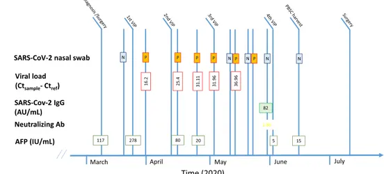
Figure 1. Laboratory parameters at diagnosis and during treatment.
Abbreviations: AFP, alpha-fetoprotein; AU, arbitrary units; Ct, cycle threshold; IgG, immunoglobulin G; N, negative; P, positive; PBSC, peripheral blood stem cells; VIP, etoposide, ifosfamide, cisplatin.
No part of this article may be reproduced, stored, or transmitted in any form or for any means without the prior permission in writing from the copyright holder. For information on purchasing reprints contact [email protected]. For permission information contact [email protected].
© 2020 AlphaMed Press
The Oncologist 2021;25:1–3 www.TheOncologist.com
The SARS-CoV-2 viral loads in nasopharyngeal swabs were retrospectively estimated using the cycle threshold (Ct) method (Ctsample-Ctref). Ct values increased from 16.2 Ct (high viral load) to 36.96 Ct (low viral load) after cycle 3, suggesting that viral clearance may have occurred during cycle 3 and that subsequent positive swabs did not repre-sent active viral replication, but only residual RNA detec-tion. To evaluate the presence of viable SARS-CoV-2, swabs collected before cycle 4 were inoculated into cell cultures (Vero-E6 cells); the cell culture method showed neither virus isolation nor cytopathic effect, further reinforcing the concept of complete viral clearance [4].
In conclusion, this patient with a poor-risk GCT underwent multimodal curative treatment despite persistent COVID-19 infection. Our report reinforces the notion that chemotherapy could be safely given in patients who need prompt interven-tion, without necessarily worsening the course of COVID-19 [5].
ACKNOWLEDGMENTS
This research was partially supported by funds from Ricerca Corrente, Fondazione IRCCS Policlinico San Matteo, Pavia, Italy. C.C. is supported by a European Society for Medical Oncology Clinical Research Fellowship (2019–2020).
PAOLOPEDRAZZOLI
Medical Oncology, Fondazione IRCCS Policlinico San Matteo, Pavia, Italy; Department of Internal Medicine and Medical Therapy, University of Pavia, Pavia, Italy
DAVIDRONDONOTTI
Medical Oncology, Department of Translational Medicine, University of Eastern Piedmont, University Hospital“Maggiore della Carità,” Novara, Italy
CARLOCATTRINI
Medical Oncology, Department of Translational Medicine, University of Eastern Piedmont, University Hospital“Maggiore
della Carità,” Novara, Italy; Department of Internal Medicine and Medical Specialties, University of Genoa, Genoa, Italy SIMONASECONDINO
Medical Oncology, Fondazione IRCCS Policlinico San Matteo, Pavia, Italy
PAOLORAVANINI
Unit of Microbiology and Virology, Department of Laboratory Medicine, University Hospital“Maggiore della Carità,” Novara, Italy
ANTONIOPIRALLA
Virology Unit, Fondazione IRCCS Policlinico San Matteo, Pavia, Italy
PIERPAOLOSAINAGHI
Internal Medicine, Department of Translational Medicine, University of Eastern Piedmont, University Hospital“Maggiore della Carità,” Novara, Italy
DIEGOBRUSTIA
Infectious Diseases, University Hospital“Maggiore della Carità,” Novara, Italy
CRISTINABOZZOLA
Pathology Unit, University Hospital“Maggiore della Carità,” Novara, Italy
MARISAGARIGLIO
Molecular Virology, Department of Translational Medicine, University of Eastern Piedmont, University Hospital“Maggiore della Carità,” Novara, Italy
CHRISTIANCOSSANDI
Neurosurgery Unit, University Hospital“Maggiore della Carità,” Novara, Italy
GIOVANNIROSTI
Medical Oncology, Fondazione IRCCS Policlinico San Matteo, Pavia, Italy
ALESSANDRAGENNARI
Medical Oncology, Department of Translational Medicine, University of Eastern Piedmont, University Hospital“Maggiore della Carità,” Novara, Italy
Figure 2. Radiologicalfindings. Computed tomography (CT) scan showing the mediastinal mass at baseline (A) and after two cycles of chemotherapy (B); CT scan performed after two cycles of chemotherapy showing interstitial pneumonia (C, D).
© 2020 AlphaMed Press
Letters to the Editor
Disclosures
The authors indicated nofinancial relationships.
REFERENCES
1. Nappi N, Ovviano M, Rescigno P et al. Management of germ cell tumors during the outbreak of the novel coronavirus disease-19 pandemic: A survey of international expertise centers. The Oncologist 2020;25:e1509–e1515.
2. Honecker F, Aparicio J, Berney D et al. ESMO Consensus Conference on testicular germ cell cancer: Diagnosis, treatment and follow-up. Ann Oncol 2018;29:1658–1686.
3. Rosti G, Secondino S, Necchi A et al. Primary mediastinal germ cell tumors. Semin Oncol 2019;46:107–111.
4. La Scola B, Le Bideau M, Andreani J et al. Viral RNA load as determined by cell culture as a management tool for discharge of SARS-CoV-2 patients from infectious disease wards. Eur J Clin Microbiol Infect Dis 2020;39: 1059–1061.
5. Pinato DJ, Zambelli A, Aguilar-Company J et al. Clinical portrait of the SARS-CoV-2 epidemic in European cancer patients. Cancer Discov 2020;10: 1465–1474.
http://dx.doi.org/10.1002/onco.13647
© 2020 AlphaMed Press