1
Scuola Normale Superiore
Laboratorio di Biologia
Multilevel investigation of Tau pathology:
from the cytoplasm to the nucleus
Candidate:
Giacomo Siano
Supervisor:
Cristina Di Primio, PhD
Professor Antonino Cattaneo
3
Abstract
Tau protein has been discovered in 1975 from brain tissues and its main function in neurons is to bind and stabilize microtubules. This observation was followed by the identification of Tau as one of the main actors able to induce neuronal toxicity in a group of neurodegenerative disorders named tauopathies. From these discoveries, the scientific community has invested great efforts to elucidate the mechanisms involving Tau and to find a way to prevent its pathological effects. Tau toxicity is due to its displacement from microtubules, progressive aggregation of the protein and spreading in several brain areas causing neuronal dysfunction
and death. These are considered the central events leading to
neurodegeneration. However, recently it has been demonstrated that Tau is located not only on microtubules or in the cytoplasmic aggregates but also in other subcellular regions, in particular in dendrites and in the nuclear compartment where it exerts functions related to synaptic transmission and genome protection, respectively.
In order to investigate the dynamics of Tau from physiological conditions to destabilization and aggregation, we developed a FRET-based biosensor, the Conformational Sensitive Tau (CST), able to determine the conformational changes of Tau during the progression of the pathology. We showed that in physiological conditions, in living cells, Tau binds microtubules with a paperclip conformation. After drug treatments or tauopathy-related mutations, the conformation of Tau opens indicating an impairment of Tau binding to tubulin. Finally, by treating cells with different kinds of Alzheimer’s aggregates, the CST displaced from microtubules and formed FRET-positive intracellular inclusions demonstrating that it is a powerful tool to study also aggregation. The CST employment allowed the characterization of a particular mutation associated to Pick’s disease, Q336H. Remarkably, we found that this mutation induces a closer conformation of Tau and a higher affinity to tubulin, an effect that is opposite to previously discovered mutations.
We applied the CST to develop a cell-based aggregation assay to screen compounds impairing Tau pathology. A first screening identified the ERK kinase inhibitor PD-901 as a compound reducing Tau aggregation. Moreover, to test the
4
efficacy of therapeutic compounds in vivo, a transgenic zebrafish expressing the CST is under development to establish a zebrafish-based aggregation platform. The signal of the CST is detectable not only in the cytoplasm but also in the nucleus, however, the FRET analysis in this compartment revealed that nuclear Tau conformation is probably more open and relaxed.
We investigated the nuclear function of Tau and we found that the increase in the soluble pool of Tau enhances its translocation into the nucleus and, concomitantly, the nuclear Tau alters the expression of VGluT1, a disease-relevant gene, indicating that Tau has a role in gene expression modulation. We observed that the increase in VGluT1 expression mediated by Tau causes neuronal hyperexcitability in hippocampal primary neurons, an event typical of the first stages of AD. We found that this Tau function is impaired by the P301L mutation and by pathological aggregation.
To identify other possible genes modulated by Tau we performed an RNAseq experiment and we found a global gene expression alteration that strongly resembles the late mild cognitive impairment. The investigation of molecular mechanisms involving nuclear Tau indicates that it competes with HDAC1 for the binding with TRIM28, a nuclear protein involved in heterochromatin formation. This competition causes the delocalization of HDAC1 from the nucleus modifying the chromatin structure and leading to VGluT1 increased levels, suggesting that Tau modulates the gene expression by altering the chromatin condensation. In conclusion, the CST developed in this study allows to follow in real time the pathological process depicting the early and the late stages of aggregation; thus, it is now at the bases of two screening platforms for the drug discovery and validation in reporter cells and in a transgenic model. In addition, this study demonstrated for the first time that Tau in the nuclear compartment modulates the expression of genes probably by altering the chromatin structure and this role seems to be strongly related to mild cognitive impairment stages when Tau is destabilized and partially aggregated.
7
Index
Abstract ... 3 Introduction ... 8 1.1 Dementia ... 8 1.2 Tau ... 8 1.2.1 Gene ... 9 1.2.2 Protein ... 101.3 Tau function and localization ... 11
1.3.1 Tau nuclear function ... 13
1.4 Tau pathology ... 16
1.4.1 Tau mutations ... 17
1.4.2 Tau post-translational modifications ... 19
1.4.3 Tau aggregation ... 24
1.4.4 Other mechanisms of cellular toxicity mediated by Tau ... 26
1.4.5 Tau secretion and spreading ... 27
1.5 Molecular tools to investigate Tau by imaging... 28
1.6 Tauopathy animal models ... 29
1.7 Therapeutic approaches against Tau pathologies ... 33
1.8 Förster Resonance Energy Transfer ... 34
1.9 Aim of the thesis ... 35
Materials and methods ... 38
2.1 Chimeric constructs cloning... 38
2.2 Cell Culture, transfections and treatments ... 39
2.3 Transgenic zebrafish ... 40
2.4 Silencing mediated by lentiviral shRNAs ... 41
2.5 Tau Purification and Aggregation ... 41
2.6 Cell Survival ... 42
2.7 Coimmunoprecipitation... 42
2.8 Western Blot ... 42
2.9 Immunofluorescence and immunohistochemistry ... 43
2.10 Real-time PCR ... 44
8
2.12 Transcriptome analysis ... 45
2.13 Image Acquisition and Analysis ... 46
2.14 FRET and FRAP Experiments ... 47
2.15 Patch-clamp recordings and electrophysiological data analysis ... 49
2.16 Statistical analysis ... 50
Results ... 51
3.1 Tau conformations in live cells by the CST biosensor ... 51
3.2 Pathological point mutations alter Tau mobility and conformation ... 57
3.3 The pathological mutation Q336H increases the stability of Tau on MTs ... 60
3.4 CST is sensitive to Tau aggregation ... 61
3.5 Development of a CST-based screening platform in vitro ... 63
3.6 Development of an in vivo screening platform based on the CST ... 71
3.7 CST is detectable in cellular nuclei and does not display FRET signal ... 73
3.8 Nuclear Tau modulates the VGluT1 expression ... 74
3.9 Pathological conditions impair Tau nuclear function in modulating VGluT1 expression ... 80
3.10 Tau nuclear function is mediated by chromatin remodelling ... 83
3.11 Tau protein causes a global alteration of gene expression and modulates pathways related to L-MCI ... 88
Discussion ... 91
4.1 Development of a Tau conformational sensor and application in vitro and in vivo 91 4.2 The role of Tau in gene expression ... 100
9
Chapter 1
Introduction
1.1 DementiaDementia is a general term referring to several different neurodegenerative diseases characterized by mental dysfunctions. The urgency of resolving these pathologies is deeply felt since these diseases have a big impact on our society, with more than 50 million people affected worldwide (Prince et al., 2015). Among the most common dementias, tauopathies represent a conspicuous part. Tauopathies group multifactorial diseases which often differ for development and symptoms, but all of them are characterized by the alteration in structure and function of Tau protein.
1.2 Tau
Tau is a microtubule associated protein (MAP) discovered in 1975, when it was isolated from porcine brain and identified as a factor able to induce tubulin polymerization in vitro (Weingarten et al., 1975). Tau is mainly expressed in the central nervous system (CNS) but recently it was found also in other regions of the organism, heart, kidney, lung, testis, pancreas and in fibroblasts (Gu et al., 1996; Ingelson et al., 1996; Sergeant et al., 2005; Vanier et al., 1998). It was also discovered to be expressed in some cancers as breast cancer (Bonneau et al., 2015). Even if Tau can be found in different tissues and conditions, currently it is mainly studied because of its involvement in tauopathies since, in these pathologies, the protein forms toxic aggregates in neurons ultimately leading to dementia.
10
1.2.1 Gene
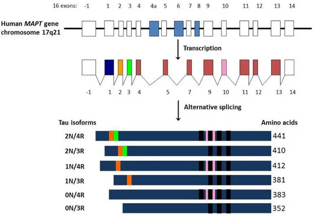
Figure 1.1: MAPT gene and splicing isoforms. Adapted from Luna-Munoz et al., 2013.
Tau protein is encoded by the microtubule associated protein Tau (MAPT) gene located on chromosome 17q21. MAPT contains 16 exons whose alternative splicing yields to six isoforms of the protein. Upstream of exon 1 there are consensus binding sites for transcription factors like AP2 and SP1 (Andreadis et al., 1992; Sadot et al., 1996). The alternative splicing involves exons E2, E3 and E10 leading to isoforms ranging from 352 to 441 aminoacids (Figure 1.1). Splicing of exons E2 or E3 generates isoforms carrying 2, 1 or none particular sequences at the N-terminal end of the protein, while the splicing of E10 determines the presence of a repeated aminoacidic sequence at the C-terminal end (Goedert and Jakes, 1990; Goedert et al., 1989b, 1989a). The expression of different Tau isoforms is specific of brain development. The shortest Tau isoform of 352aa, often referred as foetal Tau 0N3R (splicing of E2, E3 and E10), is characteristic of embryonal brain and is progressively lost during brain development. All the other isoforms are expressed in the adult brain but an additional isoform including E4a was also found to be specific of the peripheral nervous system (PNS) (Goedert et al., 1989a, 1992).
11
1.2.2 Protein
Tau protein is a hydrophilic polypeptide which appears as a random coiled protein (Cleveland et al., 1977; Hirokawa et al., 1988). It shows very small content in secondary structures (Mukrasch et al., 2009). When bound to microtubules (MTs) Tau shows a preference to assume a loop-like conformation in which the N- and the C-terminal ends are close (Jeganathan et al., 2006; Di Primio et al., 2017). Moreover, in pathological conditions, Tau assumes a β-sheet secondary structure which leads to formation of amyloidogenic aggregates (Daebel et al., 2012).
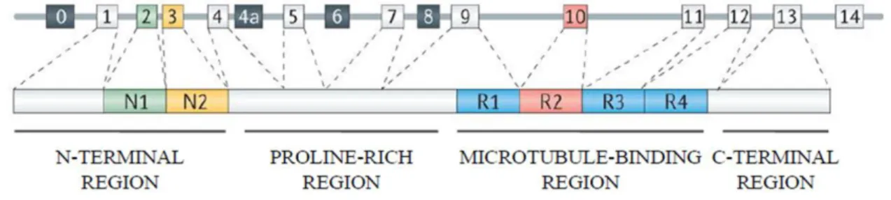
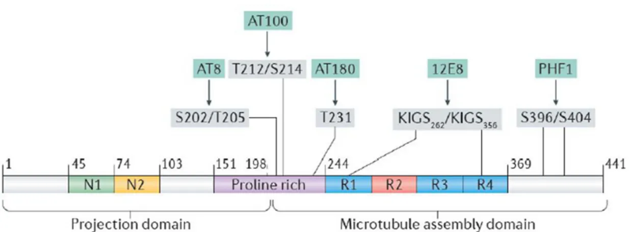
Figure 1.2: Tau protein domains. Adapted from Wang and Mandelkow, 2016.
Tau protein can be divided in two large domains: the projection domain and the microtubule binding domain. The projection domain contains the amino-terminal region (NTD) with high proportion of acidic residues and the proline-rich region (PRD); the microtubule-binding domain is subdivided into a basic true tubulin binding domain with three or four repeats (MTBD) and the acidic carboxy-terminal region (CTD) (Figure 1.2). The six Tau isoforms differ from each other for the presence of three or four microtubule-binding repeats of 31 or 32 residues (isoform 3R or 4R) and for the presence or absence of one or two inserts (29-amino acids) at the N-terminal portion of the protein (Avila et al., 2004; Binder et al., 1985). The projection domain plays several roles such as the determination of space between axonal microtubules, the interaction with other proteins and cation binding (Chen et al., 1992; Hirokawa et al., 1988). Motifs identified in this region include the KKKK sequence involved in heparin binding and the PPXXP/PXXP motifs in the proline-rich region for the binding of Tau with proteins containing SH3 domains such as the tyrosine kinase Fyn (Ittner et al., 2010; Lau et al., 2016; Mondragón-Rodríguez et al., 2012). The repeats of the microtubule-binding domain can be divided in two parts, one composed of 18 residues containing the
12
minimal region with microtubule binding capacity, while the second region composed of 13/14 residues is known as the inter repeat (Avila et al., 2004). Tau isoforms bind MTs by the MTBD with different affinity depending on the number of repeats: higher for Tau 4R isoforms, lower Tau 3R (Lu and Kosik, 2001).
1.3 Tau function and localization
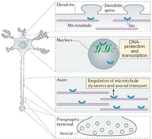
Figure 1.3: Tau subcellular localization and function. Adapted from Wang and Mandelkow,
2016.
Tau was originally found associated to MTs as a protein factor able to induce and stabilize the tubulin polymerization (Weingarten et al., 1975), however, in recent years other functions of Tau have been identified in several subcellular compartments (Figure 1.3). Tau is mostly located in axons, where it interacts with MTs stabilizing them and promoting their assembly (Weingarten et al., 1975). MTBD binds specific pockets in β-tubulin at the inner surface of the MTs while the proline-rich regions, positively charged, are bound to the negatively charged MT-surface; the acidic N-terminal domain branches away from the MT-surface probably because of electrostatic repulsion. Moreover, the β-tubulin pockets of adjacent filaments may be occupied by different repeats of the same MT-binding
13
domain causing crosslinking of three or four dimers (Amos, 2004; Kar et al., 2003). The projection domain determines spacing between MTs in the axon and may increase the axonal diameter. It interacts with other cytoskeletal components like spectrin and actin filaments allowing Tau-stabilized MTs to interconnect with neurofilaments restricting the flexibility of MTs lattices (Kolarova et al., 2012). Besides regulating MT assembly and stabilization, several evidences associate Tau with the regulation of axonal transport. Tau was seen to detach the cargoes from kinesin without influencing the speed of movement along MTs (Trinczek et al., 1999). In vitro, Tau is able to interfere with the dynamics of motor proteins and knock-down of Tau protein is able to increase transport velocity in iPSC-derived dopaminergic neurons (Beevers et al., 2017; Dixit et al., 2008). Moreover, in SHSY5Y cells, Tau is also able to bind p150 subunit of dynactin and to stabilize its binding to MTs, thereby promoting dynein transport (Dixit et al., 2008; Magnani et al., 2007).
Tau protein also localizes in dendrites (Figure 1.3). It has been proposed that it can be found in spines since in brain tissues Tau co-immunoprecipitates with PSD-95 (Ittner et al., 2010). Moreover, upon synaptic activation, Tau moves from the dendritic shaft to spines in cultured neurons and hippocampal slices (Frandemiche et al., 2014). At the synapse, Tau also interacts with Fyn, a tyrosine kinase from the Src-family involved in protein trafficking, by the proline-rich domain which binds SH3 domains of several proteins. Tau is necessary for Fyn localization at the post-synaptic compartment. There, Fyn phosphorylates NMDA subunit NR2B thereby stabilizing its interaction with PSD-95 (Ittner et al., 2010; Lau et al., 2016; Mondragón-Rodríguez et al., 2012). In addition, Tau has a role in signal transmission and synaptic plasticity. Tau reduction results in resistance to pathological network hyperexcitability in a variety of models (DeVos et al., 2013; Holth et al., 2013). It has also been demonstrated that Tau KO shows impaired LTP (Ahmed et al., 2014), but this finding is still debated. Tau is also localized at the axon terminal upon stimulation with NGF (Yu and Rasenick, 2006). Tau is able to potentiate NGF and EGF signalling thus increasing activation of the downstream pathway and influencing neurites extension (Leugers and Lee, 2010). Tau has also been described to have a role in other signalling pathways such as PLC pathway in vitro (Hwang et al., 1996).
Post-14
translational modifications seem to alter Tau distribution, for example phosphorylation in the proline-rich region localizes Tau protein mainly in the somatodendritic compartment, whereas, if the proline-rich region is dephosphorylated or if the phosphorylation occurs in the C-terminal domain, Tau localizes in the distal axonal region (Dotti et al., 1987; Mandell and Banker, 1996).
It has been recently discovered that Tau is involved in miRNA activity. As a matter of fact, Tau interacts with the DEAD box RNA helicase DDX6 involved in translation repression and mRNA decay as well as in the miRNA pathway. Tau/DDX6 interaction increases the silencing activity of the miRNA let-7a, miR-21 and miR-124 affecting the expression of their targets (Chauderlier et al., 2018). Tau has also been localized in many organelles where it can exert non-canonical functions such as ribosomes, mitochondria and endoplasmic reticulum (Tang et al., 2015). Moreover, Tau has been found in the nucleus where it is involved in several functions (Loomis et al., 1990).
1.3.1 Tau nuclear function
In 1990 Tau protein was identified in the nucleus of neuroblastoma cells and in 1995 its nuclear localization was confirmed in human brains. At the beginning, nuclear Tau was detected by immunofluorescence experiments which showed Tau labelling in the nucleus and nucleolus. This evidence was confirmed by subcellular fractionation of chromatin nuclear fraction (Loomis et al., 1990; Rady et al., 1995). The interaction between Tau and chromatin has been further investigated in vitro and in vivo. In vitro experiments demonstrated that the binding of Tau to DNA is mediated by the second half of the proline-rich domain and the R2 of the MTBD (Hua and He, 2003; Qi et al., 2015). Moreover, while hyperphosphorylation of Tau reduces the affinity to DNA, the hypophosphorylated Tau has a higher affinity (Qi et al., 2015).
Other studies clarified that Tau has higher affinity to specific DNA sequences determining also its preferential localization when bound to chromatin. Biophysical studies identified GC-rich sequences as more affine to Tau and demonstrated that the interaction stabilizes the DNA structure when altered by physical agents as high temperature (Vasudevaraju et al., 2012). Fluorescence in
15
situ hybridization (immune-FISH) in non-neuronal cells revealed that nuclear Tau binds AT-rich α-satellites and colocalizes with nucleolin, supporting the fact that nuclear Tau is localized in the internal periphery of nucleoli (Sjöberg et al., 2006). In addition, by a ChIP-on-chip experiment on primary neurons, it was recently demonstrated that Tau is highly enriched in intergenic chromatin regions even if it is also bound to promoter regions, with a high specificity for the GAGA motifs (Benhelli-Mokrani et al., 2018).
An open question in nuclear Tau field regards possible different functions of Tau isoforms and the effect of post-translational modifications in the nuclear compartment. In adult mice brains 1N Tau isoform is overrepresented in soluble nuclear fraction, while 2N Tau isoform is overrepresented in chromatin-bound fraction (Liu and Götz, 2013). Tau can be found either in a phosphorylated and non-phosphorylated state in the nucleus, probably depending on the sub-nuclear localization. In fact, nucleolar Tau appears to be mainly non-phosphorylated, while in the rest of the nucleoplasm, it can also be phosphorylated (Loomis et al., 1990; Sultan et al., 2011). Moreover, nuclear Tau phosphorylation at specific residues, such as the AT100 epitope, increases with aging, with higher affinity to heterochromatin, suggesting an epigenetic role for Tau (Gil et al., 2017).
One of the first evidence about the function of nuclear Tau was on DNA protection and stability. First in vitro experiments showed that Tau is able to increase the dsDNA melting temperature stabilizing its structure and protects DNA from oxidative stress induced by hydroxyl radicals (Hua and He, 2003). This result was further supported by Wei et al. demonstrating that Tau binds the minor groove of the double helix preventing damage by peroxidation (Wei et al., 2008). Recently in vivo results reported that non-phosphorylated nuclear Tau levels increase in neurons after oxidative stress and hyperthermic conditions which should induce the formation of ROS. By Comet and TUNEL assays and ɣH2AX immunofluorescence detection in these conditions, it has been observed that Tau protects the genome of cortical and hippocampal neurons from damage. Moreover, it seems that Tau also protects RNA molecules in cytoplasmic and nuclear compartments (Sultan et al., 2011; Violet et al., 2014). Intriguingly expression of mutated Tau in residues related to tauopathies is strongly associated to chromosome aberrant recombination and aneuploidy in mouse and
16
human samples, supporting the key role of nuclear Tau in genome stability (Rossi et al., 2008, 2013, 2014).
Tau is also involved in chromatin remodelling and gene expression regulation even if the mechanisms are still unclear. A first observation in Tau knock-out mouse models revealed the altered expression of several genes, intriguingly some of them regulates themselves (Gómez de Barreda et al., 2010; Oyama et al., 2004). Increase in nuclear Tau levels is able to induce global chromatin relaxation in Tau overexpressing drosophila and mice, indicating Tau as a factor responsible for the loss of heterochromatin in AD human brains. These alterations result in an increased transcription of genes that are heterochromatically silenced in controls. This observation was shown to be disease relevant since heterochromatin restoration in Drosophila is able to recover the locomotor impairments shown in Tau overexpressing individuals (Frost et al., 2014). More recently, it has been demonstrated that increased Tau levels induce transcription of transposable elements in Drosophila brains and in human AD and PSP brains. Dysregulation of transposable elements is due to both the loss of heterochromatin and the reduction in piwi elements which prevents transposon expression and genomic integration (Guo et al., 2018; Sun et al., 2018). Moreover, it has been demonstrated that Tau not only reduces heterochromatin but it has also a repressive role. As a matter of fact, Tau interacts with TIP5, a transcriptional repressor of rDNA, in the nucleolus and Tau depletion results in an increase of 45S-prerRNA synthesis, suggesting that Tau might be involved in rDNA silencing (Maina et al., 2018). In addition, by ChIP-on-chip experiments, a transcriptional repressive function has been observed towards promoters bound directly by Tau (Benhelli-Mokrani et al., 2018). Other studies support the possible role of Tau in chromatin remodelling. A recent work demonstrated that Tau interacts with the nuclear protein TRIM28 which is able to shuttle it to the nucleus. This was shown tracking the Tau-TRIM28 complex exploiting bimolecular fluorescence complementation and was confirmed observing that TRIM28 overexpression was able to boost Tau shuttling to the nucleus (Rousseaux et al., 2016). Tripartite motif-containing 28 (TRIM28), also known as transcriptional intermediary factor 1b (TIF1b) and KAP1 (KRAB-associated protein-1), is a nuclear protein mainly involved in transcriptional
17
regulation and chromatin remodelling. TRIM28 acts as a co-repressor of KRAB zinc finger proteins, moreover it interacts with histone deacetylase complexes and methyl transferases (Iyengar and Farnham, 2011). Since Tau is strongly associated to chromatin remodelling and it interacts with TRIM28, it would be interesting to further investigate the nature of this interaction to find out whether TRIM28 or one of its interactors could mediate nuclear Tau functions.
1.4 Tau pathology
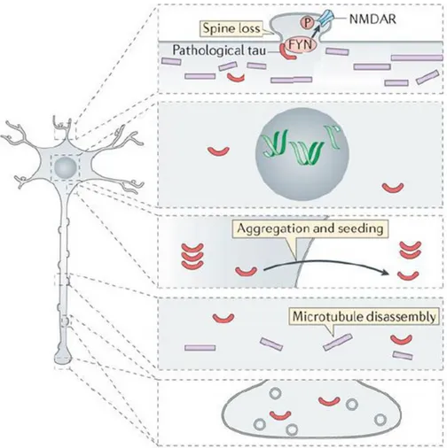
Figure 1.4: Tau pathological changes in subcellular compartments. Adapted from Wang and
Mandelkow, 2016.
As stated above, Tau protein is a key player in a group of neurodegenerative diseases named tauopathies and among these Alzheimer’s disease is one of the most common and studied. Tauopathies can differ for the progression and symptoms, but all these diseases show a common hallmark that is the pathological destabilization and aggregation of Tau protein. Tauopathies include Alzheimer’s disease (AD), Frontotemporal dementia with parkinsonism-17
18
(FTDP-17), Pick disease (PiD), progressive supranuclear palsy (PSP), corticobasal degeneration (CBD), agyrophilic grain disease (AGD) and Huntington disease (HD), and most of them show both familiar and sporadic forms (Lee et al., 2001). In the first steps of these pathologies, whose mechanisms are still not clear, MTs-bound Tau is post-translationally modified. These modifications lead to Tau displacement from MTs with its increase in the cytoplasmic pool. Post-translational modifications change Tau molecular properties and structure causing the association of Tau monomers into oligomers and then in amyloidogenic aggregates. The molecular mechanisms inducing the toxic effect of Tau aggregation are still debated even if it is strongly supported that the aggregation causes cellular stress and damage to neuronal and non-neuronal cells.
1.4.1 Tau mutations
Figure 1.5: Localization of the main MAPT mutations. Adapted from Wang and Mandelkow,
2016
The processes involved in the pathogenesis of tauopathies are still unclear. These diseases can be sporadic or familial and genetic analyses of Tau in patients with familial tauopathies demonstrated that specific mutations are associated to the insurgency of Tau destabilization and Tau failure. Familial tauopathies like FTDP-17 can be caused by several intronic and exonic mutations in MAPT gene such as Δ280K, P301L, P301S and E10+3 (Figure 1.5) (Bugiani et al., 1999; Hutton et al., 1998; Rizzu et al., 1999; Spillantini et al., 1998a). Several studies show that these mutations can induce Tau alterations, making it more prone to destabilization and aggregation. Exonic missense or
19
deletion mutations destabilize Tau binding to MTs and decrease the ability to induce MT assembly (Goedert et al., 1999). Intronic mutations can affect alternative splicing of E10 thus increasing 4R Tau as observed in patients’ brains (Spillantini et al., 1998a). The discovery of mutations altering MAPT splicing suggests that disruption of 3R:4R ratio is sufficient to cause neurodegeneration and dementia, conversely maintaining the physiological 3R:4R ratio appears to be crucial for normal brain functions. The mechanisms around the dementia caused by isoform imbalance is still obscure, but it is hypothesized that it could somehow interfere with Tau binding to MTs thus increasing Tau soluble pool (Liu and Gong, 2008).
An inversion polymorphism arising about 3 million years ago of approximately 900kb on chromosome 17q21 generated two haplotypes, H1 and H2 encompassing MAPT locus (Stefansson et al., 2005). Genome association analyses showed that H1 haplotype is associated with increased risk for PSP and CBD (Baker et al., 1999; Di Maria et al., 2000). In time, variations of each haplotype generated sub-haplotypes which differentially influence the insurgency of tauopathies. For example, the H1c sub-haplotype has been associated with increased risk for PSP and AD (Baker et al., 1999; Myers et al., 2005). In order to explain the pathogenic mechanisms of these haplotypes it has been hypothesized that they can affect Tau expression or Tau splicing. As a matter of fact, reporter genes under H2 promoter show a reduction in transcriptional activity compared to the H1 promoter (Kwok et al., 2004). Moreover, H1/H2 heterozygous neuroblastoma cell line models and human brains indicate that the H1 chromosome induce a significantly higher expression of transcripts including E10 (Caffrey et al., 2006).
Since Tau mutations are strongly associated to Tau pathological behaviour, they are experimentally employed to induce, both in a cellular context and in vivo, Tau destabilization and aggregation since wild-type Tau is not able to aggregate spontaneously. In particular, mouse models expressing mutated Tau are largely used to clarify the neurotoxic mechanisms mediated by Tau aggregation and to simulate the progression of tauopathies even if a “perfect” model able to represent all the characteristics of a specific tauopathy is not available yet
20
(Bondulich et al., 2016; Eckermann et al., 2007; Kremer et al., 2011; Lewis et al., 2000; Schindowski et al., 2006; Yoshiyama et al., 2007).
Intriguingly, mutations in Tau protein seem not only to affect Tau interaction with tubulin leading to aggregation as canonically described (Goedert et al., 1999). Recent studies showed that pathological mutations, along with genomic instability and neuronal damage, cause also an increased probability of tumorigenesis suggesting a role for Tau also in other tissues of the organism (Rossi et al., 2008, 2013, 2014, 2018). Moreover, the P301S mutation associated to FTDP and PiD affects the interaction with DDX6, resulting in an impairment of miRNAs silencing activity (Chauderlier et al., 2018).
1.4.2 Tau post-translational modifications
As described above, Tau protein is exposed to several post-translational modifications that influence both its physiological and pathological behaviour. In particular, it is commonly assumed that Tau modifications, above all hyperphosphorylation and cleavage, are the first events leading to aggregation (Pevalova et al., 2006; Wang and Mandelkow, 2016).
Phosphorylation:
Figure 1.6: Relevant Tau phosphorylation residues. Adapted from Wang and Mandelkow,
2016.
Phosphorylation is the most characterized post-translational modification of Tau. 79 putative serine and threonine phosphorylation sites have been identified. During embryonic development it has been demonstrated that Tau protein is much more phosphorylated than in post-natal period. Tau physiological
21
phosphorylation seems to control MT dynamics during normal neurite growth and maturation. In tauopathies Tau becomes hyperphosphorylated leading to the formation of amyloidogenic aggregates. Moreover, in recent years, changes in body temperature have been demonstrated to be environmental factors able to alter Tau phosphorylation leading to a predisposition in developing tauopathies (Bretteville et al., 2012; Planel et al., 2004; Tournissac et al., 2019; Vandal et al., 2016; Whittington et al., 2013). By mass spectrometry and antibody detection experiments all six Tau isoforms have been identified in aggregates and have been demonstrated to be phosphorylated at 40 different residues (Avila et al., 2004; Pevalova et al., 2006).
Among critical sites involved in Tau aberrant activity implicated in AD and in other tauopathies, AT8 epitope (Ser199/Ser202/Thr205) has an important role. The phosphorylation of these three residues is sufficient to cause MTs remodelling and instability, diminished mitochondrial transport, cell death and neurodegeneration (Shahpasand et al., 2012). A similar effect is observed by phosphorylation of residues Thr212/Thr231/Ser262 (Alonso et al., 2010). In vitro kinetic studies of the binding of unphosphorylated and hyperphosphorylated Tau to tubulin suggest that Ser199/Ser202/Thr205, Thr212, Thr231/Ser235, Ser262/Ser356, and Ser422 are among the critical phosphorylation sites that convert Tau to pathological molecule that sequesters normal microtubule-associated proteins from MTs (Figure 1.6) (Abraha et al., 2000; Alonso et al., 2004; Gong et al., 2005; Haase et al., 2004; Sengupta et al., 1998). The phosphorylation of these residues depends on the activity of both kinases and phosphatases, whose equilibrium plays a key role in Tau pathology.
Kinases:
GSK3β
Glycogen synthase kinase-3 (GSK3β) is expressed ubiquitously but it can be found at high levels in the brain where it localizes predominantly in neurons. It belongs to the PDPK class (proline directed protein kinases), and is a serine/threonine-specific kinase. GSK3β influences Tau behaviour both in physiological and pathological conditions (Pei et al., 1997; Pevalova et al., 2006). Brains of AD patients show a higher immunoreactivity against GSK3β than
22
control brains, with the kinase predominantly colocalizing with NFTs (Pei et al., 1997). Among the residues phosphorylated by GSK3β, Thr231 has been observed to be strongly related to the beginning of AD. This epitope is an example of primed phosphorylation because it occurs after Ser235 phosphorylation. The modification of Thr231 causes a conformational change in Tau affecting its stability and affinity to MTs (Cho and Johnson, 2003; Daly et al., 2000). Preclinical studies employing GSK3β inhibitors revealed that the block of kinase activity decreases aggregation and protects against axonal degeneration in TauP301L mice (Noble et al., 2005).
Cdk5
Cyclin-dependent kinase 5 (Cdk5) is a serine/threonine kinase of the PDPK class. It contributes to phosphorylation of human Tau on Ser202, Ser205, Thr212, Thr217, Ser235, Ser396 and Ser404, epitopes phosphorylated in AD brains. Cdk5 interacts with p35 to exacerbate its function. It has been seen that the conversion of p35 in p25 due to pathological conditions causes prolonged activation and mislocalization of Cdk5 and hyperphosphorylation of Tau (Cruz et al., 2003; Lee et al., 2000; Maccioni et al., 2001; Tsai et al., 2004). The knockdown of Cdk5 in AD mouse models strongly decreases the number of neurofibrillary tangles in the hippocampus (Piedrahita et al., 2010).
ERK1/2
Extracellular signal-regulated kinases isoforms 1 and 2 (ERK1/2) belong to the PDPK class of kinases. Analysis on post-mortem AD brains showed that they predominantly colocalize with NFTs (Perry et al., 1999). Consistently, the administration of an ERK2 inhibitor was able to reduce the levels of abnormally phosphorylated Tau and to rescue motor deficits in a mouse model of tauopathy (Le Corre et al., 2006).
JNK
C-Jun amino-terminal kinase (JNK) belongs to the PDPK group of kinases. JNK is able to hyperphosphorylate Tau, moreover immunohistochemical analysis of phopho-JNK showed increased activation of the kinase in the hippocampi of AD patients (Zhu et al., 2001).
23
Other kinases
Other kinases have an important role in the pathogenesis of tauopathies. PKA is a ubiquitous serine/threonine kinase activated by cAMP. It phosphorylates Tau at Ser214, Ser217, Ser262, Ser396/404 and at Ser416 (Andorfer and Davies, 2000; Carlyle et al., 2014; Zheng-Fischhöfer et al., 1998). CaMKII too seems an important factor inducing Tau aberrant hyperphosphorlation. The function of this kinase is to regulate important neuronal functions including neurotransmitter synthesis and release, modulation of ion channel activity, synaptic plasticity and gene expression. The kinase phosphorylates Tau at Ser262, Ser356, Ser409 and Ser416 that are phosphorylated in brains of AD patients. The phosphorylation of all these residues by PKA and CamKII is strongly associated to Tau pathology since this mechanism influences Tau binding on MTs, cleavage and aggregation (Andorfer and Davies, 2000; Carlyle et al., 2014; Singh et al., 1996; Steiner et al., 1990; Zheng-Fischhöfer et al., 1998).
Phosphatases
Several studies have shown that three major protein phosphatases, PP5, PP2B and PP2A, can dephosphorylate Tau protein. PP2B influences the dynamics of MTs and microfilaments. PP5 is associated with MTs and dephosphorylates Tau in the neuronal cytoplasm. PP2A is localized on MTs and can dephosphorylate Tau by direct interaction or indirectly, regulating the activity of several Tau kinases (Liu et al., 2005; Pevalova et al., 2006). The inhibition or the absence of these phosphatases in cells cause the hyperphosphorylation of Tau suggesting a possible role of these proteins during tauopathy development. Remarkably, in AD brains the impaired activity of PP2A has been observed (Gong et al., 1993).
Other Tau post-translational modifications
Tau undergoes several other post-translational modifications which could influence its functions such as acetylation, glycation, glycosylation and truncation.
As to post-translational modifications, studies on the composition of aggregates revealed that Tau is cleaved at different sites. Tau truncation is characterized by a progressive cleavage of the protein in specific residues which leads to smaller
24
and smaller fragments prone to aggregation (Gamblin et al., 2003; Guillozet-Bongaarts et al., 2005; Novak et al., 1993; Rissman et al., 2004). Several enzymes have been identified as able to cleave Tau such as caspases but also calpains, thrombin, cathepsins and the aminopeptidase PSA. In particular, in tauopathies, these enzymes are deregulated and target Tau protein inducing and enhancing its destabilization and aggregation (Arai et al., 2006; Cataldo et al., 1997; Nakanishi et al., 1997; Rao et al., 2008; Saito et al., 1993). The study of the progressive Tau cleavage revealed that some residues are cut in early stages of tauopathies and other in late stages. A caspase-cleaved Tau species, cleaved at Asp421, was identified in NFTs (Gamblin et al., 2003). Tau conformational studies suggest that this cleavage is an early event in the pathogenesis of AD (Guillozet-Bongaarts et al., 2005; Rissman et al., 2004). Intriguingly, caspase cleavage at Asp421 can be inhibited by phosphorylation at Ser422 suggesting that some phosphorylations are protective (Guillozet-Bongaarts et al., 2006). Truncated Tau at Glu391 is found in NFTs in tauopathy brains and it is associated to a late stage of the disease. In Tau aggregates a 12KDa fragment from His268 to Glu391containing the MTBD has been identified, and it is referred to as the PHF-core (Novak et al., 1993). Further experiments showed that phosphorylation and truncation work synergically and together they influence Tau aggregation. As a matter of fact, in cells with or without GSK3β, transfected Tau fragment aggregates only in presence of the kinase (Cho and Johnson, 2004). Another post-translational modification whose importance is becoming relevant is lysine acetylation, which has been revealed to affect Tau properties. This modification neutralizes charges in the MT-binding domain interfering with Tau binding to MTs. Moreover, acetylation of Lys280 increases cytosolic Tau fraction and it’s correlated with Tau hyperphosphorylation. It is present mostly in intracellular NFTs rather than in pre-tangles or extracellular aggregates and it precedes Tau truncation (Cohen et al., 2011; Irwin et al., 2012).
Glycosylation is the covalent bonding of sugars to the side chain of a polypeptide which can occur at the amine group of an asparagine (N-glycosylation) or at the hydroxyl radical of serine or threonine (O-glycosylation). These modifications seem to have opposite effect, as O-glycosylation is decreased in AD brain, while N-glycosylation is increased (Wang et al., 1996). Phosphorylation and
O-25
glycosylation target the same residues, suggesting a mutually exclusive competition. As a matter of fact, in AD patients, a negative correlation has been reported between O-glycosylation level and Tau phosphorylation (Liu et al., 2009). However, it has been reported that the biological effect at each phosphorylation site is different (Gong et al., 2005). Moreover, deglycosylation of PHFs converts them in bundles of straight filaments and successive dephosphorylation results in the release of Tau monomers (Wang et al., 1996). Oxidation too seems to increase aggregation of monomeric Tau protein because it produces disulphide cross-linking between cysteine residues. Isoforms of Tau containing three repeats (3R) have only one cysteine residue in the MTs binding domain with respect to 4R isoforms which have two cysteines. For this reason, 3R Tau is more prone to aggregate rather than 4R isoform which may form intramolecular disulphide bonds (Barghorn and Mandelkow, 2002).
Other modifications are still under investigation, even if they seem to be related to late stages of Tau pathology. In PHFs and NFTs Tau is also ubiquitinated with the covalent bonding of the 76aa protein ubiquitin. Also glycation, the non-enzymatic linkage between a reducing sugar and the amino side chain of polypeptide, can be identified in PHFs isolated from AD brains while it’s not present in normal Tau (Münch et al., 2002; Yan et al., 1995).
1.4.3 Tau aggregation
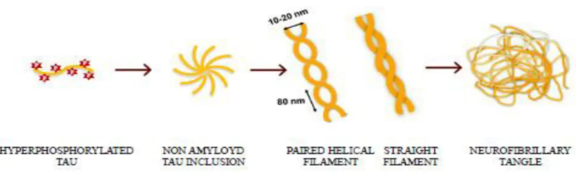
Figure 1.8: Tau aggregation process from monomers to neurofibrillary tangles.
Tau molecule has long stretches of positively and negatively charged regions which don’t allow intermolecular hydrophobic association. The β-structure, typical of amyloidogenic proteins, is present in repeated regions, in particular in R2 and R3, which can assemble by their own in filaments (von Bergen et al., 2000). Self-aggregation is inhibited by the presence of intact N- and C-terminal domains but
26
when Tau undergoes post-translational modifications, the conformational structure changes exposing the sticky repeat regions which lead to formation of aggregates (Alonso et al., 2001). Even if Tau aggregation is a common event of all tauopathies, fibrils isoform composition differs between tauopathies. In AD tangles include both 3R and 4R Tau (Sergeant et al., 1997), in PiD they are mainly made up of 3R (Delacourte et al., 1996) and in PSP, CBD, AGD and FTDP-17 they predominately comprise 4R Tau (Arai et al., 2001; Buée-Scherrer et al., 1996; Togo et al., 2002).
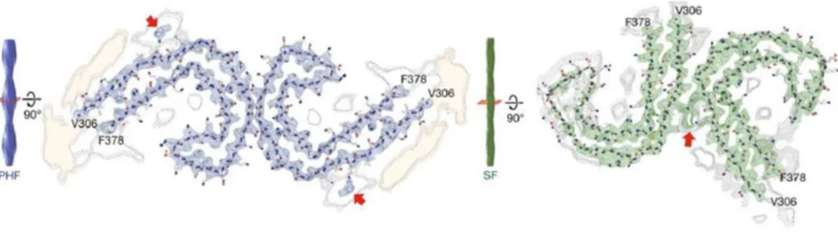
Figure 1.7: PHFs and SFs ultrastructure level. Adapted from Fitzpatrick et al., 2017.
The aggregation process seems to involve several steps. At the beginning, small Tau inclusions with low content in β-sheet structures are formed in a process called nucleation; after that other Tau molecules associate to this core to form paired helical filaments (PHFs) and straight filaments (SFs). Finally, PHFs further self-assemble to form neurofibrillary tangles (NFTs). The formation of Tau fibrils can be experimentally induced by the addition of preformed aggregates which mimic the nucleation step in a process named seeding. It has been demonstrated that the administration of small Tau assemblies in the extracellular medium can induce prion-like homotypic-seeding both in vivo and in vitro (Clavaguera et al., 2009; Frost et al., 2009). The smallest assembly having these seeding properties is Tau trimer (Mirbaha et al., 2015). Remarkably, in recent papers it has been demonstrated that Tau monomers isolated from tauopathy brains are able to induce Tau aggregation that is specific for the tauopathy the monomer is obtained from. This result opens to new insights suggesting that the monomer possesses peculiar pathological characteristics specific for each tauopathy independently from the oligomerization (Mirbaha et al., 2018; Sharma et al., 2018). It is thought that this occurs because nucleation is the limiting step in Tau
27
aggregation and if preformed aggregates are administrated, this step can be skipped. Electron-microscopy experiments showed that NFTs consists mainly of 10 – 20 nm PHF (KIDD, 1963) and of 15 – 18 nm straight filaments (SF) (Yagishita et al., 1981). PHFs and SFs also differ at ultrastructure level as both of them consist in two protofilaments with C-shaped subunits but with different symmetry. The N- and C-terminal ends protrude from the core of the aggregate forming the fuzzy coat (Fitzpatrick et al., 2017).
It has been demonstrated that in patients the number of NFTs correlates with the level of cognitive impairment. For this reason, it has been hypothesized that NFTs are toxic for the cell (Arriagada et al., 1992). In tauopathies, Tau is ineffective to keep the cytoskeleton organized in axonal processes because its affinity to tubulin is reduced. This loss of function is due to conformational changes and misfolding which lead to aberrant aggregation in fibrillary toxic structures inside neurons. Tau is redistributed in the somato-dendritic compartment and in isolated processes of affected neurons (Kolarova et al., 2012). The destabilization of MTs affects axonal trafficking in particular the plus-end-directed transport by kinesin (Ebneth et al., 1998). Inhibition of transport slows down exocytosis and the localization of organelles; the absence of these mechanisms causes a decrease in glucose and lipid metabolism, ATP synthesis and a loss of Ca2+ homeostasis which lead to a distal degeneration process (Futerman and Banker, 1996; Trojanowski and Lee, 1995). It has been seen that aberrant Tau not only can create aggregates sequestering normal Tau but it can also remove from MTs the two other major neuronal MAPs, MAP1 and 2 (Alonso et al., 1997).
1.4.4 Other mechanisms of cellular toxicity mediated by Tau
Increasing evidences show that NFTs are not the only toxic Tau species in tauopathies, in fact, in AD brains neuronal loss may exceed Tau aggregation (Gómez-Isla et al., 1997). Other pathological mechanisms can be involved in the neurotoxicity mediated by Tau. Dendritic Tau seems to mediate Aβ-induced neurotoxicity in AD since, in absence of Tau protein, Aβ-mediated LTP impairment is prevented (Shipton et al., 2011). Moreover, as already described, dendritic Tau is necessary for Fyn localization and scaffolding at the
post-28
synaptic compartment. There, Fyn phosphorylates NMDA subunit NR2B thereby stabilizing its interaction with PSD95. Under disease conditions, redistribution of Tau from the axon into the somatodendritic compartment is thought to increase Tau-dependent sorting of Fyn to dendrites, leading to toxic hyperexcitability caused by NMDA receptors. Indeed, the loss of Tau can prevent postsynaptic targeting of Fyn, and as a consequence NMDA-dependent excitotoxicity and memory impairment (Ittner et al., 2010; Lau et al., 2016; Mondragón-Rodríguez et al., 2012). Therefore, Fyn is important to potentiate glutamatergic signalling and it could mediate excitotoxicity in tauopathies. As described above, in vivo studies showed that the increase of Tau in the nucleus is associated to global chromatin relaxation resulting in alteration of gene expression. Intriguingly, chromatin relaxation has been shown to be neurotoxic (Frost et al., 2014). Moreover, Tau loss of function induced by mRNA knock down results in an increase of 45S-pre-rRNA synthesis in SHSY5Y (Maina et al., 2018). Other evidences suggested that oligomeric nuclear Tau has a more efficient repressive role than monomeric Tau in mouse models, supporting a global gene expression alteration that can affect neuronal homeostasis (Benhelli-Mokrani et al., 2018). Aberrant Tau influences also miRNA activity, as a matter of fact mutated Tau is less efficient in modulating the activity of miRNA let-7a (Chauderlier et al., 2018). All these evidences strongly support that in tauopathies, Tau is able to induce cellular toxicity since it alters several mechanisms crucial for the correct functioning of the cell.
1.4.5 Tau secretion and spreading
In pathological conditions, Tau aggregation starts in specific areas of the brain and during the pathology progression, Tau inclusions can be found also in other regions indicating a diffusion of amyloidogenic Tau. Its spreading occurs with a standard pattern with close or connected brain areas being affected one after the other (Braak et al., 2006). The mechanisms behind Tau spreading are still under investigation. Tau aggregates were found first intracellularly and the protein detected in the extracellular space was considered as a release from dead cells. On the contrary, further studies reported that Tau is secreted by different neuronal and non-neuronal cell types, associated to exosomes or other membrane vesicles (Kim et al., 2010; Medina and Avila, 2014; Shi et al., 2012). It was also observed that pathological post-translational Tau modifications enhance
29
its secretion suggesting that the release mechanism is linked to the progression of Tau pathology (Plouffe et al., 2012). Several mechanisms for the uptake have been proposed such as internalization of soluble Tau via receptor-mediated endocytosis, dynamin-driven endocytosis of soluble Tau aggregates or even proteoglycan-mediated macropynocytosis (Medina and Avila, 2014). Spreading could also occur trans-synaptically or via diffusion (Wang and Mandelkow, 2016).
1.5 Molecular tools to investigate Tau by imaging
Due to the key role of Tau in several neurodegenerative diseases, it is crucial to develop molecular tools providing a clear and simple interpretation of the state of Tau protein. These tools can be extremely useful for diagnosis or to screen molecules targeting pathological Tau.
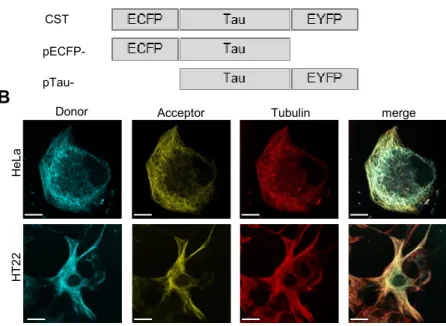
The main approaches to study Tau aggregation rely on biosensors and amyloid binding dyes. Tau biosensors are chimeric proteins containing fluorophores that provide information about the molecular state of Tau. The advantage of fluorophores is that they can be genetically controlled (Kaláb and Soderholm, 2010). An intermolecular sensor that relies on bimolecular fluorescence complementation (BiFC) has been developed. Tau proteins are fused to non-fluorescent N- or C-terminal fragments of V-YFP and when two Tau molecules interact, the fluorophore is reconstituted and the protein fluoresces providing an output for Tau aggregation (Tak et al., 2013). An intermolecular FRET biosensor developed by Kfoury et al. is commonly used in laboratories that investigate Tau aggregation. Tau fragments, prone to aggregate, are fused to either YFP or CFP and when two Tau molecules interact, a FRET positive signal is observed. The strong advantage of this biosensor is that it detects Tau aggregation by fluorescence imaging but it is also optimized for FRET cytofluorimetry (Kfoury et al., 2012). Finally, in our laboratory we developed an intramolecular FRET biosensor that will be described later (Di Primio et al., 2017).
Another method to visualize Tau aggregation is the employment of dyes such as Thioflavin T, Thiazin Red and K114. These compounds have high affinity to β-sheet structures typical of amyloidogenic aggregates and can be used in cellular cultures or tissues with the limitation that they need to be fixed by fixation methods. Amyloid binding dyes emits a fluorescence signal when bound to Tau
30
aggregates which allows to study aggregation dynamics by imaging techniques (Crystal et al., 2003; Luna-Muñoz et al., 2008; Santa-María et al., 2006).
A novel approach is the employment in pre-clinical and clinical studies of molecules for PET imaging. The interest for PET compounds is due to the possibility to accurately and specifically target Tau deposits in vivo in patients’ brains. The advances of molecular imaging in recent years provided promising Tau-specific tracers such as THK5317, THK5351, AV-1451, and PBB3. The employment of these tracers guarantees the investigation in real-time of Tau deposition patterns in vivo for different pathologies, discriminating between neurodegenerative diseases, including different tauopathies, and monitoring disease progression. In AD, the topographical distribution of tracers follows the known distribution of NTFs and is closely associated with neurodegeneration (Okamura et al., 2018; Saint-Aubert et al., 2017).
1.6 Tauopathy animal models
In order to investigate the behaviour of Tau in physiology and, above all, in pathology, several animal models based on the expression of human Tau have been generated. The main model employed for Tau studies is mouse but also D.
rerio and Drosophila are commonly used. The advantage of using animals is
obviously that the biological context is complex and closer to human, guaranteeing a higher reliability compared to the in vitro conditions. However, the main counterpart is that these models does not develop tauopathies and they need to be genetically modified with Tau molecules prone to pathological aggregation.
Several mouse models have been developed in the last 20 years and most of them are transgenic mice expressing mutated human Tau. The employment of Tau isoforms mutated in specific residues enhances the pathological behaviour of Tau, indeed, in these models Tau is unstable on MTs and forms toxic aggregates spontaneously. Each mouse usually expresses a specific mutation such as TauP301L, TauP301S or TauΔK280 and all of them lead to aggregation of Tau even if there are differences in the progression and spreading (Allen et al., 2002; Eckermann et al., 2007; Lewis et al., 2000; Terwel et al., 2005; Yoshiyama et al., 2007). Mouse models for Tau pathology can be divided into two groups: the first
31
resembles pure tauopathies expressing mutated Tau or Tau fragments, the second combines the Tau pathology with the Aβ pathology, a condition resembling Alzheimer’s disease. However, tauopathy models share common aspects of pathology development. The expression of mutated Tau causes a progressive neurodegeneration with formation of neurofibrillary tangles around the 6th month (Allen et al., 2002; Lewis et al., 2000; Terwel et al., 2005; Yoshiyama et al., 2007) but there are cases of late aggregation as for TauΔK280 model which shows mature tangles only after 24 months (Eckermann et al., 2007). Other models are based on the expression of a Tau fragment which is prone to form intracellular inclusions. In these animals spontaneous aggregation occurs at 9-10 months and progresses with age (Bondulich et al., 2016; Filipcik et al., 2012). All these models show strong neuronal damage worsening with the progression of Tau toxic aggregation. As a matter of fact, common features are the synaptic loss, neuronal death and cognitive deficits which become more severe with aggregation. In some cases also gliosis can be detected suggesting that the neurodegeneration gives rise to an inflammatory process. The main brain areas affected by aggregation and neuronal loss are the spinal cord, cortical areas, amygdala, hippocampus with a spreading which increases with the development of the tauopathy (Allen et al., 2002; Boekhoorn et al., 2006; Bondulich et al., 2016; Filipcik et al., 2012; Van der Jeugd et al., 2011; Kremer et al., 2011; Lewis et al., 2000; Terwel et al., 2005; Yoshiyama et al., 2007). Mutated Tau is also employed for the development of AD mouse models expressing human APPSWE as well. Remarkably, in these animals the presence of both toxic proteins causes a severe Tau and Aβ aggregation suggesting that there is a cooperative effect in the formation of toxic aggregates. Moreover, neuronal and synaptic loss are present and coupled with gliosis. Intriguingly, the brain areas first involved in these processes are slightly different. Indeed, the hippocampus and cortical areas show pathological aggregation followed by spreading in other regions (Lewis et al., 2001; Oddo et al., 2003; Schindowski et al., 2006). Even if a lot of models have been developed and characterized, there is still the lack of a “perfect” model mimicking all the aspects of tauopathies. However, the employment of these animals has been valuable to study pathological mechanisms with cognitive readouts and to test treatments impairing or enhancing Tau toxicity.
32
Even if the mammalian animal model is the most employed, the long experimental time required to develop the disease, the difficulties in handling and the failure of therapies efficient in human patients induced the scientific community to explore alternative models for specific applications.
During the last decade, Drosophila gained attention as a model system for common human neurodegenerative brain disorders. Most basic molecular and
cellular mechanisms are highly conserved between humans
and Drosophila and ~70% of all human disease genes have an evolutionary conserved fly homolog, providing a reliable model (Bier, 2005; Bilen and Bonini, 2005). Many Drosophila models have been generated using human Tau. Some are based on wild-type Tau isoforms whereas others express mutated forms such as TauR406W, TauV337M, and TauP301L. To explore specific mechanisms of Tau toxicity or dysfunction, transgenes with targeted mutations and truncations were also generated, with constructs which abolish or mimic Tau phosphorylation or proteolytic cleavage. Together, these models explore the great diversity of tauopathies (Gistelinck et al., 2012). Expression of Tau protein in flies results in neurotoxicity which can be evaluated by morphological readouts of the animal. Roughening of the eye is the most commonly used external phenotype to evaluate toxicity of Tau in Drosophila (Gistelinck et al., 2012). Moreover, Drosophila notum harbours around 200 bristles, which are sensory organs connected at the base with the dendrite of a sensory neuron. Tau toxicity can be quantified by simply counting bristles and overexpression of different variants of Tau leads to bristle loss (Yeh et al., 2010). Other common readouts are the lifespan and pupal lethality, as a matter of fact flies overexpressing Tau show a reduced lifespan and increased death of larvae (Talmat-Amar et al., 2011). All these readouts can be used to easily evaluate the toxic effect of Tau and also to screen genes or drugs altering the toxicity of the protein. Moreover, neuronal cell death can be detected by TUNEL or immunostaining of activated cleaved caspase-3 which indicate increased apoptosis in brains of Tau-expressing flies (Ali et al., 2012; Colodner and Feany, 2010; Talmat-Amar et al., 2011). Neuronal degeneration in Drosophila can also be observed by the presence of vacuoles in brain tissues and can be used as a quantitative readout (Ali et al., 2012; Mershin, 2004; Talmat-Amar et al., 2011). Alterations in axonal transport has also been
33
observed in flies expressing Tau bearing pathological mutations and PTMs and this causes locomotor defects and altered release of neurotransmitters and neurohormones (Gistelinck et al., 2012). Finally this model can be applied for behavioural tests, in particular olfactory learning and memory in Tau-expressing flies is strongly impaired and almost a complete loss of mushroom bodies is observed suggesting a relevant toxic effect of Tau (Kosmidis et al., 2010; Mershin, 2004). The significant damage caused by Tau in Drosophila and the easy readouts led to a relevant use of this model for the identification of Tau mechanisms and interactors related to transport and transmission and for genetic screenings (Gistelinck et al., 2012).
Another animal model which has been considered to study Tau pathological mechanisms is Danio rerio (zebrafish). This animal offers several distinct advantages over other vertebrate models: the easy maintenance in laboratory due to its simple habitat conditions, the large clutch size with hundreds of eggs, the transparency of embryos allowing an easy imaging analyses, the feasibility of the genetic manipulation, the vertebrate neural structural organisation and the presence of several orthologue genes similar to humans. Moreover, the small size of larvae allows the high-throughput screenings of neuroactive compounds (Saleem and Kannan, 2018). The zebrafish model has been employed for the study of tauopathies by expressing wild-type or mutated forms of Tau. Interestingly, the pathological features develop much earlier in zebrafish as compared with other available rodent models. Tau expression in zebrafish shows early pathological hallmarks like hyperphosphorylation and conformational changes within the first 2 days of embryonic development. After few days, the larvae develops substantial neurodegeneration displaying pathological features including neurofibrillary tangles by 5 weeks of development (Paquet et al., 2009). Tau aggregation can be observed after Tau overexpression in neurons and these cellular inclusions are positive for pathological phosphorylations in residues related to Tau aggregation and for structural antibodies and dyes (Bai and Burton, 2011; Bai et al., 2007; Paquet et al., 2009, 2010). Moreover, Tau expression in neurons disrupts the cellular cytoskeletal structure (Tomasiewicz et al., 2002). The stereotypic escape response, a behavioural test employed to observe the motility and the response to an external stimuli, has been also
34
evaluated and at 48 hpf the escape is highly reduced or absent in Tau-expressing larvae (Paquet et al., 2009). Due to the peculiar characteristics of the model and to the effect of Tau overexpression which spontaneously leads to neurodegeneration, zebrafish is considered a promising model to study pathological mechanisms and to develop screenings to impair Tau mediated toxicity.
1.7 Therapeutic approaches against Tau pathologies
Tauopathies are complex multifactorial diseases and the development of therapies has been slow and often inconclusive. Current approaches do not rely on disease modifying drugs, but rather on symptomatic ones and aim to manage behavioural symptoms. Several symptomatic drugs for AD treatment are approved by the Food and Drug Administration and they mainly rely on cholinesterase inhibitors, to prevent the breakdown of acetylcholine described in the brain of AD patients (Birks, 2006) and, at late stages, on NMDA antagonists, to prevent excitotoxicity (Wang and Reddy, 2017). However, these treatments can be unsatisfactory as they cannot halt the progress of the pathology. For these reasons, many efforts have been spent to study the two main lesions in AD, i.e. amyloid plaques (APs) and NFTs in order to come up with a disease modifying treatment. However, all the attempts targeting APs failed in blocking or curing the disease (Makin, 2018; Wischik et al., 2014). Consequently, an increasing interest toward the role of Tau in the etiology of tauopathies has arisen. Recently active or passive immunization showed great potentialities in preventing Tau aggregation, leading to promising clinical trials that are still in progress. As to the passive immunization, the injection of selected antibodies against pathological Tau has been considered and several approaches have been exploited leading to clinical trials (Novak et al., 2018a; West et al., 2017). Recent evidences showed that the reduction of total Tau in adult brains doesn’t affect physiological functions and prevents toxicity caused by Tau aggregation. This study opened to a new approach employing ASOs molecules to target Tau expression and reduce the aggregation in patient’s brains (Devos et al., 2017; DeVos et al., 2018). Actually the most promising therapeutic intervention is the active immunization by the AAD1vac, a vaccine against pathological Tau. Preclinical studies on mice models revealed that this vaccine specifically reduces
35
Tau pathological aggregates in the brain and currently it is employed in a clinical trial for AD. The phase II for the evaluation of side effects in patients has recently concluded and further investigations are in progress to determine the therapeutic effect (Kontsekova et al., 2014; Novak et al., 2018c, 2018b, 2018a).
Another possibility to treat Tau pathology is to target hyperphosphorylation since Tau phosphorylation is a key event in the pathology. Many kinase inhibitors have been approved for the treatment of cancer and other diseases, opening to the possibility of a therapeutic application of existing drugs also for neurodegenerative diseases. Among these molecules, PD-901 is a potent inhibitor of ERK pathway whose effect on Tau pathology was previously described. It is currently in clinical trial for the treatment of lung cancer and sold
tumors (ClinicalTrials.gov Identifier: NCT02022982; NCT02039336;
NCT03905148). Another interesting kinase inhibitor is D-JNKI-1, a cell-penetrating peptide that inhibits JNK pathway which is currently in clinical trial for the treatment of acute hearing loss (ClinicalTrials.gov Identifier: NCT02809118; NCT02561091).
1.8 Förster Resonance Energy Transfer
Förster or fluorescence resonance energy transfer (FRET), is a mechanism of energy transfer between two light-sensitive molecules described in 1948 by Theodor Förster (Förster, 1948). Generally, an excited fluorophore can lose its exceeding energy by emitting a photon or by non-radiative dissipation. In the FRET mechanism, if two fluorophores are sufficiently close each other, one can transmit its energy to the other. The excited fluorophore, the donor, can return to its ground state by non-radiative transfer of energy from its excited dipole to the dipole of the acceptor one. The acceptor fluorophore in turn can return to its ground state by different mechanisms including photon emission, non-radiative dissipation or again energy transfer to another acceptor molecule. No light photons are transferred to the acceptor and no acceptor fluorescence is required for resonance energy transfer to occur. In order for FRET to happen some conditions must be considered. The donor emission spectrum must overlap the acceptor excitation spectrum. Moreover, FRET efficiency is influenced by the distance (R) of the molecules involved. The efficiency decreases at the 6th power
36
of R and increases with R0, the distance at which the probability of the energy
transfer between the donor and the acceptor is 50% (Förster distance): E = 𝑅06 / 𝑅06+ 𝑅6. The orientation of the dipoles also influences FRET efficiency infact
no FRET occurs when the dipoles are perpendicular while it’s maximal for parallel dipoles (Kaláb and Soderholm, 2010; Shrestha et al., 2015). However, it has been demonstrated that in a biological context, the impact of dipoles orientation is less significant than the parameters described above since both donor and acceptor fluorophores can acquire all possible random orientations during the donor’s lifetime (Shrestha et al., 2015). The choice of the fluorophores is fundamental for an analysis based on FRET technique. The most commonly used donor-acceptor pairs in biology are synthetic organic dyes and fluorophores. However, also other molecules have been introduced such as synthetic nanoparticles, non-natural autofluorescent aminoacids, genetically encoded protein tags targeted by synthetic dyes and bioluminescent donors. Synthetic organic dyes are small molecules with favourable photochemical and spectral properties respect fluorophores. The advantage of fluorophores is that they can be genetically controlled outweighing the fact that are large molecules (Kaláb and Soderholm, 2010). FRET is a useful technique to study protein interactions and conformational changes in the same molecule and it has also been applied in several Alzheimer’s disease studies. Takahashi et al exploited FRET strategy to construct a sensor detecting Aβ oligomers using CFP and YFP respectively as donor and acceptor fluorophores (Takahashi and Mihara, 2012). Kinoshita et al used FRET to visualize the interaction between APP and β-secretase from the surface of cells to the endosomal compartment (Kinoshita et al., 2003). In another study the method has been employed for the detection of Tau aggregation by the expression of Tau repeat domains fused to CFP and YFP (Holmes et al., 2014). Other studies employs FRET to study localization of Tau protein on MTs (Nouar et al., 2013). These examples are indicative of the different applications in which FRET can be used in a biological context, to study the interaction between proteins or to detect conformational changes inside the protein.
1.9 Aim of the thesis
This work is focused on the study of molecular mechanisms involved in Tau pathology. First, a Tau intramolecular biosensor named Conformational Sensitive