A full list of authors and affiliations appears at the end of the paper.
Diverse microbial ecosystems underpin life in the sea. Among these microbes are many unicellular eukaryotes that span the diversity of the eukaryotic tree of life. However, genetic tractability has been limited to a few species, which do not represent eukaryotic diversity or environmentally relevant taxa. Here, we report on the development of genetic tools in a range of pro-tists primarily from marine environments. We present evidence for foreign DNA delivery and expression in 13 species never before transformed and for advancement of tools for eight other species, as well as potential reasons for why transformation of yet another 17 species tested was not achieved. Our resource in genetic manipulation will provide insights into the ancestral eukaryotic lifeforms, general eukaryote cell biology, protein diversification and the evolution of cellular pathways.
T
he ocean represents the largest continuous planetary ecosys-tem, hosting an enormous variety of organisms, which include microscopic biota such as unicellular eukaryotes (protists). Despite their small size, protists play key roles in marine biogeo-chemical cycles and harbor tremendous evolutionary diversity1,2.Notwithstanding their significance for understanding the evolution of life on Earth and their role in marine food webs, as well as driv-ing biogeochemical cycles to maintain habitability, little is known about their cell biology including reproduction, metabolism and signaling3. Most of the biological knowledge available is based on
comparison of proteins from cultured species to homologs in genet-ically tractable model taxa4–7. A main impediment to
understand-ing the cell biology of these diverse eukaryotes is that protocols for genetic modification are only available for a small number of spe-cies8,9 that represent neither the most ecologically relevant protists
nor the breadth of eukaryotic diversity.
The development of genetic tools requires reliable information about gene organization and regulation of the emergent model spe-cies. Over the last decade, genome4–6 and transcriptome
sequenc-ing initiatives7 have resulted in nearly 120 million unigenes being
identified in protists10, which facilitates the developments of genetic
tools used for model species. Insights from these studies enabled the phylogenetically informed approach7 for selecting and developing
key marine protists into model systems in the Environmental Model Systems (EMS) Project presented herein. Forty-one research groups took part in the EMS Project, a collaborative effort resulting in the development of genetic tools that significantly expand the number of eukaryotic lineages that can be manipulated, and that encompass multiple ecologically important marine protists.
Here, we summarize detailed methodological achievements and analyze results to provide a synthetic ‘transformation roadmap’ for creating new microeukaryotic model systems. Although the organ-isms reported here are diverse, the paths to overcome difficulties share similarities, highlighting the importance of building a well-connected community to overcome technical challenges and accel-erate the development of genetic tools. The 13 emerging model species presented herein, and the collective set of genetic tools from the overall collaborative project, will not only extend our knowl-edge of marine cell biology, evolution and functional biodiversity, but also serve as platforms to advance protistan biotechnology.
Results
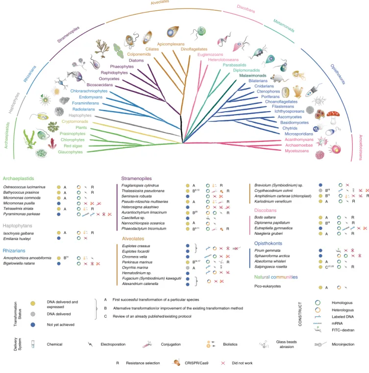
Overview of taxa in the EMS initiative. Taxa were selected from multiple eukaryotic supergroups1,7 to maximize the potential of
cel-lular biology and to evaluate the numerous unigenes with unknown functions found in marine protists (Fig. 1). Before the EMS initia-tive, reproducible transformation of marine protists was limited to only a few species such as Thalassiosira pseudonana, Phaeodactylum tricornutum and Ostreococcus tauri (Supplementary Table 1). The EMS initiative included 39 species, specifically, 6 archaeplastids, 2 haptophytes, 2 rhizarians, 9 stramenopiles, 12 alveolates, 4 disco-bans and 4 opisthokonts (Fig. 1). Most of these taxa were isolated from coastal habitats, the focus area of several culture collections7.
More than 50% of the selected species are considered photoauto-trophs, with another 35% divided between heterotrophic osmotro-phs and phagotroosmotro-phs, the remainder being predatory mixotroosmotro-phs. Almost 20% of the chosen species are symbionts and/or parasites of marine plants or animals, 5% are associated with detritus and several are responsible for harmful algal blooms (Supplementary Table 2).
While some transformation systems for protists have been devel-oped in the past8,9,11, the challenge for this initiative was to develop
genetic tools for species that not only require different cultivation conditions but are also phenotypically diverse. It should be noted that not all main lineages were explored. For example, amoebo-zoans did not feature in this aquatic-focused initiative, in part because they tend to be most important in soils, at least based on current knowledge, and manipulation systems exist for members of this eukaryotic supergroup, such as Dictyostelium discoideum12.
The overall EMS initiative outcomes are summarized in Fig. 1 and Table 1. We provide detailed protocols for 13 taxa, for which no transformation systems have been previously reported (category A) and eight taxa, for which existing protocols9,11,13–21 were advanced
(category B; Figs. 2, 3 and 4, Table 1, Supplementary Tables 1–5 and Methods). We also review an already published EMS transforma-tion protocol22 in one species (category C), and we discuss
unsuc-cessful transformation attempts for 17 additional taxa (Fig. 1 and Methods). Finally, we synthesize our findings in a roadmap for the development of transformation systems in protists (Fig. 5).
Archaeplastids. Prasinophytes are important marine green algae dis-tributed from polar to tropical regions23. They form a sister group to
Genetic tool development in marine protists:
emerging model organisms for experimental
cell biology
chlorophyte algae, and together, these two groups branch adjacent to land plants, collectively comprising the Viridiplantae, which are part of the Archaeplastida1,23 (Fig. 1). Genome sequences are available for
the picoprasinophytes (<3 µm cell diameter) tested herein, specifi-cally, Micromonas commoda, M. pusilla, Ostreococcus lucimarinus and Bathycoccus prasinos. As part of the EMS initiative, we report on genetic tools for Bathycoccus, a scaled, nonmotile genus, and Micromonas, a motile, naked genus with larger genomes than Bathycoccus and Ostreococcus22. We also report on genetic tools for Tetraselmis striata
and O. lucimarinus. The latter was transformed based on an adapted homologous recombination system for O. tauri24,25.
O. lucimarinus (RCC802) and B. prasinos (RCC4222) were trans-formed using protocols adapted from O. tauri24,25. Briefly, using
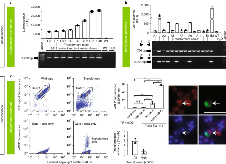
electroporation for transfer of exogenous genes, O. lucimarinus was transformed using a DNA fragment encoding the O. tauri high-affinity phosphate transporter (HAPT) gene fused to a luciferase gene and a kanamycin selection marker (Table 1 and Supplementary Table 3), which resulted in transient luciferase expression 24 h after electroporation (Table 1 and Fig. 3a). After 2 weeks of growth in low-melting agarose plates containing G418 (1 mg ml−1), 480 colo-nies were obtained, picked and grown in artificial seawater with the antibiotic neomycin. Of these, 76 displayed luminescence ≥2.5-fold above background (80 relative luminescence units (RLU)), with widely variable levels (200–31,020 RLU), likely reflecting either variations in the site of integration and/or the number of integrated genes (Fig. 3a, Supplementary Fig. 1 and Methods).
The O. tauri construct did not work in B. prasinos, while the use of the B. prasinos histone H4 and HAPT sequences in an oth-erwise identical construct and conditions was successful. Although luciferase expression was not detected 24 h after electroporation, 48 G418-resistant colonies were obtained 2 weeks later, 20 being luminescent when grown in liquid medium. Analysis of 14 resistant transformants revealed that the luciferase sequence was integrated into the genome of five luminescent clones, and one nonlumines-cent clone (Fig. 3b and Methods), suggesting that the chromatin context at integration sites in the latter was not favorable to lucif-erase expression.
Although transformation methods successful for Bathycoccus and Ostreococcus failed in Micromonas, Lonza nucleofection was success-ful with M. commoda (CCMP2709) (Table 1 and Fig. 3c) using two different codon-optimized plasmids, one encoding the luciferase gene (NanoLuc, Promega) flanked by an exogenous promoter and terminator sequence from the 5′ and 3′ untranslated regions (UTRs) of histone H3 in Micromonas polaris (CCMP2099), and the other encoding an enhanced green fluorescent protein (eGFP) gene flanked by endogenous promoter and terminator sequences from ribosomal protein S9 (Supplementary Table 5). Sensitivities to antibiotics were established (Supplementary Table 3). Constructs did not include a selectable marker, as we aimed to introduce and express foreign DNA while developing conditions suitable for transfection that supported robust growth in this cell wall-lacking protist (Table 1). Transformants revealed a significantly higher level of eGFP fluorescence than wild-type cells, with 1.3% of the population showing fluorescence per cell 45-fold higher than both the nontransformed portion of the culture and the wild-type cells (Fig. 3c and Methods). Additionally, the RLU was 1,500-fold higher than controls when using the luciferase-bear-ing construct, such that multiple experiments with both plasmids confirmed expression of exogenous genes in M. commoda.
T. striata (KAS-836) was transformed using microprojectile bom-bardment (Supplementary Fig. 2a). Two selectable marker genes were tested, consisting of a putative promoter and 5′ UTR sequences from the T. striata actin gene and either the coding sequences of the Streptoalloteichus hindustanus bleomycin gene (conferring resistance to zeocin) or the Streptomyces hygroscopicus bar gene (conferring resistance to glufosinate) (Table 1, Supplementary Fig. 2a and Methods). The terminator sequence was obtained from the
T. striata glyceraldehyde-3-phosphate dehydrogenase gene. Linearized plasmids were coated on gold particles and introduced into T. striata cells by using the PDS-1000/He Particle Delivery System (Bio-Rad). Transformants were successfully selected on half-strength f/2 at 50% salinity agar plates containing either 150 μg ml−1 zeocin or 150 μg ml−1 glufosinate.
Haptophytes (incertae sedis). Haptophytes are a group of photosyn-thetic protists that are abundant in marine environments and include the principal calcifying lineage, the coccolithophores. Genome sequences are available for Emiliania huxleyi6 and Chrysochromulina tobin26, and there is one report of nuclear transformation of a
calci-fying coccolithophore species27 but transformation of E. huxleyi, the
most prominent coccolithophore, has not been achieved yet27. Here,
as part of the EMS initiative, a stable nuclear transformation system was developed for Isochrysis galbana, a species that lacks coccoliths, but represents an important feedstock for shellfish aquaculture28.
I. galbana (CCMP1323) was transformed by biolistic bom-bardment with the pIgNAT vector, which contains nourseothricin (NTC) N-acetyltransferase (NAT), (for nourseothricin resistance) driven by the promoter and terminator of Hsp70 from E. huxleyi (CCMP1516). Twenty-four hours after bombardment, cells were transferred to liquid f/2 medium at 50% salinity containing 80 µg ml−1 NTC and left to grow for 2–3 weeks to select for trans-formants (Table 1). The presence of NAT in NTC-resistant cells was verified by PCR and PCR with reverse transcription (RT–PCR) (Fig. 4a, Supplementary Fig. 2b and Methods) and the sequence was verified. To confirm NTC resistance was a stable phenotype, cells were subcultured every 2–4 weeks at progressively higher NTC con-centrations (up to 150 µg ml−1) in the above-mentioned media. Cells remained resistant to NTC for approximately 6 months, as con-firmed by PCR screening to identify the presence of the NAT gene. Rhizarians. Rhizarians include diverse nonphotosynthetic protists, as well as the photosynthetic chlorarachniophytes that acquired a plastid via secondary endosymbiosis of a green alga4. Uniquely, they
represent an intermediate stage of the endosymbiotic process, since their plastids still harbor a relict nucleus (nucleomorph). Here, we report on an advanced transformation protocol for the chlorarach-niophyte Amorphochlora (Lotharella) amoebiformis for which low-efficiency transient transformation has previously been achieved using particle bombardment14.
A. amoebiformis (CCMP2058) cells were resuspended in 100 µl of Gene Pulse Electroporation Buffer (Bio-Rad) with 20–50 µg of the reporter plasmid encoding eGFP-RubisCO fusion protein under the control of the native rbcS1 promoter and subjected to electro-poration (Table 1). Cells were immediately transferred to fresh ESM medium and incubated for 24 h. Transformation efficiency was estimated by the fraction of cells expressing eGFP, resulting in 0.03–0.1% efficiency, as enumerated by microscopy, showing an efficiency up to 1,000-fold higher than in the previous study14
(Table 1). Stable transformants were generated by manual isolation using a micropipette, and a transformed line has maintained eGFP fluorescence for at least 10 months without antibiotic selection (Figs. 2 and 4b and Methods).
Stramenopiles. Stramenopiles are a diverse lineage harboring impor-tant photoautotrophic, mixotrophic (combining photosynthetic and phagotrophic nutrition) and heterotrophic taxa. As the most studied class in this lineage, diatoms (Bacillariophyceae) were early targets for the development of reverse genetics tool11,29. Diatoms are
estimated to contribute approximately 20% of annual carbon fixation30 and, like several other algal lineages, are used in
bioen-gineering applications and biofuels31. Although other cold-adapted
eukaryotes have, to our knowledge, yet to be transformed, here we present a protocol for the Antarctic diatom Fragilariopsis cylindrus32.
A transformation protocol has also been developed herein for Pseudo-nitzschia multiseries, a toxin-producing diatom33. Here
we also present work for nondiatom stramenopiles, including a transformation protocol for the eustigmatophyte Nannochloropsis oceanica, and an alternative protocol for the labyrinthulomycete Aurantiochytrium limacinum20, both of which are used for
bio-technological applications. Furthermore, we report on advances for CRISPR/Cas-driven gene knockouts in Phaeodactylum tricornutum8,13 and a more efficient bacterial conjugation system for Thalassiosira pseudonana13.
Microparticle bombardment was used on F. cylindrus (CCMP1102) that was grown, processed and maintained at 4 °C in 24 h light. Exponential phase cells were harvested onto a 1.2 µm membrane filter that was then placed on an 1.5% agar Aquil plate for bombardment with beads coated with a plasmid containing zeocin resistance and eGFP, both controlled by an endogenous fucoxanthin chlorophyll a/c binding protein (FCP) promoter and terminator (Table 1, Supplementary Table 3 and Methods)34. Transformation
was performed using 0.7 µm tungsten particles and the biolistic particle delivery system PDS-1000/He (Bio-Rad). Rupture disks for
Homologous Heterologous Labeled DNA mRNA FITC–dextran
Resistance selection CRISPR/Cas9
Electroporation Conjugation Microinjection
Chemical Biolistics Delivery System CO NSTRU CT Glass beads abrasion R Bodo saltans Diplonema papillatum Eutreptiella gymnastica Naegleria gruberi Pirum gemmata Sphaeroforma arctica Abeoforma whisleri Salpingoeca rosetta R R R R R R R R R R
Did not work
Fragilariopsis cylindrus Thalassiosira pseudonana Seminavis robusta Pseudo-nitzschia multiseries Heterosigma akashiwo Aurantiochytrium limacinum Caecitellus sp. Nannochloropsis oceanica Phaeodactylum tricornutum R R Breviolum (Symbiodinium) sp. Crypthecodinium cohnii Amphidinium carterae (chloroplast) Karlodinium veneficum Ostreococcus lucimarinus Bathycoccus prasinos Micromonas commoda Micromonas pusilla Tetraselmis striata Pyramimonas parkeae Pico-eukaryotes Amorphochlora amoebiformis Bigelowiella natans Isochrysis galbana Emiliania huxleyi AAAA AAAA R R R
DNA delivered and expressed DNA delivered Not yet achieved
Transformation Status AAAA Archaeplastids Discobans Haptophytans Rhizarians R Euplotes crassus Euplotes focardii Chromera velia Perkinsus marinus Oxyrrhis marina Hematodinium sp.
Fugacium (Symbiodinium) kawagutii Alexandrium catenella
AAAA
Alveolates
Opisthokonts
Stramenopiles
Natural communities
O O O CTGGTGAAA G TCTGGCA G TTCGAGCCCAATGCC ATCGG TAGTTTCTCAACGGGTT CTGGAGCTG CTGGT G AAAGT CTGG CAGTTCGAGCCCAATGCCAT TAGTTTCTCAACGGGTT CTGGAGCTG A GG G T C TTC ACAG TCA CTGGT G A A AGT CTGG CAGTTCGAGCCCAATGCCAT TAGTTTCTCAACGGGTT CTGGAGCTG A GG G T C TTC ACAG TCA A A A A A B14 A B9,13 A B15 B19 A A C21,22 A A A B18 B20 A B9,11 B16,17 A
A First successful transformation of a particular species
B Alternative transformation/or improvement of the existing transformation method C Review of an already published/existing protocol
Ciliates Chlorarachniophytes Poriferans Bilaterians Cnidarians Ctenophores Mycetozoans Plants Oomycetes Diplomonadids Malawimonads Acanthomyxans Chytrids Haptophytes Cryptomonads Apicomplexans Dinoflagellates Bicosoecidans Phaeophytes Raphidophytes Diatoms Foraminiferans Radiolarians Chlorophytes Prasinophytes Red algae Parabasalids Heteroloboseans Euglenozoans Microsporidians Basidiomycetes Ascomycetes Archaemoebae Glaucophytes Endomyxans Colponemids Choanoflagellates Ichthyosporeans Filastereans A rc ha epla stid s Rhiza rians Alveolates Discobans Metamon ads O pis thoko nts A m oe bo zo an s Stram enopiles Hap toph ytes
Fig. 1 | Phylogenetic relationships and transformation status of marine protists. A schematic view of the eukaryotic tree of life with effigies of main representatives. Color-coordinated species we have attempted to genetically modify are listed below. Current transformability status is schematized in circles indicating: DNA delivered and shown to be expressed (yellow, for details see text and Table 1); DNA delivered, but no expression seen (gray) and no successful transformation achieved despite efforts (blue). The details of transformation of species that belong to ‘DNA delivered’ and ‘Not achieved yet’ categories are described in Supplementary Table 5. mRNA, messenger RNA; FITC–dextran, fluorescein isothiocyanate (FITC)-conjugated dextran.
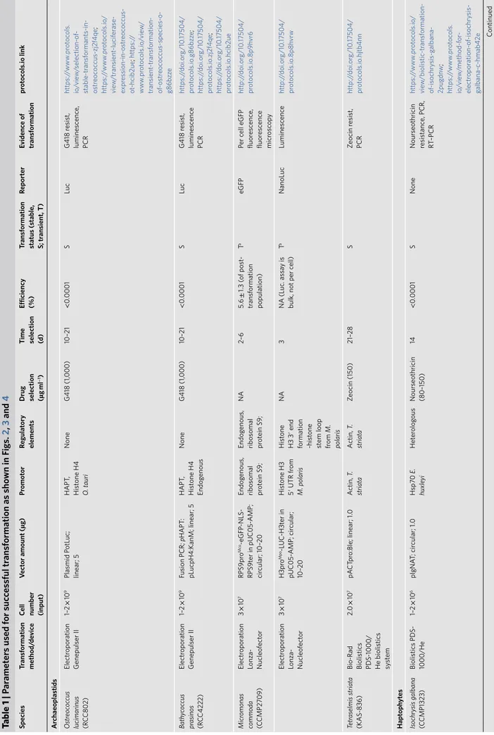
Table 1 | P ar amet er s used f or suc ces sful tr ansf ormation as sho wn in F igs. 2 , 3 and 4 species tr ansf ormation method/ de vic e
Cell number (input)
Vect or amount ( µg) Pr omot or Regulat ory elements d rug selection (µg ml − 1) time selection (d) efficiency (%) tr ansf ormation status ( stable, s; tr ansient, t) Repor ter evidenc e of tr ansf ormation pr ot oc ols.io link a rchaeoplas tids Ostr eoc oc cus lucimarinus (RCC80 2) Electr opor ation Genepulser II 1– 2 × 10 9 Plasmid P otLuc; linear; 5 HAPT , Hist one H4 O . t auri None G4 18 (1, 000) 10– 21 < 0.0 00 1 S Luc G4 18 r esist, luminesc enc e, PCR https:/ /www .pr ot oc ols. io /vie w /selection-of -stable-tr ansf ormants-in-ostr eoc oc cus-zj2f4qe ; https:/ /www .pr ot oc ols.io / vie w /tr ansient -lucif er ase-expr es sion-in-ostr eoc oc cus-ot -hcib2ue ; https:/ / www .pr ot oc ols.io /vie w / tr ansient -tr ansf ormation-of -ostr eoc oc cus-species-o-g86b zz e Bath yc oc cus pr asinos (R CC42 22) Electr opor ation Genepulser II 1– 2 × 10 9 Fusion PCR; pHAPT : pLucpH4:KanM; linear; 5 HAPT , Hist one H4 Endog enous None G4 18 (1, 000) 10– 21 < 0.0 00 1 S Luc G4 18 r esist, luminesc enc e PCR https:/ /doi. or g/10 .17 504/ pr ot oc ols.io .g86b zz e ; https:/ /doi. or g/10 .17 504/ pr ot oc ols.io .zj2f4qe ; https:/ /doi. or g/10 .17 504/ pr ot oc ols.io .hcib2ue Micr omonas commoda (CCMP2 709) Electr opor ation Lonz a-Nucleof ect or 3 × 10 7 RPS9pr o M co-eGFP -NLS-RPS9t er in pUC05-AMP; cir cular; 10– 20 Endog enous, ribosomal prot ein S9; Endog enous, ribosomal prot ein S9; NA 2– 6 5. 6 ± 1. 3 ( of post -tr ansf ormation population) T a eGFP Per c ell eGFP fluor esc enc e, fluor esc enc e micr osc op y http:/ /doi. or g/10 .17 504/ pr ot oc ols.io .8p9h vr6 Electr opor ation Lonz a-Nucleof ect or 3 × 10 7 H3pr o Mpo -L UC -H3t er in pUC05-AMP; cir cular; 10– 20 Hist one H3 5′ UTR fr om M. polaris Hist one H3 3 ′ end formation -hist one st em loop from M. polaris NA 3 NA (Luc. as sa y is
bulk, not per c
ell) T a NanoLuc Luminesc enc e http:/ /doi. or g/10 .17 504/ pr ot oc ols.io .8p8h vrw Tetr aselmis striat a (KAS-836) Bio-Rad Biolistics PD S-1000 / He biolistics sys te m 2. 0 × 10 7 pA CT pr o:Ble; linear; 1. 0 A ctin, T. striat a A ctin, T. striat a Zeocin (150) 21– 28 S Zeocin r esist, PCR http:/ /doi. or g/10 .17 504/ pr ot oc ols.io .hjtb4nn h apt oph yt es Isochry sis galbana (C CMP1323) Biolistics PD S-1000 /He 1– 2 × 10 6 pIgNA T; cir cular; 1. 0 Hsp7 0 E. huxle yi Het er olog ous Nour seothricin (80– 150) 14 < 0.0 00 1 S None Nour seothricin resistanc e, PCR, RT –PCR https:/ /www .pr ot oc ols.io / vie w /biolistic-tr ansf ormation-of -isochry sis-g albana-2pug dn w ; https:/ /www .pr ot oc ols. io /vie w /method-for -electr opor ation-of -isochry sis-galbana-c-hmab42e Continued
species tr ansf ormation method/ de vic e
Cell number (input)
Vect or amount ( µg) Pr omot or Regulat ory elements d rug selection (µg ml − 1) time selection (d) efficiency (%) tr ansf ormation status ( stable, s; tr ansient, t) Repor ter evidenc e of tr ansf ormation pr ot oc ols.io link Rhiz arians Amorphochlor a (L othar ella) amoebif ormis (C CMP2058) Electr opor ation Gene P ulser Xc ell 0.5– 1 × 10 7 GFP -Rubisc o; cir cular; 30–50 rbcS1, Endog enous rbcS1 Endog enous
Manual selection of fluor
esc ent cells a NA NA S/T GFP Fluor esc enc e, w est ern blot http:/ /doi. or g/10 .17 504/ pr ot oc ols.io .35hg q36 str amenopiles Fr agilariopsis cylindrus (CCMP110 2) Bio-Rad Biolistics PD S-1000 / He biolistics syste m 5 × 10 7 pUC:F CP:ShBle:F CP: eGFP; cir cular; 1. 0 FCP , Endog enous None Zeocin (100) 21–4 9 0. 0000 3 (30 c. f.u. per 10 8 cells ) S eGFP Zeocin r esist, fluor esc enc e, PCR, R T–PCR https:/ /doi. or g/10 .17 504/ pr ot oc ols.io .z39f8r6 Thalas siosir a pseudonana (CCMP1335) Bact erial conjug ation 4 × 10 7 TpSIl3p-eGFP in pT pP uc3; cir cular; NA Endog enous Endog enous Nour seothricin (100 in plat es, 200 in liquid cultur e) ~14 ~10 T eGFP Nour seothricin resistanc e, colon y PCR, fluor esc enc e http:/ /doi. or g/10 .17 504/ pr ot oc ols.io .nb zdap6 ; http:/ /doi. or g/10 .17 504/ pr ot oc ols.io .7ghhjt6 Pseudo-nitz schia multiseries Conjug ation 1 × 10 5 Pm_actP_eg fp_actT ; pP tPUC3 Pm actin; P t fcpB
None, other than contained in promot
er
/
term
Manual selection of fluor
esc ent cells in L G TA; zeocin (200) 24 h, 7 < 0.1 % T eGFP , shble Fluor esc enc e, vect or tar get ed PCR on gDNA http:/ /doi. or g/10 .17 504/ pr ot oc ols.io .4p zgvp6 Aur antioch ytrium limacinum (AT CC M yA -1381)
Bio-Rad Gene Pulser (165- 2076) NEP
A21 1 × 10 8 18GZ G 18GeZ G plasmid; linear; 1– 10 Endog enous GAPDH None Zeocin (100) 5–7 44 per μg of DNA S eGFP , shble Zeocin r esist., PCR, Southern, fluor esc enc e http:/ /doi. or g/10 .17 504/ pr ot oc ols.io .h3nb8me Nannochlor opsis oc eanic a (C CMP1779) Electr opor ation Genepulser II 1 × 10 9 pMOD , linear /cir cular; 0.1 –1 C MV None None 0.1 –1 20 (linear), 1– 2 (cir cular) T mT agBFP2 Fluor esc enc e, PCR, R T–PCR http:/ /doi. or g/10 .17 504/ pr ot oc ols.io .h3nb8me Phaeodact ylum tric ornutum (C CAP1055/1) Bact erial conjug ation 4 × 10 7 hCas9-2A -shble P tpBR episome 100 µl E. c oli OD 600 = 0 .9 FcpF-hCas9 psRNA – sgRNA
Cen6- Arsh4-His3 centr
omer e Phleom ycin (50) Zeocin (100) 10– 16 1.25 × 10 − 5 ≈ 500 c. f.u. S shble ( Cas9) yfp VENUS Phleo ycin resistanc e, PCR
maintained episome, PCR Cas9 tar
get sit e http:/ /doi. or g/10 .17 504/ pr ot oc ols.io .4bmgsk6 ; http:/ /doi. or g/10 .17 504/ pr ot oc ols.io .7gihjue ; http:/ /doi. or g/10 .17 504/ pr ot oc ols.io .7gnhjv e a lv eolat es Perkinsus marinus (ATC C PRA2 40) Electr opor ation LONZA -Nucleof ect or Glas s beads abr asion ( 425– 600 μm) 5– 7 × 10 7 pP mMOE-GFP; linear -cir cular (1:1); 5 Endog enous Endog enous FA CS Blasticidin (50– 200), pur om ycin (10-50), bleo (50- 200) Drug: 20–60 FACS: 3 0.0 1– 5 S GFP , mC herry Fluor esc enc e sequencing, PCR, w est ern blot https:/ /www .pr ot oc ols. io /vie w /o yst er -par asit e-perkinsus-marinus- transf ormation-u-gv9b w96 ; https:/ /www .pr ot oc ols.io / vie w /glas s-beads-based-tr ansf ormation-pr ot oc ol-for -perk -g36b yr e ; https:/ /www .pr ot oc ols.io / vie w /fluor esc enc e-activ at ed-cell-sor ting-facs-of -perkin-hh2b38e Continued Table 1 | P ar amet er s used f or suc ces sful tr ansf ormation as sho wn in F igs. 2 , 3 and 4 (c ontinued)
species tr ansf ormation method/ de vic e
Cell number (input)
Vect or amount ( µg) Pr omot or Regulat ory elements d rug selection (µg ml − 1) time selection (d) efficiency (%) tr ansf ormation status ( stable, s; tr ansient, t) Repor ter evidenc e of tr ansf ormation pr ot oc ols.io link Oxyrrhis marina (CCMP 17 88, CCMP 179 5) Electr opor ation Gene P ulser Xc ell; C hemical (CaC l2 ) 1–5 × 10 6; 1 × 10 5 Fluor esc ently labeled DNA (5– 25 µg ) or FIT C–de xtr an; mC herry NA, endog enous hsp90 NA, endog enous hsp90 NA NA 0.5–5% T mC herry Fluor esc enc e https:/ /www .pr ot oc ols.io / vie w /electr opor ation-of - oxyrrhis-marina-vcne2v e ; https:/ /www .pr ot oc ols. io /vie w /tr ansf ection-of -ale xa488-labelled-dna-int o-oxyrrhi-ha8b2h w ; https:/ /www .pr ot oc ols. io /vie w /electr opor ation-tr ansf ormation-of -fit c-de xtr an-int -3cmgiu6 ; https:/ /www .pr ot oc ols.io / vie w /c o-incubation-pr ot oc ol-for -tr ansf orming-het er otr op-hmzb4 76 Crypthec odinium cohnii (C CMP 316) Electr opor ation LONZA -Nucleof ect or Stained DNA (739 bp ); linear; 1 None None NA NA < 0.0 01 T Fluor esc enc e Fluor esc enc e https:/ /www .pr ot oc ols. io /vie w /tr ansf ection-of -crypthec odinium-c ohnii- using-label-z2 6f8he
Amphidinium carter
ae (chlor oplast) (C CMP1314) Bio-Rad Biolistics PD S-1000 /He biolistics s yst em 2.5 × 10 7 pAmpA tpBC hl; cir cular; 0. 5 Endog enous Endog enous Chlor amphenic ol (20) 3 on w ar d NA S Ab r es RT –PCR, PCR phenot ype http:/ /doi. or g/10 .17 504/ pr ot oc ols.io .4r2gv8e Karlodinium veneficum (CCMP19 75) Electr opor ation 4 × 10 5 linear -DinoIII-neo; linear; 2 Endog enous Endog enous Kanam ycin (150) 7 0. 0005 S (3 mon) NA RT –PCR, PCR https:/ /www .pr ot oc ols.io / vie w /nucleof ect or -pr ot oc ol-for -dinoflag ellat es-using-lo-qm8du9w d isc obans ( eugleno
zoans and het
er oloboseans ) Bodo salt ans (submitt ed t o AT CC ) Electr opor ation Nepa21 1– 1.5 × 10 7 Bs-EF1- α C -t erminal tagging; linear; 3–5 Endog enous Endog enous G4 18 (5) 7– 9 S GFP Fluor esc enc e, PCR, R T–PCR http:/ /doi. or g/10 .17 504/ pr ot oc ols.io .s5jeg4n ; http:/ /doi. or g/10 .17 504/ pr ot oc ols.io .7f chjiw Diplonema papillatum (A TC C 50 16 2) Electr opor ation Lonz a-Nucleof ect or 5 × 10 7 p5 7-V 5+ Neo R; linear; 3 Endog enous Endog enous G4 18 (7 5) 7– 14 ~5.5 S NA W est ern blot (r esistanc e mark er), R T–PCR http:/ /doi. or g/10 .17 504/ pr ot oc ols.io .4digs4e Naegleria gruberi (ATC C 30 22 4) Electr opor ation
Bio-Rad Gene Pulser xC
ell 5 × 10 6 pNAE G-H yG; cir cular; 4 Endog enous Endog enous Hy gr om ycin (300) Neo (700) 15– 28 80 T GFP W est ern blot (r esistanc e mark er), fluor esc enc e, http:/ /doi. or g/10 .17 504/ pr ot oc ols.io .hpub5n w ; http:/ /doi. or g/10 .17 504/ pr ot oc ols.io .7w4hpgw o pis thok onts Abeof orma whisleri (AT CC PRA -2 79) Electr opor ation Lonz a-Nucleof ect or 3 × 10 5 A whis_H2B venus + pUC19; cir cular; 1–5 + 40 carrier Endog enous Endog enous NA 10– 15 1 T Venus Fluor esc enc e, RT –PCR http:/ /doi. or g/10 .17 504/ pr ot oc ols.io .z ex f3fn Salpingoec a r osett a (A TC C PRA -390) Electr opor ation Lonz a-Nucleof ect or 4 × 10 5 SrA ctmC herry -C CTLL + pUC19; cir cular; 1– 10 + 40 carrier Endog enous Endog enous Pur om ycin (80) 10– 12 S mC herry Gene expr es sion (Luc, fluor esc enc e)/ resistanc e http:/ /doi. or g/10 .17 504/ pr ot oc ols.io .h68b9h w
For additional inf
ormation, see pr
ot
oc
ols.io link
s, Supplementary T
able 5 and Supplementary Not
e 1. F
or c
ontacting labor
at
ories w
orking with par
ticular species, see details giv
en in Supplementary T able 6. aMa y be stable but o ver gr own b y wild-t ype str
ain. NA, not applicable; NLS, nuclear localiz
ation signal. Table 1 | P ar amet er s used f or suc ces sful tr ansf ormation as sho wn in F igs. 2 , 3 and 4 (c ontinued)
used for Schizochytrium36. The highest transformation efficiency
was achieved using 1 µg of linearized 18GZG plasmid with two pulses, resulting in a time constant of ~5 ms (Table 1 and Methods). Expression of the fusion protein was confirmed by both the zeocin-resistance phenotype and the detection of eGFP (Fig. 2). Six 18GZG transformants derived from uncut and linearized plasmids were examined in detail. All maintained antibiotic resistance through-out 13 serial transfers, first in selective, then subsequently in non-selective media and then again in non-selective medium. Integration of the plasmid into the genome was confirmed by PCR as well as by Southern blots using a digoxigenin-labeled ShBle gene probe, show-ing that four transformants had integrations by sshow-ingle homologous recombination, while in two transformants additional copies of the antibiotic resistance cassette were integrated by nonhomologous recombination elsewhere in the genome (Supplementary Fig. 5).
Electroporation of N. oceanica (CCMP1779) was optimized based on observation of cells treated with fluorescein-conjugated 2,000 kDa dextran and subsequent survival (Table 1 and Methods). A sorbitol concentration of 800 mM and electroporation at between 5 and 9 kV cm−1 resulted in highest cell recovery. These conditions were used during introduction of plasmids containing the gene for the blue fluorescent reporter mTagBFP2 under the control of cytomegalovirus (CMV), the cauliflower mosaic virus 35S, or the VCP1 promoter previously described from Nannochloropsis sp.37.
Transient expression of blue fluorescence (compared to cells elec-troporated simultaneously under the same conditions without plas-mid) appeared within 2 h, lasted for at least 24 h and disappeared by 48 h in subsets of cells electroporated with mTagBFP2 under the control of CMV (Supplementary Fig. 6). The transient trans-formation was more effective when a linearized plasmid was used compared to a circular plasmid (Table 1). VCP1 did not induce blue fluorescence with a circular plasmid, while 35S gave inconsistent results with either circularized or linearized plasmids.
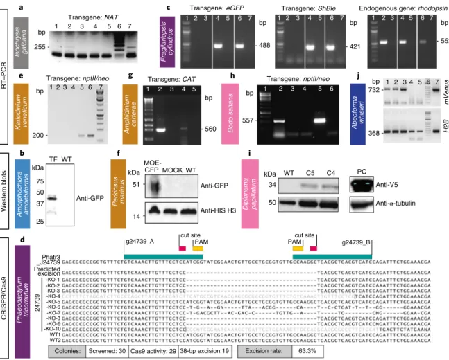
For P. tricornutum (CCAP1055/1), we adapted the CRISPR/Cas9 system8 for multiplexed targeted mutagenesis. Bacterial
conjuga-tion13 was used to deliver an episome that contained a Cas9 cassette
and two single-guide RNA (sgRNA) expression cassettes designed to excise a 38 basepair-long domain from the coding region of a nuclear-encoded, chloroplastic glutamate synthase (Phatr3_J24739) and introduce an in-frame stop codon after strand ligation (Table 1 and Methods). The GoldenGate assembly was used to clone two expres-sion cassettes carrying sgRNAs into a P. tricornutum episome that contained a Cas9–2A-ShBle expression cassette and the centromeric region CenArsHis (Supplementary Fig. 7). After their addition to a P. tricornutum culture, plates were incubated in a growth chamber under standard growth conditions for 2 d and transformed P. tricornu -tum colonies began to appear after 2 weeks. Only colonies maintain-ing Cas9–2A-ShBle sequence on the delivered episome were able to grow on selection plates because Cas9 and ShBle were transcription-ally fused by the 2A peptide38 (Supplementary Fig. 7). Gel
electropho-resis migration and sequencing of the genomic target loci confirmed the 38 bp-long excision and premature stop codon (Fig. 4d).
Alveolates. This species-rich and diverse group comprises cili-ates, apicomplexans and dinoflagellates (Fig. 1). As a link between apicomplexan parasites and dinoflagellate algae, perkinsids are key for understanding the evolution of parasitism, and also have potential biomedical applications17. Techniques currently exist
for transformation of only a small number of ciliates, perkinsids and apicomplexans39. Here, we present a transformation protocol
for Karlodinium veneficum (CCMP1975), a phagotrophic mixo-troph that produces fish-killing karlotoxins40. Experiments were
also performed on Oxyrrhis marina (CCMP 1788/CCMP 1795), a basal-branching phagotroph that lacks photosynthetic plastids and Crypthecodinium cohnii (CCMP 316), a heterotroph used in food supplements. For both of these taxa, evidence of DNA delivery 1,350 and 1,550 pounds per square inch (psi) gave the highest
col-ony numbers with efficiencies of 20.7 colcol-ony forming units (c.f.u.) per 108 cells and 30 c.f.u. per 108 cells, respectively. Following bom-bardment, the filter was turned upside down and left to recover for 24 h on the plate, then cells were rinsed from the plate/filter and spread across five 0.8% agar Aquil plates with 100 µg ml−1 zeocin. Colonies appeared 3–5 weeks later. PCR on genomic DNA showed that 100 and 60% of colonies screened positive for the bleomycin gene (ShBle) for zeocin resistance and the gene encoding eGFP, respectively. As confirmed by fluorescence-activated cell sorting (FACS) and microscopy, eGFP was localized to the cytosol and was distinguishable from plastid autofluorescence (Fig. 2). Additional confirmation by PCR and RT–PCR (Fig. 4c) revealed that the ShBle and eGFP genes were present in the genomes of transformants after multiple transfers (>10) 2 years later, indicating long-term stability.
Bacterial conjugation methods were improved in T. pseudonana (CCMP1335) using the silaffin precursor TpSil3p (Table 1 and Methods) as the target gene. TpSil3p was fused to eGFP flanked by an FCP promoter and terminator, cloned into a pTpPuc3 episomal backbone and transformed into mobilization plasmid-containing EPI300 E. coli cells (Lucigen). The donor cells were grown in super optimal broth with catabolite repression (SOC) medium at 37 °C until OD600 of 0.3–0.4, centrifuged and resuspended in 267 μl SOC medium. Next, 200 μl of donor cells were mixed with T. pseudonana cells, cocultured on predried 1% agar plates, dark incubated at 30 °C for 90 min, then at 18 °C in constant light for 4 h, followed by selection in 0.25% agar plates containing 100 µg ml−1 NTC. Colonies were observed after 2 weeks, inoculated into 300 μl L1 medium and supplemented with 200 µg ml−1 NTC to reduce the number of false positives. Positive transformants were identified by colony PCR screening (Supplementary Fig. 3) and epifluorescence microscopy (Fig. 2).
The diatom P. multiseries (15093C) and other members of this genus form buoyant linear chains with overlapping cell tips during active growth, and were unconducive to punctate colony forma-tion on agar, where their growth is generally poor. To address this challenge, a low-gelation-temperature agarose seawater medium (LGTA) was developed to facilitate growth, antibiotic selection and cell recovery. P. multiseries exhibited growth inhibition at rela-tively low concentrations under NTC, formaldehyde and zeocin (Supplementary Table 3). Biolistic transformation of two other P. species had been demonstrated at low efficiency35. To complement
this approach and explore potentially higher efficiency methods for transformation with diatom episomal plasmids, we modified the existing conjugation-based method13. The published
conjuga-tion protocol was modified to enhance P. multiseries postconjuga-tion viability by reducing SOC content. An episomal version of the Pm_actP_egfp_actT expression cassette was transfected into E. coli EPI300+pTAMOB and used for conjugation (Table 1 and Methods). After 48 h in L1 medium, cells were plated in LGTA and eGFP-pos-itive cells were observed 7 d later (Fig. 2). PCR revealed the pres-ence of plasmids in all eGFP-positive colonies (Supplementary Fig. 4). Similarly, conjugation with the episome pPtPUC3 (bleo-mycin selection marker)-containing bacterial donors was followed under zeocin selection (200 μg ml−1). After 7 d, only viable cells (based on bright chlorophyll fluorescence) contained the episome, as confirmed by PCR. Propagation of transformants after the first medium transfer (under selection) has so far been unsuccessful.
Stable transformation of A. limacinum (ATCC MYA-1381) was achieved by knock-in of a resistance cassette composed of ShBle driven by 1.3 kb promoter and 1.0 kb terminator regions of the endogenous glyceraldehyde-3-phosphate dehydrogenase gene carried in a pUC19-based plasmid (18GZG) along with the native 18S ribosomal RNA gene, and by knock-in of a similar construct con-taining a eGFP:ShBle fusion (Supplementary Fig. 5). Approximately 1 × 10⁸ cells were electroporated, adapting the electroporation protocol
was achieved (Table 1, Supplementary Results, Supplementary Fig. 15 and Methods), a goal recently achieved for C. cohnii using electroporation19. Additionally, we report on improved
transforma-tion systems for Perkinsus marinus (PRA240) and Amphidinium carterae (CCMP1314) chloroplast, published recently as part of the EMS initiative15.
K. veneficum (CCMP1975) was transformed based on electro-poration and cloning the selectable marker gene aminoglycoside 3′-phosphotransferase (nptII/neo; note that nptII/neo is used synon-ymously with amino 3′-glycosyl phosphotransferase gene conferring resistance to kanamycin, neomycin, paromomycin, ribostamycin, butirosin and gentamicin B) into the backbone of the dinoflagellate-specific expression vector DinoIII-neo41, which confers resistance
to neomycin and kanamycin (Table 1). In brief, DinoIII-neo was linearized and electroporated using the Nucleofector optimization pulse codes, buffer SF/Solution I (Lonza), and 2 μg μl−1 of linearized DinoIII-neo. Electroporated cells were selected under 150 μg ml−1 kanamycin 3 d postelectroporation. Fresh seawater with kanamy-cin was added every 2 weeks to the cultures and new subcultures were inoculated monthly. After 3 months, DNA and RNA were iso-lated from the resistant cultures as previously reported42 and cDNA
was synthesized using random hexamers. Out of 16 transforma-tions, two cell lines (CA-137, DS-138) showed stable growth under kanamycin selection. CA-137 developed dense cultures after 3 months, and the resistance gene was detected in both DNA and RNA by nested PCR and RT–PCR, respectively (Fig. 4e, Supplementary Fig. 8 and Methods).
We improved the transformation protocol16,17 of P. marinus, a
pathogen of marine mollusks, fish and amphibians43 (Supplementary
Table 5). We coexpressed two genes and efficiently selected tran-sient and stable transformants using FACS (Table 1, Figs. 2 and 4f, Supplementary Fig. 9 and Methods). In addition, we established the integration profile of ectopic DNA once introduced into the P. marinus genome. We did not see evidence of integration through homologous recombination and observed a propensity for plasmid fragmentation and integration within transposable elements sites. An optimized alternative protocol for transformation using glass bead abrasion was also developed. Two versions of the previously published Moe gene promoter16 were tested. Whereas the 1.0 kb
pro-moter version induced expression after 2 or 3 d, the truncated ver-sion (0.5 kb) took 7 d for expresver-sion to be detected. Resistance genes to zeocin, blasticidin and puromycin have all been shown to confer resistance to transformed P. marinus; however, selection regimes are still relatively slow and inefficient, indicating further room for improvement17.
We also report a vector for the transformation of the A. carterae chloroplast, a photosynthetic dinoflagellate. A. carterae, like other dinoflagellates with a peridinin-containing chloroplast, contains a fragmented chloroplast genome made up of multiple plasmid-like minicircles40. The previous transformation protocols made use
of this to introduce two vectors based on the psbA minicircle15.
Here, we show that other minicircles are also suitable for use as vec-tors. We created an artificial minicircle, using the atpB minicircle as a backbone, but replacing the atpB gene with a codon-optimized
Wild-type
Fluorescence
Bright field Bright field Bright field Bright field
Transformed cells Fluorescence
Wild-type Transformed cells
Fluorescence Fluorescence Amorphochlora amoebiformis Fragilariopsis cylindrus Thalassiosira pseudonana Perkinsus marinus Bodo saltans Naegleria gruberi Abeoforma whisleri Salpingoeca rosetta Pseudo -nitzschi a multiseries Aurantiochytrium limacinum
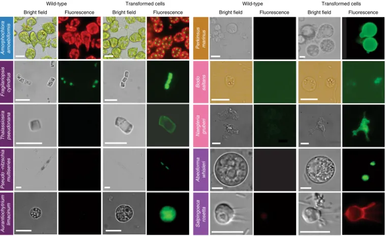
Fig. 2 | epifluorescence micrographs of transformed marine protists. Representative images of transformants and wild-type cell lines of ten selected protist species. Colored boxes behind species names refer to phylogenetic supergroup assignments given in Fig. 1. Representative data of at least two independent experiments are shown. The fluorescent images show the expression of individual fluorescent marker genes introduced via transformation for all organisms shown, except in the case of A. amoebiformis. For this, red depicts the natural autofluorescence of photosynthetic pigments in the cell, while the additional green spheres in the transformant fluorescence panel shows introduced GFP fluorescence (see Supplementary Fig. 15c for a trace of these different regions in the cell). Scale bars are as follows: 10 µm for A. amoebiformis, T. pseudonana, A. limacinum, B. saltans, N. gruberi, A. whisleri and S. rosetta; 15 µm for P. marinus; 20 µm for F. cylindrus and 100 µm for P. multiseries.
chloramphenicol acetyltransferase (Table 1 and Methods). This circular vector was introduced by biolistics to A. carterae (Supplementary Fig. 10a). Following selection with chloramphen-icol, we were able to detect transcription of the chloramphenicol acetyltransferase gene via RT–PCR (Fig. 4g). This result suggests that all 20 or so minicircles in the dinoflagellate chloroplast genome would be suitable for use as artificial minicircles, thus providing a large pool of potential vectors.
Discobans. This diverse group, recently split into Discoba and Metamonada44, includes heterotrophs, photoautotrophs and
preda-tory mixotrophs, as well as parasites. Discobans include parasitic kinetoplastids with clinical significance, such as Trypanosoma bru -cei, T. cruzi and Leishmania spp., for which efficient transformation protocols are available45. However, such protocols are missing for
aquatic species. Here, we describe available transformation pro-tocols for the kinetoplastid Bodo saltans and the heterolobosean Naegleria gruberi. The former was isolated from a lake, but iden-tical 18S rRNA gene sequences have been reported from marine environments46. The latter is a freshwater protist that represents a
model organism for closely related marine heterolobosean amoe-bas. Furthermore, we provide advanced methods that build on pre-vious EMS results18 for the diplonemid Diplonema papillatum.
B. saltans (ATCC 30904) was transformed with a plasmid con-taining a cassette designed to fuse an endogenous EF1-α gene with eGFP for C-terminal tagging. This cassette includes downstream of eGFP, a B. saltans tubulin intergenic region followed by the selectable marker nptII/neo gene, conferring resistance to neomycin. EF1-α genes exist in tandem repeats. The homologous regions that flank the cassette were chosen as targets for inducing homology-directed repair; however, they target only one copy of the gene. As transcription in B. saltans is polycistronic46, insertion of the tubulin
intergenic region into the plasmid is essential for polyadenylation of the EF1-α/GFP fusion and trans-splicing of the nptII/neo gene (Supplementary Table 5). Selection of transfected cells began with 2 µg ml−1 of neomycin added 24 h after electroporation, and this concentration was gradually increased over 2 weeks to 5 µg ml−1 (Table 1 and Methods). Cells were washed and subcultured into fresh selection medium every 4 d, and neomycin-resistant cells emerged 7–9 d postelectroporation. The eGFP signal was detected 2 d post-electroporation, albeit with low intensity. This may be due to the inefficient translation of eGFP since it has not been codon-optimized for B. saltans (Fig. 2). Genotyping analysis 9 months posttrans-fection confirmed the presence of the nptII/neo gene and at least partial plasmid sequence (Fig. 4h and Supplementary Fig. 10b). However, plasmid integration into the B. saltans genome through
c Micromonas commod a All High 0 2 4 6 8 10 Transformatio n efficiency (% cells ) eGFP fluorescence (520/35 nm ) 0 20 40 60 No puls e Transformed (eGFP) Pulse EW-113 ***P < 0.001 100 101 102 103 104 101 102 103 104 100 100 101 102 103 104 101 100 102 103 104 101 102 103 104 100 101 100 102 103 104 Chlorophyll fluorescenc e eGFP fluorescence Gate 1 Transformed
Gate 1 cells only Gate 1 cells only
Transformed cells Wild-type Gate 1 b Bathycoccus prasinos a Ostreococcus lucimarinus Transformant name
G418 resistant and luminescent clones WT
Ladde r Luminescence Luminescence (RLU) 3,299 bp Transformant name 1 2 3 4 Luminescence (RLU) Fluorescenc e
Forward angle light scatter (FALS)
756 bp 2,204 bp A1 A3 A5 A7 A9 A11 Ladde r H2O H2O All trans. High (eGFP) Non-trans. NS *** *** *** *** *** 0 500 2,000 2,500 5,000 G5 B7 G6-1 H2 C2 G6-2 B10 C10 WT 15,000 25,000 35,000 WT B2 B1 100 101 102 103 104 100 101 102 103 104
Fig. 3 | Various methods were used to demonstrate successful transformation in different archaeplastid species: luminescence and fluorescence. a–c, Luminescence (a,b) and fluorescence (by FACS and epifluorescence microscopy) (c) were used to verify expression of introduced constructs in three archaeplastids: O. lucimarinus (a), B. prasinos (b) and M. commoda (c). For the latter, red in the image depicts the natural autofluorescence of photosynthetic pigments in the cell, while green shows introduced eGFP fluorescence and blue shows the DAPI-stained nucleus; the overlay shows colocalization of eGFP and nucleus signals. See Supplementary Fig. 15d for a trace of these different regions in the cell. NS, not significant; trans., transformed. Representative data of at least two independent experiments are shown. For a detailed figure description see Supplementary Notes 2.
homologous recombination is still unconfirmed. This suggests either off-target plasmid integration or episomal maintenance.
For N. gruberi (ATCC 30224) two plasmids were designed. The first one carried the hygromycin B resistance gene (hph) with an actin promoter and terminator, along with an HA-tagged eGFP driven by the ubiquitin promoter and terminator. The second plas-mid carried the nptII/neo gene instead. For each individual circular plasmid, 4 μg was electroporated (Table 1 and Methods). About 48 h after electroporation, dead cells were removed from the suspension and viable cells were washed with PBS. Afterward, 300 μg ml−1 of hygromycin B or 700 μg ml−1 of neomycin was added to the fresh media. One to 4 weeks later, resistant clones were recovered and expression of eGFP and/or hygromycin was confirmed by western blotting (Supplementary Fig. 11). Expression of eGFP was observed by epifluorescence microscopy (Fig. 2 and Supplementary Fig. 11) with ~80% of transformants maintaining hygromycin B or neomy-cin resistance in addition to expressing eGFP.
D. papillatum (ATCC 50162) was transformed by electroporation using 3 μg of a SwaI-linearized fragment (cut from p57-V5+NeoR plasmid) containing the V5-tagged nptII/neo gene flanked by partial regulatory sequences derived from the hexokinase gene of the kinetoplastid Blastocrithidia (strain p57) (Table 1 and Methods) using a published protocol18. About 18 h after
electropor-ation, 75 μg ml−1 G418 was added to the medium and after 2 weeks, seven neomycin-resistant clones were recovered. Transcription of
nptII/neo was verified in four clones by RT–PCR (Supplementary Fig. 12) and the expression of the tagged nptII/neo protein was confirmed in two clones by western blotting using the α-V5 anti-body (Fig. 4i).
Opisthokonts. The opisthokont clade Holozoa includes animals and their closest unicellular relatives choanoflagellates, filastere-ans, ichthyosporeans and corallochytreans. The establishment of genetic tools in nonmetazoan holozoans promises to help illumi-nate the cellular and genetic foundations of animal multicellular-ity47. Genomic and transcriptomic data are available for multiple
representatives characterized by diverse cell morphologies, some of which can even form multicellular structures46. Until recently, only
transient transformations had been achieved for some opistokonts such as the filasterean Capsaspora owczarzaki48, the ichthyosporean Creolimax fragrantissima49 and the choanoflagellate Salpingoeca rosetta21. Through the EMS initiative, we report on evidence for
transient transformation of the ichthyosporean Abeoforma whisleri, isolated from the digestive tract of mussels, and review a recently published stable transformation protocol for S. rosetta achieved by using the selectable puromycin N-acetyl-transferase gene (Fig. 2)22.
All A. whisleri life stages are highly sensitive to a variety of meth-ods for transformation. However, we developed a 4D-nucleofection-based protocol using 16-well strips, wherein PBS-washed cells were resuspended in 20 μl of buffer P3 (Lonza) containing 40 μg of carrier
-h
Bodo saltans Abeoforma
whisleri Karlodinium veneficum e 3 4 # 5 " c Fragilariopsis cylindrus Isochrysis galbana a Amphidinium carterae RT–PCR d Phaeodactylum tricornutum
g24739_A cut sitePAM PAMcut site g24739_B
Colonies: Screened: 30 Cas9 activity: 29 38-bp excision:19 Excision rate: 63.3%
Transgene: eGFP Transgene: ShBle Endogenous gene: rhodopsin
bp - - 421 -bp bp 1 2 3 4 5 6 7 -255 200 PC WT Anti-α-tubulin C5 C4 34 i 50 Diplonem a papillatu m f Perkinsus marinus Amorphochlor a amoebiformi s b kDa 75 50 37 25 TF WT Anti-GFP Western blot s kDa 51 14 Anti-GFP Anti-HIS H3 Anti-V5 kDa MOE-GFP MOCK WT CRISPR/Cas9 7 6 5 4 3 2 1 1 2 3 4 5 6 7 1 2 3 4 5 6 7 -557 bp 1 2 3 4 5 6 j 1 2 3 4 5 6 7 bp 1 2 3 4 5 6 7 bp -bp Phatr3 _J24739 -KO-1 -KO-2 -KO-3 -KO-4 -KO-5 -KO-6 -KO-7 -KO-8 -KO-9 -KO-10 WT1 WT2 Predicted excision 24739 1 2 3 4 5 g - 560 bp Transgene: CAT mVenus H2B Transgene: NAT Transgene: nptII/neo Transgene: nptII/neo -732 368 488 553
Fig. 4 | Various methods were used to demonstrate successful transformation in different species: Rt–PCR, western blot and sequencing. a–j, Western blot, RT–PCR or sequencing (in case of Cas9-induced excision by CRISPR) were used to verify expression of introduced constructs in one haptophyte:
I. galbana (a), one rhizarian—A. amoebiformis (b), two stramenopiles—F. cylindrus (c) and P. tricornutum (d), three alveolates—K. veneficum (e), P. marinus
(f) and A. carterae (g), two discobans—B. saltans (h) and D. papillatum (i) and one opisthokont—A. whisleri (j). Note that nptII/neo is used synonymously with amino 3′-glycosyl phosphotransferase gene (aph(3′)) conferring resistance to kanamycin and neomycin. Representative data of at least two independent experiments are shown. For a detailed figure description see Supplementary Notes 2.
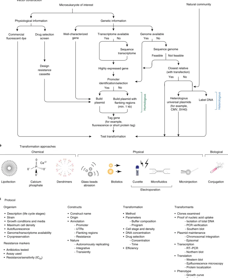
b a c Transcriptome available Well-characterized gene Drug selection screen Design resistance cassette Sequence transcriptome Genetic information Physiological information
Microeukaryote of interest Natural community
Genome available Commercial fluorescent dye No Yes Yes Yes No No Sequence genome Closest relative (with transfection) Promoter identification/selection Homologous Tag gene (for example, fluorescence or short protein tag)
Build plasmid
Build plasmid with flanking regions
(min. 1 kb) Highly expressed gene
Test transformation No Yes Not feasible Feasible Heterologous Heterologous universal plasmids (for example, CMV, SV40) Label DNA Organism
• Description (life cycle stages) • Strain
• Growth conditions and media • Maximum cell density • Autofluorescence • Genome/transcriptome availability • Cryopreservation Resistance markers • Antibiotics tested • Assay used • Resistance/sensitivity (IC50) Constructs • Construct name • Origin • Annotation - Promoter - UTRs - Flanking regions - Resistance • Nature - Autonomously replicating - Integrative - Transiently Transformants • Clones examined • Proof of nucleic acid uptake
- Isolation of total DNA - PCR verification - Southern blot • Plasmid maintenance - Chromosomal integration - Episomal • Transcription - RT–PCR - Northern blot • Translation - Western blot - Epifluorescence microscopy - Protein localization • Phenotype - Growth curve - Description Transformation • Method • Parameters - Buffer composition - Program • Cell stage and density • DNA concentration • Drug selection - Concentration - Time • Efficiency Transformation approaches Vector construction Protocol Physical Chemical Lipofection Calcium phosphate Ca+2
Dendrimers Glass beads
abrasion
Cuvette
Electroporation
Biolistics Microfluidics Conjugation
Biological P 0 0– 0– 0– Microinjection
Fig. 5 | ‘transformation roadmap’ for the creation of genetically tractable protists. a, Vector design and construction for microeukaryotes of interest and a natural community. b, Transformation approaches. Different symbols represent methods (for example chemical, physical or biological) for introducing DNA/RNA/protein into a living cell. c, Protocol. Key methodological steps for successful transformation are listed in an abbreviated form (for particular examples, see Table 1 and text).
plasmid (empty pUC19) and 1–5 μg of the reporter plasmid (A. whisleri H2B fused to mVenus fluorescent protein, mVFP) (Table 1 and Methods), and subjected to code EN-138 (Lonza). Immediately after the pulse, cells were recovered by adding 80 μl of marine broth (Gibco) before plating in 12-well culture plates previ-ously filled with 1 ml marine broth. After 24 h, ~1% of the culture was transformed based on the fraction of cells expressing mVFP in the nucleus (Figs. 2 and 4j).
Microbial eukaryotes in natural planktonic communities. Model organisms are typically selected based on criteria such as relative ease of isolation and asexual cultivation in the laboratory; however, these attributes may not correlate with the capacity for uptake and expression of the exogenous DNA. We explored whether natural marine planktonic pico- and nanoeukaryote communities would take up DNA in a culture-independent setting. Microbial plankton from natural seawater was concentrated and electroporated with plasmids containing mTagBFP2 under the control of CMV or 35S promoters (Supplementary Results and Methods). In most trials, blue fluorescent cells were rare if detected at all (compared to con-trol samples). However, in one natural community tested, a pho-tosynthetic picoeukaryote population exhibited up to 50% of cells with transient expression of blue fluorescence when the CMV pro-moter was used (Supplementary Fig. 13). This suggests it might be possible to selectively culture eukaryotic microorganisms based on capacity to express exogenous DNA.
discussion
The collaborative effort by the EMS initiative facilitated identifica-tion and optimizaidentifica-tion of the steps required to create new protist model systems, which culminated in the synthetic transformation roadmap (Fig. 5). Our genetic manipulation systems for aquatic (largely marine) protists will enable deeper insights into their cell biology, with potentially valuable outcomes for aquatic sciences, evolutionary studies, nanotechnology, biotechnology, medicine and pharmacology. Successes and failures with selectable markers, transformation conditions and reporters were qualitatively com-pared across species (Supplementary Tables 3 and 4, Table 1, Figs.
2–4 and Methods).
For some of the selected species, the first step was to identify cultivation conditions for robust growth in the laboratory to either generate high cell densities or large culture volumes for obtaining sufficient biomass required for a variety of molecular biology exper-iments. Unlike established microbial model species, cultivation of marine protists can be challenging, especially under axenic condi-tions or for predatory taxa that require cocultivation with their prey. Nevertheless, 13 out of 35 species were rendered axenic before the development of transformation protocols. For the remaining spe-cies, we were unable to remove bacteria and therefore had to make sure that transformation signals were coming from the targeted pro-tist rather than contaminants (Supplementary Table 2). Subsequent steps included the identification of suitable antibiotics and their corresponding selectable markers (Table 1 and Supplementary Table 3), conditions for introducing exogenous DNA (Table 1 and Supplementary Table 4) and selection of promoter and terminator sequences for designing transformation vectors (Table 1, Methods, Supplementary Table 5 and Supplementary Notes 1).
As exemplified in the model systems provided herein (Table 1
and Figs. 2–4), a variety of methods were used to test whether exog-enous DNA was integrated into the genome or maintained as a plas-mid, and whether the introduced genes were expressed. Approaches to show the former included inverse PCR, Southern blotting and whole genome sequencing, whereas approaches to demonstrate the latter included various combinations of PCR, RT–PCR, western blotting, epifluorescence microscopy, FACS, antibody-based meth-ods and/or growth assays in the presence of antibiotics to confirm
transcription and translation of introduced selection and reporter genes (for example, eGFP, YFP, mCherry). For fluorescent mark-ers, it was first ensured that the wild-type, or manipulated controls cells, had no signals conflicting with the marker (Figs. 2 and 3c), an important step because photosynthetic protists contain chlorophyll and other autofluorescent pigments. Overall transformation out-comes for each species were parsed into three groups according to the level of success or lack thereof (A, first transformation protocol for a given species; B, advanced protocol based on previous work and C, published protocol based on the EMS initiative) and are dis-cussed according to their phylogenetic position (Fig. 1).
Our studies did not result in a universally applicable protocol because transformability and a range of other key conditions var-ied greatly across taxa and approaches, such as intrinsic features of the genome and differences in cellular structure and morphol-ogy. In general, electroporation proved to be the most common method for introducing exogenous DNA stably into cells. This approach was used for naked cells and protoplasts, yet frequently also worked, albeit with lower efficiency, on cells protected by cell walls. Linearized plasmids were most effective for delivery, and 5′ and 3′ UTR-containing promotors of highly expressed endog-enous genes provided the strongest expression of selective reporters and markers. If successful, teams usually continued with fluores-cence-based methods. Furthermore, large amounts of carrier DNA usually facilitated successful initial transformations (for example, M. commoda, A. whisleri) or improved existing protocols (S. rosetta21).
We also provide the contact details of all coauthors who are assigned to particular species (Supplementary Table 6).
Some lineages were difficult to transform, especially dinofla-gellates and coccolithophores. Here, even if DNA appeared to be delivered (Supplementary Table 5), expression of the transformed genes could not be confirmed. Examples include the dinoflagel-lates C. cohnii, Symbiodinium microadriaticum and the coccolitho-phore E. huxleyi. Thus, at least these three species need concerted future efforts.
The combination of results presented herein together with pre-viously published protocols from the EMS initiative50 significantly
expands the segment of extant eukaryotic diversity amenable to reverse genetics approaches. Out of the 39 microbial eukaryotes selected for the initiative, exogenous DNA was delivered and expressed in more than 50% of them. The transformation systems enable us to shed light on the function of species-specific genes, which likely reflect key adaptations to specific niches in dynamic ocean habitats.
online content
Any methods, additional references, Nature Research reporting summaries, source data, extended data, supplementary informa-tion, acknowledgements, peer review information; details of author contributions and competing interests; and statements of data and code availability are available at https://doi.org/10.1038/s41592-020-0796-x.
Received: 26 June 2019; Accepted: 2 March 2020; Published online: 6 April 2020
References
1. Worden, A. Z. et al. Rethinking the marine carbon cycle: factoring in the multifarious lifestyles of microbes. Science 347, 1257594 (2015).
2. de Vargas, C. et al. Eukaryotic plankton diversity in the sunlit global ocean. Science 348, 1261605 (2015).
3. Collier, J. L. & Rest, J. S. Swimming, gliding, and rolling toward the mainstream: cell biology of marine protists. Mol. Biol. Cell. 30, 1245–1248 (2019).
4. Curtis, B. A. et al. Algal genomes reveal evolutionary mosaicism and the fate of nucleomorphs. Nature 492, 59–65 (2012).
5. Armbrust, E. V. et al. The genome of the diatom Thalassiosira pseudonana: ecology, evolution, and metabolism. Science 306, 79–86 (2004).
6. Read, B. A. et al. Pan genome of the phytoplankton Emiliania underpins its global distribution. Nature 499, 209–213 (2013).