Ecdysone-Based System for Controlled Inducible Expression
of Metabotropic Glutamate Receptor Subtypes 2, 5, and 8
PATRICK. M. DOWNEY, GIANLUCA LOZZA, ROBERTA PETRÒ, ENRICA DIODATO, CHIARA FOGLIA, FEDERICA BOTTAZZOLI, ROSSELLA BRUSA, TATIANA ASQUINI,
ANGELO REGGIANI, and MARIAGRAZIA GRILLI
Stable and inducible expression of human metabotropic glutamate receptor types 2, 5, and 8 was achieved in HEK293 cells using the ecdysone inducible system. Treatment of the respective cell lines with ponasterone A resulted in time- and concen-tration-dependent induction of receptor expression. In all cases, the functional activation of receptors was determined by mea-suring increases in intracellular calcium. The physiologically Gαi-coupled receptors mGluR2 and mGluR8 were successfully coupled to phospholipase C activation using the chimeric G protein Gαq/o. The pharmacological properties of recombinant re-ceptors were characterized and proved to be similar to native rere-ceptors. Our data suggest that the ecdysone system has a num-ber of characteristics that make it well suited for expressing mGluRs and that the combined use of this system and chimeric G proteins allows receptors to be characterized using a rapid and straightforward Ca2+
assay. (Journal of Biomolecular Screening 2005:841-848)
Key words: metabotropic glutamate receptors, inducible expression, chimeric G proteins
INTRODUCTION
G
LUTAMATE IS GENERALLY RECOGNIZEDas being the mostim-portant excitatory neurotransmitter in the mammalian CNS. It is intimately involved in a wide range of neuronal and glial pro-cesses, by the activation of both ionotropic and metabotropic (mGluR) receptor subtypes.1
These latter receptors are G protein– coupled receptors (GPCRs) and belong to a group of receptors that includes the Ca2+
sensing receptor, the GABABreceptors, and a
large number of putative olfactory, pheromone, and taste recep-tors.2
The mGlu receptors are widely distributed throughout the CNS, and there is now substantial evidence for an important role of mGluRs in normal brain functions as well as in neurological and psychiatric disorders.
The metabotropic glutamate receptors form a family of 8 sub-types that are subdivided into 3 groups based on their sequence homology, pharmacology, and transduction mechanisms. Group I receptors (mGluR1 and mGluR5) couple through Gαq/11proteins
to the activation of phospholipase C, resulting in phosphoinositide hydrolysis, the release of calcium from intracellular stores and the
activation of protein kinase C. Groups II (mGluR2 and mGluR3) and III (mGluR4, mGluR6, mGluR7, and mGluR8) are negatively coupled to adenyl cyclase (AC) through Gαi/Gαoproteins, thereby
inhibiting cyclic AMP (cAMP) formation and cAMP-dependent protein kinase activation.3
Group I receptors usually act postsynaptically to increase neuronal excitability, whereas the group II and III receptors most often act presynaptically to reduce neurotransmitter release, including glutamate.4
Excessive glutamatergic neurotransmission has been shown to underlie many CNS diseases and to play an important role in the pathophysiology of diseases such as depression, anxiety, and chronic pain, which represent key areas of interest for drug development. Accordingly, mGluR group I antagonists or group II/III agonists could represent useful agents for the treatment of these conditions. Indeed, group I antagonists have been shown to possess antidepressant activity in preclinical models5
and to be very effective agents in the treatment of prolonged and chronic pain.6-8
Studies using specific group II agonists suggest that such compounds may be useful in the treat-ment of inflammatory pain,9,10
chronic pain,11
and anxiety.12
Less is known about the role of group III mGluRs, principally due to the lack of potent and selective ligands. However, studies using knock-out mice show that animals lacking mGluR8 display a marked in-crease in anxiety-related behavior, possibly suggesting that mGluR8 agonists may also produce anxiolysis.13
In support of this notion, specific group III agonists have recently been shown to produce anxiolytic and antidepressant-like effects.14
The metabotropic glutamate receptors thus represent a potential
Schering-Plough Research Institute, Neurobiology Research, San Raffaele Sci-ence Park, Milan, Italy.
Received Mar 21, 2005, and in revised form Jun 20, 2005. Accepted for publica-tion Jul 7, 2005.
Journal of Biomolecular Screening 10(8); 2005 DOI: 10.1177/1087057105280285
goldmine of drug targets for the treatment of many CNS disorders, and their involvement in depression, anxiety and chronic pain makes them particularly appealing targets.
An important prerequisite in the search for selective mGluR modulators is the development of specific clonal cell lines for each receptor type. This has become even more important since the dis-covery of allosteric modulatory sites located in the 7 TM region of mGluRs. These sites are less conserved among groups and sub-types and thus make it possible to identify new chemical entities with improved selectivity within the family of metabotropic gluta-mate receptors. Functional cell-based assays for single mGluRs permit chemical libraries to be screened in the search for potent agonists and antagonists as well as the more selective allosteric modulators.
One of the problems in isolating cell lines that express gluta-mate receptors is the fact that glutagluta-mate is released from cells into the culture medium and hence causes desensitization and/or down-regulation of the receptor.15
To circumvent this problem, we used an inducible system, the ecdysone system. This system is based on the ability of ecdysone, an insect steroid, to potently induce tran-scription via the activation of a heterodimeric receptor composed of the ecdysone receptor (EcR) and the ultraspiracle protein (USP).16
Responsiveness to ecdysone in mammalian cells is recre-ated by transfection of EcR, the retinoid X receptor (RxR; the mammalian homologue of USP), an ecdysone-responsive re-porter, and application of ecdysone or a synthetic analogue such as ponasterone A. Expression of a given cDNA can be placed under the control of the EcR/RxR heterodimer, which activates transcrip-tion in the presence of the exogenous inducing agent. Our results suggest that this system is very well suited to the development of functional mGluR receptor assays and, in combination with the use of chimeric Gαq/oproteins, allows the use of a single assay
platform to analyze all receptor types.
MATERIALS AND METHODS Molecular biology
cDNA was synthesized from human hippocampal mRNA (Clontech, Mountain View, CA) using the superscript kit accord-ing to the manufacturer’s (Invitrogen, Paisley, UK) instructions. The coding sequences of mGluR2 and mGluR5b were amplified from hippocampal cDNA by PCR using a proofreading enzyme PfuTurbo (Stratagene, La Jolla, CA). A plasmid
pCMV6-XL5-mGluR8 encoding the human pCMV6-XL5-mGluR8a cDNA was acquired from Origene (Rockville, MD), and the hmGluR8a coding sequence was amplified from this plasmid by PCR. All amplified products were completely sequenced on both DNA strands to make sure that sequence errors had not been introduced in the amplification pro-cess. The mGluR coding sequences were subcloned into the ex-pression vector pIND (Invitrogen) using appropriate restriction enzymes. A chimeric G protein Gαq/oconstructed by replacing the
last 5 amino acids of the Gαqalpha subunit with those of the Gαo
protein as previously described17
was a kind gift of Dr. F. Monsma (Schering-Plough Research Institute, Kenilworth, NJ). The chime-ric Gαq/ocoding sequence was subcloned into pCDNA3-Hygro
us-ing appropriate restriction enzymes. A pIND vector expressus-ing mGluR8a and the Gαq/oas a single transcript was constructed by
inserting an IRES sequence between mGluR8a and Gαq/o. The
pIRESneo2 vector plasmid (Clontech) was digested with XhoI and
SmaI to remove the Neor
coding sequence and polyA sequence; they were replaced by Gαq/ocDNA, obtained from a
pCDNA3.1-Gαq/oplasmid through XhoI-SmaI cleavage. Then, the IRES-Gαq/o
cassette was subcloned into a pIND-mGluR8a plasmid as a NotI fragment, just downstream from the receptor cDNA previously cloned into pIND as a HindIII-NotI fragment. The mGluR5b and mGluR8a sequences were identical to those previously re-ported,18,19
whereas the amplified mGluR2 cDNA was identical to the variant reported by Kowal and others.20
Isolation of clonal cell lines
The HEK-293EcR cell line (Invitrogen), stably expressing the heterodimeric ecdysone receptor from the pVgRXR plasmid, was maintained in DMEM high-glucose medium, supplemented with 10% fetal bovine serum (FBS), 10,000 U/mL penicillin, 10 mg/ mL streptomycin, 2 mM GlutaMAX™ (Invitrogen), and 400µg/ mL Zeocin™ (Invitrogen) at 37° C, in an atmosphere contain-ing 5% CO2. Stable transfections with mGluRs and chimeric G
protein–coding vectors were performed using calcium phosphate according to standard protocols. Stably transfected clones were obtained after selection with 550µg/mL Geneticin®
(Invitrogen)
(mGluR5b and mGluR8a-Gαq/o cell lines) and 550 µg/mL
Geneticin®
(Invitrogen) and 200µg/mL hygromycin B (mGluR2-Gαq/o cell line). All cell culture media and reagents were from
Invitrogen (Paisley, UK). The sensitivity to DMSO was tested for all cell lines; no signs of toxicity were observed when DMSO was added up to a final concentration of 1%.
Western blotting
Cells were plated in 6-well dishes in complete DMEM and in-duced by adding various amounts of a 1 mM stock of ponasterone A (Invitrogen). Ponasterone A was dissolved in 100% ethanol, and ethanol levels were compensated in all induced cultures. After a 24-h induction period, cells were washed with ice-cold phosphate-buffered saline, detached from the dishes using a scraper, and re-suspended in lysis buffer (NaCl 150 mM, Tris-HCl 50 mM, Triton 1%, NP40 1%, SDS 0.2%, EDTA 2 mM, PMSF 1 mM, NaF 50 mM, and protease inhibitors). Samples were collected on ice and centrifuged at 10,000g for 15 min at 4° C and were subsequently stored at –80° C. Immunoblotting was performed using a poly-clonal anti-mGluR5 antibody (Upstate, Waltham, MA) 0.5µg/mL and a monoclonal anti-GAPDH antibody (Chemicon, Temecula, CA) 1µg/mL, following standard procedures.
Measurement of intracellular calcium transients
Cells from clonal cell lines were seeded into black, clear-bottom, 96-well plates at a density of 60,000 cells/well, in DMEM high glucose supplemented with 5% dialysed FBS, 3 U/mL glutamic-pyruvic transaminase (Sigma, St. Louis, MO), 5 mM so-dium pyruvate, 5µM ponasterone A. Following 24-h incubation, the cells were loaded with a fluorescent calcium indicator, either Calcium 3®
or FLIPR®
calcium assay kit, both supplied by Molec-ular Devices (Sunnyvale, CA). The dyes were dissolved in the as-say buffer, which consisted of Hanks’ balanced salt solution (HBSS; Gibco Life Technologies, Carlsbad, CA) buffered with 20 mM Hepes solution (Sigma). To avoid dye bleaching, 2.5 mM probenecid (Sigma) was added to the calcium indicator solution. Cells were loaded with the dye for 2 h at 37° C in a CO2incubator.
A fluorometric imaging plate reader (FLIPR 384; Molecular De-vices) was used to measure intracellular calcium by increases in fluorescence upon agonist stimulation following 30-s baseline measurement or 10 min antagonist administration.
Compounds
Agonists. (1S,3R-ACPD),
1-aminocyclopentane-1S,3R-dicar-boxylic acid; (2R,4R-APDC), (2R,4R)-4-aminopyrrolidine-2,4-dicarboxylic acid; (L-AP4), L(+)-2-amino-4-phosphono butyric acid; (L-CCG 1), (2S,3S,4S)-α-(carboxycyclopropyl) glycine; (CHPG), (RS)-2-Chloro-5-hydroxyphenylglycine; (DCG-IV), (2S2′R,3′R)-2-(2′3′-dicarboxycyclopropyl) glycine; (DCPG), (S)-3-4-Dicarboxyphenylglycine; (DHPG), 3,5-dihydro-phenylglycine; L-Glutamate; (NAAG), N-acetylaspartyl-glutamate; (PPG), (R,S)-4-phosphonophenylglycine; quisqualic acid.
Antagonists. (LY341495),
(2S)-2-Amino-2-[(1S,2S)-2-carboxycycloprop-1-yl]-3-(xanth-9-yl) propanoic acid; (MPEP), Methylphenylethynylpyridine; (MTEP) 3-[(2-methyl-1,3-thiazol-4-yl)ethyl]pyridine.
All agonists and antagonists were purchased from Tocris
Cookson (Bristol, UK). Compounds were dissolved in HBSS or DMSO depending on their solubility. The final concentration of DMSO in all samples was compensated.
Data analysis
EC50/IC50values were determined by nonlinear regression
anal-ysis using the software Prism 4.0 (Graphpad, San Diego, CA). Data were fitted as sigmoidal concentration-response curves and analyzed by a 4-parameter logistic equation. The Z′ factor,21
a di-rect measure of assay quality, was measured using the following equation: ′ = − + − + + − − Z c c c c 1 (3σ 3σ ) µ µ ,
whereσc+andσc–are the standard deviations of positive and
nega-tive control samples, respecnega-tively, andµc+andµc–are the means of
positive and negative controls, respectively. We tested the robust-ness of our mGluR5 assay in its ability to demonstrate both agonism and antagonism. Calculating the Z′ in the agonism assay, the positive control was vehicle + quisqualate (100 nM), whereas the negative control was vehicle only. We used 48 wells of a 96-well plate for the positive and negative controls, respectively, with 3 replicate plates per day, and we performed the assay on 3 consec-utive days. In determining the Z′ for the antagonism assay, we used a similar protocol but used vehicle + quisqualate (100 nM) as the positive control and vehicle + MPEP (50 nM) + quisqualate (100 nM) as the negative control; again, we used 3 half-plates for posi-tive and negaposi-tive controls, respecposi-tively, with 3 replicate plates per day, and we performed the assay over 3 consecutive days. The final Z′ for the assay is the average of the antagonism and agonism determinations.
RESULTS
The ecdysone system was used to establish heterologous induc-ible expression of representative mGlu receptors from all 3 groups, mGluR5b (group I), mGluR2 (group II), and mGluR8a (group III). In this system, transgene expression is under the control of a mini-mal heat shock promoter, which contains 5 copies of the ecdysone/ glucocorticoid responsive element (5X E/GRE).16The level of
transcription of the gene of interest can be simply modulated by the addition of the inducer ponasterone A, a synthetic analogue of ecdysone. Optimal 293EcRclonal cell lines expressing each mGlu
receptor were obtained and pharmacologically and biochemically characterized.
mGluR5 inducible cell line
mGluR5b is a Gαq-coupled receptor; its activation thus leads to
an increase in intracellular calcium, which can be measured using a FLIPR. Clonal cell lines expressing mGluR5 were initially prescreened for the stimulation of calcium mobilization in re-sponse to quisqualate following 24-h induction with 5 µM ponasterone A. A single clonal cell line referred to as 2A4 was cho-sen for further study. To determine how receptor density can be modulated in this system, cells were induced for 24 h with increas-ing concentrations (1, 3, and 5µM) of ponasterone A, and mGluR5 protein immunoreactivity was determined by Western blotting. The mGluR5 protein could not be detected in uninduced cells, indicating near total absence of transcription, but it is visible after incubation with 1µM ponasterone A, and the amount of immuno-reactive signal progressively increases with increasing concentra-tions of inducer (see Fig. 1, inset). The functional coupling of the receptor was tested by measuring quisqualate-induced Cai
2+
mobi-lization after ponasterone A induction. As shown in Figure 1, quisqualate (0.001-3µM) elicited a concentration-dependent in-crease in Cai
2+
, with maximal changes in relative fluorescence units (RFUs) of 8100± 210, 10,200 ± 47, and 11,700 ± 308 (n = 3)
after 1, 3, and 5µM ponasterone, respectively. Corresponding ago-nist EC50(log M) values of –7.38 at 1µM, –7.40 at 3 µM, and –7.51
at 5µM inducer concentration were obtained. In the absence of in-duction by ponasterone A, no significant functional response was detected following quisqualate stimulation, indicating tight regu-lation of recombinant receptor expression. A small amount of sig-nal was noted, however, at higher quisqualate concentrations; we believe that this is probably due to a low level of unregulated tran-scription as no signal was observed in our nontransfected cell lines when challenged with equivalent concentrations of quisqualate.
A detailed characterization of the pharmacological properties of the hmGluR5b receptor was undertaken using reference agonists and antagonists. As shown in Figure 2a, all characterized agonists were highly efficacious, eliciting maximum responses comparable to those observed with the endogenous agonist L-glutamate. The rank order of agonist potency for examined com-pounds was quisqualate > L-glutamate > DHPG > 1S-3R-ACPD > CHPG, in agreement with that determined for the native rat recep-tor22
and the recombinant human receptor.23
EC50values were
cal-culated as reported in Table 1. To examine the effect of antagonists, cells were pretreated with the noncompetitive mGluR5 selective antagonists MPEP and MTEP and the competitive antagonist LY 341495. All antagonists produced a concentration-dependent inhi-bition of quisqualate-induced calcium mobilization, as shown in Figure 2b. The following rank order of potency was determined: MPEP > MTEP > LY341495; IC50values were calculated against
an EC80concentration of quisqualate and are reported in Table 1. Calcium-coupled group II and III inducible cell lines
To use intracellular calcium transients as an assay readout for group II and III receptors, we cotransfected hmGluR2 and hmGluR8a receptors with the chimeric G protein Gαq/o, which has
been shown to effectively couple with all Gαi/o-coupled mGlu
re-ceptors, at least in transient transfections.24,25
Because stable overexpression of the chimeric Gαq/o protein could potentially
cause alterations in normal cell physiology, we tried 2 different ex-perimental approaches. In our mGluR2 cell line, we constitutively expressed the chimeric G protein but kept the metabotropic recep-tor under the control of the inducible promoter, whereas with our mGluR8 cell line, both receptor and G protein were inducible. In the latter case, inducible expression of both the mGlu receptor and chimeric G protein was achieved by inserting an IRES sequence between the receptor and the G protein coding sequences, thus en-suring that they were expressed as a single transcript under the control of the inducible promoter.
mGluR2-inducible cell line
Following cotransfection of mGluR2 and Gαq/o, clonal cell
lines were prescreened for DCG-IV-induced Cai 2+
mobilization following induction with ponasterone A (5µM). A single clone, referred to as 2q/o, which revealed agonist-dependent Cai
2+
mobi-lization when induced with negligible background levels in the ab-sence of induction, was expanded and characterized further. Clone 2q/o gave very robust calcium signals, with maximal changes in RFUs in response to DCG-IV of up to 45,000 and a calculated EC50value of 130 nM. This cell line showed excellent
signal-to--9 -8 -7 -6 -5 0 2500 5000 7500 10000 12500 MGLUR5 GAPDH 0 1 3 5 MGLUR5 GAPDH 0 1 3 5 MGLUR5 GAPDH 0 1 3 5 Quisqualate (Log M) RF U
FIG. 1. Concentration response curves (mean± SEM, n = 3) for the ef-fect of quisqualate on calcium mobilization assay using 293EcRcells ex-pressing mGluR5b without induction (¢) and induced with 1µM (p), 3 µM (q), and 5µM (u) concentrations of ponasterone A. The inset shows Western blot analysis, displaying the extent of immunoreactive mGluR5b protein 24 h after induction with 0, 1, 3, and 5µM concentrations of ponasterone A.
Table 1. Functional Activities of Agonists and Antagonists Examined in 293EcRCell Lines 2q/o, 2A4, and 8q/o Expressing
mGluR2, mGluR5, and mGluR8, Respectively
EC50µM
Agonist mGluR2 mGluR5 mGluR8
L-glutamate 1.6 (1.1-2.4) 1.5 (1.1-2.2) 8.8 (5.8-11) Quisqualate >1000 0.03 (0.02-0.05) >1000 DHPG 4.2 (2.8-5.7) DCG-IV 0.13 (0.08-0.18) 1S,3R-ACPD 3.1 (2.2-4.2) 15.7 (11.6-21.2) 2R,4R-APDC 1.3 (0.9-1.8) L-CCG I 0.14 (0.08-0.2) 2.8 (1.6-4.9) L-AP4 1.6 (0.9-2.9) PPG 4.7 (2.7- 8.1) DCPG 0.8 (0.4-1.6) NAAG >1000 IC50µM at IC50µM at IC50µM at
Antagonist 300nM DCG-IV 100 nM Quisqualate 14µM Glutamate
MPEP >100 0.02 (0.01-0.03) >100
MTEP 0.09 (0.06-0.14)
LY341495 0.0012 (0.0009-0.0015) 35.3 (22.4-48.2) 0.13 (0.08-0.16)
A description of the abbreviations used for all agonists and antagonists can be found in the Materials and Methods section under Compounds. Data are the mean EC50values of agonists and IC50values of antagonists determined from 3 independent experiments per-formed in quadruplicate. Values are given with corresponding 95% confidence limits. Each dose-response curve consisted of 8 different drug concentrations. All antagonist IC50 values were determined at agonist concentrations corresponding to the EC80of the spe-cific agonist, 300 nM DCG-IV for mGluR2, 100 nM quisqualate for mGluR5, and 14µM L-glutamate for mGluR8.
background ratio, as no significant DCG-IV-stimulated calcium responses were detected in uninduced cells, as shown in the inset in Figure 3. To show that the pharmacology of the receptor is unal-tered in this system where the receptor couples through an exoge-nous chimeric G protein, mGluR2-coupled calcium signaling was measured in response to a number of reference agonists for metabotropic glutamate receptors. As shown in Figure 3, the group II selective agonist DCG-IV as well as all the group II preferred agonists including L-CCG-I, 2R,4R-APDC, and 1S,3R-APDC were highly efficacious, eliciting maximum responses comparable to those observed with the endogenous agonist L-glutamate. In contrast, the addition of the group I agonist quisqualate or the
mGluR3-specific agonist NAAG did not elicit an increase in intracellular calcium. The rank order of agonist potency in the re-combinant cell line expressing hmGluR2 and the chimeric Gαq/o
protein was DCG-IV ~ L-CCG-I > L-glutamate ~ 2R,4R-APDC > 1S,3R-APDC. Calculated EC50values for each agonist at the
re-combinant hmGluR2 receptor are summarized in Table 1. These data are in agreement with previous work undertaken on the native rat receptor26
as well as the physiologically coupled recombinant hmGluR2 where cAMP or GTPγS assays were used.20,27
We also tested the reference compound LY341495, which acts as an antag-onist at all mGluRs but has the highest affinity for the group II re-ceptors. The compound tested in the presence of an EC80
concen-tration of DCG-IV resulted in a concenconcen-tration-dependent inhibition of agonist-induced Cai
2+
mobilization, as shown in Figure 4, giving an estimated IC50of 1.2 nM.
mGluR8-inducible cell line
Following transfection of the 293EcRcell line with the
induc-ible mGluR8a-Gq/oconstruct, clonal cell lines were isolated and
characterized for L-glutamate-dependent Cai
2+mobilization after
induction with 5µM ponasterone A. A single clone referred to as 8q/o was chosen for further studies. When induced, clone 8q/o gave robust concentration-dependent intracellular calcium in-creases after being stimulated with L-glutamate and undetectable agonist-induced signal in the absence of induction (Fig. 5, inset). A detailed pharmacological analysis was performed on the recombi-nant cell line using a number of well-characterized agonists, and the rank order of potency was determined as DCGP > AP4 > L-CCG-I > PPG>L-glutamate, as depicted in a representative
experi-FIG. 2. (a) Effect of various agonists on Ca2+mobilization in cell line 2A4 expressing mGluR5b. Agonists are quisqualate (¢), L-glutamate (p), DHPG (u), 1S,3R-ACPD (q) and CHPG (). Values are means± SEM of quadruplicate determinations from a typical experiment. (b) Antago-nism by MPEP (¢), MTEP (p), and LY341495 (q) of the agonist-stimu-lated calcium mobilization in the mGluR5b expressing cell line 2A4. Cells were pretreated for 10 min with the various antagonists over the indi-cated concentration ranges before a challenge with an EC80(100 nM)
con-centration of quisqualate. Inhibition curves were constructed from the per-centage responses pooled from 3 independent experiments performed in quadruplicate. Values are means± SEM.
-10 -9 -8 -7 -6 -5 -4 -3 -2 0 2000 4000 6000 8000 10000 Agonist (Log M) RFU -10 -9 -8 -7 -6 -5 -4 0 25 50 75 100 MPEP MTEP LY341495 Antagonist (Log M) % Inhibition -10 -9 -8 -7 -6 -5 -4 -3 0 10000 20000 30000 40000 50000 Agonist (Log M) RFU -9 -8 -7 -6 -5 0 10000 20000 30000 40000 50000 No Ponasterone A 5µM Ponasterone A (DCG-IV) RFU -10 -9 -8 -7 -6 -5 -4 -3 0 10000 20000 30000 40000 50000 Agonist (Log M) RFU -9 -8 -7 -6 -5 0 10000 20000 30000 40000 50000 No Ponasterone A 5µM Ponasterone A (DCG-IV) RFU
FIG. 3. Effect of various agonists on Ca2+
mobilization in cell line 2q/o expressing mGluR2 and the chimeric G protein Gαq/o. Agonists arer
DCG-IV (group II selective),sL-CCGI (group II preferred),p L-gluta-mate,2R,4R-APDC (group II preferred),q1S,3R-ACPD (group II pre-ferred),oNAAG (mGluR3 selective), and¢quisqualate (group I selec-tive). Values are means± SEM of quadruplicate determinations from a typical experiment. Inset concentration-response curves (mean± SEM,
n = 3) for the effect of DCG-IV on calcium mobilization using cell line 2q/
o expressing mGluR2 and the chimeric G protein Gαq/o, pretreated for 24 h
in the absence (¢or presencerof ponasterone A (5µM).
a
ment in Figure 5. These results are in agreement with data obtained from hmGluR8a-expressing cell lines using cAMP assays19,28
or radioligand binding assays.29
Agonist EC50 values for our
hmGluR8a-expressing cell line were calculated and are summa-rized in Table 1. L-glutamate-induced Cai
2+
release was tested in the presence of the competitive antagonist LY341495. When an agonist concentration corresponding to an EC80value was used to
stimulate the receptor, addition of the reference antagonist resulted in a concentration-dependent inhibition of glutamate-elicited cal-cium signal with a calculated IC50 of 130 nM. As expected,
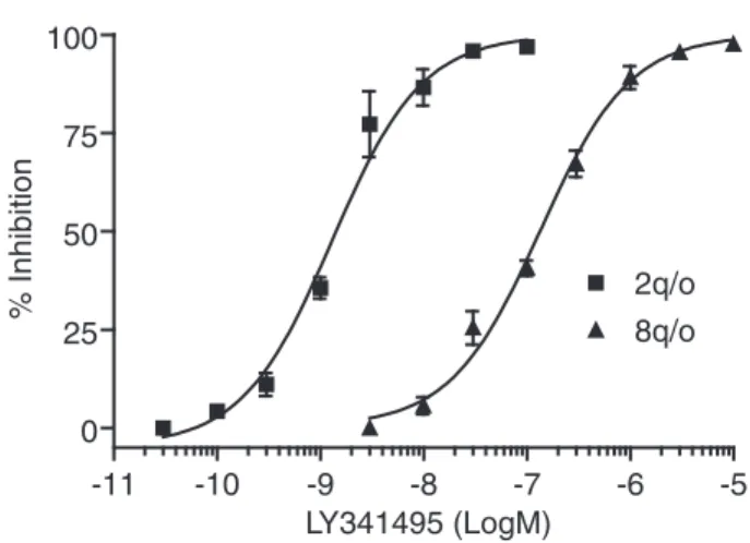
LY341495 was more potent on hmGluR2 than hmGluR8 (Fig. 4). Inducible cell line stability and assay reproducibility
To test the functional stability of the mGluR-expressing induc-ible cell lines, the biological response and the pharmacological properties of the recombinant receptors were tested at increasing passage number. We tested cell lines up to 30 passages, and no sig-nificant changes in the efficacy or potency of agonists to induce Cai
2+mobilization after ponasterone A induction was observed. In
addition, no significant response to agonists in uninduced cells was shown with increasing passage number. To determine the robust-ness of the assay, we determined the Z′ for the mGluR5 cell line 2A4; this was calculated as 0.7, showing that the assay is very re-producible. The ecdysone-inducible system would thus appear to be sufficiently robust to allow it to be used in screening assays in which very high reproducibility is essential.
DISCUSSION
Pharmacologically active compounds that are able to modulate the activity of a specific metabotropic glutamate receptor subtype
may be effective in the treatment of a wide range of CNS diseases. However, it has been traditionally very difficult to find small mole-cules acting at the agonist-binding site that achieve true subtype se-lectivity. The generation of high-throughput screening has made it possible to search for compounds that interact with allosteric sites rather than the historically targeted orthosteric site. The first such compounds described for the mGluRs were MPEP and CPCCOet, which are negative allosteric modulators selective for mGluR5 and mGluR1, respectively.30,31Recently, positive allosteric modulators
selective for mGluR1, mGluR2, mGluR4, and mGluR5 have also been described.32-35
The availability of functional cell-based assays for all members of the mGluR family makes it possible to screen chemical libraries in the search for potent agonists, antagonists, and allosteric modulators. Equally important, clonal cell lines can be used to counterscreen active compounds to determine if their activities are indeed receptor specific. This is particularly impor-tant since the revelation that the noncompetitive mGluR5 antago-nist MPEP also acts as a positive modulator of mGluR436and the
discovery of PHCCC, a compound highly related to CPCCOet that predominantly acts as a positive modulator of mGluR4 but is also a noncompetitive inhibitor of mGluR1.34
We have used the ecdysone system to functionally express the Gαqcoupled mGluR5 and in combination with the chimeric Gαq/o
protein to functionally express mGluR2 and mGluR8. In all of our cell-based assays, the receptors displayed a pharmacological pro-file for both agonists and antagonists that faithfully represented that of the native receptors. Our results show that the ecdysone sys-tem allows expression to be tightly controlled, producing barely detectable background levels and high signal-to-background ra-tios. It also allowed us to develop assays that show long-term
sta--11 -10 -9 -8 -7 -6 -5 0 25 50 75 100 2q/o 8q/o LY341495 (LogM) % Inhibition
FIG. 4. Antagonism by LY341495 of the DCG-IV and L-glutamate stimulated calcium mobilization of cell lines 2q/o and 8q/o, respectively. Cells were pretreated for 10 min with LY341495 over the indicated con-centration ranges before a challenge with an EC80concentration of
ago-nist. Inhibition curves were constructed from the percentage responses pooled from 3 independent experiments performed in quadruplicate. Cal-culated IC50values are presented in Table 1.
-9 -8 -7 -6 -5 -4 -3 0 2500 5000 7500 10000 12500 Agonist (LogM) RFU -7 -6 -5 -4 -3 -2 0 2500 5000 7500 10000 12500 No Ponasterone A 5 µM ponasterone A Glutamate (LogM) RFU -9 -8 -7 -6 -5 -4 -3 0 2500 5000 7500 10000 12500 Agonist (LogM) RFU -7 -6 -5 -4 -3 -2 0 2500 5000 7500 10000 12500 No Ponasterone A 5 µM ponasterone A Glutamate (LogM) RFU
FIG. 5. Effect of various agonists on CAi
2+ mobilization in cell line
8q/o expressing mGluR8 and the chimeric G protein Gαq/o. Agonists are (S)-3,4-DCPG (¸; mGluR8 selective), + L-AP4 (+; group III selective), L-CCG-I (s; group II-III selective), RS-PPG (*; mGluR8 selective), L-glutamate (p), and quisqualate (¢; group I selective). Values are means± SEM of quadruplicate determinations from a typical experiment. (Inset) Concentration-response curves (mean± SEM, n = 3) for the effect of L-glutamate on calcium mobilization using cell line 8q/o expressing mGluR8 and the chimeric G protein Gαq/o, pretreated for 24 h in the
bility, showing both consistent EC50values and RFUs even at high
passage numbers. Assay reproducibility was also good, and our mGluR5 assay has a calculated Z′ value of 0.7, showing remark-able across-plate uniformity. The combined use of the ecdysone system and the chimeric G protein Gαq/onot only avoids receptor
desensitization and time- and passage-dependent receptor down-regulation but also allowed us to use the same fluorescent assay format for all mGlu receptors. Having a single assay format that can be used to evaluate all mGluRs should accelerate both screen-ing and counterscreenscreen-ing activities. Here, we present data from representative receptors from each group, but the same approach should be applicable to all mGlu receptors. To express hmGluR2 and hmGluR8, we employed 2 distinct strategies: In our 2q/o cell line, the chimeric G protein was constitutively expressed, whereas in our 8q/o cell line, the expression of the chimeric G protein was inducible. Both strategies were successful, suggesting that over-expression of a chimeric G protein is not particularly deleterious for the cell. However, as the Gαq/oprotein can couple to any Gαi/
Gαo-coupled cellular receptor, limiting its expression temporally
to that of its desired target should, at least in theory, reduce any potentially undesired effects.
To date, there are very few reports of the ecdysone system being used to express GPCRs. However, in 1 notable exception, different 5HT receptors were expressed using the Tet on and the ecdysone systems; although the authors reported higher overall expression levels with the Tet on system, the ecdysone system gave much better induction ratios with effectively undetectable background levels.37
Other groups have used the expression of reporter genes to compare different inducible systems, and their results also demon-strate that the ecdysone system produces very high induction ratios with negligible background expression.38,39
Our results not only show that the ecdysone system allows expression to be tightly controlled but also demonstrate that the system can produce very reproducible assays. We believe that the ecdysone system is an excellent system for functional receptor studies and that it can produce sufficiently robust assays to be used in screening cam-paigns in which reproducibility and assay stability are of para-mount importance.
REFERENCES
1. Nedergaard M, Takano T, Hanse AJ: Beyond the role of glutamate as a
neuro-transmitter. Nat Rev Neurosci 2002;3:748-755.
2. De Blasi A, Conn PJ, Pin J-P, Nicoletti F: Molecular determinants of
metabotropic glutamate receptor signaling. Trends Pharmacol Sci 2001; 22:114-120.
3. Conn PJ, Pin J-P: Pharmacology and functions of metabotropic glutamate
re-ceptors. Annu Rev Pharmacol Toxicol 1997;3:205-237.
4. Conn PJ: Physiological roles and therapeutic potential of metabotropic
gluta-mate receptors. Ann N Y Acad Sci 2003;1003:12-20.
5. Paul IA, Skolnick P: Glutamate and depression: clinical and preclinical
stud-ies. Ann N Y Acad Sci 2003;1003:250-272.
6. Bhave G, Karim F, Carlton SM, Gereau RW: Peripheral group I metabotropic
glutamate receptors modulate nociception in mice. Nat Neurosci 2001;4:417-423.
7. Neugebauer V: Metabotropic glutamate receptors—important modulators of
nociception and pain behavior. Pain 2002;98:1-8.
8. Fisher K, Lefebvre C, Coderre TJ: Antinociceptive effects following
intra-thecal pre-treatment with selective metabotropic glutamate receptor com-pounds in a rat model of neuropathic pain. Pharmacol Biochem Behav 2002;6677:1-8.
9. Sharpe EF, Kingston AE, Lodge D, Monn JA, Headley PM: Systemic
pre-treatment with a group II mGlu agonist, LY379268, reduces hyperalgesia in vivo. Br J Pharmacol 2002;135:1255-1262.
10. Dolan S, Nolan AM: Behavioural evidence supporting a differential role for
spinal group I and II metabotropic glutamate receptors in inflammatory hyperalgesia in sheep. Neuropharmacology 2002;43:319-326.
11. Simmons RMA, Webster AA, Kalra AB, Iyengar S: Group II mGluR receptor
agonists are effective in persistent and neuropathic pain models in rats.
Pharmacol Biochem Behav 2002;73:419-427.
12. Klodzinska A, Chojnacka-Wojcik E, Palucha A, Branski P, Popik P, Pilc A:
Potential anti-anxiety, anti-addictive effects of LY 354740, a selective group II glutamate metabotropic receptors agonist in animal models.
Neuropharmacology 1999;38:1831-1839.
13. Linden AM, Johnson BG, Peters SC, Shannon HE, Tian M, Wang Y, et al:
In-creased anxiety-related behaviour in mice deficient for metabotropic gluta-mate 8 (mGlu8) receptor. Neuropharmacology 2002;43:251-259.
14. Palucha A, Tatarczynska E, Branski P, Szewczyk B, Wieronska JM, Klak K,
et al: Group III mGlu receptor agonists produce anxiolytic and antidepres-sant-like effects after central administration in rats. Neuropharmacology 2004;46:151-159.
15. Desai MA, Burnett JP, Mayne NG, Schoepp DD: Cloning and expression of a
human metabotropic glutamate receptor 1 alpha: enhanced coupling on co-transfection with a glutamate transporter. Mol Pharmacol 1995;48:648-657.
16. No D, Yao T-P, Evans RM: Ecdysone-inducible gene expression in
mamma-lian cells and transgenic mice. Proc Natl Acad Sci U S A 1996;93:3346-3351.
17. Conklin BR, Farfei Z, Lustig KD, Julius D, Bourne HR: Substitution of three
amino acids switches receptor specificity of Gαq to that of Gαi. Nature
1993;363:274-276.
18. Minakami R, Katsuki F, Sugiyama H: A variant of metabotropic glutamate
re-ceptor subtype 5: an evolutionally conserved insertion with no termination codon. Biochem Biophys Res Commun 1993;194:622-627.
19. Wu S, Wright RA, Rockey PK, Burgett SG, Arnold JS, Rosteck PR, et al:
Group III human metabotropic glutamate receptors 4, 7 and 8: molecular cloning, functional expression, and comparison of pharmacological proper-ties in RGT cells. Mol Brain Res 1998;53:88-97.
20. Kowal D, Hsiao C, Ge A, Wardwell-Swanson J, Ghosh K, Tasse R: A
[35S]GTPγS binding assessment of metabotropic glutamate receptor
stan-dards in Chinese hamster ovary cell lines expressing the human metabotropic receptor subtypes 2 and 4. Neuropharmacology 1998;37:179-187.
21. Zhang J-H, Chung TDY, Oldenburg KR: A simple statistical parameter for
use in evaluation and validation of high throughput screening assays. J Biomol
22. Balazs R, Miller S, Romano C, de Vries A, Chun Y, Cotman CW: Metabo-tropic glutamate receptor mGluR5 in astrocytes: pharmacological properties and agonist potencies. J Neurochem 1997;69:151-163.
23. Daggett LP, Sacaan AI, Akong M, Rao SP, Hess SD, Liaw C, et al: Molecular
and functional characterization of recombinant human glutamate receptor subtype 5. Neuropharmacology 1995;34:871-886.
24. Gomeza J, Mary S, Brabet I, Parmentier M, Restituito S, Bockert J, et al:
Cou-pling of metabotropic glutamate receptors 2 and 4 to Gα15, Gα16 and
chime-ric Gαq/iproteins: characterization of new antagonists. Mol Pharmacol
1996;50:923-930.
25. Parmentier ML, Joly C, Restituito S, Bockaert J, Grau Y, Pin J-P: The G
protein-coupling profile of metabotropic glutamate receptors, as determined with exogenous G proteins, is independent of their ligand recognition domain.
Mol Pharmacol 1998;53:778-786.
26. Knoflach F, Woltering T, Adam G, Mutel V, Kemp JA: Pharmacological
prop-erties of native metabotropic glutamate receptors in freshly dissociated golgi cells of the rat cerebellum. Neuropharmacology 2001;40:163-169.
27. Flor PJ, Lindauer K, Puttner I, Ruegg D, Lukic S, Knopfel T, et al: Molecular
cloning, functional expression and pharmacological characterization of the human metabotropic glutamate receptor type 2. Eur J Neurosci 1995;7:622-629.
28. Thomas NK, Wright RA, Howson PA, Kingston AE, Schoepp DD, Jane DE:
(S)-3,4-DCPG, a potent and selective mGlu8a receptor agonist, activates metabotropic glutamate receptors on primary afferent terminals in the neona-tal rat spinal cord. Neuropharmacology 2001;40:311-318.
29. Malherbe P, Kratzeisen C, Lundstrom K, Richards JG, Faull RLM, Mutel V:
Cloning and functional expression of alternative spliced variants of the human metabotropic glutamate receptor 8. Brain Res Mol Brain Res 1999;67:201-210.
30. Gasparini F, Lingenhohl K, Stoehr N, Flor PJ, Heinrich M, Vranesic I, et al:
2-Methyl-6-(phenylethynyl)-pyridine (MPEP), a potent, selective and systemi-cally active mGluR5 receptor antagonist. Neuropharmacology 1999;38:493-1503.
31. Litschig S, Gasparini F, Ruegg D, Stoehr N, Flor PJ, Vranesic I, et al:
CPCCOEt, a non-competitive mGluR1 antagonist, inhibits receptor signaling without affecting glutamate binding. Mol Pharmacol 1999;55:453-461.
32. Knoflach F, Mutel V, Jolidon S, Kew JN, Malherbe P, Vieira E, et al: Positive
allosteric modulators of metabotropic glutamate 1 receptor: characterization, mechanism of action and binding site. Proc Natl Acad Sci U S A 2001;98: 13402-13407.
33. Schaffhauser H, Rowe BA, Morales M, Chavez-Noriega LE, Yin R, Jachec C,
et al: Pharmacological characterization and identification of amino acids in-volved in the positive modulation of metabotropic glutamate receptor subtype 2. Mol Pharmacol 2003;64:798-810.
34. Maj M, Bruno V, Dragic Z, Yamamoto R, Battaglia G, Inderbitzin W, et al:
(-)-PHCCC, a positive allosteric modulator of mGluR4: characterization, mecha-nism of action, and neuroprotection. Neuropharmacology 2003;45:895-906.
35. O’Brien JA, Lemaire W, Chen T-B, Chang RSL, Jacobson MA, Ha SN, et al:
A family of highly selective allosteric modulators of the metabotropic gluta-mate receptor subtype 5. Mol Pharmacol 2003;64:731-740.
36. Mathiesen JM, Svendsen N, Brauner-Osborne H, Thomsen C, Ramirez MT:
Positive allosteric modulation of the human metabotropic glutamate receptor 4 (hmGluR4) by SIB-1893 and MPEP. Br J Pharmacol 2003;138:1026-1030.
37. Van Craenenbroeck K, Vanhoenacker P, Leysen JE, Haegeman G: Evaluation
of the tetracycline and ecdysone-inducible systems for expression of neuro-transmitter receptors in mammalian cells. Eur J Neurosci 2001;14:968-976.
38. Senner V, Sotoodeh A, Paulus W: Regulated gene expression in glioma cells:
a comparison of three inducible systems. Neurochem Res 2001;5:521-524.
39. Xu Z, Mizuguch H, Mayumi T, Hayakawa T: Regulated gene expression from
adenovirus vectors: a systematic comparison of various inducible systems.
Gene 2003;309:145-151.
Address reprint requests to:
Patrick M. Downey Schering-Plough Research Institute San Raffaele Biomedical Science Park Via Olgettina 58 20132 Milano, Italy E-mail: [email protected]