cytology is not reliable. It is important to assess preoperatively if there is suspect of angiolipoma to make a precise (and less invasive) surgical planning.
REFERENCES
1. Arenaz Bua J, Luaces F, Lorenzo Franco A, et al. Angiolipoma in head and neck: report of two cases and review of the literature. Int J Oral Maxillofac Surg 2010;39:610Y625
2. Altug HA, Sahin S, Sencimen M, et al. Non-infiltrating
angiolipoma of the cheek: a case report and review of the literature. J Oral Science 2009;51:137Y139
3. Gonzales-Crussi F, Enneking WF, Arean V. Infiltrating angiolipoma. J Bone Joint Surg 1966;48:1111Y1114
4. Alvi A, Garner C, Thomas V. Angiolipoma of the head and neck. J Otolaryngol 1998;27:100Y103
5. Kaneko T, Tokushige H, Kimura N, et al. The treatment of multiple angiolipomas by liposuction surgery. J Dermatol Surg Oncol 1994;20:90Y95
6. Mesollela M, Di Martino M, Laguardia M, et al. Angiolipoma of the larynx. Otolaryngol Head Neck Surg 2007;136:142Y143 7. Rushin JM, Flick MW. Pathologic quiz case 1. Arch Otolaryngol
Head Neck Surg 1989:115:878Y881
Conservative Surgical Treatment
of Tongue Hemangiopericytoma
Giovanni Borello, MD,* Francesco Arcuri, MD,Þ Matteo Nicolotti, MD,Þ Matteo Brucoli, MD,Þ Filippo Farri, MD,* Paolo Aluffi Valletti, MD*
Abstract: Hemangiopericytoma is a vascular tumor that is believed to arise from the Zimmermann’s pericytes, smooth muscles cells localized around the blood vessels. This tumor presents as a slowly enlarging painless mass with a clear predilection for the musculo-skeletal system. The aim of this work was to introduce a peculiar case of a tongue hemangiopericytoma managed by conservative surgical treatment.
Key Words: Hemangiopericytoma, mesenchymal tumor, neoplasm of the tongue
H
emangiopericytoma (HPC) is a vascular tumor described ini-tially by Stout and Murray1in 1942; it is believed to arise from Zimmermann’s pericytes, smooth muscles cells localized around the blood vessels. Hemangiopericytomas of the cervicofacial region ac-count from 9.4% to 28%; it can occur at any age, without any sexpredilection. The most frequently involved sites reported in the liter-ature are nasal and paranasal region, oral cavity, parotid gland, orbit, scalp, and the infratemporal fossa.2
This tumor presents as a slowly enlarging painless mass with a clear predilection for the musculoskeletal system. The diagnosis of certainty can be troublesome because of the close likeness to other spindle cell neoplasms such as malignant fibrous histiocy-toma, solitary fibrous tumor (SFT), synovial sarcoma, and mesen-chymal chondrosarcoma.3,4
The aim of this work was to introduce a peculiar case of a tongue HPC managed by a conservative surgical treatment.
CLINICAL REPORT
On December 2010, a 77-year-old woman was referred to our de-partment by her general practitioner for a painless mass in the apex of the tongue, localized in the left paramedian region, which had appeared 4 years before (Fig. 1).
There was no history of breathing or swallowing difficulty. Oral examination revealed a bulging mass with normal mucosa. The re-mainder head and neck examination did not reveal any lymphade-nopathy or pathological findings.
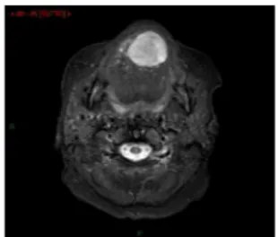
A magnetic resonance imaging was performed to evaluate the dimension of this neoplasm. A vascular flow was detected in the apex of the tongue of 3-cm diameter (Fig. 2).
The lesion was approached under general anesthesia through a transoral paralateral glossotomy. To preserve the tongue’s function, the tumor was resected encapsulated (Fig. 3).
Microscopically, the tumor was composed of hyaline stroma and spindle cells distributed in sheets and cords with voluminous, round, and ovoid nuclei. The architectural pattern was ‘‘staghorn’’ or ‘‘antler-like.’’ The mitotic rate was low (G1 per 10 high-power fields). There was no evidence of necrosis or cytological atypia. The lesion was positive for CD34 and negative for actin and synaptophysin.
On the basis of this pathological finding, the diagnosis of HPC was made. The postoperative course was uneventful; the patient was discharged 7 days after the surgical procedure, and good mucosal healing was observed after 20 days. At 1-year follow-up, clinical and radiological examination did not reveal pathological relapse (Fig. 4).
DISCUSSION
Although HPC occurs more frequently in the adults, a minority rate is reported for the pediatric population; there are 2 forms of this soft tissue tumor: the congenital/infantile and the adult. These 2 variants present different histological features and clinical behavior. The pediatric form (occurs at birth or first year of life) occurs more frequently in the subcutaneous tissue of the head and neck region or the extremities with a large size; however, it is associated to a more benign clinical behavior than the adult variant.5
From the Departments of *Otorhinolaryngology and †Maxillo-Facial Sur-gery, Azienda Ospedaliera Maggiore della Carita`, University of Piemonte OrientaleBAmedeo Avogadro,[ Novara, Italy.
Received February 2, 2012.
Accepted for publication February 26, 2012.
Address correspondence and reprint request to Giovanni Borello, MD, S.C.D.U. Otorhinolaryngology, Ospedale Maggiore della Carita`, Corso Mazzini 18, 28100 Novara, Italy; E-mail: [email protected] The authors report no conflicts of interest.
Copyright* 2012 by Mutaz B. Habal, MD ISSN: 1049-2275
DOI: 10.1097/SCS.0b013e318252d3e7 FIGURE 1. Clinical view of the lesion: neoplasm of hard consistency.
Brief Clinical Studies The Journal of Craniofacial Surgery
&
Volume 23, Number 4, July 2012e292
* 2012 Mutaz B. Habal, MDThe pathological diagnosis requires microscopic examination and immunohistochemical and ultrastructural techniques. Microscopi-cally, it is characterized by staghorn or antler-like vessels; HLA-DR, factor VIII, and vimentin are usually positive, whereas S100 protein, CD34, smooth muscle actin, and Leu-7 are variable. Electron mi-croscopic examination confirms the origin of these cells from the pericytes, which are peculiar cells in the walls of venules and cap-illaries, separated from the endothelial cells by the basement membrane.6Y8
Distinction with SFT of the oral cavity was the most difficult differential diagnosis because SFTs can sometimes display exten-sive areas with a growth pattern that is virtually indistinguishable from HPC. However, morphologic patterns, such as herringbone for-mations, neurofibroma-like and schwannoma-like areas, and diffuse sclerosing areas, which may be present in SFT, are not generally as-sociated with HPC.9Furthermore, in our case, the pathologist de-scribed multilobular fashion, a feature usually not found in SFTs. Also, cellularity can be used for differential diagnosis: in the SFT, it varies from area to area and is inversely related to the amount of collagen; the feature observed in our case showed a homogeneous pattern of cellular distribution.
Enzinger and Smith10defined possible histologic criteria to predict the clinical outcome: mitotic activity, cellularity, necrosis, and hem-orrhage. The lesion is considered as high-grade tumor if it presents more than 3 mitotic figures per 10 high-power fields, increased cel-lularity, necrosis, and hemorrhage.
The clinical behavior of this tumor is very uncertain; some tumors are considered as benign lesions, whereas others are malignant variants with a metastatic course. The most common site of metastasis is the lung; however, it has observed that the youngest patients have the lowest rate of malignancy. The histological feature of this tumor, which is considered a borderline neoplasm and therefore necessi-tating a close follow-up, does not predict the clinical course; instead, cytological atypia, frequent mitosis, and tumor necrosis are associ-ated with malignant behavior.2,3
Given the relatively low incidence of this tumor and its unpre-dictable behavior, several doubts can arise regarding the best choice for a successful treatment. The most frequent therapy reported by the literature is surgery. Several authors have described adjuvant post-operative radiotherapy/chemotherapy for high-grade tumors or for incomplete resections. However, long-term follow-up is necessary to assess long-term outcome, detecting any potential relapses.6Y8
We treated this case with a conservative surgical procedure. We preserved the function of tongue. This peculiar case adds knowledge to the scientific literature regarding the complex field of head and neck HPC.
REFERENCES
1. Stout AP, Murray MR. Hemangiopericytoma: a vascular tumor featuring Zimmermann’ s pericytes. Ann Surg 1942;116:26Y33
2. Espat NJ, Lewis JJ, Leung D, et al. Conventional hemangiopericytoma: modern analysis of outcome. Cancer 2002;95:1746Y1751
3. Kowalski PJ, Paulino AF. Proliferation index as a prognostic marker in hemangiopericytoma of head and neck. Head Neck 2001;23:492Y496 4. Fletcher C, Path FR. Distinctive soft tissue tumors of the head and neck.
Mod Pathol 2002;15:324Y330
5. Coffin CM, Dehner LP, O’Shea PA. Vascular Tumors in Pediatric Soft Tissue Tumors. Baltimore, MD: Williams & Wilkins; 1997:40Y79 6. Nappi O, Ritter JH, Pettinato G, et al. Hemangiopericytoma:
histopathological pattern or clinicopathologic entity. Semin Diagn Pathol 1995;12:221Y232
7. Nemes Z. Differentiation markers in hemangiopericytoma. Cancer 1992;69:133Y140
8. Magid MS, Campbell WG, Ngadiman S, Godwin TA, Ward R. Infantile myofibromatosis with Hemangiopericytoma-like feature of the tongue: a case study including ultrastructure. Pediatr Pathol Lab Med 1997;17:303Y313
9. Kuo WP, Sirois DA, Pemble CW. Locally aggressive solitary fibrous tumor in the infraorbital region: a case report and review of the literature. Oral Surg Oral Med Oral Pathol Oral Radiol Endod 2001;92:308Y311 10. Enzinger FM, Smith BH. Hemangiopericytoma: an analysis of
106 cases. Hum Pathol 1976;7:61Y82
Gorham Disease in the Maxilla
Jie He, DDS, PhD, Yue He, MD, DDS,Weiliu Qiu, MD, DDS, Hanguang Zhu, MD, DDS
Abstract: Gorham disease is a rare condition that is characterized by the proliferation of thin-walled vascular channels associated with
FIGURE 3. Enucleation of the capsulated neoplasm.
FIGURE 4. Healing of the tongue 1 year after surgery. FIGURE 2. T2-weighted magnetic resonance slide of the lingual lesion.
From the Department of Oral and Maxillofacial Surgery, Shanghai Jiao-tong University, School of Medicine, Shanghai Ninth People’s Hospital, Shanghai, People’s Republic of China.
Received February 8, 2011.
Accepted for publication February 27, 2012.
Address correspondence and reprint requests to Hanguang Zhu, MD, DDS, Zhi-zao-ju Rd, No. 639, Shanghai, People’s Republic of China; E-mail: [email protected]
This study was supported by the National Natural Science Foundation of China (30600714, 30973341) and the Shanghai Rising-Star Program (07QA14039).
The authors report no conflicts of interest. Copyright* 2012 by Mutaz B. Habal, MD ISSN: 1049-2275
DOI: 10.1097/SCS.0b013e318252f1b5
The Journal of Craniofacial Surgery
&
Volume 23, Number 4, July 2012 Brief Clinical Studies* 2012 Mutaz B. Habal, MD