A
A
l
l
m
m
a
a
M
M
a
a
t
t
e
e
r
r
S
S
t
t
u
u
d
d
i
i
o
o
r
r
u
u
m
m
–
–
U
U
n
n
i
i
v
v
e
e
r
r
s
s
i
i
t
t
à
à
d
d
i
i
B
B
o
o
l
l
o
o
g
g
n
n
a
a
Dottorato di Ricerca in Scienze Farmaceutiche
XXI Ciclo (2006-2008)
CHIM/08
Coordinatore: Chiar.mo Prof. Maurizio Recanatini
Computational investigation of the Plasmodium falciparum
fatty acid biosynthetic pathway
toward the discovery of novel antimalarials
Tesi di Dottorato presentata da
Francesco Colizzi
Supervisor:
Advisor:
Chiar.mo Prof.
Maurizio Recanatini
Dr. Andrea Cavalli
Doctor Philosophiæ
2
Abstract
The structural peculiarities of a protein are related to its biological function. In the fatty acid elongation cycle, one small carrier protein shuttles and delivers the acyl intermediates from one enzyme to the other. The carrier has to recognize several enzymatic counterparts, specifically interact with each of them, and finally transiently deliver the carried substrate to the active site. Carry out such a complex game requires the players to be flexible and efficiently adapt their structure to the interacting protein or substrate. In a drug discovery effort, the structure-function relationships of a target system should be taken into account to optimistically interfere with its biological function. In this doctoral work, the essential role of structural plasticity in key steps of fatty acid biosynthesis in
Plasmodium falciparum is investigated by means of molecular simulations. The key steps
considered include the delivery of acyl substrates and the structural rearrangements of catalytic pockets upon ligand binding. The ground-level bases for carrier/enzyme recognition and interaction are also put forward. The structural features of the target have driven the selection of proper drug discovery tools, which captured the dynamics of biological processes and could allow the rational design of novel inhibitors. The model may be perspectively used for the identification of novel pathway-based antimalarial compounds.
3
“ma guai a chi cede alla tentazione di scambiare una ipotesi elegante con una certezza”
4
Contents
Prologue . . . . . . . . . . 6
1. Introduction: the type II fatty acids biosynthesis . . . . 8 2. Mechanical features of Plasmodium falciparum
acyl carrier protein in the delivery of substrates . . . . 13
3. ACP/FabZ interaction . . . . . . . 38
4. Conformational plasticity in Pf FabZ . . . . . . 48 5. Atomistic simulations discern active from inactive ligands of
the β-hydroxyacyl-ACP dehydratase of Plasmodium falciparum . 52
6
Prologue
Bad Air
Until the mid-nineteenth century, most scientists thought that noxious swamp gases caused malaria, indeed the word means “bad air” in italian.1
Malaria remains a major and growing threat to the public health and economic development of countries in the tropical and subtropical regions of the world.
This dissertation does not count for explicitly introduce the malaria burden; for this aim, the reader will find better satisfaction looking at the references listed at the end of this session.1-13 The general aspects of fatty acid biosynthesis in Plasmodium falciparum will be briefly discussed in the following pages and some recall can be found along the argumentation of each chapter.
I will focus my dissertation on the advances that the molecular simulations I carried out may have brought in understanding some molecular aspects in the biology of fatty acid biosynthesis in parasites. This biological understanding is integrated with a computational drug discovery effort aimed at the design of small molecules endowed with the capability to interfere with the fatty acid production in Plasmodium falciparum. The organization of each chapter will be likely similar to that of a scientific article. The results discussed in each chapter are used as starting point for the following one. First, the substrate delivery issue as intrinsic properties of the carrier is discussed. Then the delivery of substrates is considered from a wider standpoint which takes into account the way the carrier and its counterpart may interact, and recognize each other. Structural plasticity is found to play a critical role in each of the above steps and is therefore taken into account to address the computational medicinal chemistry part of this dissertation.
8
1. Introduction: the type II fatty acids biosynthesis
The synthesis of fatty acids is organized into two distinct biosynthetic pathways based on the enzymes involved. In mammals and fungi, the fatty acid biosynthesis machinery resides on a single multifunctional polypeptide with multiple active sites (Type I).14 This multifunctional ensemble is supposed to be a product of an evolutionary flow resulting from the fusion of genes encoding individual monofunctional enzymes.15 These monofunctional enzymes constitute the Type II system.16-18
Malaria parasite and other members of the phylum apicomplexa harbor a relict plastid, known as apicoplast, homologous to the chloroplast of plants and algae. The apicomplexan, together with plants and most bacteria use a dissociated Type II fatty acids biosynthesis (FAS II) consisting of at least nine separate polypeptides, each of which may have a dimeric, tetrameric or higher order of quaternary structure.18 A key feature of the FAS II is the presence of a small, acidic and highly conserved acyl carrier protein (ACP) that shuttles all the covalently bound fatty acyl intermediates from one enzyme to the other.
Newly synthesized ACP must be converted from its apo into holo form, by the attachment of a prosthetic group in order to participate in fatty acid biosynthesis. This posttranslational modification is catalyzed by ACP synthase which transfer a 4’-phosphopantetheine group from coenzyme A (CoA) to a conserved serine residue on the ACP. The acyl intermediates are bound through a thioester linkage to the sulfhydryl of the prosthetic group. Fatty acids biosynthesis begins at the acetyl-CoA carboxilase (ACC). The substrate acetyl-CoA is converted to malonyl-CoA and the malonate group is transferred to ACP by malonyl-malonyl-CoA:ACP transacylase (FabD) to form malonyl-ACP.
9
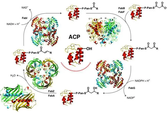
Figure 1. An insightful portrait of Type II fatty acid biosynthesis elongation cycle. Picture adapted from White et al.18. Note the central role of the Acyl carrier protein (ACP).
A Claisen condensation assisted by β-ketoacyl-ACP synthase III (FabH) initiates a cycle that elongates the acyl-ACP by two carbons unit for each cycle until a saturated fatty acid of 16 or 18 carbons is made (the elongation cycle has been insightfully depicted by White et al.18 as shown in Figure 1). The first reaction in this cycle is the NADPH-dependent reduction of β-ketoacyl-ACP to
β-hydroxyacyl-ACP by β-ketoacyl-ACP reductase (FabG), and then the β-hydroxyl intermediate is
dehydrated to yield trans-2-enoyl-ACP catalyzed by either β-hydroxydecanoyl-ACP dehydratase/isomerase (FabA) or hydroxyacyl-ACP dehydratase (FabZ, which is the unique β-hydroxyacyl-ACP dehydratase in Plasmodium). The last step of the cycle is the NADH-dependent
10
reduction of the double bond in the trans-2-enoyl-ACP intermediate by an enoyl-ACP reductase I (FabI) or by other analogue reductases. The product of FabI reduction, enoyl-ACP, is the substrate for the subsequent elongation cycle by condensation with malonyl-ACP catalyzed either by β-ketoacyl-ACP synthase I (FabB) or β-β-ketoacyl-ACP synthase II (FabF). Palmitic acid, for instance, formed by reiteration of this cycle can be either elongated by another set of enzymes or channelized for the formation of phospholipids and other molecules. All Type II systems have this basic set of enzymes to initiate and elongate acyl chains, and the diversity of products is achieved by variation on this theme.19, 20
Even if similar, different gene variations and gene expression regulate in various organisms the fatty acids chain length, branches and saturated/unsaturated balance.20
References
1. Whitfield, J., Portrait of a serial killer. Nature (News) 2002, 01 Oct.
2. Bannister, L.; Mitchell, G., The ins, outs and roundabouts of malaria. Trends Parasitol 2003, 19, (5), 209-13. 3. Breman, J. G., The ears of the hippopotamus: manifestations, determinants, and estimates of the malaria
burden. Am J Trop Med Hyg 2001, 64, (1-2 Suppl), 1-11.
4. Christophides, G. K., Transgenic mosquitoes and malaria transmission. Cell Microbiol 2005, 7, (3), 325-33. 5. Grabowsky, M., The billion-dollar malaria moment. Nature 2008, 451, (7182), 1051-2.
6. Guinovart, C.; Navia, M. M.; Tanner, M.; Alonso, P. L., Malaria: burden of disease. Curr Mol Med 2006, 6, (2), 137-40.
7. Miller, L. H.; Baruch, D. I.; Marsh, K.; Doumbo, O. K., The pathogenic basis of malaria. Nature 2002, 415, (6872), 673-9.
8. Mills, A.; Lubell, Y.; Hanson, K., Malaria eradication: the economic, financial and institutional challenge.
Malar J 2008, 7 Suppl 1, S11.
9. Targett, G. A.; Greenwood, B. M., Malaria vaccines and their potential role in the elimination of malaria.
Malar J 2008, 7 Suppl 1, S10.
10. White, N. J., The role of anti-malarial drugs in eliminating malaria. Malar J 2008, 7 Suppl 1, S8. 11. White, N. J., How antimalarial drug resistance affects post-treatment prophylaxis. Malar J 2008, 7, 9. 12. White, N. J., Qinghaosu (artemisinin): the price of success. Science 2008, 320, (5874), 330-4. 13. Winzeler, E. A., Malaria research in the post-genomic era. Nature 2008, 455, (7214), 751-6.
11
14. Leibundgut, M.; Maier, T.; Jenni, S.; Ban, N., The multienzyme architecture of eukaryotic fatty acid synthases. Curr Opin Struct Biol 2008, 18, (6), 714-25.
15. Smith, S., The animal fatty acid synthase: one gene, one polypeptide, seven enzymes. FASEB J 1994, 8, (15), 1248-59.
16. Goodman, C. D.; McFadden, G. I., Fatty acid biosynthesis as a drug target in apicomplexan parasites. Curr
Drug Targets 2007, 8, (1), 15-30.
17. Goodman, C. D.; McFadden, G. I., Fatty acid synthesis in protozoan parasites: unusual pathways and novel drug targets. Curr Pharm Des 2008, 14, (9), 901-16.
18. White, S. W.; Zheng, J.; Zhang, Y. M.; Rock, The structural biology of type II fatty acid biosynthesis. Annu
Rev Biochem 2005, 74, 791-831.
19. Heath, R. J.; Rock, C. O., The Claisen condensation in biology. Nat Prod Rep 2002, 19, (5), 581-96. 20. Lu, Y. J.; Zhang, Y. M.; Rock, C. O., Product diversity and regulation of type II fatty acid synthases.
12
2. Mechanical features of Plasmodium falciparum acyl carrier
protein in the delivery of substrates
The Acyl Carrier Protein (ACP) is a key element in the biosynthesis of fatty acids being responsible for the acyl group shuttling and delivery within a series of related enzymes. The molecular mechanism of the delivery process is poorly known, and its characterization is essential for the in-depth understanding of the biosynthetic machinery. A steered molecular dynamics approach has been applied to shed light on the putative delivery pathway, suggesting the small α3-helix to act as gatekeeper for the transfer process. Preventing the delivery mechanism would be an innovative strategy for the development of pathway-based antimalarial compounds.
Plasmodium falciparum (Pf) infections are the most widespread and lethal form of Malaria.
Despite the centenary effort to eradicate or control the disease, more than one third of the human population lives in endemic areas with an estimated half billion of infections annually resulting in about two millions of death.1-3
The recently disclosed type II fatty acid synthesis (FAS-II) pathway of Pf is offering attractive targets potentially enabling the discovery and development of efficacious and selective antimalarial agents. FAS-II system relies on a dissociative process that exploits a series of individual enzymes that are, indeed, structurally different from the multifunctional type I fatty acid synthase (FAS-I) system of humans.4,5 A key feature of the FAS-II is the presence of a small (~ 9 kDa), acidic and highly conserved Acyl Carrier Protein (ACP) that shuttles all the acyl intermediates from one enzyme to the other. The acyl substrates are bound to ACP through a flexible arm formed by the serine-bound prosthetic phosphopantetheine (4’-PP) group (Figure 1a).6 Nevertheless, ACPs are also known to play a fundamental role in numerous other
13
biosynthetic pathways in which acyl transfer steps are required.7-9 The geometric properties of several conformationally distinct ACPs have been determined by X-ray crystallography and NMR.10-15 These experimental data have suggested that structural plasticity is an intrinsic feature of this protein family, providing a possible explanation for ACPs’ capability to recognize multiple enzyme partners and transiently deliver the acyl group to the active sites of these enzymes.16
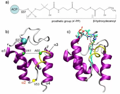
Figure 1. a) 2D structure of the acylated 4’-phosphopantetheine (4’-PP) prosthetic group in the
hydroxydecanoyl-ACP. b) Overall fold of ACPs; the α3-helix (residues 57-62) is depicted in yellow tube. c) Binding mode of the β-hydroxylated substrate (carbon atoms in cyan) after the MD equilibration compared to the decanoyl substrate (carbon atoms in light green) reported in E. coli crystal structure (pdb code: 2fae).
ACP structures are composed of four α-helices delimiting a lipophilic core that forms a binding pocket for fatty acids. Helix α2 has highly conserved residues and plays a major role in ACP-protein interactions (Figure 1b)17 whereas the short α3-helix does not have a conserved folding among the published ACP structures and, accordingly, this protein portion was observed to experience a helix-loop conformational equilibrium in PfACP.14 Moreover, the analysis of
14
experimental and computational studies suggested the protein region between helices α2 and α4 to be part of the putative ACP/enzyme interface.18-26 The functional implication of the above mentioned structural features are yet unrevealed when observed from the standpoint of the delivery of substrates.
X-ray structures of acylated ACPs have shown the thioester-bound acyl-chain to be embedded into a tunnel-like hydrophobic cavity (Figure 1c), which can harbor acyl substrates of different length.12,15 Notably, the reactive center of the acyl substrate is buried in the hydrophobic core of the carrier, and thus inaccessible to catalytic activities of FAS-II enzymes. Therefore, delivering the substrate from the inner ACP core to the FAS-II enzyme active site is a mandatory event for each biosynthetic step. The molecular understanding of ACP’s ability to deliver substrates together with the ability of drug designers to interfere with this process might be a new strategy for the development of innovative FAS-II inhibitors that could behave as pathway modulators rather than single-enzyme blockers.
Limited structural information about the complex of ACP with biological counterparts and the mechanism of substrate delivery is currently available.18,27,28 Recently, Leibundgut et al.,27 addressed the delivery issue discussing the crystal structure of yeast fatty acid synthase (FAS-I type) system with its ACP stalled at one catalytic domain. They have suggested a general switchblade-like mechanism in which the 4’-PP arm delivers the acyl chain flipping from the ACP core into the catalytic domain active site.27 However, even though ACPs share the same structural motif in the acyl-substrate binding region, when compared to the FAS-II ACP, the yeast ACP domain (~ 18 kDa) has four additional C-terminal α-helices, which take part in the interaction with the catalytic domain. Therefore, differences in the recognition and delivery process between FAS-I and FAS-II systems are likely to occur. Furthermore, substrate delivery
15
is a dynamic process, and the detailed but static X-ray picture requires to be complemented with other approaches that are able to directly capture the dynamics of the biomolecule under investigation.
In the present work, the dynamics of the delivery mechanism of the β-hydroxydecanoyl substrate by PfACP was computationally investigated using a Steered Molecular Dynamics (SMD) approach. Analyzing the trajectories and the force profiles, we were able to identify the lowest resistance pathway for the substrate delivery process. In addition, our simulations pointed out both the role of α3-helix as gatekeeper for the substrate transfer process, and the effect of the substrate β-hydroxylation on the enzyme recognition.
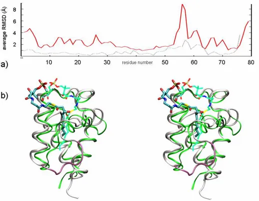
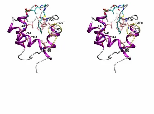
Figure 2. a) Root mean square fluctuation (RMSf) of the PfACP backbone along the whole MD simulation (red
line), and RMSf of up-to-date available crystal structures of E. coli ACP after Cα superposition (dotted gray line). b) Stereo view of the β-hydroxydecanoyl-ACP (white ribbon) as it appears after 8 ns of MD. The prosthetic group (cyan) lies on the mouth of the fissure between helices α2 and α3. The highly flexible portion going from 53 to 57 is magenta. The decanoyl-ACP crystal structure (in green, pdb code: 2fae) is shown for comparison.
16
In SMD simulations, which have been widely and successfully applied to explore the properties of non-equilibrium processes of biomolecules,29,30 a moving harmonic potential is used to induce a motion along a reaction coordinate. The free end of a virtual spring is moved at constant velocity, while a set of “pulled atoms”, attached to the other end of the spring, are subject to steering forces. The applied forces are determined by the extension of the spring, and can be monitored throughout the entire simulation. If the pulled atoms can easily advance along the selected reaction coordinate, the applied force is small and its profile is rather flat. Conversely, if the pulled atoms encounter hindrance along the pathway, the force increases to allow the pulled atoms overcoming energy barriers, thus resulting in quite relevant drops in the force profile. Based on the magnitude of the exerted force, it is possible to determine how easily a pathway can be coursed.29,30
The simulations of the covalently bound β-hydroxydecanoyl-ACP complex were performed in explicit water, using the CHARMM31 force field and the program NAMD32 [see Appendix (Ap) for details]. To get insights into intrinsic properties of the system, the complex was first simulated for 8 ns of unrestrained MD. The average value of RMSD calculated for the backbone along the whole simulation time was 2.9 Å. Nonetheless, the region including residues 53-62 showed high flexibility (RMSf up to 8 Å, red plot in Figure 2a) reflecting the higher ratio of conformational diversity observed by superimposing ACP crystal structures (dotted grey plot in Figure 2a). During the free MD simulations the β-hydroxyacyl moiety of the bound ligand behaved like a fishing float inside the lipophilic core, and the acyl moiety fluctuated assuming several conformations, according to the existence of multiple low-occupancy conformers observed in the crystal structure of decanoyl-ACP.15 The substrate β-hydroxyl group was able to interact with several polar residues (mainly the backbone of D59, A60, and I63) lining the
17
entrance of the ACP binding cavity. As a result, the β-hydroxydecanoyl was less buried in the protein core when compared to the experimentally reported decanoyl analogue. This had the consequence that the prosthetic group (4’-PP) was more relaxed and able to fluctuate among several conformations. Figure 2b shows the prosthetic group protruding towards the solvent, self-docking at the upper fissure between helices α2 and α3. In such a conformation, the FAS-II enzymatic counterparts might still recognize the conserved α2-helix, selectively interact with the prosthetic group, and establish connections with protein segment 53-62. Roujenikova et al.15 proposed that the fairly flexible prosthetic arm may adopt a set of different conformations, likely related to the carried acyl substrate, to allow optimal interaction of acyl-ACP intermediates to partner enzymes. However, the MD simulations did not provide any mechanistic explanation of the delivery process. This is likely a consequence of high potential energy barriers implicated, which cannot be sampled by ns-time-scale simulations. To overcome this drawback, we applied SMD (Figure 3) to drive the substrate along several putative reaction coordinates (see Figure 3a).
18
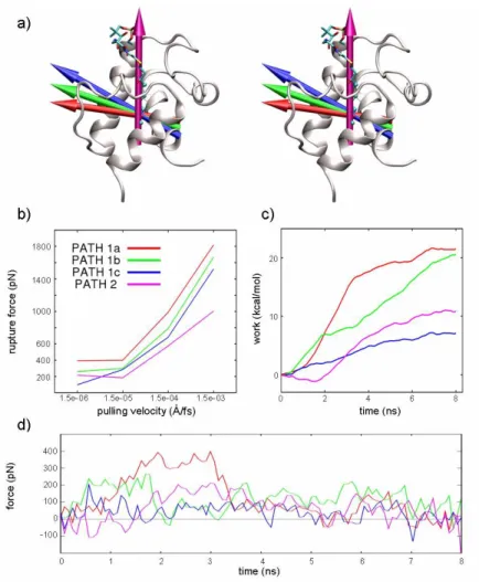
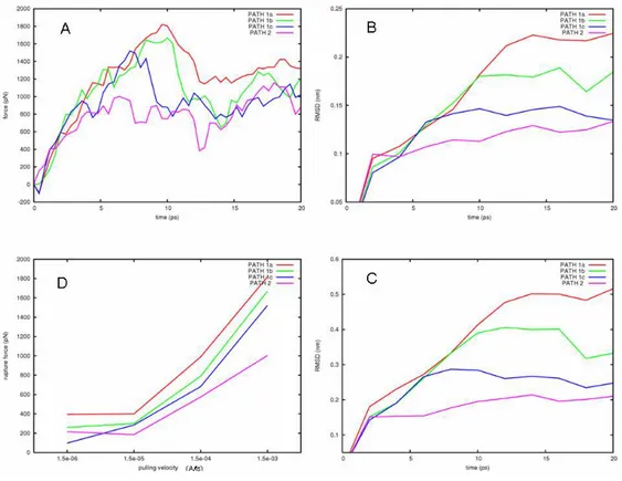
Figure 3. a) Acyl-ACP and the delivery pathways investigated. The arrows represent the pulling directions of the
applied forces. b) Correlation of the pulling velocity with the rupture force. At 1.5e-06 Å/fs the process is close to equilibrium. c) Mechanical work done on the system calculated by numerical integration. d) Force profiles of the investigated pathways at 1.5e-06 Å/fs. For sake of clarity the curves are obtained averaging the forces every 30 points.
Based on the inspection of the structural features of the reported acyl-ACPs, and on the aforementioned experimental data,18-26 several delivery pathways were initially considered. However, some of them were discarded because of either they generated unlikely structural distortion or were scarcely in agreement with the available experimental data.18-26 Two main putative delivery pathways were finally investigated (Figure 3a): the first one accounting for the substrate exposure through the fissure formed by helices α2 and α3 (path 1); the second one
19
accounting for a sword-unsheathed-like mechanism, in which the substrate is unthreaded away in a parallel direction with respect to the major axis of the binding pocket (path 2). Since for path 1 a protein conformational rearrangement was required, and considering critical the role of the pulling direction, three slightly different sub-paths (path 1a, 1b, and 1c) were investigated. For path 2, preliminary investigations revealed that no relevant protein conformational rearrangements were required, and that slight modifications of xyz components of the pulling direction provided very similar force profiles (see Ap). Hence, we assumed that the chosen pulling direction in path 2 was likewise representative of the sword-unsheathed-like mechanism.
In SMD, the pulling velocity (v) largely influences both the results of simulations and the profile of the applied forces. As v decreases, the pulled atoms have more time to sample the conformational space and to search for lowest resistance path along the selected direction. At the same time, the non pulled atoms can relax following the movement of the pulled atoms, and thus reducing the friction rate of the process. As shown in Figure 3b, a lowering of the pulling velocity resulted in a decrease of the observed rupture force likely because of the reduction of non-equilibrium effects.29,33 Remarkably, at the pulling velocity of 1.5e-06 Å/fs, a plateau was observed, pointing to this pulling rate as the more appropriate one to minimize the friction influence on the delivery process. Moreover, at such a slow pulling velocity, some spontaneous and SMD-unrelated conformational changes of ACP could also be sampled.
In Figure 3d and 3c, the force profile and the mechanical work done during each simulated pathway are shown, respectively. For each pathway, the force profile correlated well with the rupture and formation of interaction between the outgoing substrate and the binding site residues; a detailed description of the events is reported in Ap.
20
The system in both path 1a and 1b encountered relatively high hindrance in the early ns of simulations because the substrate moved against the deep portion of the cleft formed by α2- and
α3-helix, and lined by the α2-α3 loop. Here, the α3-helix could not spontaneously follow the
pulled atoms and as a consequence the exerted force increased.
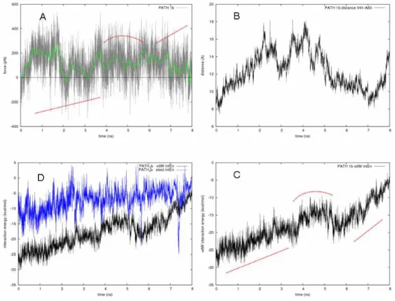
In path 1c (Figures 3 [blue line] and 4), the force profile was much flatter than in paths 1a and 1b. We observed an initial force peak (~200 pN, Figure 3d) due to the breaking of an H-bond network involving the substrate β-OH and the backbone oxygen of A60 and I63. Then, the whole process evolved with a force less than 100 pN. A strong electrostatic interaction involving the β-OH group and the side chain of D59 occurred between ~1.5-2.5 ns (Figure 4c). Concertedly, a slow “gate opening” was manifested by the increasing distance between V41 and A60 (see also Figure 1b). The flatness of the applied force profile indicated that both the pulled substrate and the constraint point were concertedly moving, and the decreasing of the overall vdW contacts meant that the substrate was leaving the binding cavity quite slowly. The lack of a significant rupture point in the force profile suggested that the substrate did not encounter any relevant resistance despite the remarkable shift of the α3-helix. This was possible because, during the substrate pulling, the ACP α3-helix could spontaneously rearrange, allowing the substrate exposure. This observation was in good agreement with both free MD and experimental data,
18-26
which pointed to α3-helix as one of the most dynamic segments of ACP structure (Figure 2a). Noteworthy, in the same simulation time scale, in the free MD, it was sampled both the helix folding of residues 19-26 and the partial unfolding of the short α3-helix (see Ap).
21
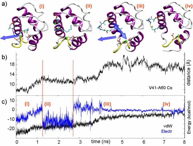
Figure 4. a) Snapshots isolated from the SMD trajectory sampled in Path 1c at (i) 300 ps, (ii) 1.6 ns, (iii) 4.2 ns, and
(iv) 7 ns. The blue arrow in (i) shows the pulling direction and in (iii), the double end arrow indicates the opening movement of the fissure formed by helices α2 and α3. b) Evolution of the distance between Cα of V41 and A60. c) Electrostatic and vdW interaction energies between the β-hydroxydecanoyl moiety and the protein residues during the SMD.
Path 1 delivery hypothesis was then compared to path 2, to investigate if a sword-unsheathed-like mechanism could provide a flatter force profile. As shown in Figure 3d (magenta line), the unthreading of the substrate had a magnitude of forces similar to that registered for path 1b and higher than that observed in path 1c. Such a mechanism poorly complied with an ACP-enzyme interface comprising both α2-helix and its connection to α4-helix. However, a sword-unsheathed-like mechanism could be proposed for enzymes, whose interactions with ACP more extensively involve the α1-α2 loop connection.16,20
22
In summary, our simulations suggest that the substrate delivery through the cleft between α2- and α3-helix is a feasible pathway that exploits intrinsic conformational plasticity of ACP (path 1c). We also show that ACP portion including the loop connecting helices α2 and α3 and the α3-helix itself can play a critical role in both the delivery and, as previously reported, the recognition processes.18-26 As hypothesized on the basis of X-ray experiments,12,15 we also point out the ability of the prosthetic group to function as label for the acyl-intermediates carried by the ACP. Such aspects are related to ACP plasticity and may account for a mechanism in which ACP and its enzymatic counterparts minimize the solvent exposure of the lipophilic substrate moiety. The delivery model would include an ACP/enzyme interface formed by ACP helices α2 and α3; it might be hypothesized that a slight conformational change of the α3-helix could easily allow the substrate to glide into the corresponding enzyme active site.
Can we use the above uncovered features of ACP to address the development of innovative antimalarial compounds? This study sheds light on the functional and plastic features of ACP and attempts to contextualize them in the protein-protein interactions network occurring in the FAS-II system. During the elongation cycle, ACP-enzyme interactions and the delivery of the acyl substrate to the active site are compulsory steps for fatty acid production. These processes are currently subject of drug discovery efforts. In this respect, blockers of substrate delivery would function as pathway-based antimalarial compounds rather than single FAS-II enzyme inhibitors. The design of a delivery-blocker might be achieved through a strategy that considers as template-structure for a lead-candidate that region of the α3-helix usually interacting with α2. The interaction of such a mimetic compounds with PfACP would interfere with the gate opening function managed by the α2/α3 cleft. Moreover, the low-resistance delivery pathways here investigated might be used as starting point for further studies aiming at identifying local energy
23
minima along the reaction coordinate. As matter of fact, our simulations suggest that, while the substrate transiently leaves the carrier core moving toward the catalytic partner, the ACP core itself might become a peculiar binding pocket for small molecules able to interfere with the relocation of the substrate into the carrier. Likewise, if the conformational “transient” state of the acyl-ACP is an highly populated energy minimum, the probability to block the delivery process would dramatically increase.
Methodological details
Starting from the first NMR conformer (out of 20) of the holo structure of PfACP (pdb code: 2fq0), the prosthetic group carrying a β-hydroxydecanoyl substrate was built using the Sybyl 7.3 molecular modeling suite of program (Tripos Inc., St. Louis, MO) and manually docked at the acyl binding pocket of the carrier using as template the binding solution of the high resolution X-ray structure of E. coli decanoyl-ACP (pdb code: 2fae). The covalently bound β-hydroxydecanoyl-ACP complex was first energy-minimized in gas phase for 1000 steps using the conjugated gradient method keeping fixed the backbone atoms and restraining the side chain with a 10 kcal mol-1 Å-2 spring constant. The complex was then solvated with a 10 Å thick layer of water using the solvate package of VMD and seven Na+ cations were added to neutralize the system. The simulations were performed using periodic boundary conditions and long-range electrostatics calculated by using the particle-mesh Ewald (PME) method with a charge grid spacing < 1Å. A cutoff of 10 Å was used for van der Waals and short-range electrostatic interactions with a switching function started at 8 Å to ensure a smooth cutoff. Time integration step of 2 fs was used and the length of all bonds involving hydrogen atoms was fixed using the SHAKE algorithm. The solvated system was minimized with the conjugate gradient method for 1000 steps restraining the heavy atoms of the protein with a force constant of 10 kcal mol-1 Å-2, followed by 1000 steps with a force constant of 3 kcal mol-1 Å-2 and finally fully minimizing the system for further 1000 steps. The insertion of the β-hydroxydecanoyl moiety in the PfACP core produced only slightly reorientations in the side chains of residues shaping the cavity, with the minimized structure having an average overall rmsd less than 0.5 Å. Six subsequent steps were
24
used to heat the system from 1 to 300 K. Every 15 ps the system was heated up by 50 K and correspondingly the alpha-carbon were gradually unrestrained (till 2 kcal mol-1 Å-2) starting from a 6 kcal mol-1 Å-2 harmonic restraint force constant. In these equilibration steps, constant volume was maintained and the temperature was controlled by Langevin dynamics with a dumping coefficient of 5 ps-1. The system was subsequently switched to the isothermal-isobaric (NPT) ensemble once the temperature was stabilized at 300 K. During the switching, a soft harmonic restraint of 2 kcal mol-1 Å-2 was still applied to the Cα atoms and gradually turned off in the next 60 ps. The constant pressure control was applied using the Nosé-Hoover Langevin piston method and 1 atm was set as target pressure. The system evolved for further 100 ps and the final state was used as starting point for both SMD study and free MD production.
All simulations were performed with the molecular dynamics program NAMD 2.6 (Phillips et al., 2005) using the CHARMM22 force field (MacKerell et al., 1998) for proteins and the TIP3P model for all water in the system (Jorgensen et al., 1983). CHARMM compliant parameters for prosthetic group together with the β-hydroxydecanoyl substrate were generated using the paratool plugin implemented in VMD (Humphrey et al., 1996. http://www.ks.uiuc.edu/Research/vmd/). Missing parameters were estimated from similar terms within the force field using an empirical/additive approach. Mulliken charges were calculated at the HF/6-31G* level of theory and refined by analogy in order to preserve consistency with CHARMM style charges (i.e., always qH = 0.09).
SMD simulations were performed at constant pulling velocity using a steering velocity of 1.5e-06 Å/fs with a spring constant of 5 kcal mol-1 Å-2 (several velocities were further considered as discussed later in the Appendix). Variation of the constant K of the harmonic restrain influences the profile/fluctuation of the applied forces. Here, we used a value of 5 kcal mol-1 Å-2 that coupled with an appropriate v gave a force profile in which it was still possible to notice a drop of the forces, but at the same time permitted the thermal fluctuation of the pulled atoms to be similar to the perturbation arising from the pulling force.
Using as reference coordinates the structure of the E. coli decanoyl-ACP (pdb code: 2fae subunit A) the x, y and z- components of the normalized pulling direction were: for path 1a, -0.9388, 0.2181, 0.2665; for path 1b, -0.9198, 0.2927, 0.2611; for path 1c, -0.8512, 0.4658, 0.2416; for path 2, 0.0326, 0.9300, 0.3640. To avoid shifting of the system during pulling, in path 1, the alpha-carbons of protein going from residue 1 to 35 were harmonically restrained to their initial
25
position using a 1 kcal mol-1 Å-2 spring constant and, in path 2 the restraints were applied to residue portions 1-16 and 48-52.
The value of the exerted force (F) were outputted every (dt) 1 ps of simulation and the work ' ) ' ( ) ( ' 0 F t vdt t
W =
∫
t done on the system during the SMD was calculated by numerical integration; the pulling velocity (v) was 1.5e-06 Å/fs.CHARMM force field topology for prosthetic group and substrate can be found at http://pubs.acs.org/doi/suppl/10.1021/ci800297v/suppl_file/ci800297v_si_001.pdf
Appendix
Pulling velocity and force profiles
To explore the influence of the pulling velocity on the behavior of the system we tested four decreasing v values starting from 1.5e-03 Å/fs (0.003 Å/timestep) leading to 1.5e-06 Å/fs (0.000003 Å/timestep). For the higher velocity used it was possible to correlate the RMSD of the protein with the magnitude of the rupture force, calculated as mean of three individual simulation for each path (Figure 1Ap-a), required for the substrate exposure along each investigated pathway. As shown, starting from the most “perturbative” pulling direction path 1a, the protein was subjected to lower conformational rearrangement going toward the path 2 direction (Figure 1Ap-b). The reported RMSD of the whole protein actually reflected the deviation whose residues 57 to 62 were subject (Figure 1Ap-c). This means that at higher velocities path 1a pushed the substrate in the deep portion of the cleft formed by α2 and α3 helices and lined by their α2-α3 loop connection. The protein could only suffer this rustling having not the possibility to give into the pushing atoms. As the pulling direction became less drastic as it was in path 1b and 1c, the substrate could find less opposition to its flowing, leading to a decrease of the exerted force as well as of the RMSD. In path 2, the conformational shift of α3-helix was not remarkable and the substrate could be easily threaded away.
As the pulling velocity is lowered one should expect a decreasing trend of the rupture force because of the reduction of non-equilibrium effects. In Figure 1Ap-d this trend is shown, and a
plateau was reached when v approached to 1.5e-06 Å/fs. Around this velocity the necessary
26
an in-depth inspection of the system was required. Remarkably, at the lower pulling velocity used, the substrate took about 6-8 ns to be extracted from the binding cavity. This was the same simulation time scale we used in the free MD, where it was possible to sample both the helix folding of residues 19-26, and the partial unfolding of the short α3-helix (see Figure 9Ap-d). That means that, meanwhile the pulling force was applied to the substrate atoms, the ACP system was able to undergo some intrinsic conformational changes normally occurring without any constraint.
Figure 1Ap. Effects of the pulling velocity on the force profiles. A) Force profile obtained for the higher velocity
(1.5e-03 Å/fs); calculated as mean of three individual simulation for each path. B) RMSD of the alpha-carbons of the whole protein during the steering process and C) RMSD of the protein portion 57-62. Path 1a induces the wider movement respect to the other paths. D) Computed rupture force as a function of several pulling velocities.
27
Figure 2Ap. Effects of modification of pulling direction on the force profiles in PATH 2. Force profiles obtained
using a pulling velocity of 1.5e-03 Å/fs, calculated as mean of three individual simulation run.
Further details on the pathways
In Figure 3Ap, we report on the force profiles of the four pathways (paths 1a-c and path 2).
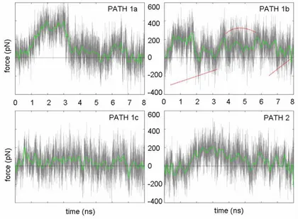
Figure 3Ap. Force profiles of pathways investigated at 1.5e-06 Å/fs. The green thick curve is obtained averaging
28
PATH 1A. In Figure 4Ap, the path 1a is summarized in terms of force profile, V41-A60 distance between their alpha-carbons, and electrostatic and van der Waals interaction energy between ACP and the β-hydroxydecanoyl moiety of the substrate. The plateau observed at 1.5-3 ns (Figure 4Ap-a) was due to the breaking of H-bond interaction between the β-hydroxyl group of the substrate and A60 and I63 backbone. Then, the acyl chain followed the unfolding of α3-helix, being it still able to transiently establish an H-bond interaction with A60 and I63 (Figure 4Ap-d).
Figure 4Ap. PATH 1A. A) Force profile obtained at v = 1.5e-06 Å/fs. B) Evolution of the distance between the
alpha-carbons of V41 and A60 during SMD simulations. C) Electrostatic and van der Waals interaction energy between the β-hydroxydecanoyl moiety and the protein during SMD simulations. D) Magnification of the van der Waals interaction energy contribution.
29
PATH 1B. In Figure 5Ap, the path 1b is summarized. As shown in Figure 5Ap-b, an opening/closure of the α2-α3 fissure was observed. The process could be divided in three steps: approach, transit, and escape. During the early stage of the simulation the acyl end was trapped into a sub-pocket toward the α1-helix and formed by F29, L47, L44, and I8; this explained the force increasing despite both electrostatic and vdW interactions remained constant. Around 1.6 ns the pulling force released the acyl group from the sub-pocket and slowly it moved towards the fissure formed by the juxtaposition of α2 and α3 helices. In the simulation time-range from ~3.7-6.5 ns, the substrate protruded into the fissure formed by I41, I44, I55 and A60, and by L40 and I63 (Figure 6Ap). Furthermore, the side chain of F29 underwent a conformational change; F29 acted as lid maintaining the cavity core water-free. Finally, in the third step, the substrate completely left the binding cavity as suggested by both the decreasing of interaction energies and forces magnitude.
Figure 5Ap. PATH 1B. A) Force profile obtained at v = 1.5e-06 Å/fs. B) Evolution of the distance between the
alpha-carbons of V41 and A60 during SMD simulations. C) Electrostatic and van der Waals interaction energy between the β-hydroxydecanoyl moiety and the protein during SMD simulations. D) Magnification of the van der Waals interaction energy contribution.
30
Figure 6Ap. Stereo view of the transient state sampled in Path 1b (see text). The conformation of F29 related to the
fully embedded substrate is shown in transparent pink stick. Note the β-OH group of the substrate exposed to putative metabolizing enzyme activity.
31
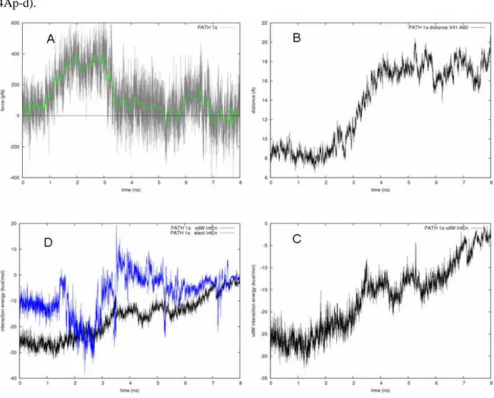
PATH 1C. In Figure 7Ap, the path 1c is summarized. This pathway is in-depth described in the paper.
Figure 7Ap. PATH 1C. A) Force profile obtained at v = 1.5e-06 Å/fs. B) Evolution of the distance between the
alpha-carbons of V41 and A60 during SMD simulations. C) Electrostatic and van der Waals interaction energy between the β-hydroxydecanoyl moiety and the protein during SMD simulations. D) Magnification of the van der Waals interaction energy contribution.
32
PATH 2. The unthreading of the substrate is summarized in Figure 8Ap. At the beginning of the simulation the force average fluctuated to both positive and negative values reflecting the floating behavior of the substrate. The β-OH of the substrate was found to interact with the backbone carbonyl group of I63 (Figure 8Ap-d). As the pulling evolved, another H-bond was formed with A60 backbone and the exerted forces increased till about 3.8 ns (Figure 8Ap-d). At 4 ns the acyl moiety started to leave the ACP core as revealed by the decreasing vdW contacts. The outgoing of the substrate was related to a decreasing of the V41-A60 distance reflecting the core contraction as the substrate left its location. During the outgoing process the prosthetic group was, finally, harbored in the mouth of α2-α3 helix cleft.
Figure 8Ap. Path 2. A) Force profile obtained at v = 1.5e-06 Å/fs. B) Evolution of the distance between the
alpha-carbons of V41 and A60 during SMD simulations. C) Electrostatic and van der Waals interaction energy between the β-hydroxydecanoyl moiety and the protein during SMD simulations. D) Monitored distances between H-bonding moieties.
33 Monitored features of the free MD simulation
Figure 9Ap. Monitored features of the free MD. A) Time dependence of the RMSD calculated for the Cα B) Evolution of distance between the alpha-carbons of V41 and A60 during the molecular dynamics simulation. C) Electrostatic and van der Waals interaction energy between the β-hydroxydecanoyl moiety and the protein during the simulation. D) Time dependence of the radius of gyration calculated for the loop portion 19-26 that completed the α helix folding around the 6th ns of simulation. Vice versa, the small α3 helix partially unfolded into a loop at the beginning of the simulation.
References
(1) Breman, J. G. The ears of the hippopotamus: manifestations, determinants, and estimates of the malaria burden. Am. J. Trop. Med. Hyg. 2001, 64, 1-11.
(2) Greenwood, B. M.; Bojang, K.; Whitty, C. J.; Targett, G. A. Malaria. Lancet 2005, 365, 1487-1498. (3) Snow, R. W.; Guerra, C. A.; Noor, A. M.; Myint, H. Y.; Hay, S. I. The global distribution of clinical
34
(4) Surolia, A.; Ramya, T. N.; Ramya, V.; Surolia, N. 'FAS't inhibition of malaria. Biochem. J. 2004, 383, 401-412.
(5) Lu, Y. J.; Zhang, Y. M.; Rock, C. O. Product diversity and regulation of type II fatty acid synthases.
Biochem. Cell Biol. 2004, 82, 145-155.
(6) White, S. W.; Zheng, J.; Zhang, Y. M.; Rock, C. O. The structural biology of type II fatty acid biosynthesis.
Annu. Rev. Biochem. 2005, 74, 791-831.
(7) Shen, B.; Summers, R. G.; Gramajo, H.; Bibb, M. J.; Hutchinson, C. R. Purification and characterization of the acyl carrier protein of the Streptomyces glaucescens tetracenomycin C polyketide synthase. J.
Bacteriol. 1992, 174, 3818-3821.
(8) Brozek, K. A.; Carlson, R. W.; Raetz, C. R. A special acyl carrier protein for transferring long hydroxylated fatty acids to lipid A in Rhizobium. J. Biol. Chem. 1996, 271, 32126-32136.
(9) Sweet, C. R.; Williams, A. H.; Karbarz, M. J.; Werts, C.; Kalb, S. R. et al. Enzymatic synthesis of lipid A molecules with four amide-linked acyl chains. LpxA acyltransferases selective for an analog of UDP-N-acetylglucosamine in which an amine replaces the 3"-hydroxyl group. J. Biol. Chem. 2004, 279, 25411-25419.
(10) Kim, Y.; Prestegard, J. H. Refinement of the NMR structures for acyl carrier protein with scalar coupling data. Proteins 1990, 8, 377-385.
(11) Xu, G. Y.; Tam, A.; Lin, L.; Hixon, J.; Fritz, C. C. et al. Solution structure of B. subtilis acyl carrier protein. Structure 2001, 9, 277-287.
(12) Roujeinikova, A.; Baldock, C.; Simon, W. J.; Gilroy, J.; Baker, P. J. et al. X-ray crystallographic studies on butyryl-ACP reveal flexibility of the structure around a putative acyl chain binding site. Structure 2002, 10, 825-835.
(13) Wong, H. C.; Liu, G.; Zhang, Y. M.; Rock, C. O.; Zheng, J. The solution structure of acyl carrier protein from Mycobacterium tuberculosis. J. Biol. Chem. 2002, 277, 15874-15880.
(14) Sharma, A. K.; Sharma, S. K.; Surolia, A.; Surolia, N.; Sarma, S. P. Solution structures of conformationally equilibrium forms of holo-acyl carrier protein (PfACP) from Plasmodium falciparum provides insight into the mechanism of activation of ACPs. Biochemistry 2006, 45, 6904-6916.
(15) Roujeinikova, A.; Simon, W. J.; Gilroy, J.; Rice, D. W.; Rafferty, J. B. et al. Structural studies of fatty acyl-(acyl carrier protein) thioesters reveal a hydrophobic binding cavity that can expand to fit longer substrates.
J. Mol. Biol. 2007, 365, 135-145.
(16) Kim, Y.; Kovrigin, E. L.; Eletr, Z. NMR studies of Escherichia coli acyl carrier protein: dynamic and structural differences of the apo- and holo-forms. Biochem. Biophys. Res. Commun. 2006, 341, 776-783. (17) Zhang, Y. M.; Marrakchi, H.; White, S. W.; Rock, C. O. The application of computational methods to
explore the diversity and structure of bacterial fatty acid synthase. J. Lipid Res. 2003, 44, 1-10.
(18) Parris, K. D.; Lin, L.; Tam, A.; Mathew, R.; Hixon, J. et al. Crystal structures of substrate binding to Bacillus subtilis holo-(acyl carrier protein) synthase reveal a novel trimeric arrangement of molecules resulting in three active sites. Structure 2000, 8, 883-895.
35
(19) Flaman, A. S.; Chen, J. M.; Van Iderstine, S. C.; Byers, D. M. Site-directed mutagenesis of acyl carrier protein (ACP) reveals amino acid residues involved in ACP structure and acyl-ACP synthetase activity. J.
Biol. Chem. 2001, 276, 35934-35939.
(20) Zhang, Y. M.; Rao, M. S.; Heath, R. J.; Price, A. C.; Olson, A. J. et al. Identification and analysis of the acyl carrier protein (ACP) docking site on beta-ketoacyl-ACP synthase III. J. Biol. Chem. 2001, 276, 8231-8238.
(21) Worsham, L. M.; Earls, L.; Jolly, C.; Langston, K. G.; Trent, M. S. et al. Amino acid residues of Escherichia coli acyl carrier protein involved in heterologous protein interactions. Biochemistry 2003, 42, 167-176.
(22) Gong, H.; Byers, D. M. Glutamate-41 of Vibrio harveyi acyl carrier protein is essential for fatty acid synthase but not acyl-ACP synthetase activity. Biochem. Biophys. Res. Commun. 2003, 302, 35-40. (23) Keatinge-Clay, A. T.; Shelat, A. A.; Savage, D. F.; Tsai, S. C.; Miercke, L. J. et al. Catalysis, specificity,
and ACP docking site of Streptomyces coelicolor malonyl-CoA:ACP transacylase. Structure 2003, 11, 147-154.
(24) Zhang, Y. M.; Wu, B.; Zheng, J.; Rock, C. O. Key residues responsible for acyl carrier protein and beta-ketoacyl-acyl carrier protein reductase (FabG) interaction. J. Biol. Chem. 2003, 278, 52935-52943.
(25) Zhang, L.; Liu, W.; Xiao, J.; Hu, T.; Chen, J. et al. Malonyl-CoA: acyl carrier protein transacylase from Helicobacter pylori: Crystal structure and its interaction with acyl carrier protein. Protein Sci. 2007, 16, 1184-1192.
(26) Liu, W.; Du, L.; Zhang, L.; Chen, J.; Shen, X. et al. Helicobacter pylori acyl carrier protein: expression, purification, and its interaction with beta-hydroxyacyl-ACP dehydratase. Protein Expr. Purif. 2007, 52, 74-81.
(27) Leibundgut, M.; Jenni, S.; Frick, C.; Ban, N. Structural basis for substrate delivery by acyl carrier protein in the yeast fatty acid synthase. Science 2007, 316, 288-290.
(28) Rafi, S.; Novichenok, P.; Kolappan, S.; Zhang, X.; Stratton, C. F. et al. Structure of acyl carrier protein bound to FabI, the FASII enoyl reductase from Escherichia coli. J. Biol. Chem. 2006, 281, 39285-39293. (29) Izrailev, S.; Stepaniants, B. I.; Isralewitz, B.; Kosztin, D.; Lu, H. et al. Steered molecular dynamics. In
Computational Molecular Dynamics: Challenges, Methods, Ideas; Springer Ed., 1999; pp 39-64.
(30) Sotomayor, M.; Schulten, K. Single-molecule experiments in vitro and in silico. Science 2007, 316, 1144-1148.
(31) MacKerell Jr., A. D., Bashford, D., Bellott, M., Dunbrack Jr., R.L., Evanseck, J., et al. All-atom empirical potential for molecular modeling and dynamics studies of protein. J. Phys. Chem. B 1998, 102, 3586 -3616. (32) Phillips, J. C.; Braun, R.; Wang, W.; Gumbart, J.; Tajkhorshid, E. et al. Scalable molecular dynamics with
NAMD. J. Comput. Chem. 2005, 26, 1781-1802.
(33) Niu, C.; Xu, Y.; Luo, X.; Duan, W.; Silman, I. et al. Dynamic mechanism of E2020 binding to acetylcholinesterase: a steered molecular dynamics simulation. J. Phys. Chem. B 2005, 109, 23730-23738.
36
3. ACP/FabZ interaction
The role of ACP’s α3-helix in the delivery of substrate is discussed in the context of the protein-protein interaction between PfACP and PfFabZ. Several experimental observations reporting on the ACP residues involved in interaction with enzymatic counterparts were taken into account and the frame of the recognition and delivery process is also outlined.
Whereas the α2-helix of ACP is thought to play the major role in ACP-enzyme interactions,1-4 other ACP portions are likely involved in the protein-protein binding process. Residues going from 57 to 62 (PfACP numeration) and forming the short α3-helix do not have a conserved secondary structure among the available ACPs. NMR investigations on PfACP have reported the
α3-helix to experience a helix-loop equilibrium which in turn formed a longer loop connection
between α2 and α4 helices. The high mobility of the ACP portion including residues 53 to 62 was sampled in simulations5 and is consistent with structural data discussed in the previous chapter. The putative physiological role of the portion 53-62 was hypothesized in virtue of its proximity to the recognition α2-helix and considering its contribution in forming the boundary of the cavity pocket hosting the acyl substrate. Moreover, several experimental and computational studies aimed at the identification of the residues involved in the interaction of ACP with enzymatic counterparts identify this portion, together with α2-helix, as being part of the interacting ACP interface. Chemical shift perturbation protein NMR studies of the complex ACP/FabA in Escherichia coli have indicated residues E53 and E60 as mediator of the interaction.6 In the X-ray crystal structure of Bacillus subtilis ACP-AcpS complex,2 residues I54, D56 and E60 of ACP are within an interacting distance from the AcpS. Zhang et al.,4 described the “outsider” mediation of I54 in the ACP-FabG interaction and hypothesized a role for the
α2-37
α3 loop in the conformational change associated with ACP binding. Using an environmentally
sensitive fluoroprobe, the interacting residues of selectively labeled myristoil-ACP with HlyC were found to be located in the α1-α2 loop, α2 helix and α2-α4 loop I54, D56 and K61.7 Figure 1 resume the above experimental observation.
Figure 1. Crystal structure of E.coli decanoyl-ACP (pdb code: 2fae). The localizations of residues (black arrows) involved in the interactions with enzymatic counterparts are shown.
Typically consisting of 70-100 residues, ACP belongs to a broad family of conserved carrier protein. The acyl substrates are bound to ACP through a flexible arm formed by the serine-bound prosthetic phosphopantetheine (4′-PP) group. ACP multiple sequence alignment (Figure 2a) revealed a high degree of identity centered on the serine linking the 4′-PP group. Conserved residues highlight their evolutionary functional importance: Asp36, Ser37, Leu38, Glu42, and Glu48, I55, D57 and position 58 which is either occupied by Glu or Asp. The lipophilic residues
38
are mainly located in core region of the carrier while charged residues tend to be at the protein surface. Importantly, lipophilic amino acids line the core pocket of ACP and allow optimal hosting of the substrate acyl chain. However, hydrophobic patches are usually present at protein interfaces8, 9 and in the specific case of FAS-II they may account for selectivity among different ACP-interacting enzymes. The negatively charged residues strongly influence the electrostatic properties of ACP surface. The negative electrostatic potential distribution is focused on both α2 and α3 helices, therefore increasing the capability of ACP to interact via its α2-α3 face with the positively charged residues lining the active site entrance of FAS-II enzymes.3
The functional role of the ACP portion including helices α2 and α3 is also suggested by an evolutionary conservation analysis of ACPs sequence (Figure 2d).10-16 Enzymatic activity as well as protein-protein interactions are mediated by clusters of evolutionarily conserved residues which are spatially related to each other. Accounting for the phylogenetic relations between aligned protein and for the stochastic nature of evolution,17 it is possible to assign a conservation score for each residue of a protein sequence/structure. Figure 3d shows how the most conserved regions (deep purple) of ACPs included the α2 and α3 helices suggesting their connection with the biological function that the carrier protein has to absolve.
Concordantly, enzymatic and in vivo experiments have illustrated how acyl-ACPs from different species can be interchangeably used as substrate by E. coli FAS-II system. Since charged and lipophilic residues are conserved on ACPs surface, then ACP/protein interactions are likely achieved through such a conserved set of electrostatic and/or hydrophobic contacts.
39
Figure 2. Acyl carrier protein from an evolutionary point of view. a) Sequence alignment of ACP homologues using the structural matrix implemented in BODIL.18 b) 3D structure and c) electrostatic
potential surface for the same view.19 The range was from -8 (red) to 8 (blue) kbT/e. d) amino acids
are colored by their conservation level, with turquoise and purple indicating the most variable and conserved residues respectively.
Whereas the α2-helix has been widely postulated to represent a “recognition helix,1, 3 the functional role of α3-helix in the delivery5 as well as in the recognition and interaction of the enzymatic counterparts is here outlined for the first time.
In the ACP/FabZ interaction, the relevance of a negative charge distribution on ACP surface is confirmed by the observation that the binding tunnel of PfFabZ is surrounded by basic residues such as R178, K180, K181 and K199 (Figure 3). Favorable long range Coulombic electrostatic
40
forces increase the rate of association rate of a protein complex,20, 21 and the affinity of the ACP/enzyme complexes is thought to be strongly affected by electrostatic contributions.
Figure 3. Electrostatic potential surface of PfFabZ. The contours were drawn at 8 kbT/e (with red
negative and blue positive charge). In the cartoon model the catalytic residues Glu147 and His133 are shown.
We used a combination of docking algorithms to elucidate the putative ACP/FabZ mode of interaction. Typically, a protein-protein docking protocol is composed by two subsequent phases. In the first, called global search, the configurational space is sampled thoroughly using efficient posing algorithms combined with low resolution energy functions. In the second phase, or local search, the configurational sampling is limited to the surrounding of the top ranked outcomes generated by the global search and a more accurate scoring function is generally be used.
In figure 4 is shown a converged outcome of a global search docking of PfACP against PfFabZ. Given two interacting counterparts, the algorithms aimed at finding docking transformations that yielded good molecular shape complementarity between the macromolecules.22, 23 Several top ranked docking poses were further refined using a more detailed energy function and accounting for side chain flexibility.24 However, only for the pose shown in Figure 4a it was observed the
41
typical energy funnel (Figure 4b) which is related to the presence of a stable energy minimum surrounded by a broad region of attraction.
Figure 4. Interaction model for the PfACP/PfFabZ complex. a) Is shown the protein complex which generated b) the energy funnel. Protein-protein docking calculations were performed using the ZDOCK,22 PatchDock23 and RosettaDock24 servers.
The binding mode is consistent with a delivery mechanism in which the α3-helix is the gatekeeper of the process. The ACP/FabZ interface is formed by ACP helices α2 and α3 and it might be hypothesized that a slight conformational change of the α3-helix could easily allow the substrate to move into the corresponding enzyme active site.
To gain further insight in the interaction process between the two macromolecules, preliminary molecular dynamics simulations were performed. Trajectories of the binding event were
42
generated in vacuum to reduce the simulation time and to enhance the attractive electrostatic contribution to the association of ACP and PfFabZ. Starting from the refined binding model, ACP molecule was translated away from PfFabZ in the direction connecting their center of mass (c.o.m). In turn, ACP’s c.o.m resulted about 15 Å farther its original docked position. Using a dielectric constant (ε) of 1 and 80, five independent short time (100ps) MD simulations were performed for each ε value with a cut off 20Å. Few alpha-carbons of FabZ were restrained to their original position so that only ACP was freely movable. One of the best ranked docking solutions, which not generated any energy funnel, was used as comparison term.
Because of the short simulation time used, the association of ACP and FabZ was sampled only for the lowest value of ε. The distance between the c.o.m was monitored along the simulations (Figure 5) and only the funneled binding mode yielded to associating trajectories. Interestingly, the trajectories converged into the original docking pose.
43
What can be argued from such simulations? α3-helix of ACP is a small flexible protein portion containing an high density of charged residues (D57, E59 and K61). The following scenario might be hypothesized: the approaching ACP points its α2 and α3 helices towards the active site of FabZ. The active site of FabZ is surrounded by positively charged residues that would have a long-rage attractive effect on the acid residues present on ACP surface. The dynamics of α3-helix might be perturbed by approaching the surface of FabZ. Namely, ACP residues D57, and E59 might tend toward FabZ even before the interaction of the two surfaces. The perturbation of the α3-helix might trigger a conformational rearrangement in ACP structure5 leading to the exposure of the carried substrate.
References
1. Byers, D. M.; Gong, H., Acyl carrier protein: structure-function relationships in a conserved multifunctional protein family. Biochem Cell Biol 2007, 85, (6), 649-62.
2. Parris, K. D.; Lin, L.; Tam, A.; Mathew, R.; Hixon, J.; Stahl, M.; Fritz, C. C.; Seehra, J.; Somers, W. S., Crystal structures of substrate binding to Bacillus subtilis holo-(acyl carrier protein) synthase reveal a novel trimeric arrangement of molecules resulting in three active sites. Structure 2000, 8, (8), 883-95.
3. Zhang, Y. M.; Marrakchi, H.; White, S. W.; Rock, C. O., The application of computational methods to explore the diversity and structure of bacterial fatty acid synthase. J Lipid Res 2003, 44, (1), 1-10.
4. Zhang, Y. M.; Wu, B.; Zheng, J.; Rock, C. O., Key residues responsible for acyl carrier protein and beta-ketoacyl-acyl carrier protein reductase (FabG) interaction. J Biol Chem 2003, 278, (52), 52935-43.
5. Colizzi, F.; Recanatini, M.; Cavalli, A., Mechanical features of Plasmodium falciparum acyl carrier protein in the delivery of substrates. J Chem Inf Model 2008, 48, (12), 2289-93.
6. Hill, R. B. A NMR Approach to the Study of Protein-Protein Interactions: The Interaction of 4-fluorodecanoyl-Acyl Carrier Protein with Bloch Dehydrase. Yale University, New Haven, CT, 1995.
44
7. Worsham, L. M.; Earls, L.; Jolly, C.; Langston, K. G.; Trent, M. S.; Ernst-Fonberg, M. L., Amino acid residues of Escherichia coli acyl carrier protein involved in heterologous protein interactions. Biochemistry 2003, 42, (1), 167-76.
8. Bogan, A. A.; Thorn, K. S., Anatomy of hot spots in protein interfaces. J Mol Biol 1998, 280, (1), 1-9.
9. Keskin, O.; Gursoy, A.; Ma, B.; Nussinov, R., Principles of protein-protein interactions: what are the preferred ways for proteins to interact? Chem Rev 2008, 108, (4), 1225-44.
10. Lichtarge, O., Getting past appearances: the many-fold consequences of remote homology. Nat Struct Biol 2001, 8, (11), 918-20.
11. Lichtarge, O.; Sowa, M. E., Evolutionary predictions of binding surfaces and interactions. Curr Opin Struct Biol 2002, 12, (1), 21-7.
12. Madabushi, S.; Yao, H.; Marsh, M.; Kristensen, D. M.; Philippi, A.; Sowa, M. E.; Lichtarge, O., Structural clusters of evolutionary trace residues are statistically significant and common in proteins. J Mol Biol 2002, 316, (1), 139-54.
13. Armon, A.; Graur, D.; Ben-Tal, N., ConSurf: an algorithmic tool for the identification of functional regions in proteins by surface mapping of phylogenetic information. J Mol Biol 2001, 307, (1), 447-63.
14. Glaser, F.; Pupko, T.; Paz, I.; Bell, R. E.; Bechor-Shental, D.; Martz, E.; Ben-Tal, N., ConSurf: identification of functional regions in proteins by surface-mapping of phylogenetic information. Bioinformatics 2003, 19, (1), 163-4.
15. Goldenberg, O.; Erez, E.; Nimrod, G.; Ben-Tal, N., The ConSurf-DB: pre-calculated evolutionary conservation profiles of protein structures. Nucleic Acids Res 2009, 37, (Database issue), D323-7.
16. Landau, M.; Mayrose, I.; Rosenberg, Y.; Glaser, F.; Martz, E.; Pupko, T.; Ben-Tal, N., ConSurf 2005: the projection of evolutionary conservation scores of residues on protein structures. Nucleic Acids Res 2005, 33, (Web Server issue), W299-302.
17. Pupko, T.; Bell, R. E.; Mayrose, I.; Glaser, F.; Ben-Tal, N., Rate4Site: an algorithmic tool for the identification of functional regions in proteins by surface mapping of evolutionary determinants within their homologues. Bioinformatics 2002, 18 Suppl 1, S71-7.
18. Lehtonen, J. V.; Still, D. J.; Rantanen, V. V.; Ekholm, J.; Bjorklund, D.; Iftikhar, Z.; Huhtala, M.; Repo, S.; Jussila, A.; Jaakkola, J.; Pentikainen, O.; Nyronen, T.; Salminen, T.; Gyllenberg, M.; Johnson, M. S., BODIL: a molecular modeling environment for structure-function analysis and drug design. J Comput Aided Mol Des 2004, 18, (6), 401-19.
45
19. Nicholls, A.; Sharp, K. A.; Honig, B., Protein folding and association: insights from the interfacial and thermodynamic properties of hydrocarbons. Proteins 1991, 11, (4), 281-96.
20. Schreiber, G.; Fersht, A. R., Rapid, electrostatically assisted association of proteins. Nat Struct Biol 1996, 3, (5), 427-31.
21. Selzer, T.; Albeck, S.; Schreiber, G., Rational design of faster associating and tighter binding protein complexes. Nat Struct Biol 2000, 7, (7), 537-41.
22. Chen, R.; Li, L.; Weng, Z., ZDOCK: an initial-stage protein-docking algorithm. Proteins 2003, 52, (1), 80-7.
23. Schneidman-Duhovny, D.; Inbar, Y.; Nussinov, R.; Wolfson, H. J., PatchDock and SymmDock: servers for rigid and symmetric docking. Nucleic Acids Res 2005, 33, (Web Server issue), W363-7.
24. Lyskov, S.; Gray, J. J., The RosettaDock server for local protein-protein docking. Nucleic Acids Res 2008, 36, (Web Server issue), W233-8.
46
4. Conformational plasticity in Pf FabZ
This little chapter resumes and augments some conformationals studies which I started during my master thesis and completed at the early stage of my PhD. It might be considered a trait
d’union between the previous and of the following chapter.
Crystal structure ascertainment of β-hydroxyacyl-ACP dehydratase (FabZ) from diverse organisms suggested that a certain extent of structural plasticity may play a critical role influencing the capabilities of the enzyme both to interact with ACP but also to harbor acyl-substrates with different chain length. Hence, the hypothesis is investigated using Langevin dynamics simulations.
PfFabZ is the β-hydroxyacyl-ACP dehydratase that catalyzes the third step in chain elongation
during fatty acid biosynthesis. The biological relevant form of PfFabZ is a dimer, with two active sites symmetrically formed at the interface. The catalytic residues are His133 and Glu147’ (where ’ indicates the residue of the symmetric chain). Together with His98’ they form the only hydrophilic site in the otherwise completely hydrophobic active site.
Previous studies1 have shown (Figure 1) that the effects of conformational changes of few residues lining the active site pocket of PfFabZ may have large effect on the shape of the binding tunnel. In particular, the conformation of Phe169 highly influenced the extension of the binding tunnel and accounts for the capability of PfFabZ to metabolize substrate of different chain length. Such conformational change was thought to be likely induced upon binding of hindered ligands. The inhibitory activity of the bioflavonoid molecule, amentofalvone,2 was interpreted as proof of the concept that PfFabZ might undergo various conformational change upon ligand binding (Figure 1).