European Journal
of Case Reports in
Internal Medicine
DOI: 10.12890/2020_001484 European Journal of Case Reports in Internal Medicine © EFIM 2020
Doi: 10.12890/2020_001484 - European Journal of Case Reports in Internal Medicine - © EFIM 2020
Two Cases of Cardiac Implantable Electronic Device Placement via
Persistent Left Superior Vena Cava
Syed Haseeb Raza Naqvi1, Ishfaq Ahmed1, Pir Sheeraz Ali1, Maqsood Alam1, Jehan Zab2, Han Naung Tun3,4
1 National Institute of Cardiovascular Diseases, Karachi, Sindh, Pakistan
2 Department of Adult Cardiology, Chaudhry PervaizElahi Institute of cardiology Multan, Punjab , Pakistan 3 Heart and Vascular Centre, Victoria Hospital Yangon, Myanmar
4 ESC Clinical and Research Working Groups, European Society of Cardiology, France
Received: 11/01/2020 Accepted: 21/01/2020 Published: 24/03/2020
How to cite this article: Naqvi SHR, Ahmed I, Ali PS, Alam M, Zab J, Naung Tun H. Two cases of cardiac implantable electronic device placement via persistent left superior vena cava. EJCRIM 2020;7: doi:10.12890/2020_001484.
Conflicts of Interests: The Authors declare that there are no competing interests. This article is licensed under a Commons Attribution Non-Commercial 4.0 License
ABSTRACT
Persistent left superior vena cava (PLSVC) is the most common variation of anomalous venous return to the heart and present in 0.1–0.5% of the general population. The left anterior cardinal veins typically obliterate during early cardiac development but failure of involution results in PLSVC. It is an asymptomatic congenital anomaly, usually discovered while performing interventions through the left subclavian vein or during cardiovascular imaging. PLSVC can be associated with cardiac arrhythmias and congenital heart disease. We present two cases of PLSVC: first, a 68-year-old male who presented with complete heart block, for which a temporary pacemaker was initially inserted followed by a permanent pacemaker; second, a 53-year-old female with a history of hypertension and ischemic cardiomyopathy with a left ventricular ejection fraction of 25%, and a survivor of sudden cardiac death, who underwent an implantable cardioverter-defibrillator (ICD) for secondary prevention.
Both cases of PLSVC were detected incidentally during the transvenous approach to the heart. PLSVC was suspected by the unusually left medial position of the lead, while cineflouroscopy showed the venous trajectory toward the coronary sinus and drainage into the right atrium. It is technically difficult to cross the wire through the tricuspid valve when coming from the PLSVC and coronary sinus without making a loop in the right atrium, which is known as a wide loop technique.
PLSVC is an uncommon anomalous anatomical variant and should be recognized appropriately by specialists who frequently carry out procedures through the left subclavian vein, such as implantation of permanent pacemaker, ICD and cardiac resynchronization therapy. It should also be recognized that wide loop formation of the right ventricular lead in the right atrium is helpful to cross the tricuspid valve and to affix the lead in the right ventricle.
LEARNING POINTS
• Persistent left superior vena cava is an anatomical variant that should be recognized by specialists who frequently carry procedures through the left subclavian vein (e.g. implantation of a permanent pacemaker, implantable cardioverter-defibrillator and cardiac resynchronization therapy).
• Maneuvers like wide loop formation of the right ventricular lead in the right atrium is helpful to cross the tricuspid valve and to affix the lead in the right ventricle.
• The cardiac imaging specialist should also suspect and rule out PLSVC on encountering a dilated coronary sinus on any imaging modality
KEYWORDS
European Journal
of Case Reports in
Internal Medicine
DOI: 10.12890/2020_001484 European Journal of Case Reports in Internal Medicine © EFIM 2020
INTRODUCTION
Persistent left superior vena cava (PLSVC) is a cardiac anomaly which is present in 0.1–0.5% of the general population[1], and the most
common variation of anomalous venous return to the heart[2]. Left anterior cardinal veins typically obliterate during early cardiac
development but failure of involution results in PLSVC[1]. Being an asymptomatic disorder, PLSVC is usually discovered while performing
interventions through the left subclavian vein or during cardiovascular imaging. PLSVC can also be associated with cardiac arrhythmias such as atrial fibrillation and congenital heart disease (e.g. atrial septal defect, ventricle septal defect, coarctation of aorta, coronary sinus ostial atresia, cor triatriatum, etc.)[3,4].
CASE DESCRIPTION Case 1
A 68-year-old male presented to the emergency department with a 3 hour history of dizziness, vertigo and two episodes of syncope. The patient had a history of hypertension and diabetes, for which he was taking enalapril 10 mg daily and metformin 500 mg twice daily. Electrocardiogram (ECG) on arrival showed sinus rhythm with a ventricular rate of 36 beats/minute and clear atrioventricular dissociation. A diagnosis of complete heart block was made following ECG. Informed consent was obtained and a temporary pacemaker inserted utilizing a right subclavian vein approach without complication. During his 3 day hospital stay, reversible causes of complete heart block were ruled out. His echocardiogram revealed normal cardiac chamber dimensions and function without valvular abnormalities. His left ventricular ejection fraction was 55%.
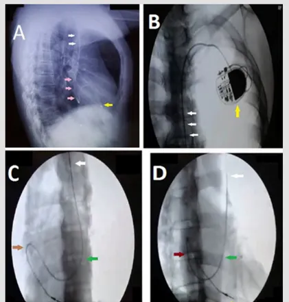
As the patient was still pacemaker-dependent, a permanent pacemaker (PPM) implantation was undertaken. For placement of the PPM (Fig. 1), venous access was achieved through the left subclavian vein and a 7 French sheath passed. While introducing the guidewire it was noticed on cineflouroscopy that instead of crossing the spine toward the right side, the guidewire bent smoothly toward the left side of the mediastinum, raising the suspicion of a PLSVC. To confirm this, the guidewire was pulled back and contrast injected, which showed a PLSVC opening into the coronary sinus. The guidewire was reintroduced gently and the sheath passed through the PLSVC. The lead was passed through the PLSVC and a loop formed in the right atrium to cross the tricuspid valve. The lead was affixed in the right ventricle and the pacemaker started pacing when attached to the pulse generator. On the first day after implantation, the pacemaker showed appropriate sensing and pacing.
Figure 1. Imaging of single chamber VVI PPM in the following views. A: Chest X-ray left lateral view showing the pacemaker’s right ventricular lead placed in the right ventricle (yellow arrow) coming through the PLSVC (white arrows) and coronary sinus (pink arrows).
B: anteroposterior cineflouroscopy view showing a single chamber permanent pacemaker device implanted on the left side of the upper chest (yellow arrow) and right ventricular lead passing through the PLSVC (white arrows). C: left anterior oblique cineflouroscopy view showing the lead tract – PLSVC (white arrow), coronary sinus (green arrow), right atrium (brown arrow) and right ventricle (yellow arrow).
D: right anterior oblique cineflouroscopy view showing the lead tract – right ventricular lead affixed in the right ventricle after making a loop in the right atrium, PLSVC (white arrow), coronary sinus (green arrow), right atrium (brown arrow) and right ventricle (yellow arrow)
European Journal
of Case Reports in
Internal Medicine
DOI: 10.12890/2020_001484 European Journal of Case Reports in Internal Medicine © EFIM 2020
Case 2
A 53-year-old female patient presented to the emergency department with sudden onset of severe palpitations, profuse sweating, dizziness and lightheadedness. While checking her vital signs, the patient collapsed suddenly with a non-palpable carotid pulse. Cardiopulmonary resuscitation was started immediately and cardiac rhythm checked on the defibrillator monitor after the first cycle, which showed a wide complex tachycardia with clear atrioventricular dissociation suggestive of monomorphic ventricular tachycardia. Immediate electrical cardioversion was carried out successfully and the patient reverted to sinus rhythm.
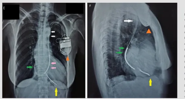
The patient had a history of hypertension, ischemic heart disease and ischemic cardiomyopathy with a left ventricular ejection fraction of 25%, for which she was taking daily aspirin 75 mg, rosuvastatin 20 mg, bisoprolol 5 mg, enalapril 10 mg and spironolactone 25 mg. As the patient survived the cardiac arrest, it was decided to insert a dual-chamber implantable cardioverter-defibrillator (ICD) as a secondary prophylaxis of sudden cardiac death. Venous access was achieved and 7 French sheath passed into the left subclavian vein. While passing the guidewire, it was observed that it was bending in the left mediastinum without resistance instead of going toward the right side and then bending in the superior vena cava. To visualize the abnormal guidewire pathway, contrast was injected that showed the PLSVC opening in the coronary sinus. The sheath was therefore passed through the PLSVC, first the right atrial lead was affixed and then the right ventricular lead was passed through the tricuspid valve after making a loop in the right atrium and affixed in the right ventricle (Fig. 2).
Figure 2. Case 2 Imaging of single chamber VVI PPM in the following views. Left: Chest X-ray PA view, both right atrial and right ventricular leads passing through PLSVC [Dual chamber ICD generator: orange arrowhead; PLSVC: white arrows; Coronary sinus: pink arrows; Right atrium: green arrow; Right ventricular: yellow arrow]
Right: Chest X-ray lateral view, both right atrial and right ventricular leads passing through PLSVC [Dual chamber ICD generator: orange arrowhead; PLSVC: white arrow; Right atrium: green arrows; Right ventricular: yellow arrow]
DISCUSSION
At the eighth week of gestation, the embryonic venous system undergoes various developments. The cephalic portion of both superior cardinal veins form internal jugular veins of each side and are anastomosed to form the brachiocephalic vein. While the caudal portion of both sides varies, the right-sided caudal portion of the cardinal vein forms the superior vena cava, while the left portion regresses to form the ligament of Marshall. If this left-sided caudal portion fails to obliterate, it can persist as a left superior vena cava.
PLSVC can be diagnosed by transthoracic and transesophageal echocardiogram with contrast venography, computed tomography venography or magnetic resonance venography, but most commonly by incidental finding during pacemaker or ICD implantation through the left subclavian vein[5]. When contrast is injected through the left subclavian vein, it appears in the right atrium first normally but in the
case of a PLSVC it appears in the coronary sinus first. It should also be suspected when the coronary sinus appears dilated in any cardiac imaging modality.
It is important to note that it is technically difficult to cross the guidewire through the tricuspid valve when it comes from the PLSVC and coronary sinus without making a loop in the right atrium, which is known as a wide loop technique[6, 7]. It can cause perforation and
dissection of the thin-walled coronary sinus. Other complications reported are coronary sinus thrombus, arrhythmias, cardiac tamponade and cardiogenic shock[8].
European Journal
of Case Reports in
Internal Medicine
DOI: 10.12890/2020_001484 European Journal of Case Reports in Internal Medicine © EFIM 2020
REFERENCES
1. Tak T, Crouch E, Drake GB. Persistent left superior vena cava: incidence, significance and clinical correlates. Int J Cardiol 2002;82(1):91–93. 2. Campbell M, Deuchar DC. The left-sided superior vena cava. Br Heart J 1954;16(4):423–439.
3. Hsu L-F, Jaïs P, Keane D, Wharton JM, Deisenhofer I, Hocini M, et al. Atrial fibrillation originating from persistent left superior vena cava. Circulation 2004;109(7):828–832. 4. Postema PG, Rammeloo LAJ, van Litsenburg R, Rothuis EGM, Hruda J. Left superior vena cava in pediatric cardiology associated with extra-cardiac anomalies. Int J Cardiol
2008;123(3):302–306.
5. Uçar Ö, Çiçekçioğlu H, Kocaoğlu I, Aydoğdu S, Paşaoğlu L, Vural M. Persistent left superior vena cava with absent right superior vena cava: a case report and review of the literature. Cardiovasc J Afr 2010;21(3):164–166.
6. Biffi M, Boriani G, Frabetti L, Bronzetti G, Branzi A. Left superior vena cava persistence in patients undergoing pacemaker or cardioverter-defibrillator implantation: a 10-year experience. Chest 2001;120(1):139–144.
7. Dilaveris P, Sideris S, Stefanadis C. Pacing difficulties due to persistent left superior vena cava. EP Eur 2011;13(1):2.
8. Rose ME, Gross L, Protos A. Transvenous pacemaker implantation by way of an anomalous left superior vena cava. J Thorac Cardiovasc Surg 1971;62(6):965–966.
9. Couvreur T, Ghaye B. Left superior vena cava. In: Rémy-Jardin M, Rémy J, editors. Integrated Cardiothoracic Imaging with MDCT, Berlin/Heidelberg, Springer; 2009, pp. 289– 305.
Therefore, if lead placement through a PLSVC is unsuccessful or a specialist under-experienced, the right superior vena cava should be utilized for device implantation, as in 80–90% of cases both superior vena cava coexist[9].
CONCLUSIONS
Though PLSVC is an uncommon anomaly, this anatomical variant should be recognized appropriately, especially by specialists who frequently carry out procedures through the left subclavian vein such as implantation of PPM. It should also be recognized that loop formation of the lead in the right atrium is helpful to cross the tricuspid valve and to affix the lead in the right ventricle. The cardiac imaging specialist should also suspect and rule out PLSVC on encountering a dilated coronary sinus on any imaging modality.