Alma Mater Studiorum – Università di Bologna
DOTTORATO DI RICERCA IN
SCIENZE BIOCHIMICHE E BIOTECNOLOGICHE
Ciclo XXIXSettore Concorsuale di afferenza: 05/G1 Settore Scientifico disciplinare: BIO/14
TITOLO TESI
MESENCHYMAL STEM CELLS AS A DELIVERY
PLATFORM FOR PHOTOACTIVABLE
NANOPARTICLES FOR CANCER THERAPY
Presentata da: Dott. Enrico Lucarelli
Coordinatore Dottorato Relatore
Prof. Santi Spampinato Prof. Santi Spampinato
i
P
REFACE
In spite of the huge progress made in the treatment of tumors in the last thirty years, for some tumors, such as osteosarcoma, the survival rate of the patients treated with the combination of multi-agent chemotherapy and surgery has not improved. This could be explained in part because chemotherapy is not selective for tumor cells, but also kills normal cells that are in active cell division. Thus, if the toxicity of the multiagent chemotherapy is increased, normal tissue will also be damaged. In order to improve patient’s survival rate, innovative treatments that specifically target osteosarcoma cells should be developed.
Mesenchymal stem cells are currently tested in more than 500 clinical trials to treat patients affected by conditions that range from traumatic and degenerative damages of tissues to the treatment of graft versus host diseases. Mesenchymal stem cells are also attracted by alteration of the tissue architecture caused by malignant cancer growth. This tumor-homing property makes mesenchymal stem cells an ideal cellular candidate to deliver selectively therapeutic agents to tumor cells.
In this context, this PhD project concerns two proofs of concept. The first being that when mesenchymal stem cells are used as a cellular vehicle to deliver photoactivable nanoparticles, they can induce osteosarcoma cell death. Secondly, nanoparticles decorated with a photosensitizer that can be activated in the near infrared region of the visible spectrum, once photostimulated, can induce tumor growth inhibition in mice bearing tumors.
An introduction, including the related references, provides the overview of the PhD project scenario. A second section reports the general aims of the thesis and the experimental part that is divided into two chapters concerning the investigated application fields. Published or submitted for publication results, supported by their specific references, are reported in the third section. Final conclusions and future perspectives complete the present thesis.
G
ENERAL
I
NDEX
ACRONYM INDEX ... IV
INTRODUCTION ... 5
1. Osteosarcoma ... 5
2. Mesenchymal stem cells ... 6
3. Mesenchymal stem cells and tumors ... 7
4. Mesenchymal stem cells as a vector of antitumor therapy ... 8
4.1 Genetically modified mesenchymal stem cells ... 8
4.1.2 Mesenchymal stem cells engineered to express antiangiogenic proteins... 13
4.1.3 Mesenchymal stem cells engineered to express proapoptotic proteins ... 13
4.1.4 Mesenchymal stem cells engineered to express proteins that enable the incorporation of radionucleotides ... 15
4.2 Mesenchymal stem cells as an anticancer cargo ... 16
4.2.1 Mesenchymal stem cells loaded with chemotherapeutic agents ... 16
4.2.2 Mesenchymal stem cells loaded with nanoparticles ... 18
4.2.3 Mesenchymal stem cells loaded with oncolytic viruses ... 19
5 Photodynamic therapy ... 20
CHAPTER 1 ... 22
Aims and objectives... 22
Methods ... 22
Photoactivable Nanoparticles ... 22
Isolation, culture and immunophenotypic characterization of human mesenchymal stem cells... 22
Human osteosarcoma cell line ... 22
FNPs uptake and photoactivation ... 23
Results and Discussion ... 24
FNPs uptake by MSCs ... 24
FNPs loaded MSCs induce OS cell death after Photactivation ... 25
Conclusions ... 27
CHAPTER 2 ... 27
Aims and objectives... 27
Methods ... 27
Photoactivalble Nanoparticles ... 27
Cell cultures ... 28
Microtissues and PDT ... 28
In vivo study ... 29
Analyses on tumor explants ... 29
Results and discussion... 29
Photoactivation of FNPs in tumor microtissues ... 29
In vivo Photodynamic therapy of solid tumors... 31
iii
PUBLICATIONS ARISING FROM THIS WORK ... 33
CONCLUSIONS AND FUTURE DIRECTIONS ... 34
ACKNOWLEDGEMENTS ... 35
A
CRONYM
I
NDEX
Adipose stem cells (ASCs) Event free survival (EFS) Extracellular vesicles (EVs) Fluorescent Nanoparticles (FNPs)Herpes simplex type 1 thymidine kinase (HSVTK) In vivo bioluminescence imaging (BLI)
Mesenchymal stem cells (MSCs) Microvesicles (MVs) Near-Infrared (NIR) Nanoparticles (NPs) Osteosarcoma (OS) Photodynamic Therapy (PDT) Photosensitizers (PS)
Sodium-iodide symporter (NIS) Stroma-derived factor 1 (SDF1) Reactive Oxygen Species (ROS)
5
I
NTRODUCTION
1. Osteosarcoma
Sarcomas are malignant tumors that originate in bones, cartilage, fat, nerves and blood vessels. Among the sarcomas, high grade osteosarcoma (OS) is the most common primary bone tumor. OS generally develops in the long bones of adolescents. Standard treatment for OS includes multi-agent neoadjuvant chemotherapy, surgery and multi-agent adjuvant chemotherapy. Radiotherapy can also be used. The 5-year event free survival (EFS) of OS patients with nonmetastatic disease at presentation is 70% (Longhi, Errani, De Paolis, Mercuri, & Bacci, 2006). Between 20-25% of the OS patients develop metastasis. In over 90% of the cases, the metastasis develops in the lungs and is the first cause of disease mortality. For patients with metastasis at presentation the EFS is below 40%. Patients with bilateral metastasis have worse outcomes than monolateral (Ferrari & Serra, 2015)(Bacci et al., 2008).
In spite of the huge effort made in the last 30 years to improve patient’s survival rate, little progress has been made and the prognosis of OS has plateaued since 1990. The major limitation of the current therapeutic protocols is the lack of tumor targeting. This could be explained in part because chemotherapy is given systemically and lacks precise specificity to target only malignant cells, it is known that chemotherapy also kills healthy cells that are in active cell division. As a consequence, healthy tissues are also damaged when the toxicity of the drugs is increased. Thus, the development of novel alternative strategies to current standard treatment is needed. In particular it is important that the innovative therapeutic agents have an increased capacity to selectively eliminate cancer cells as well as reduce the toxicity in healthy tissues. Currently, alternative treatments to standard therapy are being tested in clinical trials for OS patients, particularly therapeutic approaches that have shown to be effective for several types of advanced tumors. For example immunotherapies, such as vaccination and cytokine therapy, have been investigated for OS treatment (Uehara et al., 2015). However, these therapeutic agents are short lived, are toxic when administrated at high doses, and are constantly cleared by specialized cells and organs. Therefore, new therapeutic strategies that are able to deliver therapeutic agents directly to the tumor cells at elevated concentrations shielded from the immune system must be developed.
In 2002 Dr. Studeny published findings that mesenchymal stem cells (MSCs) can be used as delivery vehicles to selectively transport therapeutic agents to cancer cells because they have selective tropism for tumors and also because MSCs are immunosuppressive and can evade the immune system (Studeny et al., 2002). This therapeutic approach, named the
Trojan horse strategy, is important because MSCs can be exploited to deliver anticancer agents to the tumor microenvironment.
2. Mesenchymal stem cells
MSCs were first identified in 1966 as osteogenic cells (Friedenstein, 1966). MSCs are fibroblast–like cells that reside in many adult, perinatal, and fetal tissues. For example, MSCs reside in bone marrow where they represent a small population, about 0.001-0.01% of the cells (Muschler, Nitto, Boehm, & Easley, 2001). In physiological conditions, MSCs have a key role in the support of the stem cell niche. MSCs live close to hematopoietic stem cells in the bone marrow and provide for their survival (Chitteti et al., 2010). MSCs also constitute a local reservoir of cells that in time of stress or injury, can be recruited by solid organs to promote tissue repair. MSCs are functionally plastic cells that can sense clues from the local microenvironment and respond accordingly. After injury, local microenvironment changes due to hypoxia and the presence of dead or damaged cells lead to inflammation and immune response. MSCs respond to injuries, inflammation, and tumors in a way similar to that of the immune cells (Spaeth, Klopp, Dembinski, Andreeff, & Marini, 2008). MSCs can sense the pro-inflammatory cytokines released and migrate from the stem cell niche to the damaged site, where they cross-talk with the surrounding microenvironment. For example, MSCs are attracted by proteins such as stroma-derived factor 1 (SDF1) that binds to the receptor CXCR4 (Homing, Stem, Whiteman, & Ph, 2007). Once MSCs have engrafted at the damaged site, they release a spectrum of bioactive molecules that orchestrate tissue repair. MSCs first secrete soluble immunomodulatory and trophic factors that suppress both transient and continuous immune surveillance. For instance, MSCs inhibit the activity of natural killer cells, helper T cells, and cytotoxic T cells while activating the generation of regulatory T cells. After this potent immunosuppressive activity, MSCs participate actively in the support of homeostasis by active inhibition of apoptosis. The sum of these actions starts tissue remodeling and enhances the healing process (Maxson, Lopez, Yoo, Danilkovitch-Miagkova, & Leroux, 2012). Thus, it is currently believed that the therapeutic potential of these cells does not depend only on their stemness, but also on their secretory paracrine activities that include the secretion of extracellular vesicles (EVs) and exosomes.
The self-healing capacity of the human body is limited and when the injury is extensive, or the number of MSCs that migrate to the damaged tissue is insufficient, the tissue cannot be repaired by local MSCs. The ability of MSCs to migrate to areas of injury in a range of pathologic conditions suggests that they may be ideal vectors for therapeutic delivery and MSCs have been proposed as promising therapeutic tools for regenerative medicine purposes.
7 A large number of investigators have demonstrated that MSCs are easily obtained from tissue biopsies, can be easily expanded in culture, are relatively non-immunogenic and have a remarkable plasticity, thus having the potential to differentiate into cells that have the characteristics of several mesenchymal lineages. MSCs also express low levels of major histocompatibility complex I and do not express major histocompatibility complex II, which renders the allogenic use possible (Le Blanc, Tammik, Rosendahl, Zetterberg, & Ringdén, 2003). The sum of these features make MSCs the most commonly used adult stem cells in therapies for tissue engineering and regenerative medicine. 665 MSC-based clinical trials are listed in the database of the National Institute of Health (www.clincaltrials.gov) (October 31 2016). Most of the trials registered at the site are Phase I/II clinical trials in which MSCs are used to treat a vast array of conditions (Squillaro, Peluso, & Galderisi, 2016).
3. Mesenchymal stem cells and tumors
Tumors are defined as a wound that never heals (Dvorak, 1986). Malignant transformation of cells is associated with a loss of the growth inhibition and invasion of the surrounding tissues. At first, tumor growth causes changes in the microenvironment that are similar to those caused by tissue injury or stress. Local cells react to the stress and provide support with the creation of the tumor stroma. Tumor stroma provides structural support as well as biochemical and mechanical cues to tumor cells. Tumor stroma also supports the uncontrolled tumor growth, which causes the destruction of tissue organization. This sets in motion a self-feeding spiral of events. When tumor growth reaches a certain level, the body reacts with inflammation and the immune reaction, together with the hypoxic environment and growth factors, chemokines, and cytokines released by tumor cells, actively promote MSCs migration from the niche to the tumor stroma which can support tumor progression (Sung et al., 2008)(Pietras & Östman, 2010)(Spaeth et al., 2008). Beside the migration of MSCs to the tumor site caused by inflammation, a range of tumors actively attract MSCs, such as colon and pancreatic cancer, breast and ovarian cancer, gliomas, osteosarcomas and melanomas. MSCs have tropism for gliomas. MSCs migrate and localize glioma cells via platelet-derived growth factor, epidermal growth factor, and stromal-derived factor-1-mediated tropism(Pretheeban, Lemos, Paylor, Zhang, & Rossi, 2012). Furthermore, several researchers have proved that tumors provide a permissive environment for engraftment of exogenous implanted MSCs (Karnoub et al., 2007)(Nakamizo et al., 2005) (Studeny et al., 2002)(Belmar-Lopez et al., 2013).
This is relevant for the clinical application of ex-vivo expanded MSCs because when MSCs are administrated in patients, they could be engrafted in the tumor stroma and interact
with cancer cells. The concern is that once MSCs are in the tumor stroma by suppressing the immune system, they could promote tumor immune evasion and by inhibiting apoptosis, stimulating mitosis, and enhancing angiogenesis, they could cause an increase in tumor growth. In-vivo experiments were performed and resulted in the demonstration that administered MSCs engraft in cancer stroma and increase cell metastasis (Karnoub et al., 2007)(Suzuki & Sun, 2011). In contrast, administered MSCs have been shown to have an anti-tumor activity in several types of tumors (Cortes-Dericks, Froment, Kocher, & Schmid, 2016). In these publications, diverse mechanisms have been reported to be involved in the antitumor potential of MSCs. It has been observed that MSCs release soluble factors which contribute to the reduction of tumor growth and tumor progression (Khakoo et al., 2006).
In conclusion, MSCs inherent specific tropism for tumors makes MSCs ideally suited system able to have a specific and local release of therapeutic agents to the tumor site. A number of animal studies have raised concerns that the implant of MSCs can increase tumor growth and metastasis (Karnoub et al., 2007; Spaeth et al., 2008), while the implant of MSCs has been shown to be beneficial in others (Khakoo et al., 2006). While this topic is still controversial and studies are performed to elucidate why the administration of MSCs has been shown to be detrimental in several in vivo animal models, the exploitation of MSCs as a cellular vehicle is essential for the improvement of anticancer therapies and should go on.
4. Mesenchymal stem cells as a vector of antitumor therapy
The first seminal publication that proved that MSCs can be used to deliver therapeutic agents to treat cancer was published in 2002 (Studeny et al., 2002). In the following years, more than 100 papers have been published in which MSCs have been used to deliver therapeutic agents directly to the tumor. In the majority of these papers, MSCs are genetically modified to express and release proteins. MSCs have been engineered to deliver proteins that cause a direct or indirect tumor death. MSCs have been also loaded with oncolytic viruses, chemotherapeutic agents and nanoparticles that have anti-tumorigenic properties.
4.1 Genetically modified mesenchymal stem cells
MSCs can be easily genetically engineered, using the available molecular techniques to express genes of interest. MSCs have been genetically modified for both regenerative medicine purposes and to treat a wide array of diseases (D’souza et al., 2015). For example, MSCs have been genetically modified to express BMP 2 to enhance bone density. Turgeman et al demonstrated this by engineering MSCs to express BMP2 in order to enhance bone formation in osteoporotic rats (Turgeman et al., 2001).
9 MSCs have been genetically modified to deliver therapeutic agents to tumor cells as well. For example, MSCs are being engineered to express genes that improve the immune response, genes that induce cell death of tumor cells, and genes that enable radio nucleotide uptake. Viral vectors, such as lentivirus, retrovirus, and adeno-associated virus, have been used for long term transgene expression in MSCs. Non-viral gene delivery methods have been used as well. In these studies, the therapeutic efficacy of genetically engineered MSCs has been shown in diverse experimental localized and metastatic tumor types.
4.1.1 Mesenchymal stem cells engineered to express cytokines that enhance endogenous immune response to cancer cells.
Cytokines have been widely used in the innate and adaptive immune responses against tumors. However, systemic treatment with recombinant proteins is frequently associated with severe side effects in immunotherapy. Therefore, a selective delivery system that accumulates the cytokines within the tumor microenvironment may be more effective and minimize side effects.
4.1.1.1 Mesenchymal stem cells engineered to express interferons
Interferons (IFNs) display a multitude of anti-tumor biological activities, including activation of the immune response, antiproliferative and proapotiotic apoptosis effects. However, in vivo systemic administration of high doses of IFNs have undesired side effects caused by the excessive toxicity. Furthermore, IFNs have a short half life and the protein may not reach the required concentration to be effective at tumor site. The first use of MSCs as a vector for antitumor therapy was published in 2002 by Studeny (Studeny et al., 2002). In this paper, the authors used an adenoviral vector to genetically engineer MSCs to express IFN- a strong antiproliferative cytokine that is known to have anticancer properties. Results showed that genetically engineered MSCs were able to engraft in tumor stroma and to increase the survival of mice implanted with the human metastatic melanoma cell line A375SM. The efficacy of the engineered MSCs was demonstrated both when MSCs were co-implanted with the melanoma cells and also when MSCs were injected systemically after establishment of pulmonary metastasis (Studeny et al., 2002). Although the effect was temporary and not sufficient to eradicate the tumor, these striking results demonstrated that this therapeutic approach was sufficient to obtain locally a concentration of IFN- that was able to decrease the growth of such an aggressive tumor, while the same effect was not obtained with systemic infusion of IFN-. Local secretion of IFN- is preferred because it reduces systemic toxicity. In a subsequent paper, these authors demonstrated that after establishing pulmonary metastasis with the melanoma cell line A375SM and the breast tumor cell line MDA 231,
three doses of intravenously injected MSCs genetically engineered to express IFN-double mice’s survival. The same result was not obtained when MSCs expressing IFN-were implanted subcutaneously. In MDA 231, pulmonary metastasis intravenously injected MSCs colonized the tumor and participated in the formation of the tumor stroma. Only a small number of MSCs were found in the liver after necroscopy (Studeny et al., 2004). Studeny’s proof of concept demonstrated that MSCs could be used as a vector to deliver selectively therapeutic agents to the tumor and paved the way for future work.
Nakamizo et al. have investigated the tropism of labeled MSCs in vivo and demonstrated that MSCs injected in the carotid artery selectively migrate to Glioma. They showed in vitro that MSCs migrate toward Glioma. The authors performed intracranial injection of U87 and 7 days later injection in the carotid 1 x10⁶ labelled MSCs. The authors demonstrated that MSCs are found only in the tumor and were absent from the normal brain parenchyma. Results were confirmed injecting the same number of MSCs in the opposite carotid. Comparable results were obtained also when LN229 and U251 tumor cells were implanted. The authors demonstrated that the tropism is maintained even when the MSCs are injected in the contralateral hemisphere of the brain, while control fibroblasts remained in the site of the injection. With this paper the authors provided a proof of principle in vivo that MSCs expressing IFN- are effective against intracranial gliomas. They provided evidence that 1 x10⁵ MSCs expressing IFN- are necessary to increase mice survival when injected intratumorally, and they demonstrated that subcutaneous implant of 1 x10⁵ MSC expressing IFN- is not effective, implying that MSC intratumoral implants support tumor progression (Nakamizo et al., 2005).
The homing of MSCs to tumor stroma was also shown in a murine immunocompetent model of prostate cancer pulmonary metastasis. The intravenous injection of 5 x10⁵ MSCs induced to express INF- by an adenoviral vector reduced tumor growth and doubled mice’s life span (C Ren et al., 2008). In a related study, Ren et al. have confirmed preferential homing of MSCs to lung tumor. The authors also showed, in a murine model of pulmonary metastasis of melanoma, that intravenous injection of 5 x10⁵ MSCs expressing murine INF- by an adeno-associated vector can increase life span compared to control animals (Changchun Ren et al., 2008).
IFN- has been successfully used in the therapy of chronic myelogenous leukemia. Li
et al. genetically modified MSCs to express IFN- to treat Chronic Myelogenous Leukemia. In this in vitro study, cocultures of MSCs decreased K562 proliferation in 5 days because K562 cells are blocked in G1 (X. Li et al., 2006).
11 MSCs genetically engineered to express INF-are currently tested in a phase one clinical trial (NCT02530047) in which the maximal tolerated dose is investigated in patients affected by ovarian cancer. In this clinical trial, patients will receive intra-peritoneal infusion starting from 1 x10⁵ MSC/Kg once a week for 4 weeks up to 4 dose levels.
4.1.1.2– Mesenchymal stem cells engineered to express interleukin 2
The first report that MSCs can be used to deliver the antitumoral cytokine Interleukin 2 (IL-2) was published by Stagg et al. who genetically modified MSCs to express IL-2. MSCs were co-injected with the poorly immunogenic melanoma cell line B16 in C57bl/6 normal mice. The authors used two different models. In the first model, co-injection of MSCs at several doses (5 x10⁴, 5 x10⁵, 5 x10⁶ ) and B16 cells resulted in a delayed tumor growth and prolonged mice survival compared to control mice. In the second model, when IL-2 gene modified MSCs embedded in matrigel were implanted in the vicinity of a pre-established tumor, 90% of the mice failed to develop tumors. These results demonstrated that IL-2 delivery by MSCs adjacent to pre-established, low-burden tumor implants produces an immune response against the tumor that results in significantly delayed tumor growth (Stagg, Lejeune, Paquin, & Galipeau, 2004).
Nakamura et al. engineered MSCs to express IL-2 using an adenoviral vector. The efficacy of these cells was tested in a rat model established with the glioma cell line 9L, in which the efficacy of IL-2 was already proved. The authors used two models. In the first, the MSCs were co-implanted, in the second, the MSCs were intra-tumorally injected after 3 days. Results demonstrated that MSCs implanted in the contralateral hemisphere have a tropism toward syngeneic glioma in rats. They further showed that survival of glioma-bearing rats increased in both models after MSCs injection. Prolonged survival caused by MSC-IL-2 was associated with inhibition of tumor growth as measured by MRI (Nakamura et al., 2004).
4.1.1.3Mesenchymal stem cells engineered to express interleukin 12
Interleukin 12 (IL-12) is a pro-inflammatory cytokine that has several effects. Produced by antigen presenting cells, IL-12 induces the proliferation of Natural Killer and cytotoxic T lymphocytes. IL-12 induces also the production of IFN- that upregulates major histocompatibility complexes and adhesion molecules resulting in an enhanced antitumor immunity. Administration of IL-12 protein, or DNA, promotes cellular immunoregulatory responses against tumors affecting tumor growth. However, IL-12 is effective only at a high dose range that usually has toxic effects and this has hindered the clinical application of IL-12 (Meko, Yim, Tsung, & Norton, 1995). Therefore, a system that would activate the
immunological reaction directly in the cancer stroma without causing systemic toxicity would be ideal.
In 2006 Chen et al. published an interesting paper in which MSCs genetically engineered to express IL-12 of mouse origin were used as a preventive strategy. The authors inject the MSCs in the peritoneum of mice one week before the injection in the fat pad of ectopic lung, melanoma and hepatoma tumors. The treatment prevented the formation of a tumor in 95% of the cases. The authors suggested a prophylatic use of MSCs. These results suggest that the engineered MSCs are effective especially if the dimension of the tumor is small (X. C. Chen et al., 2006). In another study by the same group, the authors investigated the efficacy of MSCs engineered to express IL-12 in 3 RAT ectopic tumor models (melanoma, breast tumor, hepatoma) to test their anticancer and antimetastatic activity. They treated animals with injection of engineered 1 x10⁶ MSCs 4 times in a 20 days period. The results indicate that the treatment led to a significant reduction of the lung metastasis in the tumor models (X. Chen et al., 2008). Elazouk et al. retrovirally transduce MSC to express IL-12 and they inject the MSCs in 2 mouse melanoma models. In one model, the tumor was established subcutaneously in the flank of the mice and the mouse melanoma cells B16F10 were implanted. When the tumor reached 20mm3, 7,5 x10⁵ engineered MSCs were injected. The mice developed antibodies against the human cells and most of the human MSCs did not survive in the mice, after 7 days only 7% of the human MSCs remained. In spite of the reduced survival, injection of the engineered MSCs decreased the tumor’s volume and increased the life span of the mice. In the second model, they first injected the mice with 7.5 x10⁵ engineered MSCs and later injected 7.5 x10⁵ B16F10 cells in the tail vein to establish lung metastasis. With this prophylactic treatment they were able to decrease the number of metastases by at least 50% (Elzaouk, Moelling, & Pavlovic, 2006). Seo et al. tested several injection routes of MSCs engineered to express IL-12. When control MSCs were injected, MSCs did not alter tumor progression, while intra-tumoral injected engineered MSCs outperformed intravenous injection and decreased tumor growth and metastasis formation, and increased animal life compared to controls. Best results were obtained with intratumoral injection of engineered MSCs embedded in matrigel (Seo et al., 2011).
4.1.1.4 Mesenchymal stem cells engineered to express CXC3CL1
Soluble CXC3CL1 (Fractalkine) is a member of the CX3CL family and exists in both membrane-bound and soluble forms. The soluble form induces the migration of cells expressing the receptor CX3CR1, such as monocytes, NK cells and T cells. Intratumoral injections of CXCL1 have a strong immunostimulatory effect that causes a strong antitumoral effect (Xin et al., 2005). Xin et al. demonstrated that intravenous injected MSCs engineered
13 using an adenovirus vector CX3CCL1 in mice bearing lung metastasis of adenocarcinoma and melanoma cells localize in the tumor stroma in the lungs. Engineered MSCs decrease lung meta growth and double mice’s survival (Xin et al., 2007). In another study, the authors demonstrated that 7 days after intratracheal injection, MSCs localize within and around lung metastasis more than in the healthy lungs. In both cases, in the adenocarcinoma and lung carcinoma metastasis models, intratracheal injection of MSCS engineered to express CXC3CL1 12 days after tumor establishment decreased the number of metastases and prolonged the survival of the mice. In the adenocarcinoma model, the mice survived 5 times more than the controls, while in the lung metastasis survived 3 days longer (Xin et al., 2009).
4.1.2 Mesenchymal stem cells engineered to express antiangiogenic proteins
4.1.2.1 Mesenchymal stem cells engineered to express NK4
The blood supply is crucial for the tumor survival and for the dissemination of the metastasis. Several antinagiogenic approaches have been employed in oncology and some of them are successful. MSCs have been engineered to express antiangiogenic proteins such as NK4 that is an agonist of hepatocyte growth factor (HGF), a multifunctional growth factor that supports tumor growth, angiogenesis and lymphogenesis. NK4 antagonizes the phosphorylation of cMet by HGF. In vivo systemic administration of NK4 adenoviral vectors in mice that received subcutaneous tumors causes a strong antitumor effect, but also severe adverse effects (Maemondo et al., 2002). Kanehira et al. studied the efficacy of 1 x10⁵ MSCs engineered with an adenovirus vector to express NK4 in a mice model of pulmonary metastasis of colon carcinoma. They demonstrated a preferential homing of systemically injected engineered MSCs to tumor lung metastasis, compared to normal tissue. MSCs that expressed NK4 reduced the number of lung metastases, increased the survival of tumor bearing mice, and reduced angiogenesis and lymphogenesis (Kanehira et al., 2007). In another study, Haber et al. assessed the feasibility of therapeutic ultrasound to transfect MSCs with pDNA encoding for PEX, a protein that inhibits tumor angiogenesis in an ectopic prostatic cancer animal model. Results showed that 1 x10⁶ MSCs expressing PEX can induce a 70% decrease in tumor volume, with a single intravenous injection that can reach an 84% decrease when the injections are three (Haber, Baruch, & Machluf, 2017).
4.1.3 Mesenchymal stem cells engineered to express proapoptotic proteins.
4.1.3.1 Mesenchymal stem cells engineered to express TRAIL
Tumor necrosis factor-related apoptosis inducing ligand (TRAIL) is a membrane ligand from the tumor necrosis factor (TNF) family. TRAIL is a ligand that induces apoptosis primarily in tumor cells by binding to certain death receptors TRAIL-RI and TRAIL-RII.
Most of normal cells, MSCs included, do not express the death receptors, and therefore are not affected by TRAIL expression. TRAIL has been used in different models of cancers in
vivo, resulting in notable anti-tumor activity, making TRAIL an attractive therapeutic tool for
tumor therapy (Siegmund, Lang, & Wajant, 2016). The efficacy of intravenous delivery of recombinant TRAIL is limited by the short half-life of the protein and frequent injections are needed in order to achieve the desired effect. Therefore, the use of MSCs engineered to produce TRAIL is a promising tool to be used in treatment of several types of cancer, because MSCs can be used as a constant source of this pro-apoptotic protein.
Several authors were interested to test whether MSCs can function as a shield to protect the adenovirus from the immune system. Mohr et al. tested whether an intratumoral injection of MSCs expressing TRAIL was effective in an ectopic subcutaneous model of lung metastasis. TRAIL-expressing MSCs reduced tumor growth in the model causing diffused apoptosis (Mohr et al., 2008). Sasportas et al. investigated the effect of MSCs engineered with a lentiviral vector to express TRAIL in a preclinical model. Interestingly, MSCs survived longer when the tumor was implanted, compared to normal brain cells, suggesting that the two cell types cross talk, even if in vivo MSCs do not support tumor growth. They demonstrated that MSCs do not support the growth of the glioma in the brain but that MSCs migrate toward the glioma. Analysis of the efficacy demonstrated that co-implant of glioma and engineered MSCs in the brain of a mouse, as well as implant of the engineered MSCs 7, or 14 days after tumor implant resulted in reduction of glioma growth and increased survival of the tumor bearing mice (Sasportas, Kasmieh, Wakimoto, Hingtgen, & Water, 2009). In a similar paper, Leobinger et al. showed that lentiviral induced expression of TRAIL in MSCs was capable of reducing tumor volume in a lung metastatic tumor model obtained after the injection of the cell line MDAMB231 in the tail vein. MSCs were injected systemically 10 days after the tumor cells engraft in the tumor stroma preferentially, compared to lung parenchyma. The injection of the engineered MSCs was effective and 3 of the 8 mice were tumor free (Loebinger, Eddaoudi, Davies, & Janes, 2009). Menon et al. injected the glioma cell line U87 and five days later ipsilaterally injected 6 x10⁴ MSCs lentivirally engineered to express TRAIL. Histology performed two weeks after MSCs injection revealed that the treatment reduced the tumor volume by 81.6% and the survival was increased form 37 days to almost 60 (Menon, Shi, & Carroll, 2009). In another study, the authors used MSCs obtained from the adipose tissue, the adipose stem cells (ASCs). The authors transfected ASCs in a stable manner to express TRAIL. In vitro, they demonstrated that coculture of ASCs expressing TRAIL induced apoptosis of Ewing sarcoma cell lines by cell-to-cell contact. In
15 sarcoma models. Results indicated that when TRAIL is secreted by MSCs, it is more effective compared to the administrated TRAIL itself. They demonstrated also that cell to cell interaction was needed for ASCs expressing TRAIL to be effective. The authors investigated the importance of the timing of the injection of the ASCs in an animal model in which the Ewing sarcoma cells were injected in the muscle in the proximity of the tibia. Results demonstrated that when the ASCs were co-injected with the tumor cells, the treatment decreased the tumor volume by 75% at day 20 and more than doubled the life of tumor-bearing mice. The treatment was not effective when the ASCs expressing TRAIL were injected after the tumor was already established (Guiho et al., 2016).
4.1.3.2 Mesenchymal stem cells engineered to express TNF-
TNF- is a pleiotropic cytokine that induces tumor cell apoptosis. Tyciakova et al. tested the efficacy of the intravenous administration of 1 x10⁶ ASCs genetically modified to express TNF- in a model of melanoma lung metastasis. They could prove the homing of the engineered ASCs in the lungs (Tyciakova, Matuskova, Bohovic, & Kucerova, 2017).
4.1.3.3 Mesenchymal stem cells engineered to express HSV-TK
The herpes simplex type 1 thymidine kinase (HSVTK) has also been used as a “suicide gene” as a safety system in gene therapy experiments, allowing cells expressing the gene to be killed because HSVTK converts non-toxic gancyclovir into a toxic metabolite. In a rat experimental model of glioblastoma Miletic et al. demonstrated that MSCs induce
HSVTK gene expression by a retrovirus migrate to the tumor and prolongation of the rats’ lifespan by 70% compared to control rats. Histology revealed that no tumor cells were detectable and a scar tissue was left in the mice. The scar tissue was filled with CD8, NK, and other immune cells (Miletic et al., 2007). Currently a phase I clinical trial designed to test the safety of MSCs genetically engineered to express HSVTK in patients with advanced or recurrent or metastatic gastrointestinal adenocarcinoma is currently undergoing in Germany (NCT02008539).
4.1.4 Mesenchymal stem cells engineered to express proteins that enable the incorporation of radionucleotides
Radioactive iodine is used to as a diagnostic and therapeutic tool in patients affected by thyroid carcinoma. Taking advantage of the fact that only thyroid follicular cells express the sodium-iodide symporter (NIS), a glycoprotein that transports both Na+ and I- ions, uptaken radioactive iodine by these cells enables to visualization (gamma emission) and killing (beta emission) of thyroid carcinoma cells. In a combined strategic approach, the capacity of NIS to concentrate radioactive iodine is exploited also for other types of tumors.
In this approach, MSCs are engineered to express NIS and once MSCs are engrafted in the tumor stroma they can uptake radioactive iodine causing local death in a range of few millimiters. For example, Dwyer demonstrated in vivo that intravenous (IV) and intratumoral injection of MSCs that express NIS engraft in the tumor and can reduce the tumor size after the injection of 131I (Dwyer et al., 2011). Results presented demonstrate that the IV injection is more effective compared to the intratumoral injection. The authors explain the difference in the effectiveness of the two types of injection based on the fact that intratumoral injection of MSCs does not reach all the tumor cells as reported also by Studeny (Studeny et al., 2002), and Nakamura (Nakamura et al., 2004).
4.2 Mesenchymal stem cells as an anticancer cargo
MSCs can deliver traditional anticancer drugs without requiring genetic manipulation. MSCs can function as cellular cargo to deliver various anticancer therapeutic agents, such as chemotherapeutic drugs, nanoparticles and oncolytic viruses. Compared to the systemic infusion of these drugs, this strategy enables a selective delivery to the tumor site of elevated doses of the therapeutic agent that affects tumor cells and the associated microvessels, while keeping systemic toxicity at minimum.
4.2.1 Mesenchymal stem cells loaded with chemotherapeutic agents
The outcome for patients affected by some kind of tumor has greatly improved in the last 30 years, while for other tumors has not. The improvement of the outcome for patients affected by tumors, for example with breast and prostate cancer, is partially due to the intensive screening that enables the diagnosis of the disease at an early stage, and partially to the improvement of the treatments. For other patients affected by other tumors, such as the osteosarcoma, mesothelioma and tumor of the pancreas, little progress has been made. This can in part be explained by the reasoning that for these tumors the diagnosis is made at a later stage, and in part because chemotherapy is infused systemically and is not selective for cancer cells and kills also normal cells that are in active cell division. Thus, if these drugs were delivered directly to the tumors by MSCs, the tumor could be treated with high doses of the drugs while keeping systemic toxicity at a minimum. MSCs have a relatively slow kinetic of replication that enables MSCs to uptake high doses of the drugs and reach the target site before the drugs causes cell immobilization or death. In a first seminal paper by Pessina et al., the authors demonstrated that MSCs exposed in vitro to Paclitaxel (PTX) can uptake the drug and release it in the culture media (Pessina PLOS 2011). PTX is used to treat several types of cancers because it has antiproliferative and antiangiogenic properties. A prolonged block of the progression of mitosis can either trigger the cell to enter in G1 phase without undergoing anaphase several times, resulting in aberrant multinucleated cells, or trigger apoptosis. PTX
17 treatment can have serious side effects, therefore a selective delivery of PTX only to cancer cells can be beneficial. MSCs resistance to PTX is probably dependent on the capacity of these cells to pump out the drug and on the ability of these cells to enter a block of cell division after tubulin polymerization that does not activate the cell’s death mechanisms. P-glycoprotein is an ATP-dependent drug efflux pump that is responsible for the uptake and the efflux of drugs and controls the concentration of some drugs within the cell. MSCs are the ideal carrier of toxic drugs because MSCs overexpress P-glycoprotein. P-glycoprotein overexpression makes MSCs highly drug resistant. Therefore, MSCs can be exposed to concentrations of drugs that are lethal for cancer cells without consequences. MSCs can acquire anti-cancer activity upon exposure to high doses of chemotherapeutic agents, such as PTX, because MSCs can release the uptaken drugs in the tumor stroma. The authors demonstrated that exposure of MSCs to PTX resulted in a moderate accumulation in S and G2-M phase and in a modest induction of apoptosis without affecting their viability and functions. In vitro, the PTX released by MSC was sufficient to inhibit tumor and endothelial cell proliferation (Pessina et al., 2011)(Pascucci et al., 2014). Similar results were obtained when MSCs were exposed to other chemotherapeutic agents such as doxorubicin and Gemcitabine (L. Li et al., 2011)(Bonomi et al., 2015), while MSCs contrarily do not uptake pemetrex (Petrella et al., 2017). Other authors have demonstrated that PTX reduces, but not impairs MSCs’ ability to migrate (Duchi et al., 2013)(Pacioni et al., 2015). PTX released by MSCs is also released via exoxosmes. MSCs are known to secrete membrane microvecicles (MVs) (that range in size from 20 to 900 nm) and exoxosomes (that are more homogenous in size ranging from 20 to 200 nm) to communicate among cells. Pascucci et al. demonstrated in
vitro that PTX uptake does not alter MSCs cytokine secretion. PTX is released by MSCs via
MVs and these MVs retain their antitumor activity (Pascucci et al., 2014). Once released, PTX can penetrate tumor cells (Duchi et al., 2013), increase the number of multinucleated tumor cells (Pacioni et al., 2015), and reduce tumor cell growth (Pacioni et al., 2015; Pessina et al., 2011). In vivo coinjection of MSC loaded with PTX resulted in the marked decrease of tumor volume in mice in which three different tumor types were established: prostate cancer (DU145 cells), melanoma (B16 cells) and glioblastoma, supporting that MSCs can be used to deliver PTX to tumors (Pessina et al., 2011). Similar studies have demonstrated that MSCs loaded with PTX reduce the growth of several tumors, such as leukemia, the growth of several subcutaneous tumor xenograft models, and the growth of glioblastoma multiform in a intracranial xenograft model (Pessina et al., 2013)(Pessina et al., 2013)(Pacioni et al., 2015). When MSCs are injected intravenously, they can reduce the formation of lung metastasis established with the melanoma cell line B16. Three doses of MSCs were intravenous injected
5, 10 and 15 days after the metastasis was established. MSCs loaded with PTX were found in the metastatic mouse models and a reduction of 90% of the metastasis was reached in a syngenic mouse model (Pessina et al., 2015). Conforti et al demonstrated in vitro that MSCs maintain their immunoregulatory and anti-inflammatory properties after PTX uptake. Results showed that PTX uptake increases the MSCs ability to inhibit peripheral blood mononuclear cells (Conforti et al., 2014) .
4.2.2 Mesenchymal stem cells loaded with nanoparticles
Nanoparticles (NPs) treatment of tumors exploits the enhanced permeation of the vessels of the tumor. The blood supply of the malignant tumors is abnormal; blood and lymphatic vessels are characterized by vascular hyperpermeability which allow the
accumulation of the nanodrugs within the tumor tissue microenvironment. As a consequence of this passive mechanism, only a minor fraction of the injected NPs can accumulate in tumor cells and affect their behavior. Using MSCs as vehicle for NPs to treat tumors is advantageous for several reasons. For example, MSCs can deliver NPs directly to the tumor stroma,
improving the pharmacokinetics and the bio-distribution of the NPs. MSCs can actively engraft also in underperfused tumor stroma, a location that free NPs cannot reach. Compared with systemic perfusion of NPs using MSCs as a delivery system is also advantageous
because the amount/dose of the NPs required for the clinical treatment is inferior and because MSCs prevent excessive NPs from being cleared by specialized organs. The first proof of principles that MSCs could deliver NPs to the tumor stroma was published by Lobinger in 2009. In this case, the authors loaded MSCs with superparamagnetic iron oxide nanoparticles (SPIO) to track the cells. They utilized the fact that SPIO can be tracked with MRI. They used a 9.4 T MRI system to show that SPIO labeled MSCs home to an ectopic subcutaneous breast cancer tumor, as well as to pulmonary breast cancer metastasis. MSCs could be detected in tumor metastasis within 1 hour and histology confirmed that MSCs migrated within the tumor (Loebinger). Li et al. have developed silica NPs that contain doxorubicin. In order to promote NPs uptake by MSCs, the NPs that contained doxoubicine and were conjugate with antibodies against CD73 and CD90 are selectively expressed on the MSCs’ surface. MSCs were resistant to doxorubicine. NPs upload did not interfere with MSCs proliferation or tropism toward glioma U251 cells. The authors demonstrated that MSCs released the NPs over several days and that NPs with doxorubicine increased tumor cell apoptosis (L. Li et al., 2011).In a successive study, Sadhukha et al. engineered PLGA nanoparticles that contained PTX. The advantage of having PTX in the NPs was that MSCs overexpress P-glycoprotein and could retain the drug more once it is incorporated in the NPs. Furthermore, the MSCs enable a specific delivery to the tumor stroma of the drug. PTX was loaded at the concentration of
19 148+ 5 g/ml. The NPs released 50% of the PTX on the first day and then 5% each day. By the 9th day 90% of the PTX was released as measured by PHLC. Maximal NP uptake in MSCs was reached within 6 hours with an average 4.7 pg PTX per cell. MSCs released approximately 2% of the internalized PTX every 24 hours for 25 days. NPs internalization did not alter MSCs proliferation, survival, motility and differentiation. In in vitro co-culture of MSCs that have internalized the NPs with PTX with A549 lung adenocarcinoma and MA148, ovarian cancer cell lines resulted in growth inhibition of the tumor cells. Comparison of the bio distribution of intravenous injected NPs alone or loaded on MSCs in a mouse model of lung adenocarcinoma resulted in a selective and prolonged accumulation in the lungs when NPs are loaded on MSCs (Sadhukha, O’Brien, & Prabha, 2014). In a similar study, Wu et al. tested the efficacy of MSCs loaded with NPs in a breast cancer mouse model. The NPs tested had a core made of gold nanorod and an outlier of mesoporous nanosilica that was loaded with PTX. They demonstrated that when injected intratumorally, MSCs ensure a better distribution of the NPs throughout the tumor, compared to the injection of NPs alone. They demonstrated the efficacy of a combined strategy, photothermal activation of gold nanorods and the release of the PTX reduced tumor volume drastically (Wu et al., 2016).
4.2.3 Mesenchymal stem cells loaded with oncolytic viruses
In addition to their ability to deliver chemotherapeutic agents and nanoparticles, MSCs have been used to deliver live oncolytic viruses. Oncolytic viruses are an emerging class of therapies in oncology. Oncolytic viruses are replication-competent cells that have been genetically modified to infect and selectively replicate in cancer cells. Upon infection the virus highjacks the proteins of the cancer cells in order to replicate inside the tumor cells. Once that number of virus particles has reached a critical point, the cancer cell is lysed and the new virus particles are ready to infect neighboring cancer cells. Sequential rounds of infection can potentially eradicate the cancer. The use of oncolytic viruses, especially adenoviruses, has been proven to be safe, even if their efficacy is impaired by the limited amount of the virus that reaches the tumor cells. In intratumoral injection of oncolytic viruses, the major hurdle is that the tumor has areas of necrosis and edema that are difficult to reach with direct injection. In intravascular injection of oncolytic viruses, the immune system can destroy most of the administrated oncolytic viruses and if the dose of oncolytic viruses is increased to overcome this limitation, the treatment can became toxic for the patients (Fukuhara, Ino, & Todo, 2016). To overcome the problems associated with the delivery of oncolytic viruses, MSCs can be used as a vehicle for oncolytic viruses. The advantage is that MSCs can migrate directly to the tumor cells and are not recognized by the immune system, so they can safely deliver oncolytic viruses to the tumor. Oncolytic viruses have been reported
to have a slow kinetic that is compatible with time required by MSCs to reach the tumor site before they are destroyed by viral replication. Sonabend et al. infected MSCs with a replication competent oncolytic adenovirus (CRAd) that selectively kills tumor cells, but has a limited diffusion in the tumor far from the injection site. In this paper, they demonstrate that CRAd delivered by MSCs can infect glioma cells when injected 7 days after tumor implant in immunodeficient mice in vivo. They demonstrated that the virus can infect MSCs, that the infection does not prevent MSC migration toward glioma cells, and that the virus replicates in tumor cells (Sonabend et al., 2008). In another study, Komarova et al. implanted an ovarian cancer intraperitoneally in nude mice. After 20 days of the tumor implant they injected twice 1 x 10⁶ MSCs loaded with oncolytic virus. They demonstrated that MSCs were detectable within the tumor and that survival of the treated mice was double compared to control mice (Komarova, Kawakami, Stoff-Khalili, Curiel, & Pereboeva, 2006). Clinical trials using MSCs to deliver oncolytic viruses are currently under way. Melen et al. have treated 17 non responder patients affected by neuroblastoma and other tumors with a compassionate treatment of Oncolytic viruses ICOVIR-5, delivered by MSCs. The number of treatments varied from 4 to 70 and the number of injected cells ranged from 150 x 1 x 106 to 264 x 1 x 106. No adverse reactions were reported and the treatment was effective in a patient that responded to the therapy (Melen et al., 2016).
5
Photodynamic therapy
Photodynamic therapy (PDT) is a successful, clinically approved, and minimally invasive treatment that uses light to activate photosensitizer molecules to produce cytotoxic reactive oxygen species (ROS) that kill neighbor cells (Giribabu, 2017). Each photosensitizer is activated by light of a specific wavelength. Using photosensitizers that are activated in the near infrared spectrum, we can cause cell death also in deep tissues (Lin et al., 2015). PDT is used for a wide range of clinical applications including the treatment of the tumors. The selectivity of PDT treatment of cancer depends on the fact that photosensitizers tend to build up in tumors cells, so photoactivation can damage specifically tumor cells and the surrounding vasculature. For OS the data on the efficacy are currently limited to experimental mice and dogs. Burch et al. treated 8 dogs affected by OS of the distal radius. The dogs were treated with systemic administration of 0.4mg/kg of verteporfirin. The authors exposed the photosensitizer to 690-nm laser light for 5 minutes. The laser is on a fiber optic cable for a total light dose of 500J/cm. After 48 hours the limb was amputated. The necrotic area was measured before and after the treatment. The area of necrotic tissue was 4 times larger after the treatment. Necrosis was confirmed by histology. Untreated dogs had no necrosis. In
21 conclusion, the paper proved that PDT is feasible and effective for OS. The results are encouraging also if the efficacy of the treatment could be improved (Burch et al., 2009). The use of PDT is limited by the route of administration when the photosensitizer is injected in the blood stream and can penetrate the skin. If the patients are exposed to sun light or bright indoor light, they can develop severe burns. The distribution of the photosensitizer has been improved by the incorporation of the photosensitizer in nanoparticles (Calixto, Bernegossi, de Freitas, Fontana, & Chorilli, 2016). The distribution of the photosensitizer can be more selective if MSCs are loaded with photoactivable NPs that can be released in the tumor stroma and activated “on demand” only at the desired site to selectively kill target cells.
C
HAPTER
1
Aims and objectives
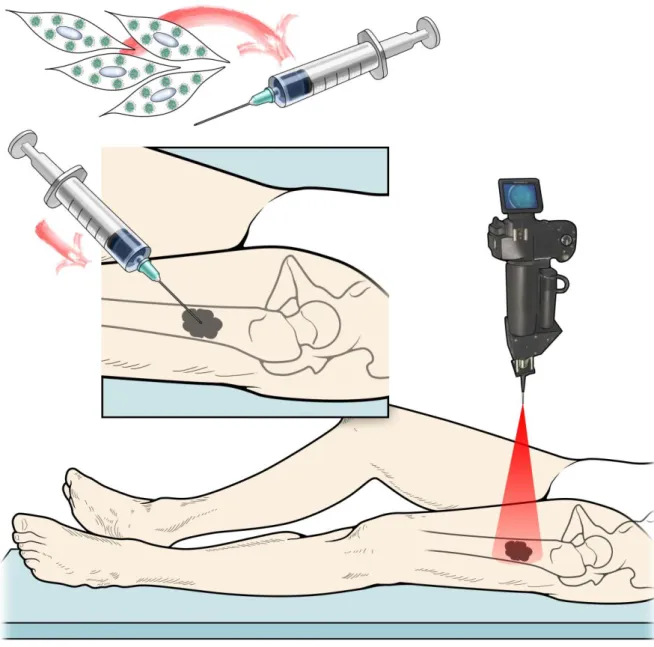
The aim of this PhD thesis is to develop an innovative strategy to be used as neodajuvant therapy for osteosarcoma patients that do not respond to current standard therapy. In our strategy we will take advantage of the tumor tropic characteristic of MSCs to make them a cellular based delivery system for photoactivable nanoparticles (FNPs). The objective of the first chapter is to demonstrate in vitro that MSCs can incorporate the FNPs and after photoactivation can release free reactive oxygen species causing the death of the tumor cells. Classical bi-dimensional cultures were intentionally selected because they are the best tools to obtain evidence at a singular cell level.
Methods
Photoactivable Nanoparticles
Fluorescent photoactivable nanoparticles (FNPs) were provided by Dr. Greta Varchi’s group at the National Research Council, Institute for the Organic Synthesis and Photoreactivity, Via Gobetti 101, 40129, Bologna, Italy. The tri-component FNPs were composed of core-shell PMMA nanoparticles post-loaded with the photosensitizer, meso-tetrakis (4-sulfonatophenyl). The external shell is decorated with primary and quaternary ammonium salts, able to electrostatically bind anionic porphyrins.
Isolation, culture and immunophenotypic characterization of human mesenchymal stem cells
MSCs were obtained from bone marrow samples obtained from 3 patients undergoing surgery at Rizzoli Orthopaedic Institute after obtaining informed consent according to a protocol approved by the Ethics Committee. MSCs were isolated through gradient separation and plastic adherence. Briefly, cells were transferred to 150-cm2 culture flasks with α-MEM supplemented with 20% lot-selected FBS and GlutaMAX™ 1%, incubated in a humidified atmosphere at 37°C with 5% CO2 and medium changed every 3-4 days. When the cells reached approximately 70-80% confluence, they were detached by mild trypsinization for 5 minutes at 37°C and counted. Then 1/3 of the cells was reseeded into a new 150-cm2 flask.
Human osteosarcoma cell line
Human osteosarcoma cells (U2OSTubRFP cells) were cultured according to the Sigma-Aldrich instructions. Briefly, the culture medium was McCoy’s 5A and was
supplemented with 10% FBS and 1% GlutaMAX™. The cells were maintained at 37°C, 95% humidity, and 5% CO2 and split 1:5 every 3 days.
23
FNPs uptake and photoactivation
To determine the cellular uptake of the nanoparticles, we analyzed the fluorescent signal from the FNPs using a BD FACSCanto II (BD Bioscience, Frankiln Lakes, NJ, USA). MSCs loaded with 45 g/ml FNPs at the indicated time points were washed three times with PBS 1X and harvested by trypsinization. After centrifugation, the loaded cells were washed twice with PBS 1X, counted with NucleoCounter® (Chemotec, Lillerod, Denmark) and 1x10⁶ MSCs suspended in fresh PBS 1X and directly analyzed.
MSCs were plated at a density of 2.5x10⁴ cells per 35 mm Glass Bottom Dishes
(MatTek Corporation) and loaded with FNPs. Photoactivation was performed using a NikonTi Eclipse microscope equipped with temperature and CO2 controllers (Okolab, Ottaviano, Napoli, Italy), a DS-QiMc-U2 12 bit camera, and an AR1 confocal laser by exposing cells to 10% of the maximum power of 405 nm laser (0.02 J/sec).
In vitro cytotoxicity of FNPs on MSCs was evaluated using a cell metabolic assay by
the reagent water soluble tetrazolium salt-1 (WST-1) following the manufacturer’s
instructions. Briefly, cells were seeded in 96-well plates at a density of 2000 cells/well in 100
l a-MEM containing 20% FBS. After overnight attachment, cells were treated with FNPs for 1 hour and then immersed in a 90 l culture medium supplemented with 10 l WST-1. After 4 hours of WST-1-incubation at 37°C and 5% CO2, the optical density of each well was measured by a microplate reader (Synergy HT, BioTek Winooski, VT, USA) set at 450 nm with a correction wavelength set at 690 nm.
In a methylene blue assay, MSCs were fixed by adding 10% formol saline to each well for 30 minutes. Cells were then stained with filtered 1% (w/v) methylene blue in 0.01 M borate buffer (pH 8.5). After 30 minutes, excess dye was removed and the remaining dye was rinsed three times in 0.01 M borate buffer (pH 8.5). The cells were examined microscopically at that point. The dye was then eluted by adding 1:1 (v/v) absolute ethanol and 0.1 M HCl to each well. The plates were gently shaken and the absorbance at 650 nm (A650) was measured using a microplate spectrophotometer (Synergy HT, BioTek Instruments Inc., Winooski, VT USA). The photometer was blanked on control wells containing elution solvent alone. Each treatment condition was in quintuplicate. A linear relationship between absorbance and cell number was verified.
For co-culture, MSCs were plated at a density of 2.5x10⁴ cells in a 6 well plate and treated with FNPs. After 1 hour of incubation, in order to eliminate any residual NPs containing medium, cells were washed three times with PBS 1X and harvested by trypsinization. After centrifugation the labelled MSCs were washed twice with PBS 1X,
counted with NucleoCounter® and 2.5x10⁴ cells reseeded in 35 mm Glass Bottom Dishes (MatTek Corporation) in complete medium. In the same plates, U2OSTubRFP were seeded in a 1:5 ratio, let adhere overnight and then imaged as described above for photoactivation and time lapse imaging.
The LIVE⁄DEAD® Viability⁄Cytotoxicity Kit quickly discriminates live from dead cells by simultaneously staining with green-fluorescent calcein-AM to indicate intracellular
esterase activity and red-fluorescent ethidium homodimer-1 (EthD1) to indicate loss of plasma membrane integrity. For our analysis and after photoactivation, cells were incubated with the staining solutions according to the manufacturer’s protocol. The apoptotic index was calculated as the percentage of ethidium positive apoptotic nuclei divided by the total number of nuclei visualized by counterstaining with Hoechst and obtained from counts of randomly chosen microscopic fields in the irradiated areas.
Results and Discussion
FNPs uptake by MSCs
As a first step, we needed to demonstrate that FNPs could be uptaken and retained by MSCs for a time that was consistent with the migration of the MSCs from the injection site to the tumor stroma. The three major concerns were: 1) that MSCs would not uptake the FNPs, 2) that the MSCs would not retain the FNPs and 3) that the FNPs would be expelled in a short time frame by the MSCs. Fluorescent FNPs uptake by MSCs cells was investigated by exposing MSCs to 45 mg/mL FNPs for 60 minutes. Flow cytometry with a FACS scan analyser was used to quantify the FNPs uptake and after 60 minutes, fluorescence signal was detected in 85% of the cells. Using the fluorescent microscope, it was clearly visible that the
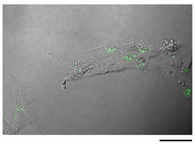
fluorescent signal of the FNPs was in the cellular cytoplasm (green in Fig. 1). FNPs tended to form aggregates and were still detectable after 72 hours. Taken together, these results confirmed that MSCs efficiently internalized the FNPs and were able to retain the FNPs for a time that was compatible with the migration of the implanted MSCs to the tumor site.
25 Figure 1. Intracellular distribution of FNPs in loaded MSCs. A confocal microscope image of cells loaded for 60 minutes with 45 g/ml FNPs and observed after 72 hours with a Nikon confocal microscope with a SFluor 40X Oil DICN2 objective. Scale bar is 50 m.
As a second step, we needed to demonstrate that FNPs did not affect MSCs survival. MSCs were exposed to 45 mg/mL FNPs for 60 minutes in the dark to avoid the activation of the photosensitizer. To evaluate the cytotoxicity, methylene blue and WST-1 assay were conducted. Both assays’ results showed no significant decrease in cell proliferation up to 7 days from the initial loading of the FNPs.
As a third step we needed to demonstrate that FNPs did not affect MSC motility. Live cell time lapse observations clearly showed that motility of loaded MSCs is not altered by the presence of intracellular FNPs when compared to unloaded cells (data not shown). We can conclude that exposure of MSCs to 45 mg/mL FNPs for 60 minutes in the dark does not affect MSC viability, nor does it affect the motility.
FNPs loaded MSCs induce OS cell death after Photactivation
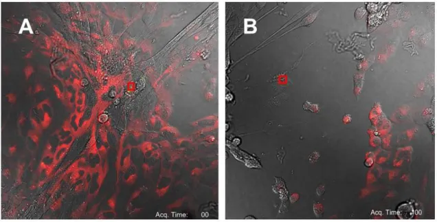
In order to test whether photoactivation of the MSCs loaded with the FNPs could induce OS cell death, we conducted a co-culture of the MSCs with the OS cell line U2OS. To be able to distinguish between the MSCs and the OS cells, we used the cell line U2OSTubRFP that is genetically engineered to express the protein tubulin in red. MSCs loaded with the FNPs without the photosensitizer were used as control. Photactivation was performed using the
laser of the confocal microscope. MSCs were subjected to illumination with a 405 nm laser (10% of the maximum power, 0.02 J/sec) for 10 seconds causing specific activation of the photosensitizer. The field of irradiation of the laser is indicated in the red square in Figure 2A. Laser light activation of a few FNPs within a single MSC resulted in rapid formation of bubble-like structures from the simulated cells in a short time frame that were indicative of cell damage and necrosis (Figure 2B). Also in a short time frame, the surrounding tumor cells that acquired a round shape detached from the plastic plate and died. Interestingly, subsequent activation of the FNPs within a single dead MSC was still able to induce cell death of the surrounding OS cells. Laser light stimulation of the control MSCs did not cause any cell damage.
Figure 2. Co-culture assay of FNPs loaded MSCs with U2OSTubRFP cells seeded in a 1:5 ratio before A) and
after B) photoactivation. The area of photostimulation is indicated in red square. Scale bar is 50 m.
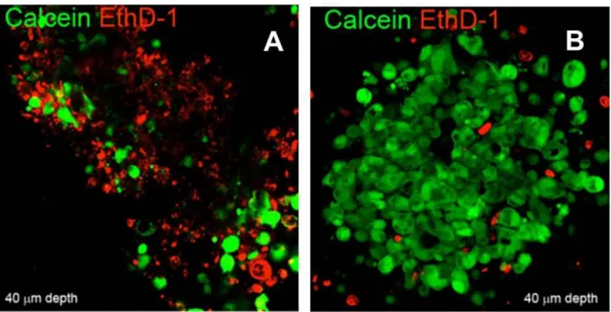
In order to quantify the effect of the photoactivation, a Live and Dead Assay was performed in the coculture of MSCs and U2OSTubRFP seeded in 1:5 ratio. Photoactivation was performed using 405 laser power at 0.16 J/sec for 1.5 minutes. After 24 hours the apoptotic index was calculated as the percentage of ethidium positive apoptotic nuclei divided by the total number of nuclei visualized by counterstaining with Hoechst obtained from counts of randomly chosen microscopic fields in the corresponding irradiated areas. The
27 percentage of apoptotic cells was 84% when the MSCs were loaded with the FNPs with the photosensitizer, while the percentage was around 4% when the MSCs were loaded with control FNPs.
Overall, our results confirm in vitro that MSCs can be loaded with photactivable nanoparticles and that photactivation is able to eliminate both vehicle cells (MSCs) and tumor cells at the same time. The fact that the MSCs are eliminated as well is very important because as we have mentioned in the introduction, there is a concern that the MSCs can promote tumor growth. Moreover, the efficacy of the second photostimulation on the same FNPs demonstrated the opportunity to induce several and repeated cycles of photostimulation that could eventually be repeated if the first was not effective in the tumor eradication.
Conclusions
The first chapter demonstrated that FNPs are efficiently uptaken by MSCs at a concentration of 45µg/ml without evident sign of toxicity or inhibition of MSCs motility. Most importantly, these results indicate that our bio-system could represent an efficient and targeted delivery strategy in killing human OS cells, suggesting a novel therapeutic avenue for the treatment of bone sarcomas.
The major limitation of the current FNPs is that the photosensitizer used in this first proof of concept can be activated in the visible spectrum and it is known that visible light does not penetrate into deep tissues.
C
HAPTER
2
Aims and objectives
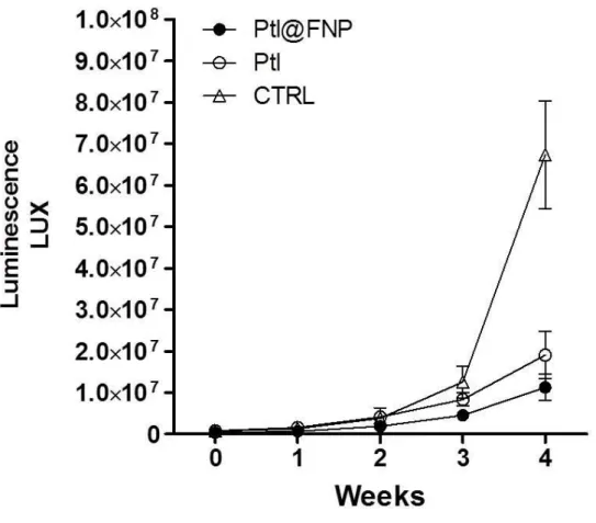
The objective of the second chapter is to demonstrate in vitro and in vivo that FNPs with a photosensitizer that can be photoactivated in near infrared spectrum can trigger tumor cell death in tridimensional tissues.
Methods
Photoactivalble Nanoparticles
The Fluorescent photoactivable nanoparticles (FNPs) were provided by Dr. Greta Varchi’s group at the National Research Council, Institute for the Organic Synthesis and Photoreactivity, Via Gobetti 101, 40129, Bologna, Italy. The particles are composed of a hydrophobic inner core of poly-PMMA covalently functionalized with allyl-fluorescein and surrounded by a hydrophilic shell of quaternary ammonium salts, suitable for the electrostatic loading of the tetra-anionic photosensitizer Tetrasulfonate aluminum phthalocyanine. FNPs
are characterized by an average hydrodynamic diameter of 80 nm. Activation of the photosensitizer by NIR-light generates cytotoxic reactive oxygen species (ROS), such as singlet oxygen (1O2), from oxygen molecules available in the surrounding environment.
Cell cultures
The androgen-independent human prostate carcinoma PC3 cell line was purchased from ATCC (CRL-1435) and cultured in a 1:1 ratio with HAM’S and F12:DMEM-hg, 10% FBS, with 1% GlutaMAX™ and 50 U ml-1 penicillin/streptomycin. RLuc-PC3 cell line, permanently transfected with RLuc-pRC-CMV vector, were provided by professor Jeronimo Blanco’s group of the Cell Therapy Group, Institute for Advanced Chemistry of Catalonia, CSIC; Networking Biomedical Research Center on Bioengineering, Biomaterials and Nanomedicine (CIBER-BBN), Barcelona, Spain.
MSCs were obtained from bone marrow samples obtained from 3 patients undergoing surgery at Rizzoli Orthopaedic Institute after obtaining informed consent according to a protocol approved by the Ethics Committee. MSCs were isolated through gradient separation and plastic adherence. Briefly, cells were transferred to 150-cm2 culture flasks with α-MEM supplemented with 20% lot-selected FBS and GlutaMAX™ 1%, incubated in a humidified atmosphere at 37°C with 5% CO2 and medium changed every 3-4 days. When the cells reached approximately 70-80% confluence, they were detached by mild trypsinization for 5 minutes at 37°C and counted. Then 1/3 of the cells was reseeded into new 150-cm2 flasks.
Microtissues and PDT
PC3 tumor tri-dimensional microtissues were prepared by co-culturing PC3 cells with MSCs at a 1:1 ratio and following an adaptation of the pellet culture method (Johnstone, Hering, Caplan, Goldberg, & Yoo, 1998). MSCs and PC3 cells, 1.25×10⁵ cells of each cell type, were mixed in a 1.5 mL polypropylene screw cap conical tube (Primo Euroclone, Milan, Italy), suspended in 0.5 mL DMEM-HG supplemented with 10% FBS, spun at 500 g for 5 minutes and the tubes, with caps partially loosened, directly incubated in humidified atmosphere at 37 °C with 5% CO2. After 72 hours, the cell pellets turn into compact spherical aggregates that easily detach from the tube and can be handled without loss of integrity. Following 3 days in culture, the microtissues were incubated for 10 minutes on a rotating plate with 18g/mL- FNPs washed with PBS and irradiated. PDT was performed using a 668±3 nm LED (XLed McManton, Italy). Cell viability was assessed with a LIVE⁄DEAD® Kit.