Journal of Microbial & Biochemical Technology
Use of particle counter system for the optimization of sampling, identification and
decontamination procedures for biological aerosols dispersion in confined environment
--Manuscript
Draft--Manuscript Number:Full Title: Use of particle counter system for the optimization of sampling, identification and decontamination procedures for biological aerosols dispersion in confined environment Short Title:
Article Type: Research Article
Section/Category: Journal of Microbial & Biochemical Technology
Keywords: Biological contamination; particle counters; air sampling; detection; identification; decontamination.
Corresponding Author: Andrea Malizia
University of Rome Tor Vergata Rome, Italy ITALY
Corresponding Author Secondary Information:
Corresponding Author's Institution: University of Rome Tor Vergata Corresponding Author's Secondary
Institution:
First Author: Michele Pazienza
First Author Secondary Information:
Order of Authors: Michele Pazienza
Mariaserena Britti Mariachiara Carestia Orlando Cenciarelli Fabrizio D'Amico Andrea Malizia Carlo Bellecci Roberto Fiorito Antonio Gucciardino Maria Rosa Bellino Corrado Lancia Corrado Lancia Pasqualino Gaudio Order of Authors Secondary Information:
Manuscript Region of Origin: ITALY
Abstract: In a CBRNe (Chemical, Biological, Radiological, Nuclear and explosive) scenario,
biological agents hardly allow efficient detection/identification because of the
incubation time that provides a lag in symptoms outbreak following their dissemination. The detection of atmospheric dispersion of biological agents (i.e.: toxins, viruses, bacteria and so on) is a key issue for the safety of people and security of environment. Another fundamental aspect is related to the efficiency of the sampling method which leads to the identification of the agent released, in fact an effective sampling method is needed either to identify the contamination and to check for the decontamination Powered by Editorial Manager® and ProduXion Manager® from Aries Systems Corporation
procedure.
Environmental monitoring is one of the ways to improve fast detection of biological agents; for instance, particle counters with the ability of discriminating between biological and non-biological particles are used for a first warning when the amount of biological particles exceeds a particular threshold. Nevertheless, these systems are not able to distinguish between pathogen and non-pathogen organisms, thus, classical "laboratory" assays are still required to unambiguously identify the particle which triggered the warning signal. In this work, a combination of commercially available equipment for detection and identification of the atmospheric dispersion of biological agents, was evaluated in partnership between the Italian Army, the Department of Industrial Engineering and the School of Medicine and Surgery of the University of Rome "Tor Vergata". The aim of this work, whose results are presented here was to conduce preliminary studies on the dynamics of biological aerosols fallout after its dispersion, to improve detection, sampling and identification techniques. This will help minimizing the impact of the release of biological agents and guarantee environmental and people safety and security.
Suggested Reviewers: Opposed Reviewers:
Rome, 14/12/2013
Andrea Malizia, PhD
Senior researcher
Deparment of Industrial Engineering
University of Rome “Tor Vergata”
Via Del Politecnico 1, 00133 Roma (Italy)
[email protected]
Tel: +39 0672597202
Subject: SUBMISSION OF A MANUSCRIPT FOR EVALUATION
Dear Editor, dear Editorial Board, Dear Reviewers
I am enclosing herewith a manuscript entitled “Use of particle counter system for the optimization
of sampling, identification and decontamination procedures for biological aerosols dispersion in
confined environment” for publication in the Journal of Microbial & Biochemical Technology.
This work is an outcome of the international post graduating courses in “Protection against CBRN
events” (NATO selected and in cooperation with OPCW; www.mastercbrn.com), and it was
conducted in partnership between the Italian Army and the University of Rome “Tor Vergata”. The
cooperation between our University and the Italian Ministry of Defense was born to give an high
level training to people with different background, working in the field of protection against
chemical, biological, radiological and nuclear events.
This work, and its results, represents an evidence of this fruitful collaboration.
The intentional or unintentional release of a biological agent is among the main concerns about
safety and security for people and environment. Unlike for the other dangerous agents such as the
radiological and chemical ones, at the moment no instrument which is able to rapidly detect and
unambiguously identify a biological agent. This represent a main issue not only from a public health
point of view, but also for that of civil and military defense.
In Italy, in case of release of biological agents, military forces and fire brigades are equipped and
trained for detection, identification and decontamination procedures of personnel and technical
equipment, and particular efforts are continuously made to optimize these procedures and find new
technologies which will allow faster and easier detection, identification and response to this kind of
threat.
The studies, whose results are presented in the submitted paper, were conducted in partnership
between the Italian Army, the Department of Industrial Engineering and the School of Medicine
and Surgery of the University of Rome “Tor Vergata”, with the aim of collecting data concerning
the dynamics of the dispersion of a biological aerosol in a confined environment and its effects on
detection and sampling procedures. The dispersion was analyzed by means of aerodynamic particle
counters and samples were collected by means of different sampling methods and instruments and
then analyzed by using both classical microbiological and molecular biology techniques.
These preliminary outcomes will contribute to implement a setup for real-time
detection/identification of biological agents releases which, in the future, could also have civilian
applications.
With the submission of this manuscript I would like to undertake that the above mentioned
manuscript has not been published elsewhere, accepted for publication elsewhere or under editorial
review for publication elsewhere.
All authors have approved the manuscript and agreed with its submission.
Thank you for consideration, kind regards
Use of particle counter system for the optimization of sampling,
identification and decontamination procedures for biological aerosols
dispersion in confined environment
Michele Pazienza1,3*, Mariaserena Britti1,3, Mariachiara Carestia1*, Orlando Cenciarelli1, Fabrizio D'Amico1,3, Andrea Malizia1,3*, Carlo Bellecci1,3, Roberto Fiorito2,3, Antonio Gucciardino3, Maria Rosa Bellino1,3, Corrado Lancia1,3 and Pasquale Gaudio1,3.
1. Department of Industrial Engineering, University of Rome “Tor Vergata”, Via del Politecnico 1 - 00133 Rome, Italy
2. Department of Bio-Medical & Prevention, School of Medicine and Surgery, University of Rome “Tor Vergata”, Via Montpellier 1 - 00133 Rome, Italy
3. International Master Courses in Protection against CBRNe events, Department of Industrial Engineering –School of Medicine and Surgery, University of Rome “Tor Vergata”, Italy
Abstract
In a CBRNe (Chemical, Biological, Radiological, Nuclear and explosive) scenario, biological agents hardly allow efficient detection/identification because of the incubation time that provides a lag in symptoms outbreak following their dissemination. The detection of atmospheric dispersion of biological agents (i.e.: toxins, viruses, bacteria and so on) is a key issue for the safety of people and security of environment. Another fundamental aspect is related to the efficiency of the sampling method, which leads to the identification of the agent released, in fact an effective sampling method is needed either to identify the contamination and to check for the decontamination procedure.
Environmental monitoring is one of the ways to improve fast detection of biological agents; for instance, particle counters with the ability of discriminating between biological and non-biological particles are used for a first warning when the amount of biological particles exceeds a particular threshold. Nevertheless, these systems are not able to distinguish between pathogen and non-pathogen organisms, thus, classical “laboratory” assays are still required to unambiguously identify the particle which triggered the warning signal. In this work, a combination of commercially available equipment for detection and identification of the atmospheric dispersion of biological agents was evaluated in partnership between the Italian Army, the Department of Industrial Engineering and the School of Medicine and Surgery of the University of Rome “Tor Vergata”. The aim of this work, whose results are presented here, was to conduce preliminary studies on the dynamics of biological aerosols fallout after its dispersion, to improve detection, sampling and identification techniques. This will help minimizing the impact of the release of biological agents, guarantee environmental, and people safety and security.
Keywords
: Biological contamination; particle counters; air sampling; detection; identification;decontamination.
Introduction
An aerosol release of biological agents (i.e. toxins, vegetative bacteria, endospores, viruses, fungal and mold spores) is the main concern about a biological attack [1], but it can also be the result of routine operation in an industrial or healthcare structure [2-7]. To prevent and manage the possible diffusion of biological agents and the subsequent contamination, there are three activities to implement : detection, identification and decontamination [8].
The dispersion may occur in an open area or in a confined environment: homes, offices and other public enclosures such as airports, shopping malls, subway stations, theaters and arenas. These inherently three-dimensional living spaces are made more complex due to the placement of doors, windows, vents, walls and furniture [9].
For this reason, it becomes essential to acquire information about the dynamics of dispersion and deposition of biological agents, which will be useful to optimize either the positioning of the detectors and samplers and the procedures for decontamination.
*Corresponding authors:
Michele Pazienza, Department of Industrial Engineering,
University of Rome “Tor Vergata”, Via del Politecnico 1 - 00133 Rome, Italy. E-mail [email protected] .
Mariachiara Carestia, Department of Industrial Engineering,
University of Rome “Tor Vergata”, Via del Politecnico 1 - 00133 Rome, Italy. E-mail: [email protected].
Andrea Malizia, Department of Industrial Engineering,
University of Rome “Tor Vergata”, Via del Politecnico 1 - 00133 Rome, Italy. E-mail: [email protected]
Manuscript
Chemical and radiological agents are relatively easy to detect and identify thanks to their intrinsic features: furthermore, fast, field portable and “user friendly” instruments are already available for their detection and identification [10-11]. On the other hand, due to the complexity of the molecules constituting the biological agents, equipment and methodologies for detection and identification of biological aerosols are still in an embryonic phase of development [12-15].
Efforts are continuously made to create a unique instrument either for detection and identification of biological aerosols basing on the dimensional and fluorescence characteristics of biological agents [16-18], but results from these efforts showed that despite the advances in discriminating between biological and non-biological molecules, the simultaneous identification of the biological agent (or agents) is still a demanding issue.
For this reason, sampling still represents a key point to guarantee the effectiveness of the identification phase which may be performed both with consolidated microbiological or immunochemical techniques or with cutting edge molecular techniques (such as microchip arrays [19]), with cutting edge molecular techniques (Real-Time PCR [20]), or with classical spectrometry technologies such as Differential Mobility Spectrometry (DMS) [21]. At this purpose, in case of release of biological agents, military forces and fire brigades are equipped and trained for detection identification and decontamination procedures of personnel and technical equipment, but particular efforts should be put into the optimization of these procedures [22].
Aerodynamic particle counters (APC) are currently used to estimate pollutants in air: a particle counter can measure the diameter and number of particles in the air with the least expenditure of time and materials, and with greater accuracy than can be obtained by any other method. Particles may be either droplets of fluid or particles of solids [23].
Discrimination among particle sizes is realized thanks to their property of scattering light from an incident light source, while discrimination among biological and non-biological particle is achieved by fluorescence measurement. Indeed, biological particles are able to emit fluorescence when excited at specific wavelengths (mainly from 250 to 500 nm) thanks to the presence of ubiquitous endogenous fluorophores, mainly aromatic aminoacids, reduced NAD, NADP, FAD and riboflavin) [24-25].
In this study, biological aerosols, produced by means of different atomizers, were released in a confined environment and the dynamics of their deposition were evaluated using an aerodynamic particle counter (APC) together with active and passive aerosol sampling. Results from these trials will help identifying best practices for air monitoring, sampling, identification and
response in case of atmospheric dispersion of biological agents.
Materials, instruments and methods
In these trials, a solution containing Saccharomyces cerevisiae, was atomized within a sealed chemical hood using two different atomizers, in different experimental conditions.
It has to be underlined that S.cerevisiae is not a recognized biological warfare agent simulant, but it was chosen to guarantee safety of operators because of its non-pathogenicity and because its structural characteristics are in between prokaryotic and eukaryotic organisms (cell wall, nucleus, double strand linear chromosome). Furthermore, all the experiments were conducted using Saccharomyces “training kits” (Idaho Technology, Inc. now supplied as BioFire Diagnostics, Inc), which are specifically designed to safely perform laboratory routines in the same experimental conditions as using BWA samples.
First of all, the deposition dynamics were evaluated by means of an aerodynamic particle counter. After evaluating the appropriate setup for the atomization and dispersion of the biological aerosol, air and surface samples were collected by means of an automatic air sampler and by swabbing Petri dishes placed on the worktop of the hood, respectively. Samples were then analyzed by Real-Time PCR assay, and by direct plating on Agar Sabouraud [26].
Biological aerosol production and aero-dispersion
A solution containing commercially available, food grade, lyophilized Saccharomyces cerevisiae at concentration of 7 g/L has been aerosolized and released for 3 minutes, within a sealed chemical hood(1,5 m3 volume)according to the three following experimental conditions:
a) using a pre compression (manual) pump at a temperature of 25°C;
b) using an automatic atomizer which releases particles in the range between 5 and 20 micron, with a flux of 0-70 mL/min at a temperature of 18°C;
c) using an automatic atomizer which releases particles in the range between 5 and 20 micron, with a flux of 0-70 mL/min at a temperature of 25°C.
Biological aerosol detection
The aerodynamic particle counter used in these trials is the Fido B2 IBAC (FLIR®) which has an air flow rate of3.8 liters per min and allows detecting particles with a diameter grater or equal to 0.7microns. Detection was performed in continuous.
Sampling methods
Two different methods were used to sample the biological aerosol. Active sampling of aerosol in air was performed by means of the portable sampler Fido 1 (FLIR®). This instrument has a sampling flow rate of 200 liters/min and can collect particles whose size ranges between 0.5 and 10.0 microns. The collected air sample is diluted in the aqueous solution (provided by the manufacturer) in a volume between 2 and 5 ml. Each air sample derived from a 5 minutes air sampling. This specific duration of sampling (5 minutes) was chosen in accordance to the volume of air in the hood (1.5 m3) to avoid re-suspension phenomena. Collected samples were then diluted in a liquid volume of 5 ml. Passive sampling was performed by swabbing opened Petri dishes positioned on the worktop of the hood. Air and surface samples were collected at time intervals of an hour, and after about 24 hours from the release.
Real-time PCR
Real-Time PCR assay was conducted using the Ruggedized Advanced Pathogen Identification Device. TheR.A.P.I.D.TM BioDetection system (Idaho Technology Inc, now supplied as BioFire Diagnostics, Inc), a ruggedized, portable Real-time PCR designed to identify biological agents especially for mobile analytical labs and fields hospitals.
DNA extraction from the samples has been performed using the “IT 1-2-3DNA Sample purification kit” (BioFire Diagnostics, Inc). The “S. cerevisiae Detection Kit for Hybridization Probe assay” (Idaho Technology, Inc., now supplied as BioFire Diagnostics, Inc.) was used for the identification of the agent; the kit consists of lyophilized reagents including primers which have specificity for S. cerevisiae and are validated according to the GMP (Good Manufacturing Practice) standards. A fragment of the target DNA is amplified using specific primers. The amplicon is detected by fluorescence using a specific pair of hybridization probes. These probes consist of two different short oligonucleotides that hybridize to an internal sequence of the amplified fragment during the annealing phase of the reaction cycle. One probe is labeled at the 5' end with LCRed 640. To avoid extension on the 3' end, it is modified by phosphorylation. The second probe is labeled at the 3' end with fluorescein.
After hybridization to the template DNA do the two probes come in close proximity, resulting in fluorescence resonance energy transfer (FRET) between the two fluorophores. The fluorescence emitted by the LCRed 640 dye is measured in channel 2 of the R.A.P.I.D. instrument. The fluorescent signal from the unknown sample is compared to the signals from the positive and negative control samples.
Real-Time PCR reactions were optimized at a temperature of 90°C for 5 sec, 35 amplification cycles at
a temperature of 60°C for 15 sec. The melting curve analysis has been performed reaching a temperature of 90°C with a ramp of 0.2°C/sec. For Real-Time PCR cycles settings, melting curve settings and sample preparation, protocols have been implemented according to the manufacturer’s instructions.
Culture media
S. cerevisiaes samples (0.1 mL from: S. cerevisiae solution just after aerosolization; 0.1 mL solution from air sampler and swab from the Petri dishes) were plated on Agar Sabouraud [26] and incubated at a temperature of 37°C for 48 hours.
Results
The first step of the trials was to evaluate different dispersion means i.e. a pre compression pump and an automatic atomizer. In the latter case, dispersion was performed at two different temperatures (18°C and 25°C) to evaluate possible effects on the dynamics of the particle diffusion due to the temperature.
Particle counts have been performed as described in the methods section. Once identified the proper dispersion model, air and surfaces samples were collected at regular time intervals. Finally, samples were analyzed for qualitative information about the presence or absence of the microorganism with Real-Time PCR and for semi-quantitative information by plating samples on Agar Sabouraud. Results from these trials are reported in the following paragraphs.
Nebulization trials and particle counts
Particle counts were performed with the Fido B2 IBAC (FLIR®); this instrument is able to discriminate between biological and non-biological particles, thus results show both total particle counts (per liter of air) and biological particle counts (per liter of air). Particle counts are further divided into small (less than or equal to 0.7 micron) and big (greater than 0.7 micron) particles. Results from particle counts are shown in Fig.1, Fig.2, Fig.3 and Fig.4.
a) Nebulization with pre compression pump at 25°C
Figure 1: Distribution of the particle counts (per liter of air) versus time. The aerosol was generated by a pre compression pump and released at a temperature of 25°C. Black squares: total big particles; red circles: biological big particles; blue squares: total small particles; magenta circles: biological small particles. b) Nebulization with automatic atomizer at 18°C
Figure 2: Distribution of the particle counts (per liter of air) time. The aerosol was generated by an automatic atomizer (released at a temperature of 18°C). Black squares: total big particles; red circles: biological big particles; blue squares: total small particles; magenta circles: biological small particles.
c) Nebulization with automatic atomizer at 25°C
Figure 3: Distribution of the particle counts (per liter of air) as function of the time after the release of the aerosol. The aerosol was generated by an automatic atomizer and released at a temperature of 25°C. Black squares: total big particles; red circles: biological big particles; blue squares: total small particles; magenta circles: biological small particles.
Figure 4: Distribution of the particle counts (per liter of air) versus time (one day after the release). The aerosol was generated by an automatic atomizer and released at a temperature of 25°C.Black squares: total big particles; red circles: biological big particles; blue squares: total small particles; magenta circles: biological small particles.
The dynamic of the particle diffusion seems to differ significantly according to the dispersion method. When the aerosol is generated with the pre-compression pump, in fact, a rapid drop of the counts for the small particles occurs just 50 minutes after the dispersion.
When the aerosol is generated by means of an automatic atomizer and released at a temperature of 18°C, the count of big and small biological particles drops to a minimum after just 15 minutes from the release. On the other hand, when the nebulization is performed with the same automatic atomizer, at a temperature of 25°C, the particle count show a more homogenous distribution of
big and small, biological and non-biological particles within 24 hours from the release of the aerosol.
For this reason, air samples and samples of deposited aerosol were collected and analyzed for this latter dispersion model.
Real-Time PCR analysis
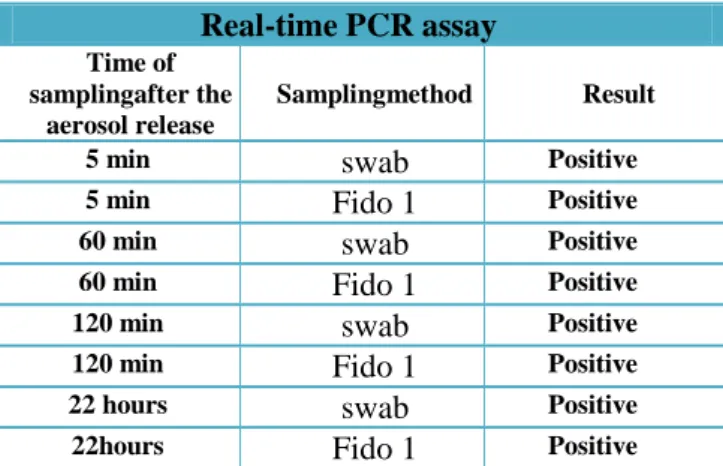
Real-Time PCR assay was performed on S. cerevisiae samples collected by means of a) the automatic air sampler Fido 1 (FLIR®) and b) swabbing Petri dishes positioned on the worktop of the sealed chemical hood. Samples were collected 5 minutes, 1 hour, 2 hour and 22 hours after the dispersion of the aerosol.
Qualitative results from Real-Time PCR assay are shown in Table 1: a positive result indicates the presence of recognizable genome target sequences from S. cerevisiae, a negative result indicates the absence of recognizable target sequences from the same organism.
Real-Time PCR assay Time of sampling
after the aerosol release
Sampling method Result
5 min swab Positive
5 min Fido 1 Positive
60 min swab Positive
60 min Fido 1 Positive
120 min swab Positive
120 min Fido 1 Positive
22 hours swab Positive
22 hours Fido 1 Positive
Table1: Real-Time PCR results for samples collected at
different time interval from the release of the biological aerosol. Samples were collected by active air sample (Fido 1) method and by passive sampling by swabbing Petri dishes placed on the worktop of the chemical hood. “Positive” indicates specific amplification of the sample; “Negative” indicates the absence of amplification.
Microbiological growth (viability assay)
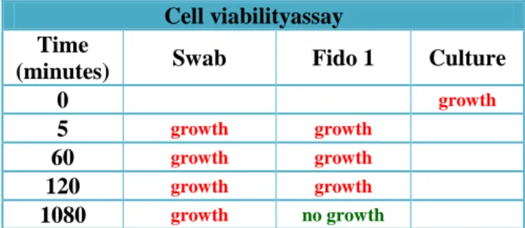
To verify the effects of nebulization and active sampling on cells viability, samples of the biological aerosol collected by means of active and passive sampling were plated on Agar Sabouraud and incubated at 37°C for 48 hours. Results are shown in Table 2.
Cell viabilityassay Time
(minutes) Swab Fido 1 Culture
0 growth
5 growth growth
60 growth growth
120 growth growth
1080 growth no growth
Table 2: Swab from Petri dishes on the worktop and 0.1 mL
of solution from air sampler plated on Agar Sabouraud.
Discussion
Preliminary outcomes from the three trials performed to evaluate the dispersion dynamic of an aerosolized solution containing liophylized S. cerevisiae, shows significant differences among the different experimental conditions. First of all, when the bio aerosol is generated by means of a pre-compressed pump, detectable big and small particles tend to rapidly decrease within 50 minutes from the release. Similar observations were made when the S. cerevisiae solution was aerosolized by means of an automated atomizer and released at a temperature of 18°C.
On the other hand, when the aerosol is produced by the same automatic atomizer, but is released at a temperature of 25°C, countable particles persist in air at least more than 100 minutes.
Countable particles can be found also after about 24 hours from the release; nevertheless it should be noticed that values at 21.45 hours (1305 minutes) are 1 order magnitude less than values obtained 5 and 10 minutes later, suggesting that some re-suspension phenomena may have occurred. This point should be taken into particular consideration when sampling occurs in small, confined environments.
Ratio between big and small particles is also different, according to which instrument produced the aerosol: the pre-compressed pump generated more big particles than small particles. An opposite result can be observed for the automatic atomizer either when the release occur at a temperature of 18 or 25°C; this result clearly depends on the technical characteristic of the atomizers and this can produce differences in the aerosol characteristics.
Real-Time PCR assay performed on air and surface samples collected at time intervals of 5, 60, 120
minutes and 22 hours after the release, showed amplification of S. cerevisiae target sequences for each of the time intervals. These preliminary results should be further investigated with the aim of correlating the particle count with sensitivity of the PCR assay [27].
Cell viability assays shows growth for all samples collected at each time interval after the release either from the surface of the hood and from air samples, with the only exception of the air sample collected one day (18 hours) after the release of the aerosol.
This result is of particular interest if compared with the positive result of the PCR for an air sample collected 22 hours after the release.
On one hand it could support the hypothesis of the re-suspension phenomenon due to the air sampling instrument; on the other hand it may suggest that Real-Time PCR is able to detect contamination from nucleic acids. In this particular case, could have been released from cells which were damaged or were viable but non-culturabele [28] as a consequence of the nebulization or the sampling process.
Conclusions
Biological contamination of open space or confined environments is a main issue from a military and civilian point of view.
In case of biological threaten towards military forces, these ones should be able to efficiently perform detection, identification and decontamination procedures to minimize risks for operators and safeguard sensible equipment.
In this study, the authors evaluated different aspects of the detection of atmospheric release of biological agents by simulating the release of a biological agent in a confined environment, starting from finding a proper methodology for the production of the aerosol, to the proper methodology for sampling and revealing the presence of contamination in air and surfaces sampling. Preliminary results suggest that the methodology for the production of the aerosol has a deep impact on the detection method based on aerodynamic particle counters.
Furthermore, classical biological assays for the detection and identification of a potential threat may not be sufficiently sensitive, beyond being time consuming, when samples are collected by means of automatic samplers. For this reason, Real-Time PCR assays may represent a useful implementation of conventional biological agents identification techniques.
At present, only the combination of several biological and non-biological techniques can help detecting and identifying the biological agents, thus, further studies are required to find a unique, integrated, fast and field portable tool, for the real-time detection and identification of biological agents.
Acknowledgments
Special acknowledgments for the realization of this work go the International Master Courses in “Protection against CBRNe events”.
(www.mastercbrn.com).
References
1. Wanger MM, Moore AW, Aryel (2006) Handbook of biosurveillance (5thedition) Elsevier Academic Press,
Burlington, MA, USA.
2. Cenciarelli O, Malizia A, Marinelli M, Pietropaoli S, Gallo R, et al. (2013) Evaluation of biohazard management of the Italian national fire brigade. Defence S&T Tech Bull 6:33-41.
3. Gallo R, De Angelis P, Malizia A, Conetta F, Di Giovanni D, et al. (2013) Development of a georeferencing software for radiological diffusion in order to improve the safety and security of first responders. Defence S&T Tech Bull 6:21-32.
4. Malizia A, Lupelli I, D'Amico F, Sassolini A, Fiduccia A, et al. (2012) Comparison of software for rescue operation planning during an accident in a nuclear power plant. Defence S&T Tech Bull 5:36-4.
5. Malizia A, Quaranta R, Mugavero R, Carcano R, Franceschi G (2011) Proposal of the prototype RoSyD-CBRN, a robotic system for remote detection of CBRN agents. Defence S&T Tech Bull 4:64-76.
6. Malizia A, Quaranta R, Mugavero R (2010) CBRN events in the subway system of Rome: Technical-managerial solutions for risk reduction. Defence S&T Tech Bull 3:140-157.
7.Cenciarelli O, Rea S, Carestia M, D’Amico F, Malizia, et al. (2013) Bioweapons and Bioterrorism: A Review of History and Biological Agents. Defence S&T Tech Bull 6(2):111-129.
8. U.S. Department of Justice (2001) An introduction to biological agent detection equipment for emergency first responders. NIJ Guide 101-00.
9. Winters WS, Chenoweth DR (2002) Modeling dispersion of chemical – biological agents in three dimensional living spaces. Sandia Report SAND 2002-8009.
10. Sferopoulos R (2009) A review of chemical warfare agent (CWA) detector technologies and commercial-off-the-shelf items. Human Protection and Performance Division DSTO Defence Science and Technology Organisation Australia DSTO-GD-0570.
11.Jopling L (2005) Chemical, biological, radiological or nuclear (CBRN) detection: A technological overview. Special Report to NATO Parliamentary Assembly, 167.
12. Seto Y (2006) On-site detection method for biological and chemical warfare agents. Bunseki Kagaku, 55:891-906.
13. Jonsson P, Elmqvist M, Gustafsson O, Kullander F, Persson R, et al. (2009) Evaluation of biological aerosol stand-off detection at a field trial. Proceedings of SPIE - The International Society for Optical Engineering 7484, art. no. 74840I.
14. Gardner CW, Wentworth R, Treado PJ, Batavia P, Gilbert G (2008) Remote chemical biological and explosive agent detection using a robot-based Raman detector. Proceedings of SPIE - The International Society for Optical Engineering 6962, art. no. 69620T.
15. Jonsson P, Kullander F, Tiihonen M, Nordstrand M, Tjærnhage T, et al. (2005) Development of fluorescence-based LIDAR technology for biological sensing. Materials Research Society Symposium Proceedings 883, art. no. FF1.6, 51-62.
16. Ho J (2002) Future of biological aerosol detection. AnalyticaChimicaActa 457:125–148
17. Ho J, Spence M, Hairston P (1999) Measurement of biological aerosol with a fluorescent aerodynamic particle sizer (FLAPS): correlation of optical data with biological data. Aerobiologia 15:281–291.
18. Pinnick RG, Hill SC, Nachman J, Pendleton D, Fernandez GL, et al. (1995) Fluorescence particle counter for detecting airborne bacteria and other biological particles. Aerosol Science and Technology, 23(4):653-664.
19. Bhatta D, Michel AA, Marti Villalba M, Emmerson GD, Sparrow IJG (2011) Optical microchip array biosensor for multiplexed detection of bio-hazardous agents. Biosensors and Bioelectronics 30:78-86.
20. Gamier L, Gaudin JC, Bensadoun P, Rebillat I, Morel Y (2009) Real-Time PCR assay for detection of a new Simulant for poxvirus biothreat agents. Applied and Environmental Microbiology 75:1614-1620.
21. Krebs MD, Zapata AM, Nazarov EG, Miller RA, Costa IS, et al. (2005) Detection of biological and chemical agents using Differential Mobility Spectrometry (DMS) technology. IEEE Sensors Journal, 5:696-702.
22. Mari G, Giraudi G, Bellino MR, Pazienza M, Garibaldi C et al. (2009) CBRN mobile laboratories in Italy. Proc. of SPIE Vol. 7304 73040W-1.
23. Zinky WR (1962) A New Tool for Air Pollution Control: The Aerosol Particle Counter. Journal of the Air Pollution Control Association, 12(12):578-583.
24. Lakowicz J (1999) R. Principles of Fluorescence Spectroscopy, 2nd ed. Kluwer Academic/Plenum Publisher: New York.
25. Teale FWJ, Weber G (1957) Ultraviolet fluorescence of the aromatic amino acids. Bioch. 65:476–482.
26. Sabouraud R (1896) Recherche des milieux de culture propres a la différenciation des espèces trichophytiques a grosse spore. Les trichophyties humaines. Paris: Masson et Cie, France; pp. 49-55.
27. Sirigul1 C, Wongwit W, Phanprasit W, Paveenkittiporn W, Blacksell SD, et al. (2006) Development of a combined air sampling andquantitative real-time PCR method for detectionof Legionella spp. Southeast Asian J Trop Med Public Health 3(3):503-507.
28. Oliver JD (2005) The Viable but Nonculturable State in Bacteria. J Microbiol 43:93-100.
Figure
Figure
Figure
Figure
Real-time PCR assay Time of
samplingafter the aerosol release
Samplingmethod Result
5 min swab Positive
5 min Fido 1 Positive
60 min swab Positive
60 min Fido 1 Positive
120 min swab Positive
120 min Fido 1 Positive
22 hours swab Positive
22hours Fido 1 Positive
Table1: real-time PCR results for samples collected at different time interval from the release of the biological aerosol.
Samples were collected by active air sample (Fido 1) method and by passive sampling by swabbing Petri dishes placed on the worktop of the chemical hood. “Positive” indicates specific amplification of the sample; “Negative” indicates the absence of amplification.
Cell viabilityassay Time
(minutes) Swab Fido 1 Culture
0 growth
5 growth growth
60 growth growth
120 growth growth
1080 growth no growth
Table 2: Swab from petri dishes on the worktop and 0,1 mL of solution from air sampler plated on Agar Sabouraud. Table
Figure 1: Distribution of the particle counts (per liter of air) versus time. The aerosol was generated by a pre compression pump and released at a temperature of 25°C. Black squares: total big particles; red circles: biological big particles; blue squares: total small particles; magenta circles: biological small particles.
Figure 2: Distribution of the particle counts (per liter of air) time. The aerosol was generated by an automatic atomizer and released at a temperature of 18°C. Black squares: total big particles; red circles: biological big particles; blue squares: total small particles; magenta circles: biological small particles.
Figure 3: Distribution of the particle counts (per liter of air) as function of the time after the release of the aerosol. The aerosol was generated by an automatic atomizer and released at a temperature of 25°C. Black squares: total big particles; red circles: biological big particles; blue squares: total small particles; magenta circles: biological small particles.
Figure 4: Distribution of the particle counts (per liter of air) versus time (one day after the release). The aerosol was generated by an automatic atomizer and released at a temperature of 25°C. Black squares: total big particles; red circles: biological big particles; blue squares: total small particles; magenta circles: biological small particles.


