UNIVERSITY OF PISA
FACULTY OF MEDICINE AND SURGERY
PhD school in:
“FISIOPATOLOGIA CLINICA E SCIENZE DEL FARMACO” School Dean: Prof. P. Miccoli
Program:
“SCIENZE CHIRURGICHE, ANESTESIOLOGICHE E
DELL’EMERGENZA”
Title:
Epigenetic biomarkers, folates and genetic susceptibility
in colorectal carcinoma
Supervisors:
Prof. Lucia Migliore
Prof. Paolo Miccoli
Candidate:
Francesca Migheli
Index
Abstract
IIntroduction
1 1. Colorectal cancer 1 1.1 Epidemiology 21.2 Genetics of colorectal cancer 4
1.3 Cytogenetics of colorectal cancer 10
1.4 Epigenetics 11
1.4.1 DNA methylation in colorectal cancer 13
1.4.2 Histone modifications in colorectal cancer 18
1.4.3 NcRNA alterations in colorectal cancer 20
1.5 Colorectal adenoma-carcinoma sequence 21
1.5.1 APC (Adenomatous Polyposis Coli) gene 23
1.5.2 MGMT (O-6-methylguanine-DNA methyltransferase) gene 25
1.5.3 CDKN2A or p16Ink4A (Cyclin-dependent kinase inhibitor 2A) gene 28
1.5.4 RASSF1A (Ras association (RalGDS/AF-6) domain family member 1) gene 30
1.5.5 hMLH1 (mutL homolog 1, colon cancer, nonpolyposis type 2) gene 31 1.6 Environmental factors that might influence epigenetic patterns in colorectal cancer 33 1.7 Genetic factors affecting DNA methylation in colorectal cancer 43
2. DNA methylation analysis techniques 45
Materials and Methods
48
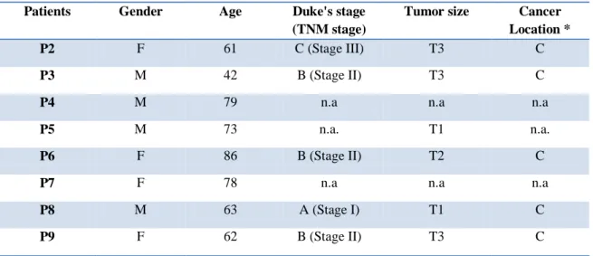
1. Study population 48
3. Extraction of genomic DNA 51 4. Genotyping 51 5. Methylation analysis 52 5.1 MS-HRM protocols 53 5.2 Pyrosequencing protocols 59 6. Statistical analysis 66
Aim of the study
67
Results
69
1. Comparison between tumor and healthy tissue in CRC patients 69
2. Correlation between methylation and clinical-pathological features 72
3. Comparison between MS-HRM and Pyrosequencing techniques 76
4. Tumor cells separation 79
5. Polymorphisms in folate metabolism genes and methylation 80
6. Folate, homocysteine and vitamin B12 values in CRC patients 86
Discussion
88
I
Abstract
The implications of DNA hypomethylation and hypermethylation in the etiology of tumorigenesis have become quite clear. Since epigenetic modifications are reversible, methylation studies are extremely promising to better characterize colorectal cancer (CRC) and to identify new tools for diagnosis and prognosis. In the frame of a wider study ongoing aimed at searching for possible correlations among genetic, epigenetic and environmental factors in colorectal cancer, this thesis took into account a cohort of CRC subjects until now recruited, focusing on the following items: 1) the validation of the MS-HRM protocol, used for the methylation analysis, by comparing it with a widely employed technique (Pyrosequencing); 2) the evaluation of the influence of an immunomagnetic method with microbeads coated with the antibody CD326+, specific for epithelial cells, to clarify if cancer epithelial cells from the surgically resected CRC tissue could give more accurate results in DNA methylation levels detection with respect to the whole CRC tissue; 3) the detection of methylation levels (performed by MS-HRM) in promoters of of APC, CDKN2A, hMLH1, MGMT and RASSF1A genes in CRC and healthy adjacent tissue specimens; 4) the analysis of the correlation among the methylation status of the chosen genes and the clinical-pathological features of the patients; 5) the analysis of the correlation among MTHFR C677T, DNMT3B C-149T polymorphisms and the methylation levels of APC, CDKN2A, hMLH1 and MGMT gene promoters. Results obtained by using MS-HRM and Pyrosequencing have shown to be quantitatively comparable. No statistically significant difference between the epithelial cells CD326+ fraction and the whole tissue about the promoter methylation of APC, CDKN2A, MGMT and hMLH1 genes was observed, although some patients showed individually a statistically significant difference between the two experimental
II
conditions regarding the degree of methylation of these genes. It was also seen, for all of the studied genes, a higher methylation level in CRC tissue respect to the healthy adjacent tissue. No statistically significant association between stage (TNM), gender, sex, tumor size, and location with regard to the methylation profile of each of the analyzed genes was found in the examined cohort. However, it was found an interesting positive association between age and both hMLH1 (P value= 0.007) and MGMT (P value= 0.03). Finally significant interactions between MTHFR 677C>T, DNMT3B -149C>T polymorphisms and gene promoter methylation were observed.
1
Introduction
1. Colorectal cancer
The digestive surface of the human large intestine is characterized by a monolayer of specialized epithelial cells that forms crypts. The first recognizable manifestation of epithelial alteration during colorectal tumor development are the Aberrant Crypt Foci (ACF), small hyper- or dysplastic lesions. Larger ACF with altered morphology, dysplastic histology and associated gene mutations are high-risk candidates for adenoma and CRC formation. According to the their architecture, adenomas may be divided in tubular, when coarsely lobulated and pedunculated, or villous, when sessile, covering a broad area directly onto the muscularis mucosae (the muscle layer underlying the epithelial lining) and submucosa (the underlying stromal layer). Villous adenomas are thought to have higher risk of malignant progression. The carcinoma in situ are advanced high dysplastic lesions still confined within the epithelial layer. Finally, malignant adeno-carcinomas are characterized by the ability to invade the surrounding tissues through the muscularis mucosae and into the stromal compartment, and migrate to distal organs where they can form metastasis. The TNM Classification of Malignant Tumours (TNM) is a cancer staging system that describes the extent of cancer in a patient’s body:
· T describes the size of the tumor and whether it has invaded nearby tissue,
· N describes regional lymph nodes that are involved,
· M describes distant metastasis (spread of cancer from one body part to another).
2
Particularly T represents the size or direct extent of the primary tumor (Tx: tumor cannot be evaluated, Tis: carcinoma in situ, T0: no signs of tumor and T1, T2, T3, T4: size and/or extension of the primary tumor); N represents the degree of spread to regional lymph nodes (Nx: lymph nodes cannot be evaluated, N0: tumor cells absent from regional lymph nodes, N1: regional lymph node metastasis present, N2: tumor spread to an extent between N1 and N3, N3: tumor spread to more distant or numerous regional lymph nodes; M represents the presence of metastasis (Mx: distant metastasis cannot be evaluated, M0: no distant metastasis, M1: metastasis to distant organs (beyond regional lymph nodes).
The Stage grouping is as follow: Stage 0: Tis N0 M0
Stage I: T1 N0 M0; T2 N0 M0: Cancer has begun to spread, but is still in the inner lining. Stage II: T3 N0 M0; T4 N0 M0: Cancer has spread to other organs near the colon or rectum. It has not reached lymph nodes.
Stage III: any T, N1-2, M0: Cancer has spread to lymph nodes, but has not been carried to distant parts of the body.
Stage IV: any T, any N, M1: Cancer has been carried through the lymph system to distant parts of the body. This is known as metastasis. The most likely organs to experience metastasis from colorectal cancer are the lungs and liver.
1.1 Epidemiology
CRC is the third most common cancer in men (663 000 cases, 10.0% of the total) and the second in women (571 000 cases, 9.4% of the total) worldwide (data observed in 2008). Almost 60% of the cases occur in developed regions. The highest percentage is observed in Australia/New Zealand and Western Europe, the lowest in Africa (except Southern Africa) and South-Central Asia. Colorectal cancer is the fourth most common
3
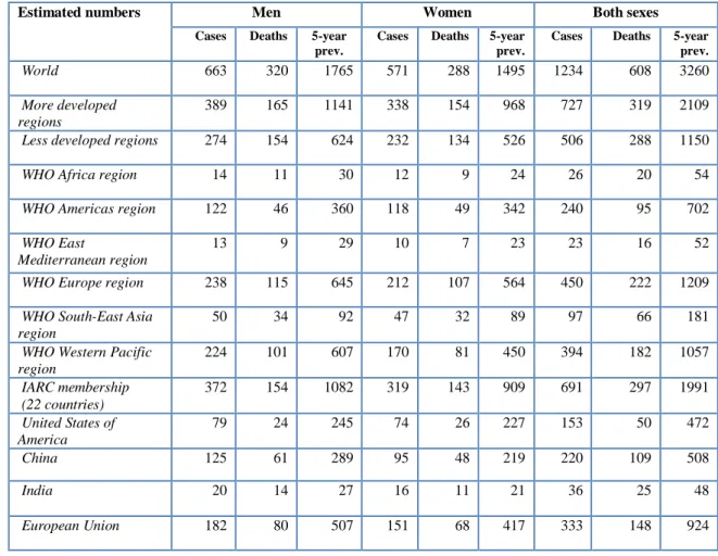
cause of death from cancer (608 000 deaths worldwide, the 8% of all cancer deaths). Mortality rates are also lower in women than in men, except in the Caribbean. There is less variability in mortality rates worldwide (6-fold in men, 5-fold in women), with the highest mortality rates in both sexes estimated in Central and Eastern Europe and the lowest in Middle Africa (Figure 1). Table 1 shows the incidence, mortality and prevalence in the world for the year 2008 (Cancer Mondial, Globocan 2008).
4
Table 1: Colorectal Cancer Incidence, Mortality and Prevalence Worldwide in 2008.
Estimated numbers Men Women Both sexes
Cases Deaths 5-year prev.
Cases Deaths 5-year prev.
Cases Deaths 5-year prev.
World 663 320 1765 571 288 1495 1234 608 3260
More developed regions
389 165 1141 338 154 968 727 319 2109
Less developed regions 274 154 624 232 134 526 506 288 1150
WHO Africa region 14 11 30 12 9 24 26 20 54
WHO Americas region 122 46 360 118 49 342 240 95 702
WHO East
Mediterranean region
13 9 29 10 7 23 23 16 52
WHO Europe region 238 115 645 212 107 564 450 222 1209
WHO South-East Asia region
50 34 92 47 32 89 97 66 181
WHO Western Pacific region 224 101 607 170 81 450 394 182 1057 IARC membership (22 countries) 372 154 1082 319 143 909 691 297 1991 United States of America 79 24 245 74 26 227 153 50 472 China 125 61 289 95 48 219 220 109 508 India 20 14 27 16 11 21 36 25 48 European Union 182 80 507 151 68 417 333 148 924
1.2 Genetics of colorectal cancer
Most cases of colorectal cancer are sporadic. Risk factors include increasing age, male sex, previous colonic polyps or previous colorectal cancer, and environmental factors (red meat, high-fat diet, inadequate intake of fiber, obesity, sedentary lifestyle, diabetes mellitus, smoking, and high consumption of alcohol). Inflammatory bowel disease (ulcerative colitis and Chron’s disease) are two-thirds of the incidence, and the risk increases with duration of illness, severity and extent of inflammation.
A number of familial syndromes are associated with a high risk of colorectal adenocarcinoma. FAP (Familial Adenomatous Polyposis) is an autosomal dominant condition due to different types of mutations in APC gene (Adenomatous polyposis
5
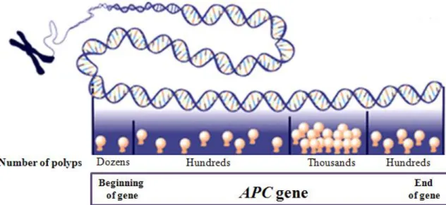
coli), which encodes a tumor suppressor that is part of the WNT signaling pathway (see below); its incidence varies from 1 in 7000 to 1 in 22000 births. The syndrome is more common in Western countries and it is characterized by more than 100 adenomatous polyps in the colon and rectum, developing after 10 years age. In addition to colonic polyps the syndrome can give rise to extraintestinal manifestations such as osteomas, epidermoid cysts, supernumerary teeth, thyroid tumors and brain tumors. By age 10 years, 15% of FAP gene carriers manifest adenomas; by age 20 years, the probability rises to 75% and by 45 years the risk becomes 90% (www.cancer.gov). The average age of CRC diagnosis if untreated is 39 years; 7% develop CRC by age 21 and 95% by age 50 (Jasperson et al., 2010). The clinical features of FAP appear to be generally associated with the location of the mutation in the APC gene and the type of mutation; for example a dense carpeting of colonic polyps is seen in patients with mutations between codons 169 and 1393. Figure 2 shows mutations in different regions of APC gene that correspond to variation in severity of symptoms.
Figure 2: Mutations in APC gene cause FAP, but mutations in different regions of the gene will have a varying effect on the nature and severity of symptoms.
The detection of APC mutation is approximately 80% using sequencing alone, such as the protein truncation assay (PTT). Studies reported whole exon deletion in 12% of
6
patients negative to APC sequencing test. Individuals at risk of FAP begin surveillance in the early teenage years (10-15 years). Colon surveillance should not be stopped in carriersof APC mutations because polyps sometime do not manifest until the fourth and fifth decades of life. Colectomy could be important when, in a FAP patient, numerous polyps have developed (>20) and when adenomas >1 cm are found or when advanced histology appears. Cyclooxigenase II inhibitors such as celecoxib and rofecoxib have been associated with a decrease in polyp size and number in FAP patients. Attenuated FAP is characterized by fewer adenomatous polyps (30, range 0-100) at colon and rectum than in classic FAP and is associated with particular subsets of APC mutations, including missense changes. Approximately 7% to 17% of patients with a FAP phenotype and without a detectable APC germline mutation carry the mutations in MYH gene.
MYH associated polyposis (MAP) occurs in about 1/18000 CRC patients. MAP is caused by biallelic mutations in MYH, a gene involved in the base excision repair pathway to defend DNA against oxidative damage. This gene product prevents G:C to T:A transversions, caused by oxidative stress. The colonic phenotype of MAP mimics attenuated FAP, including propensity for proximal colonic neoplasms. There is also an association between hyperplastic or sessile serrated polyps (Jasperson et al., 2010). Individuals with Lynch syndrome (hereditary non-polyposis colorectal cancer/HNPCC) are predisposed to various types of cancers and are the 2-4% of all CRCs; in particular this syndrome occurs in about 1/300 people with colorectal cancer. Endometrial cancer is the most common extracolonic malignancy associated with Lynch syndrome; also other cancers such as gastric, ovarian, biliary, urinary tract, small bowel, brain are associated with HNPCC. It is an autosomal dominant condition caused by mutations in one of several DNA mismatch repair (MMR) genes. The mean age of CRC diagnosis in
7
HNPCC syndrome mutation carriers is 44 years, compared with 64 years in sporadic cancer. Lifetime CRC risk is estimated to be 50-80%. Colon cancers arise in Lynch syndrome at a younger age of onset and a more proximal location compared to sporadic neoplasms; these types of tumor are poorly differentiated, mucinous. They are also characterized by a high level of microsatellite instability (MSI-H), a feature of cancers with mutations in MMR, necessary for maintaining genomic fidelity by correcting single base mismatches and insertion-deletion loops that form during DNA replication. Mutations in hMLH1 and hMLH2 account for the up to 90%. Mutations in hMSH6 account for approximately 10% and mutations in hPMS2 are detected in rare occasions. The research criteria for defining Lynch syndrome families were established by the international collaborative group (ICG) meeting in Amsterdam in 1990 and are known as the Amsterdam criteria.
Amsterdam criteria:
1) One member diagnosed with colorectal cancer before age 50 years.
2) Two affected generations
3) Three affected relatives, one of them a first degree relative of the other two
4) FAP should be excluded
5) Tumor should be verified by pathological examination
To increase sensitivity, the Amsterdam criteria were revised to obtain Amsterdam criteria II:
1) There should be at least three relatives with a Lynch syndrome associated cancer (colorectal cancer or cancer of the endometrium, small bowel, ureter, or renal pelvis).
2) One should be a first-degree relative of the other two
8
4) At least one should be diagnosed before age 50 years
5) Familial adenomatous polyposis should be excluded in the colorectal cancer cases
6) Tumor should be verified by pathological examination.
Another set of clinical criteria (Bethesda Guidelines) can be used to identify families with germline MMR mutations:
1) Colorectal cancer diagnosed in an individual younger than 50 years.
2) Presence of synchronous, metachronous colorectal, or other Lynch syndrome-associated tumor (i.e endometrial, stomach, etc) in an individual regardless of age.
3) Colorectal cancer with MSI-high pathologic associated features diagnosed in an individual younger than 60 years
4) Colorectal cancer or Lynch syndrome associated tumor diagnosed in at least one first-degree relative younger than 50 years.
5) Colorectal cancer or Lynch syndrome-associated tumor diagnosed at any in two first degree, second-degree relatives.
Genetic tests typically start with analysis of hMLH1 and hMLH2 by sequencing. Another approach for identifying Lynch syndrome, is to perform tumor testing when any of the Bethesda guidelines are identified. Approximately 90% Lynch syndrome associated CRC will have MSI-H, making this analysis very sensitive; sporadic MSI-H CRCs could be the result of somatic hypermethylation of the hMLH1 promoter region. Tumor testing with IHC utilizes four antibodies specific for hMLH1, hMSH2, hMSH6, hPMS2 proteins to evaluate tumor for MMR deficiency. Surveillance decrease both incidence and related deaths. Colonoscopies at 3-years intervals have been shown to decrease the risk of CRC by 50% and prevent CRC deaths. The reduction in CRC risk
9
and death is expected to be even greater when screening intervals are reduced to every 1-2 years. Screening colonoscopy in affected individuals should be initiated by 20-25 years of age and repeated every 1-2 years. Subtotal colectomy with ileorectal anastomosis is advised with the appearance of colon cancer. Population-based studies have led to estimates that 0.8%-2.3% of all CRC cases meet the Amsterdam criteria I and II for Lynch syndrome. A subset of these (40-70%) do not have MMR deficiency and have been termed familial colorectal cancer type X (that have lower CRC risk than HNPCC and do not exhibit MSI). Familial CRC arises from a number of different, lower-penetrance susceptibility genes than those associated with the well-defined, but rare inherited syndromes. No specific genetic markers are available for common familial CRC, then screening and surveillance are based on family history. Patients with a single first-degree relative older than age 60 years with colon cancer should receive average-risk colon cancer screening, but starting at age of 40 year; patients who have one relative with CRC before 60 years or two first degree relatives with CRC should be screened every 5 years by colonoscopy, starting at age 40 years, or at age 10 years younger than the earliest case in the family; patients with only second or third degree relatives with CRC should receive average-risk screening. The recommendations for screening average risk persons include these options: fecal occult blood screening each year, followed by colonoscopy; double contrast barium enema every 5-10 years; colonoscopy every 10 years.
Rare Colon Cancer Syndromes: Peutz-Jeghers syndrome (PJS) is an early-onset autosomal dominant disorder characterized by small bowel and histologically distinctive hamartomatous polyps. Individuals with PJS have an estimated 81-93% lifetime risk of cancer. Germline mutations in the STK11 gene at chromosome 19p13.3 (tumor suppressor gene) have been identified in approximately half of PJS families; a
10
study reported that 85% of individuals with PJS developed cancer by age 70. Juvenile polyposis syndrome (JPS) is a genetically heterogeneous, rare, childhood-onset, autosomal dominant disease due to germline mutations in the MADH4 gene (SMAD4/DPC4) in approximately 15-20% of cases and to mutations in BMPR1A gene in approximately 25-40% of cases. HPP is a rare condition characterized by multiple and/or large hyperplastic polyps of the colon. The World Health Organitation’s criteria for HPP include 30 cumulative hyperplastic polyps of any size distributed throughout the colon; >5 hyperplastic polyps proximal to the sigmoid colon with at least two >10 mm in diameter; or at least 1 hyperplastic colonic polyp in an individual with a first-degree relative with HPP. These conditions increase risk of CRC, which occurs on average in the 50s or 60s. The inheritance of HPP is weak, although both recessive and dominant transmission patterns have been proposed. Curiously, individuals with biallelic MUTYH mutations have been shown, on occasion, to meet criteria for HPP (Cunningham et al., 2010; Jasperson et al., 2010; www.cancer.gov).
1.3 Cytogenetics of colorectal cancer
For colorectal cancers, the acquisition of genomic instability is considered a key hallmark. Three major molecular subtypes can be recognized: MIN (or MSI, for “microsatellite instability”), CIN (for “chromosomal instability”) and CIMP (for “CpG islandmethylator phenotype”). MIN-CRC accounts for approximately 15%–20% of sporadic colorectal cancers. The characteristics of the three pathways (MIN, CIN, and CIMP) are not completely defined and thus they are not mutually exclusive; it is believed that a tumor can occasionally show features of multiple pathways, although the extent and nature of this overlap remains to be determined. CIN is the most common type of genomic instability observed in colon cancer and occurs in 80%–85% of
11
colorectal tumors. It occurs mainly in non-MIN cancers (or MSS for “microsatellite stable”) which are proficient for mismatch repair. CIN CRC show several forms of genomic instability, characterized mainly by chromosomal rearrangements and numerical abnormalities at a greatly increased rate compared with normal cells. The most recurrent aberration found in all cytogenetic studies performed, either in primary tumors or in colon cancer cell lines or in fixed colorectal cancer tissue blocks is 18q (Figure 3). (see the review by Migliore et al., 2011).
Figure 3: The most frequent aberrations found in CRC (Migliore et al., 2011).
1.4 Epigenetics
Epigenetics is defined as heritable changes in gene expression that are not accompanied by changes in DNA sequence. An epigenetic modification is DNA methylation, a covalent addition of a methyl group (CH3) to the nucleotide cytosine. In mammals, most of the DNA CpG sites are methylated (90%–98%), but there are specific CpG-rich areas of DNA where most CpGs are not methylated (CpG islands); a few genes are imprinted genes, regulated by methylation of the CpG islands in their promoter, and the markings are stably replicated during cell division, but are reversed when inherited
12
through an individual of the opposite sex. CpG islands are associated with promoter regulatory regions of almost all housekeeping genes as well as with half of tissue-specific genes (Esteller et al., 2011). Promoter hypomethylation has been associated with an increased gene transcription. DNA hypermethylation occurs at specific regulatory sites in the promoter regions or repetitive sequences. A heavy density of cytosine methylation in the CpG islands of the tumor suppressor gene promoters can lead to a complete block of transcription, and many types of cancer use this mechanism to inactivate tumor suppressor genes. Another critical epigenetic mechanism refers to chemical modifications of the histone tails. Histones, besides being DNA-packaging proteins, can regulate the underlying DNA sequences through complex posttranslational modifications of their N-terminal tails, such as aminoacid-specific acetylation, methylation, or phosphorylation. Histones are acetylated on lysine residues at their amino termini by histone acetyltransferases (HATs), and acetylated histones are deacetylated by HDACs. The opposing effects of HATs and histone deacetylases (HDACs) regulate gene expression through chromatin modification. The HDAC-mediated removal of acetyl groups from lysine residues in the amino termini of histones leads to chromatin condensation and transcriptional inactivation of the involved DNA (Ropero et al., 2007; Yang et al., 2007). This transcriptional inactivation can contribute to suppression of tumor suppressor gene expression and enhanced tumorigenesis. Non-coding RNA (ncRNAs) are involved in the regulation of many important biological processes; the most widely studied class of ncRNAs are miRNAs, which are involved in post-transcriptional gene silencing by controlling mRNA translation into proteins. Alterations (genetic or epigenetic) of genes coding these ncRNAs can modify the expression profile of the ncRNAs and thus alter the mechanism they regulate.
13
1.4.1 DNA methylation in colorectal cancer
Almost 30–40% of proximal site colon tumors and a 3–12% of distal colon and rectal tumors are characterized by a high CIMP, in which numerous CpG islands are methylated and several tumor suppressor genes or ncRNA are inactivated. Altered promoter DNA methylation seems to correlate with deregulation of DNA methyltransferases. De novo DNA methyltransferase 3B (DNMT3B) is generally repressed in human colorectal cancer cell lines (CCL) and primary tumors by aberrant DNA hypermethylation of its distal promoter. DNMT3B distal promoter region was unmethylated in nomal colon tissue and densely hypermethylated in certain colon cancer cell lines. The CpG sites located 764 bp upstream and 208 downstream the TSS (transcription start site) were hyper-and hypomethylated respectively and showed no differences between healthy and tumor samples. The CpG site 352bp upstream of the TSS was completely unmethylated in non–tumorigenic colon tissues, while it was hypermethylated to different degree in most colon CCL analyzed (HCT15, Sw480, Co115, HT29). None of the tumors overexpressing DNMT3B showed DNMT3B promoter hypermethylation. 25% of primary colon tumors with an unmethylated DNMT3B promoter overexpressed DNMT3B. At the epigenome level, DNMT3B promoter hypermethylation was associated with the hypomethylation of gene promoters usually hypermethylated in the healthy colon. Forced DNMT3B overexpression in cancer cells restored the methylation levels of these promoters in the healthy colon (Huidobro et al., 2012). Some genes such as hLMH1, MGMT and TSP1 showed an increase in methylation during all the stages of the disease. Other genes such as RASSF1A or TIMP3 seem more methylated in the last stages or in metastases. However, sometimes conflicting results are found, with the same gene studied. This could be because of the analysis of different CpG sites in the same gene depending on the
14
different methods used to assess methylation [Pyrosequencing, vs Methylation Specific Polymerase chain reaction or vs COBRA]. Moreover, differences in age, tumor type or heterogeneity, or different exposure to environmental factors (diet or microorganisms) could also explain the contrasting methylation pattern of specific genes found in various studies. Leong and co-workers (Leong et al., 2011) found an inverse relationship between methylation of 10 tumor suppressor genes and chromosomal aberrations; then if the frequency of methylation is less prevalent in advanced disease, it is possible that the advanced rectal cancers are driven via the CIN pathway, whereas the early cancers were driven via the methylation pathway. Some genes could have a dual role to promote or suppress tumor formation depending on tumor type and molecular context; the receptor tyrosine kinase-like orphan receptor 2 (ROR2), a transmembrane protein that participates in Wnt signaling, is frequently silenced by promoter hypermethylation in human colon cancer; instead in osteosarcoma cells suppressed expression of ROR2 inhibits cell invasiveness (Lara et al., 2010). SPARCL1 protein was over-expressed in the early stages and weakly expressed in the metastatic tumors; however, in a study of non-small cell lung carcinoma, SPARCL1 mRNA levels were found to be down-regulate. SPARCL1 protein could therefore play a dual role of oncogene in colorectal carcinoma and tumor suppressor in lung cancer (Zhang et al., 2011). The discovery of methylation marks in CRC was considered of fundamental importance to correlate methylation status of specific genes with early diagnosis and prognosis. For instance, it was shown that aberrant methylation of APC, MGMT, RASSF2A, and Wif-1 genes is associated to tumor initiation, but not to tumor progression (Lee et al., 2009). Several epigenetically silenced genes have an important role in colorectal carcinogenesis; APC2, a homolog of APC1 tumor suppressor gene involved in Wnt signaling pathway, showed a frequency of 95.5% methylation in its promoter. ECAD, a Ca+-dependent
15
adhesion molecule that mediates intercellular contacts, was hypermethylated in 40.8% of primary CRCs. Loss of ECAD expression is then associated with the invasion and metastasis (Naghibalhossaini et al., 2011). Lind and colleagues (Lind et al., 2011) found promoter hypermethylation of the CNRIP1, FBN1, INA, MAL, SNCA, and SPG20 genes was frequent in both CRCs (65–94%) and adenomas (35–91%), whereas normal mucosa samples were rarely (0–5%) methylated; the sensitivity of at least two positives among the six markers was 94% for CRCs and 93% for adenoma samples, with a specificity of 98%. Methylation of IGFBP3, EVL, FLNC, and CD109 genes is associated with an eightfold increase in mortality risk relative to that of patients with no DNA methylation of these genes (Carmona et al., 2011). Moreover, hypermethylation of CDH13 and FLBN3 genes is associated with poor prognosis in CRC (Wang et al., 2011). Looking for novel prognostic biomarkers for CRC, recent studies showed that tumors that have silenced genes in the extracellular matrix remodeling pathway show worse survival (Carmona et al., 2011; Yi et al., 2011). Invasive screening modalities, including colonoscopy, are not ideal for application to the asymptomatic population. Therefore, active investigations are now underway to discover noninvasive biomarkers, such as those found in stool, which could supplement or supplant colonoscopic screening. DNA methylation changes have also been observed in plasma as a marker of circulating tumor DNA. Less is known about changes in WBC DNA methylation levels and cancer risk, although studies are rapidly emerging. Several studies measuring overall WBC global DNA methylation in different cancer types including colon, bladder, stomach, breast, head and neck cancer have found an elevated risk (sometimes statistically significant) for cancer between those in the lowest quantile of global DNA methylation compared to those in the highest quantile (Terry et al., 2011). In plasma, cell-free methylated DNA AQ3 has been reported to be a useful biomarker of
16
noninvasive blood screening for the detection of CRC. Septin 9 (SEPT9) and vimentin (VIM) genes have been analyzed in blood/serum samples and stool samples and reported sensitivity and specificity range of 68–77% and 83–94%, respectively (Chen et al., 2005; Devos et al., 2009). Among genes that have been proven to be promising for an early diagnosis of CRC, besides SEPT9 (Grützmann et al., 2008), there are also ALX4 and TMEFF2 (He et al., 2010). Loss of SMAD4, a tumor suppressor gene frequently inactivated in pancreatic and CRCs, correlates significantly with decreased survival in colon cancer patients. High SMAD4 expression, however, is significantly associated with increased survival, especially in colon cancer patients who has undergone potential curative surgery (Isaksson-Mettävainio et al., 2011). Figure 4 shows some genes methylated during the different stages of carcinogenesis and figure 5 shows the most studied genes which have been found methylated in CRC, with their possible functions in cancer initiation and progression.
17
Figure 4: Methylation of genes during CRC stages (Migheli and Migliore, 2012). NORMAL EPITHELIUM
MGMT , P16 (respectively methylated in 20% 18% of samples); MSP assay (Krakowczyk et al., 2008)
MGMT, P16 (respectively methylated in 59%, 53% of samples); (MSP) (Krakowczyk et al., 2008)
CANCER
METASTATIC CANCER Adenomatous polyps
ADENOMA
hMLH1 (12,3 % methylated samples with more then 20% of methylation and loss of expression); samples principally MSI-H and of proximal tumor site (PYROSEQUENCING) (Gay et al., 2011)
hMLH1 (all methylation negative samples with less then 20% of methylation); PYROSEQUENCING assay (Gay et al., 2011)
hMLH1, hMSH2 and MGMT respectively methylated in 1.8%, 8.0%, 33.9% of salmples; MSP assay (Lee et al., 2011 ) hMLH1, hMSH2, MGMT (respectively methylated in 0%, 5.4%, 10.7% of samples); MSP assay (Lee et al., 2011 )
hMLH1, MSH2, MGMT (respectively methylated in 1,8%, 13.4% and 47.3% of samples; MSP (Lee et al., 2011 ) The highest methylation detected was for MGMT gene (47.1%) followed by 35.3% for HIC-1 and 5.9% for RASSF1A gene;
MSP assay (Abouzeid et al., 2011)
High frequency of methylation at MGMT, RASSFA, and HIC-1 (respectively in 25%, 47.2%, and 41.7% of samples); MSP assay (Abouzeid et al., 2011).
67% of samples had methylation in EVL/hsa-miR-342; MSP assay (Grady et al., 2008)
86% of samples methylated in EVL/hsa-miR-342; MSP assay (Grady et al., 2008) Methylated EVL/hsa-miR-342 in 12% of normal mucosa from cancer-free controls and 56% of histologically
normal colorectal mucosa from individuals with concurrent colorectal adenocarcinoma (‘field defect’); MSP assay (Grady et al., 2008)
Extensive (including two methylation-sensitive regions) and partial (one of two regions) SFRP2 methylation levels found in 61.7% and 24.8% of samples; COBRA (Takeda et al., 2011) .
Extensive (including two methylation-sensitive regions ) and partial (one of two regions) SFRP2 methylation levels were in 8.7 % and 37.9 % of samples; COBRA (Takeda et al., 2011) .
Extensive (including two methylation-sensitive regions ) and partial (one of two regions) SFRP2 methylation levels were in 3.9 % and 39.9 % of normal colonic mucosa from CRC patients (N-Cs), and in 0 % and 30.6 % of colonic mucosa from subjects with no
18
Figure 5: The methylation pattern of many genes from normal and tumor colonic mucosa from CRC patients has been extensively studied. Here we report the most studied genes and the pathways in which they are involved (Migheli and Migliore, 2012).
1.4.2 Histone modifications in colorectal cancer
Little is known about patterns of histone modification alteration in human tumors and even less in CRC. Chromatin remodeling is, together with methylation, a key mechanism for gene regulation and consists of modifications at conserved lysine residues on the tails of histone proteins; lysine acetylation allows the transcription by weakening the association of the histone with DNA and allows transcription factor binding. Lysine methylation can be associated with both active and repressed regions of DNA; trimethylation of histone H3 lysine 4 active transcription, instead methylation of H3K9 and H3K27 appears at transcriptionally silent gene promoters (Jones et al., 2007). Hypomethylation alone cannot turn on silenced genes, instead increased Histone H3 acetylation with localized hypomethylation allows long-term reversion of
19
epigenetically silenced genes; a study showed as CDO1, HSPC105 and MAGEA3 were still expressed 10 days post 5-aza-dC treatment, in fact they had localized hypomethylation at the transcriptional start site and an increased histone H3 acetylation (Mossman et al., 2011). Enzymes for chromatin remodeling can alter chromatin by covalent modification of histone or by using the energy from ATP hydrolysis. Included in the ATP-dependent chromatin remodeling enzyme family is the chromodomain helicase DNA-binding protein (CHD) family, which consists of nine proteins (CHD1– 9) in humans. With regard to cancer, CHD5 controls proliferation and apoptosis. In humans, CHD5 is inactivated not only by deletion but also by hypermethylation. These alterations might contribute to cancer pathogenesis by deregulating CHD-mediated chromatin remodeling (Kim et al., 2011). SOCSs and SHP1 genes seem to have a role as tumor suppressor and DNA methylation as well as histone acetylation/deacetylation could control their transcriptional regulation in CRC cells. Xiong and colleagues (Xiong et al., 2012) showed that TSA, an histone deacetylase inhibitor (HDACi), increased the mRNA levels of SOCS1 and SOCS3. The induction of SOCS1 and SOCS3 expression by TSA in human CRC cells was because of an increase in the acetylation of H3 and H4 histone proteins associated with their promoter regions. However, they did not observe any significant changes in histone acetylation of SHP1 promoter regions. Two different studies reported that the global levels of H4K12ac and H3K18ac increased in adenocarcinomas respect to the normal tissue or adenomas and that the H3K9me2 expression was also associated with the progression adenoma–adenocarcinoma (Ashktorab et al., 2009; Nakazawa et al., 2011).
20
1.4.3 NcRNA alterations in colorectal cancer
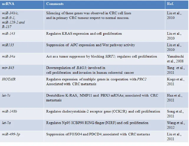
Chromosome anomalies (deletions, translocations, copy-number alterations), DNA mutations and epigenetic deregulation of the ncRNAs or of the genes involved in their biogenesis have been described in tumor progression and the best-characterized miRNAs dysregulated by DNA hypermethylation in tumors, including CRC and the functional consequences in tumoral cells, have been reviewed recently (Lopez-Serra et al., 2011). In Table 2 some recent examples of ncRNAs found dysregulated in CRC cells are shown. For instance, epigenetic alteration of miR-143 which targets KRAS can interfere with its expression and induce cell proliferation (Liu et al., 2010); epigenetic alteration of miR-148b which targets cholecystokinin-2 receptor gene (CCK2R) can lead to cell proliferation (Song et al., 2011). A small part of the miRNAs were up-regulated by 5-aza-2 deoxycytidine (5-aza-dC) treatment in CRC cells; however, different cell type like SW1116 and HT29 cell could have different drug sensitivity. DNA methylation analyses showed that the promoter region of the mir-345 gene was heavily methylated in HT29 cells (64.73%) and it was decreased to 35.78% after 5-aza-dC treatment, which results in a major 345 expression; AQ4 overexpression of mir-345 may suppress colon cancer cell invasiveness in vitro. mir-mir-345 might be involved in pathogenesis of CRC through downregulation of the expression of BAG3, one of the molecules that regulates apoptosis (Tang et al., 2011). Hox transcript antisense intergenic RNA (HOTAIR) is a long ncRNA that regulates expression of multiple genes in cooperation with PRC2; HOTAIR expression levels in CRC tissues were higher than those in corresponding noncancerous tissues; its overexpression increased the invasiveness of CRC cells, then this ncRNA might play a role in promoting metastasis of CRC. Moreover, patients with high HOTAIR expression had a significantly poorer prognosis than those with low HOTAIR expression (Kogo et al., 2011).
21
Table 2: Deregulation of ncRNAs in CRC by epigenetic, chromosome abnormalities (deletions, translocations, copy-number alterations), and DNA mutations (Migheli and Migliore, 2012).
1.5 Colorectal adenoma-carcinoma sequence
An unresolved question related to genomic instability, which has been raised in many papers, is whether CIN arises early in tumorigenesis and initiates the adenoma-carcinoma sequence or whether it is acquired during this process and facilitates the formation of colon cancer. Michor et al. developed a mathematical representation of the evolutionary dynamics of colorectal tumorigenesis and found that one or two CIN genes in the genome are enough to make sure CIN emerges early (Michor et al., 2005). However this view is not universally accepted: some authors are inclined to believe that CIN is acquired during tumorigenesis and facilitates progression to malignancy. Even in light of the latest knowledge, the question is still considered unresolved (see the recent review by Pino and Chung, 2010). Different tumors show various patterns of
22
aneuploidy. The loss of heterozygosis that occur in the first phases of the CRC cancerogenesis, as well as the alteration of methylation pattern of multiple key genes can drive the development of colorectal cancer by facilitating the acquisition of multiple tumor-associated mutations and the instability phenotype. Aberrant CpG island methylation is involved in cancer development, but it is not yet clear if it is a cause or an effect of cancer formation, which genes are methylated during the pathogenesis of individual cancers, when is the time of methylation and gene silencing, how specific methylation profiles are established, and what determines tumor typespecific methylation (Kim et al., 2010). Restricted folate diet or SNPs in one-carbon metabolism, leading to a reduction of the total amounts of DNA methylation in human tumors results in hypomethylation of repetitive DNA sequences, contributing to the origin of cancer cells by generation of chromosomal instability, reactivation of transposable elements, and loss of imprinting; moreover, hypomethylation could activate proto-oncogenes. The misincorporation of uracil into human DNA, favoured when thymidylate availability is restricted, could also increase the frequency of chromosome cleavage. On the other hand, tumor suppressor genes could gain CpG island methylation, resulting in the inactivation of these protecting proteins. Moreover epigenetic alterations could influence either cancer initiation or progression. In 1990 Fearon and Volgestein proposed a model for colorectal tumorigenesis. For this model the mutational activation of oncogenes and inactivation of tumor suppressor genes, lead to the development of CRC. These alterations generally occur in a vertical sequence that goes togheter with the clinical progression of the tumor, but probably it is more important the total accumulation of changes rather than their order. The key oncogene in this model is K-RAS, instead the tumor suppressor genes involved reside on chromosome 5q, 17p and 18q. Other somatic alterations, such as DNA methylation, are
23
involved (Leslie et al., 2002). Figure 6 shows a colorectal adenoma-carcinoma sequence model.
Figure 6: Colorectal adenoma-carcinoma sequence (according to Fearon and Volgestein model).
1.5.1 APC (Adenomatous Polyposis Coli) gene
The human APC gene is located on the long (q) arm of chromosome 5 between positions 21 and 22, from base pair 112,118,468 to base pair 112,209,532.
This gene encodes a tumor suppressor protein that acts as an antagonist of the Wnt signaling pathway. It is also involved in other processes including cell migration and
24
adhesion, transcriptional activation, and apoptosis. Defects in this gene cause familial adenomatous polyposis (FAP), an autosomal dominant pre-malignant disease that usually progresses to malignancy (see page 5). Disease-associated mutations tend to be clustered in a small region designated the mutation cluster region (MCR) and result in a truncated protein product. APC activity is correlated with its phosphorylation state. The APC gene product (2843 amino acids; 310 kDa) indirectly regulates transcription of a number of critical cell proliferation genes, through its interaction with the transcription factor β catenin. APC binding to β catenin leads to ubiquitin-mediated beta catenin destruction; loss of APC function increases transcription of β catenin targets. These targets include cyclin D, C-myc, ephrins and caspases. Particularly the APC protein normally builds a complex with glycogen synthase kinase 3-beta (GSK-3β) and axin. This complex is then able to bind β- catenin in the cytoplasm; with the help of casein kinase 1 (CK1), which carries out an initial phosphorylation of β-catenin, GSK-3β is able to phosphorylate β-catenin a second time. This targets β-catenin for ubiquitination and degradation by cellular proteosomes. This prevents it from translocating into the nucleus, where it acts as a transcription factor for proliferation genes. APC also interacts with numerous actin and microtubule associated proteins. APC itself stabilizes microtubules. Hypermethylation of APC promoter 1A, instead of mutations involving APC and beta-catenin, contributes to moderate activation of Wnt signalling in a subset of serrated adenomas (Fu et al., 2009). A study analyzed the methylation status of 10 genes in fresh-frozen tissues and corresponding plasma samples from patients from stage I and II of sporadic colorectal cancer, 276 healthy individuals, and plasma from 64 colorectal adenoma patients using MSP. The methylation was detected in 18% for p14, 34% for p16Ink4A, 27% for APC, 34% for DAPK, 32% for HLTF, 21% for hMLH1, 39% for MGMT, 24% for RARbeta2, 58% for RASSF2A, and 74% for Wif-1; the author
25
concluded that tumor-specific methylation of APC, MGMT, RASSF2A, and Wif-1 genes might be a valuable biomarker in plasma for the early detection of CRC (Lee et al., 2009).
1.5.2 MGMT (O-6-methylguanine-DNA methyltransferase) gene
Cytogenetic Location 10q26.3: Start: 131,265,448 bp from pter - End: 131,566,271 bp from pter; Size: 300,824 bases. Protein: 207 amino acids; 21646 Da.
O(6)-alkyl-guanine is the major carcinogenic lesion in DNA induced by alkylating mutagens. This DNA adduct is removed by the repair protein, O(6)-methylguanine-DNA methyltransferase. This protein is not a true enzyme since it accepts the alkyl group from the lesion in a stoichiometric reaction and the active enzyme is not regenerated after it is alkylated. The methyl-acceptor residue in the protein is cysteine. MGMT promoter methylation in normal colonic mucosa might be a predisposing factor for cancer as a field effect and an early event in colorectal carcinogenesis. MGMT promoter methylation and loss of expression have been associated with G>A mutations in a variety of genes such as KRAS, PIK3CA, TP53, and APC (Halford et al. 2005; Shen et al., 2005; Ogino et al. 2007; Nagasaka et al., 2008; Nosho et al. 2008). DNA hypermethylation for hMLH1 and MGMT DNA repair genes was reported in precursor lesions to colorectal cancer. These epigenetic alterations may be influenced by factors such as xenoestrogens, folate, and multivitamins. Detection of these changes may help determining cancer susceptibility and early diagnosis (Dumitrescu, 2012). Promoter hypermethylation status of RASSAF1A, MGMT, and HIC-1 genes were determined in
26
36 CRC, 17 adenomatous polyps, and 19 ulcerative colitis, and adjacent normal-appearing tissues using MSP assay. High frequency of methylation at MGMT, RASSF1A, and HIC-1 genes was detected in CRC patients (25%, 47.2%, and 41.7% respectively). The highest methylation observed in adenomatous polyps patients was in MGMT gene (47.1%) followed by 35.3% for HIC-1 gene and only 5.9% for RASSF1A gene; they also found an association between methylation at RASSF1A gene with gender (p = 0.005) (Abouzeid et al., 2011). Morning stool specimens were collected from 69 patients with colorectal cancer, 24 with colon adenoma, 19 with hyperplastic polyps, and 26 healthy controls. The methylation frequencies of MAL, CDKN2A and MGMT were 78.3%, 52.5% and 55.1% in colorectal cancer, 58.3%, 41.7% and 37.5% in colon adenomas, 26.3%, 15.8% and 10.5% in hyperplastic polyps, and 3.8%, 0 and 3.8% in healthy controls, respectively. Significant differences in three genes were found between colorectal cancer and hyperplastic polyp, colorectal cancer and healthy control, colon adenoma and hyperplastic polyp, colon adenoma and healthy control (all P<0.05). The diagnostic sensitivity by combining three methylation markers was 92.8% in colorectal cancer, 70.8% in colon adenomas, significantly higher than fecal occult blood test (29.0% in colorectal cancer and 25.0% in colon adenomas, all P<0.05). No significant associations existed between methylation of the three genes and clinical characteristic including sex, age, tumor location, lymphnode metastases and TNM stage (all P>0.05) (Kang et al., 2011). A study showed an association (81%, κ = 0.59, p < 0.0001) between MGMT methylation and MGMT loss. MGMT methylation and loss of MGMT were not perfectly correlated; in fact MGMT expression may be caused not only by promoter methylation but also by other mechanisms such as a gene mutation. Second, promoter methylation may be present in only one MGMT allele, and the MGMT protein may be expressed from the second allele. Third, there may be other
27
molecules such as ncRNA, that may downregulate MGMT (Shima et al., 2011). It was hypothesised that an MGMT field defect may constitute a preneoplastic event for the development of MMR-deficient tumors displaying microsatellite instability (MSI). Loss of MGMT expression was more frequent in MSI than MSS CRC (p=0.047); moreover loss of MGMT expression was associated with MGMT gene promoter methylation (p=0.03) (Svrcek et al., 2010). Methylation status of the MGMT gene was examined in primary carcinomas and the corresponding normal tissues in 48 patients with CRC using MSP; aberrant methylation was detected in 21% of primary colorectal cancers. There was no statistically significant association between abnormal methylation and gender or age, maximal tumor size, tumor extent, tumor site, histology, lymphnode metastasis, and TNM stage. All stages of colorectal cancers presented MGMT methylation, indicating that the MGMT gene has been methylated from the early stages of colorectal cancers (Hibi et al., 2009). A study observed the incidence of MGMT methylation increased along the adenoma–carcinoma sequence, and the evaluation of the contribution of MGMT methylation to the development of an advanced lesion was statistically significant. The increased incidence of MGMT methylation in normal-appearing mucosa could represent the “field defect”. Authors reported that MGMT promoter methylation occurred frequently in the apparently normal colonic mucosa of CRC patients with tumors that also showed MGMT promoter methylation; in contrast, MGMT promoter methylation was observed much less frequently in healthy subjects and in the apparently normal colonic mucosa of CRC patients with tumors that did not show MGMT methylation. In addition, they observed an association between age and promoter methylation levels. Methylation detected by MSP may not affect the expression of gene products if methylation affects only a few cells or if the methylation
28
of one copy happens with an absence of methylation in the other copy of the gene (Lee et al., 2011).
1.5.3 CDKN2A or p16
Ink4A(Cyclin-dependent kinase inhibitor 2A) gene
Cytogenetic Location 9p21.3: Start: 21,967,751 bp from pter- End: 21,995,300 bp from pter; Size: 27,550 bases.
This gene generates several transcript variants which differ in their first exons. At least three alternatively spliced variants encoding distinct proteins have been reported, two of which encode structurally related isoforms known to function as inhibitors of CDK4 kinase. It acts as a tumor suppressor and has to arrest the cell cycle in G1 and G2 phases. It binds to MDM2 and inhibits its oncogenic action by blocking MDM2-induced degradation of p53 and enhancing p53-dependent transactivation and apoptosis. It also induces G2 arrest and apoptosis in a p53-independent manner by preventing the activation of cyclin B1/CDC2 complexes. Increased expression of the p16Ink4A gene reduces the proliferation of stem cells. This reduction in the division and production of stem cells protects against cancer while increases the risks associated with cellular senescence. Analysis of p16Ink4A promoter methylation was performed by MSP assay. 42.1% of the CRC were found to have the p16Ink4A gene methylated. The methylation status was found to be associated with the gender, lymph node status, tumor stage, smoking status and tumor grade of the CRC patients. P16INK4A plays a pivotal role in tumor development and progression to advanced stages (Sameer et al., 2012). P16INK4A methylation was evaluated by Q-MSP technique in the serum of CRC patients during
29
their follow-up period. The p16Ink4A methylation decreased during two weeks after surgery. One month after surgery, in the patients with recurrence of cancer, a great increase in p16Ink4A methylation was observed, while in the disease-free patients no methylation was seen more; so methylation could sensitively reflect the recurrence status and may be useful for identifying the presence of recurrence during the follow-up of CRC patients (Nakayama et al., 2011). Colorectal cancer (CRC) screening using stool DNA has yielded a greater detection rate than conventional fecal occult blood testing. Stool samples from 31 healthy controls, 25 patients with adenomas and 30 patients with CRC were analyzed and methylation regard to ITGA4, SFRP2 and p16Ink4A promoters were observed in 36.7%, 60.0%, and 40.0% of the CRC samples and in 16.0%, 44.0%, and 24.0% of the colorectal adenomas, respectively. The methylation status had high sensitivity and specificity for the detection of colorectal adenomas and CRC; thus stool screening might be a useful non-invasive method for CRC detection (Chang et al., 2010). Aberrant methylation of p16Ink4A and hMLH1 promoters was found in 47.2% and 53.4% of tumors respectively. For adjacent normal mucosa, 30% of patients were fully unmethylated in p16Ink4A promoter, whereas hMLH1 promoter was predominantly unmethylated (76%). Methylation of p16Ink4AI correlated with gender and tumor size, whereas that of hMLH1 significantly correlated with overall survival. Concomitant methylation of CDKN2A and hMLH1 genes was associated with TNM stage and tumor size (Miladi-Abdennadher et al., 2011).
30
1.5.4 RASSF1A (Ras association (RalGDS/AF-6) domain family member
1) gene
Cytogenetic Location 3p21.31: Start: 50,367,217 bp from pter-End: 50,378,411 bp from pter; Size: 11,195 bases.
Several alternatively spliced transcript variants of this gene encoding distinct isoforms have been reported. It is a potential tumor suppressor. Isoform A interacts with CDC20, an activator of the anaphase-promoting complex (APCCdc20) that initiates chromatid separation and entrance into anaphase. The APCCdc20 protein complex targets securin for destruction, enabling sister chromatid separation. It also targets S and M-phase (S/M) cyclins for destruction, which inactivates S/M cyclin-dependent kinases (Cdks) and allows the cell to exit from mitosis. Isoform A also disrupts interactions among MDM2, DAXX and USP7, thus contributing to the efficient activation of TP53 by promoting MDM2 self-ubiquitination in cell-cycle checkpoint control in response to DNA damage. MSP was used to examine the promoter methylation status of the serum RASSF1A gene; the RASSF1A promoter hypermethylation in gastric (34.0%) and colorectal (28.9%) adenocarcinoma patients were significantly higher than those in patients with benign gastric (3.3%) or colorectal (6.7%) disease or in healthy donors (0%) (P < 0.01). Although the serum RASSF1A promoter hypermethylation frequency tended to be higher in patients with distant metastases, there was no correlation between methylation status and metastasis. There was no correlation between serum RASSF1A promoter hypermethylation and sex, age, tumor differentiation grade, surgical therapy, or serum carcinoembryonic antigen level (Wang et al., 2008). A study analyzed, by mean of Q-MSP, primary tumor and synchronous liver metastatic tissues of 75 CRC patients and
31
evaluated K-RAS and BRAF mutations. RARbeta, RASSF1A, and CDKN2A genes were methylated in 82%, 35%, and 26% of primary tumors, respectively. RASSF1A methylation status was significantly higher in liver metastasis with respect to primary tumor (P=0.000) underlying the role of this gene in liver metastatic progression. Moreover K-RAS and BRAF were mutated in 39% and 4% of cases, respectively and RASSF1A methylation resulted significantly higher in liver than in primary tumor (P=0.05) only in K-RAS wild-type patients. All Ras protein family members are GTPase; when RAS is 'switched on' by incoming signals, it subsequently switches on other proteins, which turn on genes involved in cell growth, differentiation and survival. As a result, mutations in RAS genes can lead to the production of permanently activated Ras proteins. This can cause unintended and overactive signaling inside the cell, even in the absence of incoming signals. Ras is the most common oncogene in human cancer; mutations that permanently activate Ras are found in 20-25% of all human tumors and up to 90% in certain types of cancer (Tommasi et al., 2011).
1.5.5 hMLH1 (mutL homolog 1, colon cancer, nonpolyposis type 2) gene
Cytogenetic Location: 3p21.3; from 37,034,840 to base pair 37,092,336; The hMLH1 gene is composed of 19 exons spanning in a region of 57496 bp. Protein: Aminoacids: 756. Molecular Weight: 84.6 kDa.
32
The MLH1 gene is a member of a set of genes known as the mismatch repair (MMR) genes. This protein fixes mistakes that are made during DNA replication in preparation for cell division. It contains an ATPase domain and two interaction domains, one for MutS homologs (MSH2, MSH3, MSH6) and the other for PMS2, MLH3 or PMS1. MLH1 has no known enzymatic activity. MLH1 forms a heterodimer with PMS2 known as MutLa, although it can also bind to PMS1 or MLH3. This heterodimeric complex binds to the heteroduplexes MutSa (composed of MSH2 and MSH6) or MutSb (composed of MSH2 and MSH3), which recognizes DNA lesions. The heterodimer formed by MLH1 is responsible for the recruitment of the proteins needed for the excision and repair synthesis. The repair is made by removing a section of DNA that contains mistakes and replacing the section with a corrected DNA sequence. A study evaluated the frequency of promoter methylation for hMLH1, hMSH2, and MGMT in colorectal non-tumoral mucosa adenoma, and adenocarcinoma by mean MSP assay. The hMLH1 promoter was methylated in 1.8% of the adenoma and adenocarcinoma samples. No methylation of hMLH1 was found in any corresponding non-tumoral mucosa tested. MSH2 promoter methylation was found in 8.0% of adenoma and 13.4% of adenocarcinoma samples. MGMT methylation was detected in 33.9% of adenoma and 47.3% of carcinoma samples. In the corresponding normal-appearing mucosa, methylation was seen in hMSH2 (5.4%) and MGMT (10.7%), but not in hMLH1 (Lee et al., 2011). In other study by Psofaki et al. (2010) promoter hMLH1 hypermethylation was observed in tumor samples, but not in blood samples.
33
1.6 Environmental factors that might influence epigenetic patterns in
colorectal cancer.
Studies where people migrate from low to high CRC risk areas of the world, demonstrate that changes of diet and physical activity enhance the incidence of cancer in a high-risk country even over one or two generations (Nystrom et al., 2009). Considering CpG island methylation levels in normal colorectal mucosa of different racial groups, some Authors found that African Americans had lower levels of methylation compared to Caucasians and Hispanics. Possible explanations for these findings, include lifestyle factors, genetic protection or predisposition to methylation (Wallace et al., 2010). Moreover, monozygotic twins carriers of high penetrant genetic alteration in HNPCC, associated with hMLH1 mutations, develop cancer at different ages. These observations suggest a role of the environment in epigenetic changes (Esteller et al., 2008).
It was observed that a positive association between vitamin B6 and rectal cancer risk exists in women. Among men, methionine was associated with a decreased risk of proximal colon cancer whilst among women it was inversely associated with rectal cancer (De Vogel et al., 2008).
Choline could derive from the diet, but also from de novo biosynthesis by means of an enzyme coded by the gene phosphatidylethanolamine-N-methyltransferase (PEMT). One of the choline metabolites, betaine, participates in the methylation of homocysteine to form methionine. Then choline and betaine have been hypothesized to decrease the risk of colorectal cancer. Estrogens cause a marked upregulation in PMET mRNA expression and enzyme activity, then premenopausal women have an enhanced capacity for de novo biosynthesis of choline. In choline deficient cells in culture, and in fetal
34
rodent brains from mothers fed with choline-deficient diets, methylation of the CDKN3 gene promoter decreased, resulting in overexpression of this gene which inhibits cell proliferation. Maternal diet high in choline and/or methionine and/or methyl-folate during pregnancy results in epigenetic changes in gene expression in the fetus (Zeisel et al., 2007).
Alcohol consumption was found to be a risk factor for colorectal tumorigenesis. In an in vivo study (male rats), it was observed that a decrease in RFC1 (reduced folate carrier) mRNA and protein expression correlates with alcoholism. That is a possible reason of lower blood folate levels commonly found in chronic alcoholics (Hamid et al., 2009). Alcohol in murine studies appears to reduce MTR levels; thus it could induce DNA hypomethylation (Arasaradnam et al., 2008). Alcohol consumption may in fact decrease DNA methylation in hepatic tissue by affecting folate metabolism and/or methionine synthesis, which might decrease levels of the S-adenosylmethionine (SAM), the main methyl donor of DNA. Excessive alcohol consumption has been shown to alter DNA methylation in humans. Patients suffering from alcoholism had 8–10% higher methylation in CCGG sequences respect to controls. Associations between alcohol drinking and DNA repetitive element methylation levels have shown inverse associations between Alu and alcohol drinking. However, several studies did not find an association between alcohol consumption and LINE1 methylation. One study found a significant increase in HERP (homocysteine-induced ER protein) promoter methylation in patients with alcohol dependence (Terry et al., 2011). Moreover in a Dutch study, higher frequency of promoter methylation of APC1A, P14, hMLH1, MGMT, and RASSF1A genes was observed in presence of a low-folate/high-alcohol diet.
35
Tobacco could also influence CRC risk. The methylation levels for MGMT, RAR-b, and SST decrease in the following sequence: nonsmokers without colorectal adenomas > smokers without colorectal adenomas > nonsmokers with colorectal adenomas > smokers with colorectal adenomas. Smoking predisposes to diminished methylation of several genes, which, in turn, contribute to colorectal adenoma development (Paun et al., 2010). A study reported that the risk of CIMP-high tumor, among smokers of 20 or more cigarettes per day, was higher among those with low folate and low fiber intake and those who had greater long-term alcohol consumption, although without statistical significance. Moreover, among women alcohol and cigarette smoking were associated with risk of CIMP-high tumors; women who smoked 20 or more cigarettes per day and consumed little or no alcohol did not have an increased risk of a CIMP-high tumors (Slattery et al., 2007). The cellular modifications due to exposures to the chemicals present in cigarette smoke have been widely investigated and include DNA adducts, gene mutations, micronuclei, chromosome aberrations, sister chromatin exchanges and DNA strand breaks; DNMTs seem to bind DNA damage sites, which results in altered methylation patterns on these regions, suggesting a molecular mechanism for the generation of aberrant DNA methylation by exposure to chemicals such as those present in cigarettes. Only a few studies have investigated overall 5-mC content and smoking, and found no difference by cigarette smoking patterns. More research has focused on the effect of smoking on the methylation levels of repetitive elements. LINE-1, Alu and AluYb8 methylation did not differ between current and never/former smokers. It has been suggested that chemicals affecting epigenetic marks could have a larger impact when exposure occurs in utero or earlier in life, a time when epigenetic modifications are being established. Overall levels of genomic DNA methylation have been investigated in several studies in children and adults. Prenatal exposure to cigarette
36
smoke has been associated with an increase in the overall blood levels of DNA methylation in adulthood. LINE-1, Sat2 and Alu methylation was analyzed in adults and children prenatally exposed to smoking. LINE-1 and Sat2 levels were lower in exposed individuals when compared to unexposed ones. However, other elements such as LINE-1 do not seem to be affected by exposure to cigarette smoke. Several genes were investigated in blood samples from adults in a cohort of individuals born preterm that were exposed to smoking, with lower levels found for IGF2 methylation (see review by Terry et al., 2011).
In a study in vitro on HT 29 cells, green tea was found to inhibite DNMT1 causing CpG demethylation and reactivation of previously methylated genes (hMLH1, MGMT, P16INK4A). There is strong evidence that polyphenols from tea, soft fruits and berries, vegetables, apples, and even from wine, are potent anti-carcinogenic agents in vitro and in animal models that prevent DNA instability. Several polyphenols are potent inhibitors of DNMT activity in vitro, capable of reversing DNA hypermethylation and reactivating tumor suppressor genes activity. Polyphenols inhibit DNMT activity and DNA methylation in two ways: by direct insertion into the binding pocket of DNMT (competitive inhibition) or indirectly by decreasing intracellular SAM concentrations. Tea components (especially green and black tea) and specific soya isoflavones inhibit DNMT activity in human cancer cells. DNMT 1 inhibition demethylates CpG islands in the promoter regions of silenced tumor suppressor genes including p16Ink4A, retinoic acid receptor b, MGMT, mMLH1 and GST. Green tea (EGCG) can reactivate retinoic acid receptor b in prostate and breast cancer cells, p16Ink4A in colon cancer cells and GST in prostate cells. Genistein could reverse DNA hypermethylation and reactivate p16Ink4A, retinoic acid receptor b and MGMT gene expression (Duthie, 2011).