Creative Commons Non Commercial CC BY-NC: This article is distributed under the terms of the Creative Commons Attribution-NonCommercial 4.0 License (http://www.creativecommons.org/licenses/by-nc/4.0/) which permits non-commercial use, reproduction and distribution of the work without further permission provided the original work is attributed as specified on the SAGE and Open Access pages (https://us.sagepub.com/en-us/nam/open-access-at-sage).
https://doi.org/10.1177/0394632017722332 International Journal of
Immunopathology and Pharmacology 2017, Vol. 30(3) 238 –252 © The Author(s) 2017 Reprints and permissions:
sagepub.co.uk/journalsPermissions.nav DOI: 10.1177/0394632017722332 journals.sagepub.com/home/iji
Introduction
Multiple sclerosis (MS) is a long-lasting progres-sive autoimmune neurological disorder of the cen-tral nervous system.1 Immune cells invasion, axonal injury, and myelin sheath deformation are the com-mon hallmarks of MS, which eventually produce neurological disability.2 Based on heterogeneous
Human periodontal ligament stem
cells secretome from multiple sclerosis
patients suppresses NALP3 inflammasome
activation in experimental autoimmune
encephalomyelitis
Thangavelu Soundara Rajan1, Sabrina Giacoppo1,
Francesca Diomede2, Placido Bramanti1, Oriana Trubiani2
and Emanuela Mazzon1
Abstract
Research in recent years has largely explored the immunomodulatory effects of mesenchymal stem cells (MSCs) and their secretory products, called “secretome,” in the treatment of neuroinflammatory diseases. Here, we examined whether such immunosuppressive effects might be elicited due to inflammasome inactivation. To this end, we treated experimental autoimmune encephalomyelitis (EAE) mice model of multiple sclerosis (MS) with the conditioned medium or purified exosomes/microvesicles (EMVs) obtained from relapsing-remitting-MS patients human periodontal ligament stem cells (hPDLSCs) and investigated the regulation of NALP3 inflammasome. We noticed enhanced expression of NALP3, Cleaved Caspase 1, interleukin (IL)-1β, and IL-18 in EAE mouse spinal cord. Conversely, hPDLSCs-conditioned medium and EMVs significantly blocked NALP3 inflammasome activation and provided protection from EAE. Reduction in NALP3, Cleaved Caspase 1, IL-1β, and IL-18 level was noticed in conditioned medium and EMVs-treated EAE mice. Pro-inflammatory Toll-like receptor (TLR)-4 and nuclear factor (NF)-κB were elevated in EAE, while hPDLSCs-conditioned medium and EMVs treatment reduced their expression and increased IκB-α expression. Characterization of hPDLSCs-conditioned medium showed substantial level of anti-inflammatory IL-10, transforming growth factor (TGF)-β, and stromal cell–derived factor 1α (SDF-1α). We propose that the immunosuppressive role of hPDLSCs-derived conditioned medium and EMVs in EAE mice may partly attribute to the presence of soluble immunomodulatory factors, NALP3 inflammasome inactivation, and NF-κB reduction.
Keywords
inflammasome, mesenchymal stem cells, multiple sclerosis, NALP3, regenerative medicine, secretome
Date received: 3 April 2017; accepted: 16 June 2017
1 IRCCS Centro Neurolesi “Bonino Pulejo,” Messina, Italy 2 Stem Cells and Regenerative Medicine Laboratory, Department
of Medical, Oral and Biotechnological Sciences, “G. d’Annunzio” University of Chieti-Pescara, Chieti, Italy
Corresponding author:
Emanuela Mazzon, IRCCS Centro Neurolesi “Bonino Pulejo,” Via Provinciale Palermo, Contrada Casazza, 98124 Messina, Italy. Email: [email protected]
clinical indices, MS has been classified into four subtypes: relapsing-remitting (RR), primary pro-gressive, secondary propro-gressive, and progressive relapsing.3 About 2.5 million people are affected by MS worldwide.4 It is predominant in women than in men and that RR type is the most common form of MS with 85% occurrence.5,6 However, few immu-nomodulatory drugs are available only for RR form, and no medicine is available yet for progressive form.7
In recent years, mesenchymal stem cells (MSCs), owing to their role in regenerative medicine, has drawn a center of attraction toward the treatment for MS.8 MSCs are pluripotent stromal cells, greatly emphasized for their self-renewal capacity and the ability to differentiate into various kinds of cell types such as neural cells, chondrocytes, and adipocytes.9 Human MSCs are largely present in adult tissues including bone marrow, adipose, den-tal, and placenta.10 In particular, neural crest MSCs from human dental tissues such as periodontal liga-ment, dental pulp, and gingiva attained greater attention owing to the minimal invasive procedure involved in surgical dental tissue explants removal, remarkable differentiation ability toward neuro-genic and other cell lineages, its cost effectiveness, and free from ethical concerns.11 Indeed, our research group and others have demonstrated the immunosuppressive and regenerative effects of dental MSCs in in vivo models including MS, muscular dystrophy, myocardial infarction, dental tissue–associated and connective tissue damages, and neurotrauma.12,13
At molecular level, evidence shows that the ther-apeutic effects of MSCs, particularly immunomod-ulatory effects, might be promoted by the presence of small membranous exosomes/microvesicles (EMVs).14 EMVs contain a pool of soluble cytokines, proteins, nucleic acids, and lipids. Upon receiving pathological signals, such as inflamma-tory cues, these bioactive molecules are homed into the damaged site, where they suppress pro- inflammatory responses, inhibit apoptosis, and induce tissue-specific precursor cells differentia-tion via paracrine signaling.15 Indeed, regenerative potential of EMVs and other secretions, also called as “secretome,” has been investigated in preclinical and clinical studies associated with miscellaneous diseases including neurodegenerative diseases.16–18 Recently, we demonstrated the immunosuppressive role of human periodontal ligament stem cells
(hPDLSCs)–derived conditioned medium (CM) and purified EMVs in mice experimental autoim-mune encephalomyelitis (EAE) models.19 Given the substantial immunoregulatory role of stem cells–derived EMVs in MS, one might presume that EMVs may have a modulatory role against inflammasomes activation in MS development. Inflammasomes are a multicomplex of scaffold proteins localized in cytoplasm, activated during sterile inflammatory diseases including neuroin-flammatory disorders, metabolic diseases, and ath-erosclerosis.20 Under appropriate stimuli, the nucleotide-binding oligomerization domain-like receptors (NLRs) assembled in the inflammasomes form a complex with apoptosis-associated speck-like protein containing a C-terminal caspase recruit-ment domain (ASC) and regulate inactive procaspase-1 processing into active cleaved cas-pase-1, which in turn modifies the synthesis of active pro-inflammatory cytokines interleukin (IL)-1β and IL-18 from their respective precursor forms.21 Among other NLRs, the NACHT, LRR, and PYD domains–containing protein 3 (NALP3; also known as NLRP3) is a key regulator and that NALP3 inflammasome pathway has been widely studied in various autoimmune diseases including MS.22 Nevertheless, modulation of NALP3 path-way with respect to the mechanistic actions of EMVs has not been well documented in MS.
In this study, we investigated the regulation of NALP3 inflammasome pathway in EAE mice administered with hPDLSCs-derived CM and puri-fied EMVs obtained from RR-MS patients. Expression of NALP3 inflammasome pathway proteins NALP3, inactive and active caspase-1, IL-1β, and IL-18 was analyzed in the spinal cord. In addition, we examined transcription factor NF-κB signaling modulation in terms of NALP3 inflammasome activation.
Materials and methods
Ethics statement for human sampling
The procedure and informed agreement from human periodontal ligament biopsies were per-formed according to the approved guidelines of Medical Ethics Committee at the Medical School, “G. d’Annunzio” University, Chieti, Italy (no. 266/17.04.14). The formal consent form was signed by all subjects before samples collection
was carried out. The Department of Medical, Oral and Biotechnological Sciences and the Laboratory of Stem Cells and Regenerative Medicine are certi-fied in accordance with the quality standard ISO 9001:2008 (certificate no. 32031/15/S).
hPDLSCs culture establishment
Human periodontal ligament biopsies were per-formed in healthy donors and RR-MS patients (n = 5 for each). Before sampling, each subject was pretreated with 0.2% chlorhexidine solution (1 min). Sample tissues were collected from hori-zontal fibers present in the periodontal ligament of premolar teeth by root scaling. Periodontal tissue samples were cut into small pieces and washed with phosphate-buffered saline (PBS; LiStarFish, Milan, Italy). Then, the specimens were plated with human mesenchymal stem cells growth medium–chemically defined (MSCGM-CD) (Lonza, Basel, Switzerland). The medium was replaced with the fresh medium twice a week. Once 80% confluence was attained, hPDLSCs spontaneously migrated from the tissue explants were trypsinized and subcultured. hPDLSCs at second passage were used for the experiments.23 Morphological assessment, stem cell surface mark-ers expression, and mesengenic differentiation ability were carried out as reported previously.12 Preparation of hPDLSCs-CM
CM of hPDLSCs (15 × 103°cells/cm2) grown in MSCGM-CD was harvested after 72 h of incuba-tion. hPDLSCs-CM was centrifuged at 1200 r/min for 5 min (4°C), and the resulting supernatant was further centrifuged at 3000 r/min for 3 min (4°C) to obtain the secondary supernatant. Then, 1 mL of secondary supernatant was mixed with 3 mL of ice-cold ice acetone, followed by overnight incubation (4°C). Afterward, hPDLSCs-CM:acetone mix was centrifuged at 16,000 r/min for 12 min (4°C) (Centrifuge 5804 R; Eppendorf, Milan, Italy). The resulting suspension was lysated in radioimmuno-precipitation assay buffer and protein estimation was quantified by Bradford assay.
Preparation of hPDLSCs-EMVs
EMVs were purified from hPDLSCs-CM using a commercial agglutinating agent Exoquick TC (System Biosciences, Mountain View, California,
USA) as per the manufacturer’s instructions. In brief, 10 mL of hPDLSCs-CM was added with 2 mL of Exoquick TC and the mix was incubated for overnight (4°C). Then, the hPDLSCs-CM:Exoquick TC mix was centrifuged at 1500g for 30 min (4°C) and the resulting EMVs pellet was resuspended in 200 μL PBS. Protein quantifi-cation was performed in EMVs to confirm their release from hPDLSCs.
Enzyme-linked immunosorbent assay
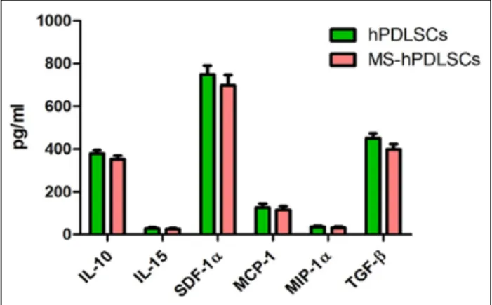
Supernatants from hPDLSCs and MS-hPDLSCs- CM, plated at a density of 106°cells/mL, were col-lected for cytokines/chemokines detection of IL-10, IL-15, stromal cell–derived factor 1α (SDF-1α), monocyte chemoattractant protein-1 (MCP-1), mac-rophage inflammatory protein-1α (MIP-1α), and transforming growth factor (TGF)-β after 24 h of culture. Cytokines/chemokines release was meas-ured by enzyme-linked immunosorbent assay (ELISA) according to the manufacturer’s protocol (eBioscience, San Diego, California, USA).
Animals
A 12-week-old, male C57BL/6 mice weighing 20– 25 g (Harlan, Milan, Italy) were kept in separate ventilated cages. Food and water were supplied ad libitum. The housing room was maintained under constant temperature and humidity conditions. All animals were subjected into 12-h/12-h light/dark cycle.
Ethics statement for animal use
Mice were maintained appropriately in accordance with the European Organization Guidelines for Animal Welfare. All the experimental procedures were performed under the guidelines approved by the Ministry of Health “General Direction of ani-mal health and veterinary drug” (Authorization 621/2015- D.lgs 26/2014). The experiments were designed in such a way to reduce the total number of mice required for the study.
EAE induction
Mice were anesthetized with a mixture of tiletamine and xylazine (10 mL/kg, intraperitoneal (i.p.)). Subsequently, EAE was induced in mice using Myelin Oligodendrocyte Glycoprotein peptide (MOG) 35–55 (MEVGWYRSPFSRVVHLYRNGK;
% peak area by high-performance liquid chromatog-raphy (HPLC) ⩾ 95, AnaSpec, EGT Corporate Headquarters, Fremont, CA, USA) as reported by Paschalidis et al.24 In brief, mice were immunized subcutaneously in the flank with 300 μL of emulsion (300 μg of (MOG) 35–55 in Complete Freund’s Adjuvant (CFA) with 300 μg of heat-killed Mycobacterium tuberculosis H37Ra (Difco Laboratories Sparks, MD, USA)). An i.p. injection of Bordetella pertussis toxin (500 ng in 100 μL; Sigma-Aldrich, Milan, Italy) was administered immediately after (MOG) 35–55 injection and after 48 h. After 14 days of EAE induction, active encepha-litogenic responses in EAE-induced mice were iden-tified with the visible pathological signs such as tail flaccidity and loss of hind legs movement.
Experimental design
Mice were arbitrarily divided into following groups (N = 30 total animals): (a) naïve (N = 5): normal mice with no (MOG) 35–55 or other immunization, (b) EAE (N = 10): mice subjected to EAE with no other treatment, (c) EAE + hPDLSCs-CM (N = 5): EAE mice intravenously (i.v.) injected (tail) with RR-MS patients–derived hPDLSCs-CM (≈1600 μG of hPDLSCs-CM/mouse) after 14 days of EAE induction, (d) EAE + hPDLSCs-EMVs (N = 5): EAE mice i.v. injected (tail) with RR-MS patients– derived hPDLSC-EMVs (≈24 μG of hPDLSCs-EMVs/mouse) after 14 days of EAE induction, (e) naïve + hPDLSCs-CM (N = 5): normal mice i.v. injected with RR-MS patients–derived hPDLSCs-CM (≈1600 μG of hPDLSCs-CM/mouse) after 14 days of EAE induction, and (e) naïve + hPDLSCs-EMVs (N = 5): normal mice IV injected with RR-MS patients–derived hPDLSCs-EMVs (≈24 μG of hPDLSCs-EMVs/mouse) after 14 days of EAE induction.
On 28th day after EAE induction, mice were sacrificed with Tanax (5 mL/kg body weight; i.p.). Spinal cord tissues were collected and processed for biochemical analyses.
Clinical disease score evaluation. From day 14 after EAE
immunization, MS pathological symptoms such as hind limb paralysis and tail tonus reduction were noticed in EAE mice. The severity of encephalito-genic responses was assessed using a 0–10 scoring system as reported by Campbell et al.,25 where score 0 represents no disease condition and score 10 represents EAE-associated mortality. To obtain
more disease parameters and to perform statistical analysis,26 we used a 0–10 scoring system in our study. Gait disturbances, righting reflexes impair-ment, tail tonicity, and limb tonicity were calculated to obtain disease scores, daily. The scoring system used in the present is as follows. (a) Gait was calcu-lated as normal (0), marginally abnormal (+1), moderately abnormal (+2), or severe (+3). (b) Righting reflex analyses were accomplished by placing the animal on its back and recording the capacity to return rapidly to all four limbs. Righting reflex was counted as normal (0), slow (+0.5), or absent (+1). (c) Loss of tail tonicity was scored as normal (0), distal loss of tone (+0.5), or completely loss of tone (+1). (d) Limb weakness was measured by placing the animal in upturned position on a grid for 20 s and recording the ability to hold the grid. Limb tonicity was scored as normal (0), weak (+0.5), near paralysis where limb movement was limited with inability to hold the limbs under the body (+1), or paralyzed where limb movement was completely stopped (+1.5). Animal with both fore-limb and hind fore-limb paralysis was given the score 9 and animal died due to EAE was given the score 10.
Initial clinical disease score was measured on the day of EAE induction (day 0), and all the suc-ceeding measurements were documented every 24 h until sacrifice. In addition, the daily variation of the clinical score of the disease has been expressed in comparison with a day of EAE induc-tion (day 0). The value day has been reported as mean ± standard deviation (SD) of all animals for each experimental group.
Light microscopy. Spinal cord tissues (fixed in 10%
(w/v) PBS-buffered formaldehyde) were embed-ded in paraffin and sectioned into 7 μm thin slices. After processing into xylene deparaffinization and subsequent rehydration steps, sections were stained with eosin and hematoxylin. Sections were visual-ized under optical microscope (Leica microscope ICC50HD).
Immunohistochemistry. Paraffin-embedded tissue slices
were deparaffinized with xylene, rehydrated with alcohol series, and incubated in 0.01 M citrate buffer (pH 6) for 4 min to retrieve antigen. Then, the slices were incubated with 0.3% (v/v) hydrogen peroxide in 60% (v/v) methanol for 30 min to quench endogenous peroxidase and blocked with normal goat serum in PBS (2% v/v) for 20 min. Afterward, slices were incubated with
selective primary antibodies for overnight at 4°C. The primary antibodies applied for immunohisto-chemical analysis are as follows: IL-18 anti-body (1:250 in PBS v/v; Abcam, Cambridge, UK), anti-TLR4 antibody (1:100 in PBS v/v; Abcam, Cambridge, UK), anti-IκB-α antibody (1:250 in PBS v/v; Cell Signaling Technology, Leiden, The Netherlands), and anti-NF-κB antibody (1:250 in PBS v/v; Cell Signaling Technology, Leiden, The Netherlands). Then, the slices were washed with PBS and incubated with avidin/biotin blocking rea-gent (DBA, Milan, Italy) to block endogenous avi-din and biotin binavi-ding sites. Afterward, slices were incubated with universal biotinylated secondary antibody followed by avidin-horseradish peroxi-dase (HRP)-conjugated solution (Vectastain ABC kit, Vector Laboratories, Burlingame, California, USA) according to the manufacturer’s instructions. Slices were then incubated with hydrogen perox-ide/DAB kit (Vectastain DAB kit, Vector Laborato-ries, Burlingame, California, USA) according to the manufacturer’s instructions. Counterstaining was performed with nuclear fast red. To verify nonspe-cific background immunostaining, slices were incu-bated with either primary or secondary antibody alone. No staining was noticed in these controls. Slices were visualized using LEICA DM 2000 combined with LEICA ICC50 HD camera. Images were acquired using Leica Application Suite V4.2.0 software.
Western blot analysis. Spinal cord tissues were
homog-enized using ice-cold lysis buffer with following ingredients: 10 mM Tris–HCl pH 7.4, 0.32 M sucrose, 2 mM ethylenediaminetetraacetic acid (EDTA), 1 mM ethylene glycol-bis(β-aminoethyl ether)-N,N,N′,N′-tetraacetic acid (EGTA), 50 mM NaF, 5 mM NaN3, 10 mM 2-mercaptoethanol, and protease inhibitor tablets (Roche Applied Science, Monza, Italy). Homogenates were clarified by cen-trifugation at 1000g for 10 min at 4°C, and the result-ing supernatant was served as cytoplasmic fraction. The pellets were further lysed using ice-cold extrac-tion buffer consisting of 10 mM Tris–HCl pH 7.4, 150 mM NaCl, 1 mM EDTA, 1 mM EGTA, 1% Triton X-100, and protease inhibitors. Homogenates were clarified by centrifugation at 15,000g for 30 min at 4°C. The resulting supernatant was served as nuclear fraction. Protein concentration was assayed by using Bio-Rad Protein Assay (Bio-Rad, Segrate, Milan, Italy). Proteins were subjected to sodium dodecyl sulfate–polyacrylamide gel electrophoresis,
followed by blotting with polyvinylidene difluoride (PVDF) membranes (Immobilon-P Transfer mem-brane, Millipore, Vimedrone, Milan, Italy). Then, membranes were incubated in blocking solution (5% skimmed milk in 1× PBS) for 45 min at room temperature. Subsequently, membranes were incu-bated with selective primary antibodies for over-night at 4°C. The primary antibodies used were NALP3 (1:500; R&D Systems, Minneapolis, USA), Pro/cleaved caspase 1 (1:500; Abcam, Cambridge, UK) and IL-1β (1:500; Cell Signaling Technology, Leiden, The Netherlands). Later, membranes were incubated with appropriate secondary antibodies conjugated with HRP (1:2000; Santacruz Biotech-nology Inc., Dallas, Texas, USA) for 1 h at room temperature. The relative protein expression was observed using enhanced chemiluminescence kit (Luminata Western HRP Substrates, Millipore, Vimodrone, Milan, Italy). To assess equal loading of proteins, membranes were stripped and reprobed with HRP-conjugated GAPDH (glyceraldehyde 3-phosphate dehydrogenase) antibody (1:1000; Cell Signaling Technology, Leiden, The Netherlands). Images of protein bands were captured by Chemi-Doc™ MP System (Bio-Rad, Segrate, Milan, Italy), and relative densitometric level of the protein bands was evaluated with a software program (ImageJ Bethesda, Maryland, USA). Experiments were repeated for three separate times, and statistical analysis was performed on blots acquired from three separate experiments.
Statistical analysis. Data were analyzed statistically
using GraphPad Prism version 6.0 program (Graph-Pad Software, La Jolla, CA). One-way analysis of variance (ANOVA) and post hoc Bonferroni multi-ple comparison test were performed. A P value of 0.05 or less between the groups was considered with statistical significance. Study data are reported as means ± SD.
Results
RR-MS patients–derived hPDLSCs-CM and purified EMVs reduce the EAE progression
EAE-induced mice showed severe dysfunction in neurological functions. Clinical disease score assessment revealed the active encephalitogenic challenges occurred in EAE mice. We found that EAE induction caused severe phenotypic impair-ments. EAE-associated clinical disease parameters such as severe problem in gait, righting reflex
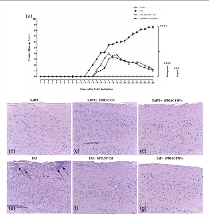
deficiency, and loss of tail and limb tonicity were noticed in EAE mice. On the contrary, EAE mice administered with hPDLSCs-CM and EMVs showed significant improvement. Progression of the disease has been remarkably reduced in the treated EAE mice (Figure 1(a)). Clinical parameter scores evaluated in naïve, EAE, EAE + hPDLSCs-CM, and EAE + hPDLSCs-EMVs groups have been reported in Table 1. E/H staining revealed marked inflammatory cells invasion in EAE mice spinal cord (Figure 1(e)). Conversely, treatment with hPDLSCs-CM and EMVs significantly termi-nated the invasion of inflammatory cells (Figure 1(f) and (g), respectively). No inflammatory cells infiltration was observed in naïve (Figure 1(b)), naïve + hPDLSCs-CM (Figure 1(c)), and naïve + hPDLSCs-EMVs (Figure 1(d)). These find-ings suggest that hPDLSCs-CM and EMVs may possess effective anti-inflammatory capacity, which in turn protect the EAE mice from inflam-matory cells infiltration.
EAE-induced NALP3 inflammasome activation was repressed by RR-MS patients–derived hPDLSCs-CM and purified EMVs
Then, we investigated whether hPDLSCs-CM and purified EMVs obtained from RR-MS patients could modulate NALP3 inflammasome activation. We noticed that in EAE mice spinal cord, NALP3 inflammasome–associated proteins were promi-nently increased. Western blot results showed that NALP3 expression was enhanced in EAE mice (Figure 2(a)). Consequently, active cleaved cas-pase 1 was increased only in EAE mice (Figure 2(b)), while its precursor protein procaspase 1 showed no changes in their expression in all mice groups. Cleaved caspase 1 activation resulted in the synthesis of active pro-inflammatory IL-1β (Figure 2(c)). In addition, Immunohistochemistry data showed increased expression of active pro-inflammatory IL-18 (Figure 2(g)) mediated by active cleaved caspase 1. Interestingly, hPDLSCs-CM and EMVs treatment inhibited NALP3 inflam-masome activation in EAE mice. Significant reduction in NALP3 and cleaved caspase 1 expres-sion was noticed in EAE mice administered with hPDLSCs-CM and EMVs (P < 0.0001 vs EAE). As expected, decreased cleaved caspase 1 resulted in substantial reduction of active IL-1β and IL-18 (Figure 2(h) and (i), respectively; P < 0.0001 vs EAE) level. NALP3 expression in naïve,
naïve + CM, and naïve + hPDLSCs-EMVs group mice were unaltered, while cleaved caspase-1 and IL-1β were undetectable in these groups. Similarly, IL-18 was undetectable in naïve (Figure 2(d)), naïve + hPDLSCs-CM (Figure 2(e)) and naïve + hPDLSCs-EMVs (Figure 2(f)) group mice. These findings suggested that RR-MS patients–derived hPDLSCs-CM and purified EMVs might elicit anti-inflammatory response in EAE mice via blocking NALP3 inflammasome activation.
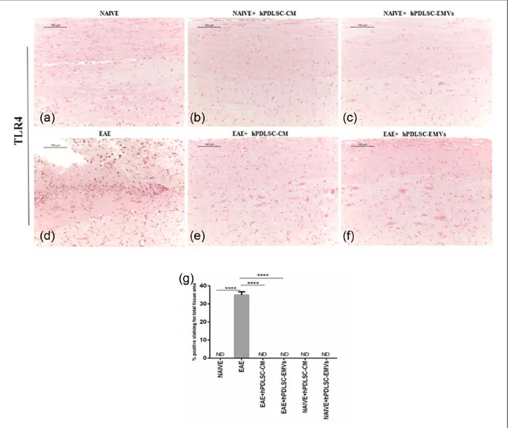
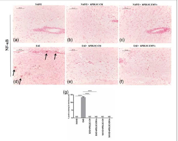
RR-MS patients–derived hPDLSCs-CM and purified EMVs prevents TLR4-mediated pro-inflammatory response of NF-κB in EAE mice Then, we evaluated the expression of pro-inflamma-tory transcription factor NF-κB signaling with refer-ence to NALP3 stimulation. Immunohistochemistry results revealed that the expression of NF-κB posi-tive regulator TLR4 and NF-κB was markedly ele-vated in EAE mice spinal cord (Figures 3(d) and 5(d), respectively), while NF-κB inhibitor protein IκB-α was absent in EAE mice (Figure 4(d)) similar to that of naïve mice (Figure 4(a)). On the contrary, EAE mice treated with hPDLSCs-CM and EMVs showed negative staining of TLR4 (Figure 3(e) and (f), respectively; P < 0.0001 vs EAE) and NF-κB (Figure 5(e) and (f), respectively; P < 0.0001 vs EAE) similar to that of naïve mice (Figures 3(a) and 5(a), respectively). Interestingly, we noticed that IκB-α was significantly enhanced after treatment with hPDLSCs-CM and EMVs (Figure 4(e) and (f), respectively; P < 0.0001 vs EAE). Negative staining of TLR4 and NF-κBwas noticed in naïve (Figures 3(a) and 5(a), respectively), naïve + hPDLSCs-CM (Figures 3(b) and 5(b), respectively), and naïve + hPDLSCs-EMVs (Figures 3(c) and 5(c), respectively). Similar negative staining was observed for IκB-α in naïve + hPDLSCs-CM (Figure 4(b)) and naïve + hPDLSCs-EMVs (Figure 4(c)). These results showed that RR-MS patients– derived hPDLSCs-CM and EMVs suppress EAE-induced NF-κB activation.
Immunoregulatory cytokines and chemokines expression in healthy subjects and RR-MS patients–derived hPDLSCs-CM
Finally, we investigated the expression of immu-noregulatory molecules present in RR-MS hPDLSCs-CM. ELISA analysis showed marked presence
of anti-inflammatory cytokines IL-10 and TGF-β (Figure 6). Substantial amount of immunoregulatory chemokine SDF-1α was noticed. In addition, immu-nomodulatory cytokine IL-15 and chemokines mono-cyte chemotactic protein-1 (MCP-1) and macrophage
inflammatory protein-1α (MIP-1α) were also present. Interestingly, expression of these immu-nomodulatory molecules present in RR-MS patients– derived hPDLSCs-CM was similar with healthy subjects–derived hPDLSCs-CM.
Figure 1. RR-MS patients–derived hPDLSCs-CM and purified EMVs reduce EAE progression. Clinical disease score evaluation
showed severe impairment in EAE mice, while (a) treatment with hPDLSCs-CM and EMVs significantly suppressed EAE
progression. ****P < 0.0001 for naïve versus EAE; ****P < 0.0001 for naïve versus EAE + hPDLSCs-CM; ***P = 0.0002 for naïve versus EAE + hPDLSCs-EMVs. P values were calculated using one-way ANOVA followed by Bonferroni multiple comparison test. Data are represented as mean ± SD. E/H staining displayed severe infiltration of inflammatory cells (black arrows) in (e) EAE spinal cord white matter, while treatment with (f) hPDLSCs-CM and (g) EMVs attenuated the invasion of inflammatory cells. Infiltration of inflammatory cells were unnoticed in (b) naïve mice and naïve mice treated with (c) hPDLSCs-CM and (d) EMVs.
Discussion
Exploring the immunoregulatory effects of adult MSCs and their secretome has become a signifi-cant research area in the therapeutic arena of MS,
a most prevalent and debilitating autoimmune dis-ease of the central nervous system, and other degenerative diseases.27,28 Dental tissues have emerged as a simple and potential alternative autologous MSCs resource due to the minimal
Table 1. Clinical parameters of EAE.
Group Incidence (%) Day of disease
onset (avg) ± SD Peak disease (avg) ± SD Cumulative disease score (CDS) ± SD Mortality
Naïve 0 0 0 0 0/5
EAE 100 13.1 ± 0.22 8.60 ± 0.50 11.97 ± 1.01 1/10
EAE + hPDLSCs-CM 100 15.3 ± 0.25 4.00 ± 0.74 3.71 ± 0.87 0/10
EAE + hPDLSCs-EMVs 100 14.9 ± 0.35 3.80 ± 0.60 3.20 ± 0.30 0/10
P value <0.0001 0.04 0.002 <0.0001 <0.0001
SD: standard deviation; EAE: experimental autoimmune encephalomyelitis; hPDLSCs-CM: human periodontal ligament stem cells–derived condi-tioned medium; EMVs: exosomes/microvesicles; ANOVA: analysis of variance.
Incidence: percentage of mice that displayed any clinical signs of disease. Day of onset: the first day mice showed clinical signs. Peak of disease: the maximum value score observed between days 0 and 28. Cumulative disease score: mean cumulative disease scores were calculated by summing the daily clinical scores of living mice divided by the number of animals in the groups. One-way ANOVA followed by Bonferroni multiple comparison test was used to determine the statistical significance of differences.
Figure 2. RR-MS patients–derived hPDLSCs-CM and purified EMVs inhibits EAE-induced NALP3 inflammasome activation. EAE
induction stimulated NALP3 inflammasomes in the spinal cord of EAE mice. Western blot results showed elevated level of (a) NALP3, (b) active cleaved caspase 1, and (c) IL-1β in EAE mice. On the contrary, significant reduction was observed in EAE mice administered with hPDLSCs-CM and EMVs. ****P < 0.0001 versus EAE. Data expressed as mean ± SD. Representative bands of three separate experiments are shown. (g) Immunohistochemistry results showed significant positive staining for IL-18 in EAE mice, while in EAE mice treated with (h) hPDLSCs-CM and (i) EMVs, negative staining was noticed as that of (d) naïve mice. Naïve mice treated with (e) hPDLSCs-CM and (f) EMVs showed negative staining for IL-18. Magnification: 10×. (j) Densitometric analysis of IL-18 expression (****P < 0.0001 vs EAE). P values were calculated using one-way ANOVA followed by Bonferroni multiple comparison test. Data are represented as mean ± SD.
invasive procedure. In addition, dental MSCs pos-sess remarkable self-renewal and differentiation capacities toward neurogenic, osteogenic, and adi-pogenic lineages.29 Recently, we demonstrated the neuroprotective and immunosuppressive role of hPDLSCs and their CM and EMVs in EAE mice.12,19 Given the considerable role of inflam-masome activation in MS and in in vivo EAE model,30 in this study, we investigated the regula-tory role of RR-MS patients hPDLSCs–derived CM and purified EMVs on NALP3 inflammas-omes activation in EAE mice.
NALP3 inflammasome activation and its sub-sequent positive regulatory role in cleaved cas-pase 1, IL-1β, and IL-18 in EAE mice have been already demonstrated in the previous studies.31,32 Likewise, in our study, we noticed that NALP3 inflammasome-linked proteins were signifi-cantly modulated. NALP3 expression was ele-vated in EAE mice spinal cord, which in turn proteolytically cleaved the inactive procaspase 1 into active cleaved caspase 1. Increased level of cleaved caspase 1 processed the synthesis of active IL-1β and IL-18 from their inactive
Figure 3. RR-MS patients–derived hPDLSCs-CM and purified EMVs prevents TLR4 expression in EAE mice. Immunohistochemistry
data revealed that (d) EAE induction markedly activated TLR4 expression in the spinal cord with strong positive staining. On the contrary, hPDLSCs-CM and EMVs treatment significantly inhibited TLR4 activation. Negative staining was observed in (e) hPDLSCs-CM and (f) EMVs-treated EAE mice as that of (a) naïve. Similar negative staining was noticed in naïve mice treated with (b) hPDLSCs-CM and (c) EMVs. Magnification: 10×. (g) Densitometric analysis of TLR4 expression (****P < 0.0001 vs EAE). P values were calculated using one-way ANOVA followed by Bonferroni multiple comparison test. Data are represented as mean ± SD.
precursor forms. Interestingly, we found signifi-cant inhibition of NALP3 inflammasome activa-tion in EAE mice treated with CM and EMVs. NALP3 expression was unaltered in CM and EMVs-administered EAE mice. These data were corroborated with the significant reduction of active cleaved caspase 1, IL-1β, and IL-18. It is interesting to mention here that treatment with interferon (IFN)-β or steroids in MS patients resulted in the reduction of IL-1β and the upreg-ulation of endogenous IL-1 receptor antago-nists.33–35 From our in vivo findings, we may
assume that IL-1β downregulation followed by IFN-β or steroids administration in MS patients may partially depend on NALP3 suppression.
Moreover, it has been reported that NALP3 prim-ing is a prerequisite for inflammasome formation. NF-κB signaling is a most common pathway involved in NALP3 priming, which in turn enhanced the cytosolic NALP3 level.36 Consequently, we assessed NF-κB pathway in EAE mice. As reported earlier,37–40 augmented expression of TLR4, an up-stream inducer of NF-κB, and NF-κB was found in EAE mice. NF-κB inhibitor protein IκB-α was
Figure 4. RR-MS patients–derived hPDLSCs-CM and purified EMVs induces IκB-α expression in EAE mice. Immunohistochemistry
data revealed negative staining for IκB-α expression in (a) naïve and (d) EAE mice spinal cord. Similarly, naïve mice administered with (b) hPDLSCs-CM and (c) EMVs displayed negative staining. Conversely, significant positive staining for IκB-α was noticed in (e) hPDLSCs-CM and (f) EMVs-treated EAE mice. Magnification: 10×. (g) Densitometric analysis of IκB-α expression (****P < 0.0001 vs EAE). P values were calculated using one-way ANOVA followed by Bonferroni multiple comparison test. Data are represented as mean ± SD.
decreased. However, CM and EMVs significantly inhibited TLR4 and NF-κB activation, while increased the level of IκB-α in EAE mice. Our results suggest that the anti-inflammatory role of hPDLSCs-CM and -EMVs in EAE mice may be due to the suppression of NF-κB stimulation, which in turn inhibited NALP3 inflammasome activation.
To understand the molecular mechanisms underlying the immunosuppressive effects of hPDLSCs-derived secretome, we studied the expression of immunomodulatory cytokines and chemokines secreted from hPDLSCs. Characterization of hPDLSCs-CM derived from
RR-MS patients showed increased level of IL-10 and TGF-β. Anti-inflammatory effects of IL-10 and TGF-β have been well documented in EAE.41– 44 Indeed, recently we demonstrated the presence of IL-10 and TGF-β in hPDLSCs-EMVs as well.19 Moreover, it has been demonstrated that IFN-β treatment in MS patients increased the synthesis of IL-10 and TGF-β, suggesting the beneficial role of these anti-inflammatory cytokines in the treat-ment of MS.45,46 Interestingly, negative regulatory role of IL-10 on NALP3 inflammasome activation has been reported earlier in lipopolysaccharide-activated macrophages.47 Thus, we believe that
Figure 5. RR-MS patients–derived hPDLSCs-CM and purified EMVs prevents NF-κB expression in EAE mice. (d)
Immunohistochemistry data displayed positive staining for the expression of inflammatory transcription factor NF-κB in EAE mice spinal cord. On the contrary, NF-κB activation was completely terminated in EAE mice treated with (e) hPDLSCs-CM and (f) EMVs. Negative staining was noticed in (a) naïve mice and naïve mice treated with (b) hPDLSCs-CM and (c) EMVs. Magnification: 10×. (g) Densitometric analysis of NF-κB expression (****P < 0.0001 vs EAE). P values were calculated using one-way ANOVA followed by Bonferroni multiple comparison test. Data are represented as mean ± SD.
IL-10 mediated NALP3 inflammasome reduction might be a crucial mechanism underlying hPDLSCs-derived CM and EMVs-mediated anti-inflammatory responses. In addition, hPDLSCs-CM showed a lesser presence of IL-15, a pro-inflammatory cytokine, which activates and promotes the survival of natural killer cells and memory CD8+ T-cells during inflammation.48 Unlike the distinguished effector role of CD4+ cells (Th1 and Th17) in EAE, the role of CD8+ T-cells in EAE is unclear as studies have demon-strated both stimulative and suppressive effects during EAE development.49 Recent studies dem-onstrated that IL-15 inhibits EAE progression by reducing CD4+ cells proliferation.49,50 Our results support this finding that the presence of IL-15 in hPDLSCs-CM, although present in low amount, may produce immunosuppressive effects by CD4+ cells inhibition in EAE. As reported in our previ-ous work, reduced expression of CD4 in the spinal cord and spleen of EAE mice treated with hPDLSCs-CM19 further support our notion on negative regulation of CD4+ cells by IL-15.
In our study, we noticed substantial amount of immunomodulatory chemokine SDF-1α in hPDLSCs-CM. CXC type SDF-1α (also called as CXCL12) is a chemokine, which acts as a potent chemoattractant for B and T cells51 and promotes neural precursor cells migration in pathological conditions such as ischemic stroke.52 In EAE, SDF-1α was addressed to be a suppressive factor
of the disease progression by redirecting the polarization of Th1 cells into IL-10 secreting Treg cells.53 Interestingly, it has been reported that intrathecal application of SDF-1α reduced the expression of NALP3 inflammasome activa-tion in rat spinal cord injury model.54 SDF-1α expression in hPDLSCs-CM from our study cor-roborated these previous studies, suggesting a potential negative regulatory during EAE pro-gression, which may depend on NALP3 inflam-masome inactivation and Treg cells activation. In addition, we observed a lesser amount of MCP-1 and MIP-1α chemokines in hPDLSCs-CM. Pro-inflammatory role of MCP-1 and MIP-1α has been reported in EAE development and progres-sion.55 Conversely, these chemokines are also involved in neural stem cells migration followed by neuroinflammatory conditions such as striatal lesion and cerebral ischemia.56 Given their low amount of secretion in hPDLSCs-CM, we assume that MCP-1 and MIP-1α may not exert consider-able immunoregulatory or neuroprotective effects in EAE mice due to the presence of substantial level of IL-10, TGF-β, and SDF-1α.
Taken together, we assume that the immuno-suppressive effects stimulated by RR-MS patients hPDLSCs-CM and -EMVs in EAE mice might occur due to the existence of these inflammation regulatory molecules and that NALP3 inflamma-some inactivation might be partly involved in the overall anti-inflammatory response. Interestingly, expression of these cytokines and chemokines in CM and EMVs derived from hPDLSCs of RR-MS patients was similar to that of hPDLSCs derived from healthy subjects, which suggest that MS pathology, per se, has no marked role in regulat-ing the expression of cytokines and chemokines. Thus, our results propose hPDLSCs as a simple autologous MSCs resource in stem cell therapy for MS patients. Furthermore, we observed that the anti-inflammatory effects triggered from CM and purified EMVs were analogous with respect to EAE suppression. Considering the require-ment of enormous amount of time and money for EMVs purification, results from our study sug-gest that hPDLSCs-CM alone could be adequate for therapeutic intervention in MS patients. However, comprehensive high-throughput analy-sis of the proteome present in both CM and puri-fied EMVs is necessary before proceeding to clinical trials.
Figure 6. Immunomodulatory cytokines and chemokines
expression in healthy subjects and RR-MS patients–derived hPDLSCs-CM. ELISA data revealed the presence of substantial amount of anti-inflammatory IL-10 and TGF-β cytokines and chemokine SDF-1α in hPDLSCs-CM obtained from normal healthy subjects and RR-MS patients. Immunomodulatory cytokine IL-15 and chemokines MCP-1 and MIP-1α were also expressed in hPDLSCs-CM.
Conclusion
In summary, our research findings suggest that the secretory molecules present in CM and puri-fied EMVs obtained from hPDLSCs of RR-MS patients modulate NF-κB level, inhibit NALP3 inflammasome activation, and exert protection in EAE mice. We conclude that hPDLSCs might be a simple and potential autologous stem cell resource in the treatment of MS patients. Both hPDLSCs-derived CM and purified EMVs exert comparable immunosuppressive effects. We pro-pose that CM alone obtained from hPDLSCs may serve as effective and economical therapeutic tool in MS treatment.
Acknowledgements
The authors thank Dr Mario Russo for his procedural assistance on intravenous injection in mice. T.S.R. and S.G. contributed equally to this article. O.T. and E.M. con-tributed equally as senior authors.
Declaration of conflicting interests
The author(s) declared no potential conflicts of interest with respect to the research, authorship, and/or publication of this article.
Funding
This study has been supported by current research funds 2016 of IRCCS-Centro Neurolesi “Bonino-Pulejo,” Messina, Italy.
References
1. Calabresi PA (2002) Considerations in the treatment of relapsing-remitting multiple sclerosis. Neurology 58: S10–S22.
2. Frohman EM, Racke MK and Raine CS (2006) Multiple sclerosis–The plaque and its pathogen-esis. The New England Journal of Medicine 354: 942–955.
3. Rieckmann P and Smith KJ (2001) Multiple sclerosis: More than inflammation and demyelination. Trends
Neuroscience 24: 435–437.
4. Ebers GC (2008) Environmental factors and multiple sclerosis. The Lancet Neurology 7: 268–277.
5. Compston A and Coles A (2002) Multiple sclerosis.
Lancet 359: 1221–1231.
6. Lublin FD, Reingold SC, Cohen JA, et al. (2014) Defining the clinical course of multiple sclerosis: The 2013 revisions. Neurology 83: 278–286.
7. Jancic J, Nikolic B, Ivancevic N, et al. (2016) Multiple sclerosis in pediatrics: Current concepts and treatment options. Neurology and Therapy 5: 131–143.
8. Dulamea A (2015) Mesenchymal stem cells in multi-ple sclerosis: Translation to clinical trials. Journal of
Medicine and Life 8: 24–27.
9. Sarugaser R, Hanoun L, Keating A, et al. (2009) Human mesenchymal stem cells self-renew and dif-ferentiate according to a deterministic hierarchy.
PLoS ONE 4: e6498.
10. Ullah I, Subbarao RB and Rho GJ (2015) Human mesenchymal stem cells: Current trends and future prospective. Bioscience reports 35: e00191.
11. Sharpe PT (2016) Dental mesenchymal stem cells.
Development 143: 2273–2280.
12. Trubiani O, Giacoppo S, Ballerini P, et al. (2016) Alternative source of stem cells derived from human periodontal ligament: A new treatment for experi-mental autoimmune encephalomyelitis. Stem Cell
Research & Therapy 7: 1.
13. Tatullo M, Marrelli M, Shakesheff KM, et al. (2015) Dental pulp stem cells: Function, isolation and appli-cations in regenerative medicine. Journal of Tissue
Engineering and Regenerative Medicine 9: 1205–
1216.
14. Pashoutan Sarvar D, Shamsasenjan K and Akbarzadehlaleh P (2016) Mesenchymal stem cell-derived exosomes: New opportunity in cell-free ther-apy. Advanced Pharmaceutical Bulletin 6: 293–299. 15. Merino-Gonzalez C, Zuniga FA, Escudero C, et al.
(2016) Mesenchymal stem cell-derived extracellu-lar vesicles promote angiogenesis: Potencial clinical application. Frontiers in Physiology 7: 24.
16. Biancone L, Bruno S, Deregibus MC, et al. (2012) Therapeutic potential of mesenchymal stem cell-derived microvesicles. Nephrology, Dialysis,
Transplantation: Official Publication of the European Dialysis and Transplant Association–European Renal Association 27: 3037–3042.
17. Salgado AJ, Sousa JC, Costa BM, et al. (2015) Mesenchymal stem cells secretome as a modulator of the neurogenic niche: Basic insights and therapeutic opportunities. Frontiers in Cellular Neuroscience 9: 249.
18. Jarmalaviciute A and Pivoriunas A (2016) Exosomes as a potential novel therapeutic tools against neurode-generative diseases. Pharmacological Research 113: 816–822.
19. Rajan TS, Giacoppo S, Diomede F, et al. (2016) The secretome of periodontal ligament stem cells from MS patients protects against EAE. Scientific Reports 6: 38743.
20. Patel MN, Carroll RG, Galvan-Pena S, et al. (2017) Inflammasome priming in sterile inflammatory dis-ease. Trends in Molecular Medicine 23: 165–180. 21. Guo H, Callaway JB and Ting JPY (2015)
Inflammasomes: Mechanism of action, role in dis-ease, and therapeutics. Nature Medicine 21: 677–687.
22. Shao B-Z, Xu Z-Q, Han B-Z, et al. (2015) NLRP3 inflammasome and its inhibitors: A review. Frontiers
in Pharmacology 6: 262.
23. Eleuterio E, Trubiani O, Sulpizio M, et al. (2013) Proteome of human stem cells from periodontal liga-ment and dental pulp. PLoS ONE 8: e71101.
24. Paschalidis N, Iqbal AJ, Maione F, et al. (2009) Modulation of experimental autoimmune encepha-lomyelitis by endogenous annexin A1. Journal of
Neuroinflammation 6: 33.
25. Campbell AM, Zagon IS and McLaughlin PJ (2012) Opioid growth factor arrests the progression of clini-cal disease and spinal cord pathology in established experimental autoimmune encephalomyelitis. Brain
Research 1472: 138–148.
26. Fleming KK, Bovaird JA, Mosier MC, et al. (2005) Statistical analysis of data from studies on experi-mental autoimmune encephalomyelitis. Journal of
Neuroimmunology 170: 71–84.
27. Uccelli A, Laroni A and Freedman MS (2011) Mesenchymal stem cells for the treatment of multiple sclerosis and other neurological diseases. The Lancet
Neurology 10: 649–656.
28. Paul G and Anisimov SV (2013) The secretome of mesenchymal stem cells: Potential implications for neuroregeneration. Biochimie 95: 2246–2256.
29. Liu J, Yu F, Sun Y, et al. (2015) Concise reviews: Characteristics and potential applications of human dental tissue-derived mesenchymal stem cells. Stem
Cells 33: 627–638.
30. Barclay W and Shinohara ML (2016) Inflammasome activation in multiple sclerosis and experimental auto-immune encephalomyelitis (EAE). Brain Pathology 27: 213–219.
31. Gris D, Ye Z, Iocca HA, et al. (2010) NLRP3 plays a critical role in the development of experimental autoimmune encephalomyelitis by mediating Th1 and Th17 responses. Journal of Immunology 185: 974–981.
32. Inoue M, Williams KL, Gunn MD, et al. (2012) NLRP3 inflammasome induces chemotactic immune cell migration to the CNS in experimental autoim-mune encephalomyelitis. Proceedings of the National
Academy of Sciences of the United States of America
109: 10480–10485.
33. Coclet-Ninin J, Dayer JM and Burger D (1997) Interferon-beta not only inhibits interleukin-1 beta and tumor necrosis factor-alpha but stimulates inter-leukin-1 receptor antagonist production in human peripheral blood mononuclear cells. European
Cytokine Network 8: 345–349.
34. Nicoletti F, Patti F, DiMarco R, et al. (1996) Circulating serum levels of IL-1ra in patients with relapsing remitting multiple sclerosis are normal dur-ing remission phases but significantly increased either
during exacerbations or in response to IFN-beta treat-ment. Cytokine 8: 395–400.
35. Dujmovic I, Mangano K, Pekmezovic T, et al. (2009) The analysis of IL-1 beta and its naturally occurring inhibitors in multiple sclerosis: The elevation of IL-1 receptor antagonist and IL-1 receptor type II after steroid therapy. Journal of Neuroimmunology 207: 101–106.
36. Cassel SL and Sutterwala FS (2010) Sterile inflam-matory responses mediated by the NLRP3 inflamma-some. European Journal of Immunology 40: 607–611. 37. Kerfoot SM, Long EM, Hickey MJ, et al. (2004)
TLR4 contributes to disease-inducing mechanisms resulting in central nervous system autoimmune dis-ease. Journal of Immunology 173: 7070–7077. 38. Reynolds JM, Martinez GJ, Chung Y, et al. (2012)
Toll-like receptor 4 signaling in T cells promotes auto-immune inflammation. Proceedings of the National
Academy of Sciences of the United States of America
109: 13064–13069.
39. Pahan K and Schmid M (2000) Activation of nuclear factor-kB in the spinal cord of experimental allergic encephalomyelitis. Neuroscience Letters 287: 17–20. 40. van Loo G, De Lorenzi R, Schmidt H, et al. (2006)
Inhibition of transcription factor NF-kappaB in the central nervous system ameliorates autoimmune encephalomyelitis in mice. Nature Immunology 7: 954–961.
41. Zhang X, Koldzic DN, Izikson L, et al. (2004) IL-10 is involved in the suppression of experimental autoim-mune encephalomyelitis by CD25+CD4+ regulatory T cells. International Immunology 16: 249–256. 42. Klose J, Schmidt NO, Melms A, et al. (2013)
Suppression of experimental autoimmune encephalo-myelitis by interleukin-10 transduced neural stem/pro-genitor cells. Journal of Neuroinflammation 10: 117. 43. Zhang X, Reddy J, Ochi H, et al. (2006) Recovery
from experimental allergic encephalomyelitis is TGF-beta dependent and associated with increases in CD4+LAP+ and CD4+CD25+ T cells. International
Immunology 18: 495–503.
44. Laouar Y, Town T, Jeng D, et al. (2008) TGF-beta signaling in dendritic cells is a prerequisite for the con-trol of autoimmune encephalomyelitis. Proceedings
of the National Academy of Sciences of the United States of America 105: 10865–10870.
45. Nicoletti F, Di Marco R, Patti F, et al. (1998) Blood levels of transforming growth factor-beta 1 (TGF-beta1) are elevated in both relapsing remitting and chronic progressive multiple sclerosis (MS) patients and are further augmented by treatment with inter-feron-beta 1b (IFN-beta1b). Clinical & Experimental
Immunology 113: 96–99.
46. Kvarnstrom M, Ydrefors J, Ekerfelt C, et al. (2013) Longitudinal interferon-beta effects in multiple
sclerosis: Differential regulation of IL-10 and IL-17A, while no sustained effects on IFN-gamma, IL-4 or IL-13. Journal of the Neurological Sciences 325: 79–85.
47. Gurung P, Li B, Subbarao Malireddi RK, et al. (2015) Chronic TLR stimulation controls NLRP3 inflam-masome activation through IL-10 mediated regula-tion of NLRP3 expression and caspase-8 activaregula-tion.
Scientific Reports 5: 14488.
48. Stonier SW and Schluns KS (2010) Trans-presentation: A novel mechanism regulating IL-15 delivery and responses. Immunology Letters 127: 85–92.
49. Yu P, Bamford RN and Waldmann TA (2014) IL-15-dependent CD8+ CD122+ T cells ameliorate experi-mental autoimmune encephalomyelitis by modulating IL-17 production by CD4+ T cells. European Journal
of Immunology 44: 3330–3341.
50. Pandiyan P, Yang X-P, Saravanamuthu SS, et al. (2012) The role of IL-15 in activating STAT5 and fine-tuning IL-17A production in CD4 T lympho-cytes. Journal of Immunology 189: 4237–4246. 51. Nanki T and Lipsky PE (2000) Cutting edge: Stromal
cell-derived factor-1 is a costimulator for CD4+ T cell activation. Journal of Immunology 164: 5010–5014.
52. Cui L, Qu H, Xiao T, et al. (2013) Stromal cell-derived factor-1 and its receptor CXCR4 in adult neurogenesis after cerebral ischemia. Restorative Neurology and
Neuroscience 31: 239–251.
53. Meiron M, Zohar Y, Anunu R, et al. (2008) CXCL12 (SDF-1alpha) suppresses ongoing experimental auto-immune encephalomyelitis by selecting antigen-specific regulatory T cells. Journal of Experimental
Medicine 205: 2643–2655.
54. Zendedel A, Johann S, Mehrabi S, et al. (2016) Activation and regulation of NLRP3 inflamma-some by intrathecal application of SDF-1a in a spi-nal cord injury model. Molecular Neurobiology 53: 3063–3075.
55. Karpus WJ and Kennedy KJ (1997) MIP-1alpha and MCP-1 differentially regulate acute and relaps-ing autoimmune encephalomyelitis as well as Th1/ Th2 lymphocyte differentiation. Journal of Leukocyte
Biology 62: 681–687.
56. Tang SK, Knobloch RA, Maucksch C, et al. (2014) Redirection of doublecortin-positive cell migra-tion by over-expression of the chemokines MCP-1, MIP-1alpha and GRO-alpha in the adult rat brain.