DOCTORAL SCHOOL IN BIOLOGY Section: Biodiversity and Ecosystem Analysis
XXVIII Cycle
Karyological evolution in Cistaceae,
with emphasis on
Cistus and Helianthemum
Evoluzione cariologica in Cistaceae,
con particolare attenzione ai generi Cistus e Helianthemum
CANDIDATE PhD STUDENT: Chiara Totta
TUTOR: Prof. Fernando Lucchese CO-TUTOR: Prof. Josep A. Rosselló
PhD SCHOOL COORDINATOR: Prof. Marco A. Bologna
ii
University, Rome, Italy.
Chiara Totta
Department of Science, Roma Tre University, Viale Marconi, 446,
00146 Rome, Italy.
e-mail: [email protected]; [email protected]
Front cover: Iconographies of Cistus heterophyllus (purple-flowered individual), Cistus
monspeliensis (white-flowered individual) and Helianthemum nummularium (yellow-flowered individual).
Thesis defense 15th February 2016 in front of the following jury of committee members:
Prof. Ferdinando Boero (Università del Salento) Prof. Giovanna Abbate (Università Sapienza di Roma) Prof. Romolo Fochetti
“Wenn [...] ich dann im hohen Grase am fallenden Bache liege, und näher an der Erde tausend mannigfaltige Gräschen mir merkwürdig werden [...] mein Freund! Wenn's dann um meine Augen dämmert, und die Welt um mich her und der Himmel ganz in meiner Seele ruhn”
“Quando [...] mi stendo nell'erba alta accanto al torrente e, cosí vicino alla terra, scopro le piante più diverse e più singolari [..] oh, amico mio!, i miei occhi si smarriscono in questa vertigine e l'universo e il cielo riposano nella mia anima”
CONTENTS
PREFACE ... vii
CHAPTER 1 General introduction ... 1
1.1 Genome evolution in plants ... 1
1.2 Organization of plant chromosomes ... 2
1.3 45S and 5S rDNA ... 2
1.4 Chromosome changes in plant tissue culture ... 3
1.5 Ancestral state reconstruction ... 4
1.6 The study systems ... 5
1.6.1 Cistaceae ... 5
1.6.2 Cistus ... 5
1.6.3 Helianthemum ... 6
1.6.4 Cistus heterophyllus ... 7
1.7 Aims of the study ... 7
References ... 9
CHAPTER 2 Temporal frames of 45S rDNA site-number variation in homoploid plant lineages: lessons from the rock rose genus (Cistus, Cistaceae) ... 15
CHAPTER 3 Trends in site-number change of 45S rDNA loci during karyological evolution in Helianthemum (Cistaceae). A genus with distinct chromosome base numbers ... 37
CHAPTER 4 Latent nuclear rDNA instability in in vitro-generated plants is activated after sexual reproduction with conspecific wild individuals ... 61
CHAPTER 5 Conclusions ... 83
PREFACE
This thesis encompasses a general introduction, three independent researches, and general conclusions.
CHAPTER 1. General introduction
The first chapter gives an overview of the basic cytogenetic concepts, followed by the description of the study systems with notes of their systematic, distribution and karyological features. For the last study system, some information about the conservation strategy are also given. At the end of the chapter, the aims of the thesis are explicitly stated.
CHAPTER 2.
I. Temporal frames of 45S rDNA site-number variation in homoploid plant lineages: lessons from the rock rose genus (Cistus, Cistaceae).
Chiara Totta, Marcela Rosato, Pablo Ferrer-Gallego, Fernando Lucchese, and Josep A. Rosselló. (in preparation).
CHAPTER 3.
II. Trends in site-number change of 45S rDNA loci during karyological evolution in Helianthemum (Cistaceae). A genus with distinct chromosome base numbers.
Chiara Totta, Marcela Rosato, Pablo Ferrer-Gallego, Fernando Lucchese, and Josep A. Rosselló (in preparation).
CHAPTER 4.
III. Latent nuclear rDNA instability in in vitro-generated plants is activated after sexual reproduction with conspecific wild individuals.
Marcela Rosato, Pablo Ferrer-Gallego, Chiara Totta, Emilio Laguna and Josep A. Rosselló. Botanical Journal of the Linnean Society, accepted for publication January 2016.
CHAPTER 5. Conclusions
The chapter includes brief synthesis of the major outcomes of this thesis, in the light of the aims stated.
CHAPTER 1
GENERAL INTRODUCTION
Understanding how species interact with each other, and when/where the diversification of clades has taken place provides hints about the process of speciation in plants. A more detailed view can only be achieved by looking at the mechanisms responsible for the reproductive isolation of species. Although the role of chromosomal rearrangements as mechanisms for plant speciation is still debated (Faria and Navarro, 2010), studies on the distribution of ribosomal RNA gene families and changes in chromosome numbers have begun to shed light about the evolutionary significance of chromosomal changes (Weiss-Schneeweiss and (Weiss-Schneeweiss, 2013).
1.1 Genome evolution in plants
Eukaryotic chromosomes are organized linear structures carrying the majority of the genetic material of the organisms and are located within the nucleus of all eukaryotic cells. They differ in size, shape and composition of DNA and proteins (Schubert, 2007). All of these features can be affected during evolution and participate at chromosomal diversification, accompanying taxa diversification and eventually speciation (Stebbins, 1971; Rieseberg, 2001; Levin, 2002; Schubert, 2007; Leitch and Leitch, 2008). Chromosomal rearrangements involving inversions, translocations, duplications, deletions, fusions and fissions result in changes in chromosome number (dysploidy and polyploidy) and chromosome structure (Schubert, 2007; Lysak and Schubert, 2013). Genome size changes and more subtle changes in sequence composition of the repetitive fraction of the genome, most commonly involving expansions or reductions of repetitive DNA sequence amounts, constitute additional sources of variation occurring during the evolutionary history of taxa (Weiss-Schneeweiss and (Weiss-Schneeweiss, 2013). Scientists have long argued over whether – and to what degree – changes in chromosome structure contribute to reproductive isolation and, ultimately, speciation.
Despite polyploidy is much more studied and often viewed as the most common type of chromosome evolution in plants and the main chromosomal driver of plant diversification, dysploidy, defined as the stepwise change of the haploid chromosome number among related species (Stebbins, 1971), is also very common across angiosperms and may frequently co-occur with polyploidy within lineages (Escudero et al., 2014). Although chromosome number change may involve the loss or gain of entire chromosomes, it is much more frequently
2
achieved via genome restructuring, resulting in centromere/chromosome number reduction or increase with or without significant loss of genetic material (unbalanced and balanced rearrangements, respectively). The majority of earlier studies considered descending dysploidy as the main trend of chromosomes number change in angiosperms (Weiss-Schneeweiss and Schneeweiss, 2013).
1.2 Organization of plant chromosomes
In short, in most angiosperm species the shape of monocentric chromosomes is determined by a centromere (primary constriction), comprised of regions of condensed chromatin, flanked by pericentromeric regions rich in heterocromatin, and telomeres that mark the ends of chromosomes and protect them from degradation (Gill et al., 2008). Based on morphological observation, quantitative chromosome maps called “ideograms” can be constructed (Fukui
et al., 1998).
The genome of most higher organisms consists of a large amount of DNA motifs repeated in hundreds or thousands of copies (Britten, 1969). For instance, the Arabidopsis genome is 25% repetitive DNA (Leutwiler et al., 1984) whereas in pea it has been estimated to reach up the 95% of the total DNA (Thompson and Murray, 1980). Some sequence motifs are extremely variable while other are highly conserved from one species to another (rRNA genes), and information about their number and chromosomal distribution provide tools to assess potential functions of particular aspects of genome architecture and evolution and for studying inter-organism relationships.
Molecular cytogenetic methods offer a powerful system for looking at the organization of DNA repeat motifs along a chromosome using in situ hybridization (ISH). With this technique, labeled probe sequences are able to localize a specific sequence to the denatured DNA of chromosomes spread on microscope slides. In plants, the use of radioactive tracer or modified nucleotides (attached to biotin, digoxigenin, or flourophores) to make ISH probes allows the microscopic visualization and localization of complementary sequences within cells and nuclei, and on individual chromosomes (Figueroa and Bass, 2010). Direct and indirect fluorescence in situ hybridization (FISH) has been broadly used over the last 30 years to localize repetitive sequences. Moreover, it can be used to observe genomic organization and chromosome structure, and to look for landmarks that allow the identification of genes, their clustering and orientation.
1.3 45S and 5S rDNA
Tandemly repeated genes encoding 45S (18S-5.8S-25S) and 5S rRNAs (rDNA) are ubiquitous and highly conserved over long evolutionary timescales (Volkov
CHAPTER 1
et al., 2007; Richard et al., 2008 Weiss-Schneeweiss et al., 2008), therefore, their variation in number and distribution can be easily detected and represents a useful model to study and compare chromosome evolution and genome organization in different taxonomic groups (Shan et al., 2003; Volkov et al., 2007; Heslop-Harrison and Schwarzacher, 2011). The 45S rDNA cistron, coding for 18S, 5.8S, and 25S rRNAs, also includes external transcribed spacer (ETS), two internal transcribed spacers (ITS1 and ITS2) and the non-transcribed intergenic spacer (IGS). Numerous 45S rDNA repeats are tandemly arranged in one or more loci in the genome known as nucleolar-organizer regions (NORs). Cytologically, the 45S rDNA appears in the nucleolus in interphase nuclei and forms secondary constrictions in mitosis/meiosis (Volkov
et al., 2007).
While FISH detects all 45S rDNA signals, including small and/or inactive ones, the silver staining (Ag-NOR) allows to identify only those loci transcriptionally active in the preceding interphase of the cell cycle (Jiménez et
al., 1988), staining both the decondensed NOR-associated proteins on chromosomes or the nucleoli, the nuclear organelle assigned to ribosomes synthesis (Medina et al., 2000), whose maximum number stained corresponds to the maximum number of active NORs in the diploid chromosome complement.
The 5S rDNA repeated unit is comparatively simple: each repeat is composed of a conserved, ca. 121 bp long coding region separated by variable length Non-Transcribed Spacer (NTS). In angiosperm, both the 45S and 5S loci mostly map independently from each other, despite exceptions are known (Sone et al., 1999; Garcia et al., 2010; Garcia and Kovařík, 2013).
Moreover, the ITS and ETS of the 45S rDNA cistron and the NTS of the 5S rDNA array are often use as molecular markers for phylogenetic analysis. Hence, the combination of such genetic and cytogenetic approaches enables the direct correlation of chromosomal loci evolution with phylogenetic relationships (Weiss-Schneeweiss et al., 2007, 2008; Weiss-Schneeweiss and Schneeweiss, 2013; Mahelka et al., 2013).
1.4 Chromosome changes in plant tissue culture
Plant tissue culture is one of the fundamental tools of plant science research and it is extensively employed in the production, conservation and improvement of plant resources. Micropropagation through plant tissue culture has become the most widely used approach for in vitro mass production of endangered species (Fay 1992; Iriondo 2001; Rao 2004; Engelmann 2010; Cruz-Cruz et al. 2013).
The growth of plant cells in vitro and their regeneration into whole plants is an asexual process, involving only mitotic division of the cell and, theoretically, it should not cause any variation. However, the special in vitro culture environments can be mutagenic and plants regenerated from organ
4
cultures, calli, protoplasts and via somatic embryogenesis sometimes exhibit phenotypic and DNA variations (Bouharmont 1994; Orbović et al., 2008), defined as somaclonal variations. Somaclonal variants may differ from the source plant permanently (heritable variation due to a genetic mechanism) or temporarily (changes result from epigenetic or physiological effects and are non-heritable and reversible) (Kaeppler et al., 2000).
Changes in ploidy level, chromosome number, nuclear DNA amount, chromosome repatterning, distribution and abundance of highly repeated sequences, and transposition of ribosomal gene families are the most frequently reported mutational aberrations detected in the in vitro culture of plant tissues (Al-Zahim et al., 1999; Hao and Deng, 2002; Mujib et al., 2007).
Transposable and retrotransposable elements are mobile DNA sequences in a genome that can induce gene mutations and contribute to genome rearrangements. Transposons account for significant portions of most plant genomes. Activation of cryptic transposable elements is another source of chromosome based somaclonal variation (Grandbastien, 1998). For instance, protoplast isolation or in vitro cell or tissue cultures disrupt normal cell function and may activate transposable elements, stress-induced enzymes or other products (Pietsch and Anderson, 2007). It is not unexpected, therefore, that repeated sequence variation has been detected among tissue culture regenerants and that this variation may be responsible for some of the observed phenotypic variability.
The in vitro culture induces major modifications of cell metabolism and gene expression, by the activation of growth- and stress-related genes. Hence, transposon research has implications for a better understanding of various areas as the genome evolution and speciation, and the possibility that the activity of plant retrotransposons is directly linked to defense responses.
1.5 Ancestral state reconstruction
To elucidate the evolutionary dynamics of rDNA site numbers in plants, they can be used as character states to trace changes during the history of organisms evolution.
Ancestral state reconstruction is an important approach to understanding the origins and evolution of key features of different living organisms (Liberles, 2007). A variety of ancestral reconstruction including parsimony and likelihood-based methods exist for biomolecular sequencing (Yang et al., 1995; Koshi and Goldstein, 1996; Elias and Tuller, 2007), multistate discrete data (Pagel, 1999, Schultz et al., 1996; Mooers and Schluter, 1999) and continuous data (Martins, 1999). Likelihood-based methods of state reconstruction (both maximum likelihood and Bayesian) have more advantages over parsimony methods. Maximum parsimony reconstruction method finds the ancestral states that minimize the number of steps of character change given the tree and
CHAPTER 1
observed character distribution, while the likelihood reconstruction finds, for each node, the state assignment that maximizes the probability of arriving at the observed states in the terminal taxa, given the model of evolution, and allowing the states at all other nodes to vary (Schluter et al., 1997; Pagel, 1999).
1.6 The study systems 1.6.1 Cistaceae
The family of Cistaceae consists typically of heliophyte shrubs, subshrubs and herbs occurring in open areas on poor soils. Distributed in temperate and subtropical regions of the northern hemisphere, the family shows the highest genus and species diversity in the Mediterranean floristic region. Usually, Cistaceae has been divided into eight genera (ca. 200 species); five of them (Cistus, Fumana, Halimium, Helianthemum, Tuberaria) are native to the Mediterranean area while the remaining three (Crocanthemum, Hudsonia,
Lechea) inhabit temperate regions in America (Arrington and Kubitzki, 2003). The two genera Cistus and Halimium were recognized as distinct by most botanists, although they share some morphological, palynological and karyological characters (Sánchez Anta et al., 1986; Ukraintseva, 1993). They hybridize in wild and in cultivation, making genus delimitation more tedious. Phylogenetic analysis (Civeyrel et al., 2011; Guzmán and Vargas, 2005) are congruent with the monophyly of the assemblage Cistus-Halimium supporting the hypothesis that the separation between Halimium and Cistus was not appropriate. Therefore Demoly (2006) proposed a new taxonomy, combining the two groups of species in the unique Cistus genus.
In literature there are many studies providing details about chromosome number of the Mediterranean species of Cistaceae, which seems to be in line with dysploid chromosome differentiation: Cistus (x=9), Helianthemum (x=5, 10, 11, 12), Fumana (x=8), Tuberaria (x=6, 7) (Proctor, 1955; Markova, 1975; Sánchez and Gallego, 1985; Sánchez et al., 1986, Gallego and Aparicio, 1992). These multiple shifts in chromosome number reflect active cytological differentiation in Cistaceae (Guzmán and Vargas, 2009b).
1.6.2 Cistus
The genus Cistus is a relatively small genus, although shows a noteworthy morphological diversification, such as to be considered one of the most characteristic genera of the Mediterranean flora. The genus (Cistus-Halimium) actually counts about 28 species, with the highest taxa diversity occurring in the western Mediterranean, particularly on both sides of the Straits of Gibraltar (Guzmán and Vargas, 2005). The Canary Islands host ten Cistus species, of which eight are endemic. Cistus is self-incompatible and outcrossing favors the
6
inter-individual and inter-specific production of hybrids. Consequently, natural hybrids are quite common when Cistus species co-occur (Danserau, 1940; Demoly and Montserrat, 1993), and intermediate morphological characters are discernible. Hence, it was hypothesized that the hybridization can be the main evolutionary force in Cistus (Dansereau, 1940; Demoly, 1996). Nonetheless, all species display a uniform diploid chromosome number of 2n=18 and variation of DNA content is not significant among them (Ellul et al., 2002).
Phylogenetic analyses performed with both nuclear and plastidial markers (Guzmán and Vargas, 2005, 2009a, 2009b; Guzmán et al., 2009; Civeyrel et
al., 2011; Fernandez-Mazuecos and Vargas, 2010) always confirm the monophyly of the entire assemblage Cistus-Halimium and separate Cistus lineage into two monophyletic groups: the white-flowered lineage, including all species with white flowers, except C. parviflorus with pale-pink petals, and the purple-flowered lineage, mainly including species endemic to the Canary Islands. Taxa formerly classified as Halimium fall in three separate clades. The geographical distribution and the genetic analysis of the haplotypes suggest that dispersion and colonization across the Mediterranean basin (Europe, North Africa) have taken place successfully after divergence and species formation, resulting from the onset of the Mediterranean climate change. The history of the Canarian purple-flowered Cistus has implied a single mainland-to island invasion event, followed by a recent rapid geographic radiation. Conversely, the white-flowered Cistus landed on the islands have not been involved in radiation processes, whereas adaptive radiation occurred throughout the Mediterranean where the group is best represented.
1.6.3 Helianthemum
Helianthemum Miller is the biggest genus in the Cistaceae family and currently includes about 110 heliophytic species and subspecies concentrated in the Mediterranean region, the bulk of which occurring in and about the Iberian Peninsula, though a few occur in the Canary and Cape Verde Islands, a single species found in the Asian steppes, and some species extend into Central and Nord Europe (Proctor, 1956; Arrington and Kubitziky, 2003). The genus is divided into two subgenera, Helianthemum and Plectolobum, whose differences led several authors to consider them as separate genera, Helianthemum and
Rhodax (Löve and Kjellqvist, 1964; Holub, 1970, Markova, 1975). As Cistus, also Helianthemum is self-incompatible, and frequent events of hybridization and introgression make the taxonomy still uncertain, due to the high even intra-population morphological variability. Thus, each subgenus has further been divided into different sections (Lopez-Gonzalez, 1992).
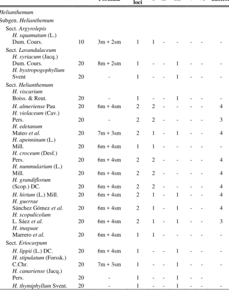
Cytologically, the subgenus Helianthemum shows different base chromosome numbers, where the most represented is x=10 (sect.
CHAPTER 1
(sect. Argyrolepis) and x=12 (sect. Caput-felis) have been registered. In contrast, the subgenus Plectolobum (sect. Atlanthemum and Pseudocistus) presents the uniform base chromosome number of x=11 (Markova, 1975, Sánchez et al., 1986; Arista et al., 1990; Izuzquiza et al., 1998). Only one tetraploid has been reported for the Bulgarian and Armenian populations of H.
lasiocarpum (2n=4x=40) with probably allopolyploid origin (Markova, 1975). Despite these observations and the multitude of reports of Helianthemum chromosome numbers, a hypothesis about the pattern of this chromosome number transition is still not available. Moreover, while Cistus has been widely investigated in terms of phylogeny and divergence times, studies about the whole Helianthemum evolutionary history are in short supply. Nevertheless, the few available data provide the monophyly of Helianthemum genus, thus rejecting the possibility of raising the subgenus Plectolobum to the Rhodax genus rank and supported the section division made by Lopez-Gonzalez (1992). 1.6.4 Cistus heterophyllus
Cistus heterophyllus is a narrowly-distributed W-Mediterranean species present in North Africa (Morocco, Algeria) and in the Iberian Peninsula (Spain) (Crespo and Mateo, 1988; Demoly and Montserrat, 1993). The European individuals are extremely endangered because of their rarity (only two populations, with about 26 and a single individual, respectively, have been reported), and threats caused by abiotic (fires, severe drought), and biotic factors (habitat transformation) (Güemes et al., 2004). In addition, nuclear and plastid DNA markers, together with morphological evidences, strongly suggest that ongoing gene flow with the related C. albidus L. is occurring in European and North African populations (Jiménez et al., 2007; Navarro et al., 2009). In 1987, a single individual showing no signs of interspecific hybridisation was found at E Spain (Valencia) (Crespo and Mateo, 1988) and recovery plans were designed to create a new population in Valencia. This specimen was multiplied through in vitro culture (Arregui et al., 1993; González-Benito and Martín, 2011) to obtain accessions suitable for reintroduction in a new site (Tancat de Portaceli, Valencia, Spain) (Laguna et al., 1998; Aguilella et al., 2010). Here, new plants arising from in vitro-generated individuals were spontaneously produced between 2012 and 2013. Actually the species is listed as Critically Endangered (CD), according to the Red List criteria of the International Union for Conservation of Nature and Natural Resources, IUCN (Moreno, 2008). 1.7 Aims of the study
Modern molecular cytogenetic offers a number of tools suitable for investigating chromosomal architecture such as nucleolar organizer regions (NORs) and 5S rDNA, centromere and telomeres, as well as for identifying
8
locations of genes and repetitive elements and for molecular karyotyping, providing interesting information about plant speciation processes. Cytogenetic features may also be useful when studying threatened or rare species, in case of their management is entrusted to micropropagation protocols, since the stress conditions generated by the in vitro tissue culture are responsible of chromosomal changes. Despite patterns of chromosome evolution using molecular cytogenetic have been established in several plant genera, studies mainly focus on the relationships within polyploid lineages, whereas little is still known about the evolutionary patterns of chromosome changes in predominantly diploid plant groups.
Cytogenetic analyses, using FISH and AgNOR staining methods, have been employed in an attempt to gather information about the karyotypes of a number of selected species and to elucidate the evolutionary dynamics of 45S rDNA site number and position within the homoploid genus Cistus (Chapter 2) and a species sample representative of the whole chromosome base number variation of the genus Helianthemum (Chapter 3). With this purpose the rDNA site number features were mapped onto available phylogenies. Furthermore, the same cytogenetic approach has been used to assess the stability of two nuclear ribosomal DNA families (5S rDNA and 45S rDNA) in the offspring of F1 experimental crosses between Cistus heterophyllus Desf. (Cistaceae) accessions generated after in vitro culture and wild individuals (Chapter 4).
The information collected serve to:
- ascertaining whether changes in the 45S rDNA site features are phylogenetically-correlated within the genus Cistus, and whether the organismal evolutionary history may affects the patterns of rDNA site changes;
- evaluating whether the 45S rDNA site changes reflect the taxonomic infrageneric ranks and the chromosome base number variations, and whether they are phylogenetically-correlated within the genus
Helianthemum;
- verifying whether the in vitro-propagated plants of C. heterophyllus have produced genetically modified or genetically unstable regenerants not present in the original genotype stock, and drowing conclusions with respect to the use of micropropagated material for the management of endangered species.
CHAPTER 1
REFERENCES
Aguilella A, Fos S, Laguna E. 2010. Catálogo valenciano de especies
amenazadas de Flora. Colección Biodiversidad, 18. Conselleria de Medi Ambient, Aigua, Urbanisme i Habitatge, Generalitat Valenciana, 364. Valencia, Spain.
Al-Zahim M, Ford-Lloyd B, Newbury H. 1999. Detection of somaclonal variation in garlic (Allium sativum L.) using RAPD and cytological analysis. Plant Cell Reports, 18: 473-477.
Arista M, Talavera S, Diaz Lifante Z. 1990. Números cromosomáticos para la flora Española: 620-642. Lagascalia, 16: 323-333.
Arregui JM, Juárez J, Laguna E, Reyna S, Navarro L. 1993. Micropropagación de Cistus heterophyllus. Un ejemplo de la aplicación del cultivo de tejidos a la conservación de especies amenazadas. Flora Silvestre, 74: 24-29.
Arrington JM, Kubitzki K. 2003. Cistaceae. In Kubitzki K. (ed.). The Families
and genera of vascular plants IV. Flowering Plants. Dicotyledons. Malvales, Capparales and Non-betalain Caryophyllales, 62-70. Springer, Berlin.
Bouharmont J. 1994. Application of somaclonal variation and in vitro selection to plant improvement. Acta Horticulturae, 355: 213-218.
Britten RJ. 1969. Repeated DNA and transcription. In EW Hanly (ed.).
Problems in Biology: RNA in Development, 187-216. University of Utah Press, Salt Lake City.
Civeyrel L, Leclercq J, Demoly JP, Agnan Y, Quèbre N, Pélissier C, Otto T. 2011. Molecular systematics, character evolution, and pollen morphology of Cistus and Halimium (Cistaceae). Plant Systematics and
Evolution, 295: 23-54.
Crespo M.B., Mateo G. 1988. Consideraciones acerca de la presencia de Cistus
heterophyllus Desf. en la Península Ibérica. Anales del Jardín Botánico
de Madrid, 45: 165-171.
Cruz-Cruz CA, González-Arnao MT, Engelmann F. 2013. Biotechnology and conservation for plant diversity. Resources, 2: 73-95.
Dansereau P. 1940. Études sur les hybrides de Cistes. Annales des Épiphytes, 6: 7-26.
Demoly JP, Montserrat P. 1993. Cistus. In: Castroviejo S et al., (eds.) Flora
Iberica, 3: 319-337. Real Jardín Botánico, CSIC, Madrid.
Demoly JP. 1996. Les hybrides binaires rares du genre Cistus L. (Cistaceae).
Anales del Jardín Botánico de Madrid, 54: 241-254.
Demoly JP. 2006. Notes taxonomiques, chorologiques et nouveautés nomenclaturales pour le genre Cistus L. elargi, incluant Halimium (Dunal) Spach (Cistaceae). Acta Botanica Gallica, 153: 309-323.
10
Elias I, Tuller T. 2007. Reconstruction of ancestral genomic sequences using likelihood. Journal of Computational Biology, 14: 216-237.
Ellul P, Boscaiu M, Vicente O, Moreno V, Rosselló JA. 2002. Intra- and interspecific variation in DNA content in Cistus (Cistaceae). Annals of
Botany, 90: 345-351.
Engelmann F. 2010. Use of biotechnology for the conservation of plant diversity. In Vitro Cellular & Developmental Biology, 47: 5-16.
Escudero M, Martín-Bravo S, Mayrose I, Fernández-Mazuecos M, Fiz-Palacios O, Hipp AL, Pimentel M, Jiménez-Mejías P, Valcárcel V, Vargas P, Luceño M. 2014. Karyotypic changes through dysploidy persist longer over evolutionary time than polyploid changes. PlosOne, 9: e85266. Faria R, Navarro A. 2010. Chromosomal speciation revisited: rearranging
theory with pieces of evidence. Trends in Ecology and Evolution, 25: 660-669.
Fay M.F. 1992. Conservation of rare and endangered plants using in vitro methods. In Vitro Cellular & Developmental Biology, 28, 1-4.
Fernández-Mazuecos M, Vargas P. 2010. Ecological rather than geographical isolation dominates Quaternary formation of Mediterranean Cistus species. Molecular Ecology, 19: 1381-1395.
Figueroa DM, Bass HW. 2010. A historical and modern perspective on plant cytogenetics. Briefings in Functional Genomics, 9: 95-102.
Fukui K, Nakayama S, Ohmido N, Yoshiaki H, Yamabe M. 1998. Quantitative karyotyping of three diploid Brassica species by imaging methods and localization of 45S rDNA loci on the identified chromosomes.
Theoretical and Applied Genetics, 96: 325-330.
Gallego AJ, Aparicio A. 1993. Karyological study in the genus Tuberaria sect.
Scorpioides (Cistaceae): Taxonomic and evolutionary inferences. Plant
Systematics and Evolution, 184: 11-25.
Garcia S, Kovařík A. 2013. Dancing together and separate again: gymnosperms exhibit frequent changes of fundamental 5S and 35S rRNA gene (rDNA) organization. Heredity, 111: 23-33.
Garcia S, Panero JL, Siroky J, Kovařík A. 2010. Repeated reunions and splits feature the highly dynamic evolution of 5S and 35S ribosomal RNA genes (rDNA) in the Asteraceae family. BMC Plant Biology, 10: 176. Gill N, Hans CS, Jackson S. 2008. An overview of plant chromosome structure.
Cytogenetic and Genome Research, 120: 194-201.
González-Benito M., Martín C. 2011. In vitro preservation of Spanish biodiversity. In Vitro Cellular and Developmental Biology, 47: 46-54. Grandbastien MA. 1998. Activation of plant retrotransposons under stress
conditions. Trend in Plant Science, 3: 181-187. Grant V. 1981. Plant Speciation. Columbia University Press.
Güemes J, Jiménez JF, Sánchez-Gómez P. 2004. Cistaceae: Cistus
CHAPTER 1
Banares A, Blanca G, Guemes J, Moreno JC, Ortiz S (eds.). Atlas y
Libro Rojo de la Flora Vascular Amenazada de Espana: Taxones prioritarios, 192-193. Madrid. Ministerio de Medio Ambiente.
Guzmán B, Lledó MD, Vargas P. 2009. Adaptive radiation in Mediterranean
Cistus (Cistaceae). PlosOne, 4: e6362.
Guzmán B, Vargas P. 2005. Systematics, character evolution, and biogeography of Cistus L. (Cistaceae) based on ITS, trnL-trnF, and
matK sequences. Molecular Phylogenetics and Evolution, 37: 644-660 Guzmán B, Vargas P. 2009a. Historical biogeography and character evolution
of Cistaceae (Malvales) based on analysis of plastid rbcL and trnL-trnF sequences. Organisms, Diversity and Evolution, 9: 83-99.
Guzmán B, Vargas P. 2009b. Long distance colonization by the Mediterranean
Cistus ladanifer (Cistaceae) despite the absence of special dispersal mechanisms. Journal of Biogeography, 36: 954-968.
Hao YJ, Deng XX. 2002. Occurrence of chromosomal variations and plant regeneration from longterm-cultured citrus callus. In Vitro Cellular
Developmental Biology Plant, 38: 472-476.
Heslop-Harrison JS, Schwarzacher T. 2011. Organisation of the plant genome in chromosomes. The Plant Journal, 66: 18-33.
Holub J. 1970. New names in Phanerogamae, I. Folia Geobotanica et
Phytotaxonomica Bohemoslovaca, 5: 435-441.
Iriondo J.M. 2001. Conservación de germoplasma de especies raras y amenazadas (Revisión). Investigación Agraria: Producción y Protección
Vegetal, 16: 5-24.
Izuzquiza Á, Mayol M, Rosselló JA, Sáez L, Boscaiu M, Riera J, Estrelles E, Güemes J. 1998. Números cromosomáticos de plantas occidentales.
Anales del Jardín Botánico de Madrid, 56: 119-120.
Jiménez JF, Sánchez-Gómez P, Rosselló JA. 2007. Evidencia de introgresión en Cistus heterophyllus subsp. carthaginensis (Cistaceae) a partir de marcadores moleculares RAPD. Anales de Biologia, 29: 95-103.
Jiménez R, Burgos M, Diaz de la Guardia R. 1988. A study of the silver staining significance in mitotic NORs. Heredity, 60: 125-127.
Kaeppler SM, Kaeppler HF, Rhee Y. 2000. Epigenetic aspects of somaclonal variation in plants. Plant Molecular Biology, 43: 179-188.
Koshi JM, Golstein RA. 1996. Probabilistic reconstruction of ancestral protein sequences. Journal of Molecular Evolution, 42: 313-321.
Laguna E, Ballester G, Escribá MC, Arregui JM, Juárez J, Navarro L. 1998. Reintroducción y reforzamientos poblacionales de especies amenazadas en la Comunidad Valenciana. Conservación Vegetal, 3: 4-5.
Leitch AR, Leitch IJ. 2008. Genomic plasticity and the diversity of polyploid plants. Science, 320: 481-483.
Leutwiler LS, Hough-Evans BR, Meyerowitz EM. 1984. The DNA of
12
Levin DA. 2002. The role of chromosomal change in plant evolution. Oxford University Press, Oxford.
Liberles DA. (ed.). 2007. Ancestral sequence reconstruction. Oxford University Press, Oxford.
López-González G. 1992. Apuntes para justificar el tratamiento del género
Helianthemum Miller, s.l. (Cistaceae) en Flora ibérica. Anales del
Jardín Botánico de Madrid, 50: 35-63.
Löve Á, Kjellqvist E. 1964. Chromosome numbers of some Iberian Cistaceae.
Portugalia Acta Biologica, A8: 69-80.
Lysák MA, Schubert I. 2013. Mechanisms of chromosome rearrangements. In
Plant Genome Diversity, 2. Physical Structure, Behavior and Evolution of Plant Genomes, (eds.), 137-147. Springer-Verlag,Wien.
Mahelka V, Kopecky D, Baum BR. 2013. Contrasting patterns of evolution of 45S and 5S rDNA families uncover new aspects in the genome constitution of the agronomically important grass Thinopyrum
intermedium (Triticeae). Molecular Biology and Evolution, 30: 2065-2086.
Markova M. Karyosystematische Untersuchungen an den Cista¢eae Bulgariens. Plant Systematics and Evolution, 123: 283-315.
Martins EP. 1999. Estimating of ancestral states of continuous characters: A computer simulation study. Systematic Biology, 48: 642-650.
Medina FJ, Cerdido A, de Cárcer G. 2000. The functional organization of the nucleolus in proliferating plant cells. European Journal of
Histochemistry, 44: 117-131.
Mooers AØ, Schluter D. 1999. Reconstructing Ancestor States with Maximum Likelihood: Support for One- and Two-Rate Models. Systematic
Biology, 48: 623-633.
Moreno JC (coord.). 2008. Lista Roja 2008 de la flora vascular española. Dirección General de Medio Natural y Política Forestal (Ministerio de Medio Ambiente, y Medio Rural y Marino, y Sociedad Española de Biología de la Conservación de Plantas). Madrid.
Mujib A, Banerjee S, Dev Ghosh P. 2007. Callus induction, somatic embryogenesis and chromosomal instability in tissue culture raised hippeastrum (Hippeastrum hybridum cv. United Nations). Propagation
of Ornamental Plants, 7: 169-174.
Navarro JA, Sánchez-Balibrea J, Barberá GG, Ferrández M, El Andalossi M. 2009. Siguiendo la huella de la hibridación en poblaciones de Cistus
heterophyllus en el Rif marroquí. Conservación Vegetal, 13: 9-10. Orbović V, Calović M, Viloria Z, Nielsen B, Gmitter FG Jr, castle WS, Grosser
JW. 2008. Analysis of genetic variability in various tissue culture-derived lemon plant populations using RAPD and flow cytometry.
CHAPTER 1
Pagel M. 1999. Inferring the historical patterns of biological evolution. Nature, 401: 877-884.
Pietsch GM, Anderson NO. 2007. Epigenetic variation in tissue cultured Gaura lindheimeri. Plant Cell, Tissue and Organ Culture, 89: 91-103.
Proctor MCF. 1955. Some chromosome counts in the European Cistaceae.
Watsonia, 3: 154-9.
Proctor MCF. 1956. Helianthemum Mill. The Journal of Ecology, 44: 675-692. Rao N.K. 2004. Plant genetic resources: Advancing conservation and use
through biotechnology. African Journal of Biotechnology, 3: 136-145. Richard GF, Kerrest A, Dujon B. 2008. Comparative genomics and molecular
dynamics of DNA repeats in eukaryotes. Microbiology and Molecular
Biology Reviews, 72: 686-727.
Rieseberg LH. 2001. Chromosomal rearrangements and speciation. Trends in
Ecology and Evolution, 16: 351-358.
Sánchez AMA, Gallego MF, Navarro AF. 1986. Aportaciones al conocimiento cariológico de las Cistáceaeas del Centro-Occidente español. Studia
Botanica, 5: 195-202.
Sánchez-Gómez P, Carrión MA, Hernández A, Guerra J. 2002. Libro Rojo de la flora silvestre protegida de la Región de Murcia. Murcia: Consejería de Agricultura, Agua y Medio Ambiente, Universidad de Murcia. Schluter D, Price T, Mooers AØ, Ludwig D. 1997. Likelihood of ancestral
states in adaptive radiation. Evolution, 51: 1699-1711.
Schubert I. 2007. Chromosome evolution. Plant Biology, 10: 109-115.
Schultz TR, Cocroft RB, Churchill GA. 1996. The Reconstruction of ancestral character states. Evolution, 50: 504-511.
Shan F, Yan G, Plummer JA. 2003. Cyto-evolution of Boronia genomes revealed by fluorescent in situ hybridization with rDNA probes.
Genome, 46: 507-513.
Sone T, Fujsawa M, Takanaka M, Nakagawa S, Yamaoka S, Sakaida M, Nishiyama R, Yamato KT, Ohmido N, Fukui K, Fukuzawa H, Ohyama K. 1999. Bryophyte 5S was inserted into 45S rDNA repeat units after the divergence from higher land plants. Plant Molecular Biology, 41: 679-685.
Stebbins GL. 1971. Chromosomal evolution in higher plants. Edwards Arnold, London.
Thompson WF, Murray MG. 1980. Sequence organisation in pea and mung bean DNA and a model for genome evolution. In: Davies JR and hopwood (eds): Fourth John Innes Symposium, 31-45. John Innes Institute, Norwich.
Ukraintseva VV. 1993. Pollen morphology of the family Cistaceae in relation to its taxonomy. Grana, 2: 33-36.
14
Volkov R, Komarova NY, Hemleben V. 2007. Ribosomal DNA in plant hybrids: Inheritance, rearrangement, expression. Systematics and
Biodiversity, 5: 261-276.
Weiss-Schneeweiss H, Schneeweiss GM, Stuessy TF, Mabuchi T, Park J-M, Jang CG, Sun B-Y. 2007. Chromosomal stasis in diploids contrasts with genome restructuring in auto- and allopolyploid taxa of Hepatica (Ranunculaceae). New Phytologist, 174: 669-682.
Weiss-Schneeweiss H, Schneeweiss GM. 2013. Karyotype diversity and evolutionary trends in angiosperms. In: Leitch IJ, Greilhuber J, Doležel J, Wendel JF (eds): Plant Genome Diversity, 2: Physical Structure,
Behaviour and Evolution of Plant Genomes, 209-230. Springer, Wien. Weiss-Schneeweiss H, Tremestsberger K, Schneeweiss GM, Parker JS, Stuessy
TF. 2008. Karyotype diversification and evolution in diploid and polyploidy South American Hypochaeris (Asteraceae) inferred from rDNA localization and genetic fingerprint data. Annals of Botany, 101: 909-918.
Yang Z, Kumar S, Nei M. 1995. A new method of inference of ancestral nucleotide and amino acid sequences. Genetics, 141: 641-1650.
CHAPTER 2
Temporal frames of 45S rDNA site-number variation in
homoploid plant lineages: lessons from the rock rose genus
(
Cistus, Cistaceae)
Chiara Totta1, Marcela Rosato2, Pablo Ferrer-Gallego3, Fernando Lucchese1, and Josep A. Rosselló2,4
1. Università degli Studi Roma Tre, Viale G. Marconi 446, 00146, Rome, Italy 2 Jardín Botánico, ICBiBE, Universidad de Valencia, c/Quart 80, E46008 Valencia, Spain
3 CIEF, Servicio de Vida Silvestre, Generalitat Valenciana, Avda. Comarques del Pais Valencia 114, E46930 Valencia, Spain
CHAPTER 2
INTRODUCTION
Genes encoding ribosomal RNA (rDNA) are universal constituents of cell genomes and are essential for organismal growth and integrity, since their products form the backbones of cytoplasmic, plastidial, and mitochondrial ribosomes (Hillis and Dixon, 1991). In contrast with the single or low-copy number of rDNA genes present in the plastidial and mitochondrial genomes, the nuclear genome harbours hundreds to several thousand copies of each ribosomal species (18S, 5.8S, 25S/26S, 5S) that are usually arranged in distinct arrays of tandemly-repeated units (Srivastava and Schlessinger, 1991).
Although rDNA is the most abundant gene family and occupies a large fraction of the nuclear genome, it is also the most unstable genomic region (Kobayashi, 2008). The reasons for this instability are not fully appreciated; however, it has been reported that rDNA loci are the predominant sites of repeated recombination (Kobayashi and Ganley, 2005). Thus, unequal recombination between homologous and homoelogous loci may trigger both intragenomic fluctuation in rDNA copy number and amplification of new arrays (Cronn et al., 1996; Tsang and Carr, 2008). Furthermore, it has been shown that rDNA arrays and neighbouring regions are one of the frequent targets for mobile element insertions (Raskina et al., 2008). Transposition may promote the evolutionary dynamics of rDNA loci not only across species radiation but also during intraspecies differentiation and domestication, producing karyological rearrangements that may be the onset of speciation processes (Pedrosa-Harand et al., 2006).
In plants, detailed knowledge about the number of rDNA loci, their genomic location, and rDNA linkage are known for a substantial number of species (Garcia et al. 2012, 2014; Roa and Guerra, 2012). However, information concerning the dynamics of rDNA loci among closely related species only becomes biologically relevant when put into an evolutionary or phylogenetic framework.
One of the best-studied systems in plants for assessing the dynamic turnover of rDNA loci is polyploidy. The rapid rearrangement of parental genomes after polyploid formation is now a well-established paradigm in plant genome evolution (Wendel, 2000). The available knowledge suggests that the fate of 45S rDNA units in plant polyploids after their genesis does not follow a uniform evolutionary pattern (Volkov et al., 2007). Because the ancestors of many polyploids have been fully identified, the synthetic reconstruction of the polyploids has been possible in many cases, and the temporal frame and exact origin of polyploid formation are usually known, the dynamic distribution patterns of rDNA loci have been confidentially assessed.
In sharp contrast with polyploids, assessing the evolutionary patterns of rDNA loci change in predominantly diploid lineages has usually received less
18
attention (e.g., Datson and Murray, 2006). Unfortunately, the conclusions drawn from previous studies lack explicitly temporal frames for rDNA loci changes. This is unfortunate since the rates of chromosomal diversification and rDNA dynamics could not be assessed, and their comparison with those known for polyploid lineages could not be established. In addition, eventual correlations between rDNA changes and known significant palaeoecological and palaeoclimatic events could be missed.
The genus Cistus (including Halimium; Cistaceae) provides a suitable case study for assessing the temporal patterns of rDNA site evolution in entirely diploid lineages. Congruent phylogenies based on a suite of plastidial and nuclear DNA markers have been obtained, and dated nodes for virtually all known species (ca. 28) are available (Guzmán and Vargas, 2005, 2009a, 2009b, 2010; Guzmán et al., 2009; Fernández-Mazuecos and Vargas, 2010, 2011; Civeyrel et al., 2011). In addition, several important evolutionary patterns and processes have been revealed for Cistus (Table 2), including colonization and asymmetric diversification of insular oceanic lineages, synchronous evolutionary histories in Mediterranean and Macaronesian species, adaptative radiation in non-insular lineages, and long-distance colonization events despite the absence of effective dispersal mechanisms.
In this study, the number and position of 45S rDNA loci have been determined in Cistus species using fluorescent in situ hybridisation (FISH) studies and Ag-NOR staining, and mapped onto dated phylogenies in an attempt to ascertain whether (a) a phylogenetically-correlated variation in rDNA loci features is present within the genus, (b) rates of rDNA site evolution are equivalent in all lineages, and (c) organismal evolutionary history may have an effect on the patterns of rDNA site or family change.
MATERIALS AND METHODS
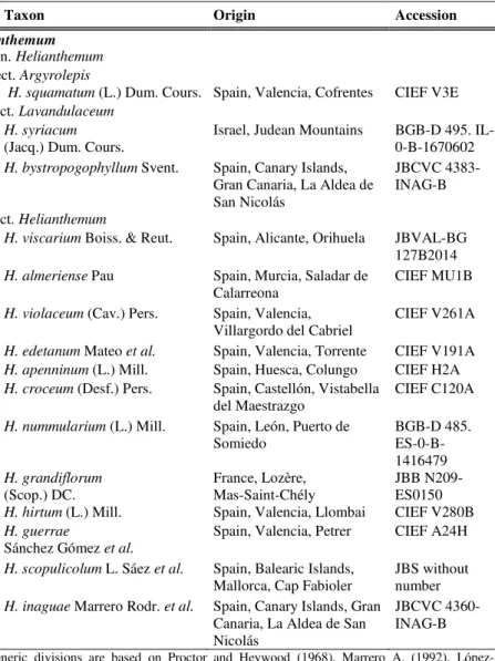
Plant materialsThirty-five taxa of the Cistaceae family belonging to Cistus, Tuberaria and
Fumana genera were analyzed in this study. Seeds were obtained from field sampling, botanical gardens, plant breeding stations and research centers. The origin of the plant analyzed material is provided in Table 1.
Cytogenetic analysis
Cytological preparations
Living plants were cultivated in pots at the CIEF greenhouses. The root tips from the plants were excised and pre-treated with 2 mM 8-hydroxyquinoline for 2 h at 4ºC, then 2 h at room temperature, fixed in an ethanol/glacial acetic
CHAPTER 2
acid (3 : 1) mixture and stored at –20ºC until required. For mitotic chromosome spreads, we followed the protocols described in Rosato et al. (2008).
Fluorescence in situ hybridisation
The 45S rDNA multigene family was localised using the pTa71 (Gerlach and Bedbrook, 1979) clones. The pTa71 probe was labelled with digoxigenin-11dUTP through a nick translation procedure (Roche, Germany). We followed the in situ hybridisation protocols of Rosato et al. (2008), except for the proteinase K pre-treatment, which was performed following Schwarzacher and Heslop-Harrison (2000). Probe detection was conducted using the method of Zhong et al. (1996) with modifications according to Galián et al. (2014).
Ag-NOR staining
Silver impregnation was carried out on 1–2 day-old chromosome preparations according to the protocol described in Rosato and Rosselló (2009).
Karyotype analysis
Chromosome measurements were made on digital images using the freeware
software MICROMEASURE v.3.3 (available at
http://www.colostate.edu/depts/biology/micromeasure). Idiograms were obtained from chromosome measurements of at least five well-spread metaphase plates.
rDNA site-number evolution
Mapping of cytogenetic features onto a phylogenetic framework was carried out following the likelihood reconstruction methods in Mesquite, version 3.04 (Maddison and Maddison, 2015), assigning to each internal node the state that maximizes the probability of obtaining the observed states in the terminal taxa under the specified model of evolution (Mk1 model, in this study). In addition, the most parsimonious reconstruction of the ancestral character states for the number of rDNA sites were also estimated using the Mesquite software.
The phylogenetic tree used as an evolutionary framework for Cistus was the backbone consensus tree obtained by several plastidial and nuclear markers, e.g. rbcL/trnL-trnF (Guzmán and Vargas, 2009a), rbcL/trnK-matK (Guzmán and Vargas, 2009b), trnL-trnF/trnS-trnG/trnK-matK/rbcL/ITS/ncpGS (Guzmán
et al., 2009), trnL-trnF/matK (Fernández-Mazuecos and Vargas, 2010),
trnS-trnG/trnK-matK (Guzmán and Vargas, 2010), and trnS-trnG/trnL-trnF (Civeyrel et al., 2011). Fumana thymifolia and T. lignosa were used as outgroups (Guzmán and Vargas, 2005, 2009a, 2009b, 2010; Guzmán et al., 2009; Fernández-Mazuecos and Vargas, 2010). Dated nodes obtained by relaxed clock methods by Guzmán et al. (2009) and Guzmán and Vargas (2009a) were indicated in the consensus tree.
20
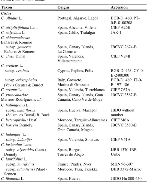
Table 1 - Cistaceae studied taxa, sources of material and seed accessions. BGB-D, Botanischer Garten Berlin-Dahlem; CBNM, Conservatoire Botanique National Méditerranéen de Porquerolles; CIEF, Centro para la Investigación y Experimentación Forestal; IJBB, Jardí Botànic de Barcelona; JBC, Jardín Botánico de Córdoba; JBCA, Jardín Botánico El Castillejo; JBCVC, Jardín Botánico Canario Viera y Clavijo; JBDO, Jardín Botánico Dunas del Odiel; MHN, Muséum d’Histoire Naturelle; RJB-CSIC, Real Jardín Botánico de Madrid.
Taxon Origin Accession
Cistus
C. albidus L. Portugal, Algarve, Lagoa BGB-D. 460. PT-0.B-0100208
C. atriplicifolium Lam. Spain, Alicante, Villena CIEF A26E
C. calycinus L. Spain, Cádiz, Trafalgar JAR-1
C. chinamadensis
Bañares & Romero subsp. gomerae
Bañares & Romero Spain, Canary Islands, La Gomera JBCVC 2674-B
C. clusii Dunal Spain, Valencia,
Villamarchante CIEF V24B
C. creticus L.
subsp. creticus Cyprus, Paphos, Polis BGB-D. 463. CY-0-B-2400300 subsp. eriocephalus Italy, Grosseto,
Marina di Grosseto
BGB-D. 469. IT-0-B-1971186 (Viv.) Greuter & Burdet
C. crispus L. Spain, Valencia, Torreblanca CIEF C67A
C. grancanariae
Marrero-Rodríguez et al. Spain, Canary Islands, Gran Canaria, Cabo Verde-Moya JBCVC 3567-B
C. halimifolius L.
subsp. multiflorus Spain, Huelva, Mazagón JBDO without number (Salzm. ex Dunal) B. Bock
C. heterophyllus Desf. Morocco, Targuist-Alhucemas CIEF M6A
C. horrens Demoly Spain, Canary Islands,
Gran Canaria, Mogana JBCVC 3580-B
C. ladanifer L.
subsp. ladanifer Spain, Valencia, Sinarcas CIEF V51A
C. lasianthus Lam. subsp. alyssoides (Lam.)
Demoly Spain, Burgos, Torres de Abajo IJBB 1370-JBB-Uribe
C. laurifolius L.
subsp. laurifolius France, Prades, Nyer MHN 96-307 subsp. atlanticus (Pitard)
Sennen Morocco, Taza, Tazekka IJBB 3372-Marroc
CHAPTER 2
Table 1 (Continued)
Taxon Origin Accession
C. monspeliensis L. Spain, Canary Islands,
Gran Canaria, Artenara JBCVC 2880/B France, Montpellier, BGB-D. 474. FR-0- Saint-Mathieu-de-Tréviers B-2042205
C. ochreatus Chr. Sm. ex Buch
Spain, Canary Islands, Gran Canaria, Artenara
JBCVC 1402/B
C. ocymoides Lam. Spain, Huelva, Aracena CIEF Hu1A
C. osbeckiifolius Webb ex Christ.
Spain, Canary Islands, Tenerife, La Orotava
JBCVC 3641/B
C. palmensis Bañares & Demoly
Spain, Canary Islands, La Palma, San Andrés y Sauces
JBCVC 3617/B
C. parviflorus Lam. Greece, Crete, Akrotiri, Chania BGB-D. 475. GR-0-B-2680597
C. populifolius L. Spain, Valencia, Serra CIEF V92A
C. pouzolzii Delile France, Alès,
Saint Jean du Gard CBNM without number
C. psilosepalus Sweet Spain, Toledo, Velada RJB-CSIC JGF.030
C. salviifolius L. Greece, Dodecanese, Karpathos BGB-D. 476. GR-0-1270207
C. symphytifolius Lam. Spain, Canary Islands,
Gran Canaria Artenara JBCVC 3656/B
C. umbellatus L.
subsp. micranthus Demoly Greece, Messinía, Spárti BGB-D. 580. GR-O-B-1901502 subsp. viscosum (Willk.)
Demoly
Spain, Valencia, Castielfabib CIEF A65A
Tuberaria
Tuberaria lignosa (Sweet) Samp.
Spain, Valencia, Sinarcas CIEF V141A
Fumana
F. clausonis Pomel Morocco, Beni Mellal IJBB 165-Marroc
F .ericifolia Wallr Spain, Valencia, Sot de Chera CIEF V273A
F. fontanesii Pomel Spain, Murcia, Sierra de Espuña
CIEF Mu4A
F. thymifolia (L.) Spach Spain, Balearic Islands, Formentera
22
RESULTS
Karyotype analysisThe chromosome numbers of thirty-five species and subspecies belonging to Cistus (30), Tuberaria (1), and Fumana (4) were determined and the idiograms for Cistus and Tuberaria samples were assessed. No departures from the previous known chromosome counts were found and the chromosome number of Cistus grancanariae (2n=18) was determined for the first time. For most species, the karyotype was composed by seven metacentric, one submetacentric and one metacentric-submetacentric chromosome pairs with slight variations in C. clusii and C. umbellatus (Table 2). In contrast, the
Tuberaria lignosa karyotype (2n=14) was characterized by the presence of two metacentric and five submetacentric chromosome pairs. In Fumana species (2n=32) idiograms could not be assessed due to difficulties in defining the centromere position of most chromosomes.
Variation in the number and localization of 45S rDNA loci
45S rDNA site-number variation in Cistus ranged from one to four, whereas two loci were present in Tuberaria, the sister clade of Cistus, and in the four analyzed species of Fumana (Table 2).
Most species of Cistus showed one or two 45S rDNA loci, accounting for the 43.3% and 46.7% on the entire sample analyzed, respectively. Higher numbers of loci were rarely present; three loci were found in C. monspeliensis and C. umbellatus subsp. viscosum, while the maximum number of 45S rDNA loci, four, was present exclusively in C. grancanariae (Figure 1).
The 45S rDNA sites were mostly localized at the sub-terminal regions of chromosomes and were associated to secondary constrictions. In a few cases, a satellite portion was observed attached to the chromosome body by a de-condensed string of labeled chromatin. No interstitial or proximal rDNA signals were observed.
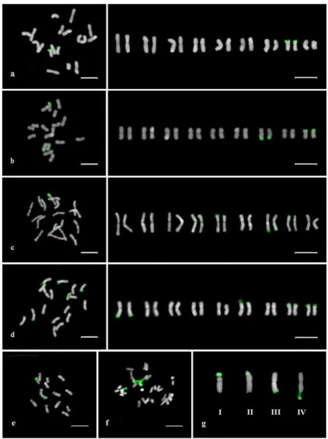
According to the localization of the 45S rDNA sites along the chromosome, four landmarks (types I-IV) were defined (Figure 1, g). Type I represented chromosomes bearing a sub-terminal 45S rDNA site on the short arm. Type II consisted of a chromosome showing one 45S rDNA site on the short arm followed by a minor satellite region. Type III was characterized by the presence of a sub-terminal 45S rDNA site on the long arm and type IV by a 45S rDNA locus located on the long arm attached by a conspicuous satellite region.
Chromosome type I was present in all Cistus accessions, whereas type III co-occurred in only seven accessions. The less represented rDNA locations
CHAPTER 2
were observed exclusively in C. lasianthus subsp. alyssoides and C.
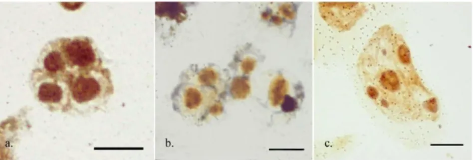
psilosepalus (type II), and in C. libanotis and C. salviifolius (type IV) (Table 2). For those species showing more than a single rDNA locus their NOR activity was assessed by silver staining, and the maximum number of interphase nucleoli was recorded. In all but one the analyzed taxa all 45S rDNA loci were transcriptionally active, since the number of the FISH signals equated the maximum number of nucleoli detected. In C. grancanariae, the maximum number of nucleoli observed was six, suggesting that one of the four 45S rDNA loci was silenced. Often odd numbers of heteromorphic nucleoli were observed (Figure 2).
24
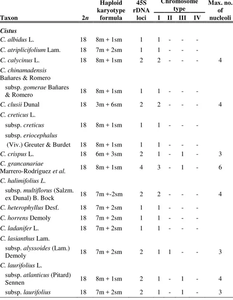
Table 2 - Karyotypic features of the analyzed species. The chromosome number, the haploid karyotype formula, the total number of 45S rDNA loci with their distribution in each rDNA-bearing chromosomal type and maximum number of nucleoli observed are indicated. Taxon 2n Haploid karyotype formula 45S rDNA loci Chromosome
type Max. no. of nucleoli I II III IV Cistus C. albidus L. 18 8m + 1sm 1 1 - - - C. atriplicifolium Lam. 18 7m + 2sm 1 1 - - - C. calycinus L. 18 8m + 1sm 2 2 - - - 4 C. chinamadensis
Bañares & Romero subsp. gomerae Bañares
& Romero 18 8m + 1sm 1 1 - - -
C. clusii Dunal 18 3m + 6sm 2 2 - - - 4
C. creticus L.
subsp. creticus 18 8m + 1sm 1 1 - - - subsp. eriocephalus
(Viv.) Greuter & Burdet 18 8m + 1sm 1 1 - - -
C. crispus L. 18 6m + 3sm 2 1 - 1 - 3
C. grancanariae
Marrero-Rodríguez et al. 18 8m + 1sm 4 3 - 1 - 6
C. halimifolius L.
subsp. multiflorus (Salzm.
ex Dunal) B. Bock 18 7m +-2sm 2 2 - - - 4
C. heterophyllus Desf. 18 7m + 2sm 1 1 - - -
C. horrens Demoly 18 7m + 2sm 1 1 - - -
C. ladanifer L. 18 7m + 2sm 1 1 - - -
C. lasianthus Lam. subsp. alyssoides (Lam.)
Demoly 18 7m + 2sm 2 1 1 - - 3
C. laurifolius L.
subsp. atlanticus (Pitard)
Sennen 18 8m + 1sm 2 1 - 1 - 4
CHAPTER 2 Table 2 (Continued) Taxon 2n Haploid karyotype formula 45S rDNA loci Chromosome
type Max. no. of nucleoli I II III IV Cistus 1C. monspeliensis L. 18 8m + 1sm 3 2 - 1 - 6 2C. monspeliensis L. 18 8m + 1sm 3 2 - 1 - 5 C. ochreatus Chr. Sm. ex Buch 18 - 1 1 - - - C. ocymoides Lam. 18 7m + 2sm 2 2 - - - 4 C. osbeckiifolius Webb ex Christ. 18 8m + 1sm 1 1 - - -
C. palmensis Bañares &
Demoly 18 8m + 1sm 1 1 - - - C. parviflorus Lam. 18 7m + 2sm 2 1 - 1 - 3 C. populifolius L. 18 7m + 2sm 2 2 - - - 4 C. pouzolzii Delile 18 7m + 2sm 2 2 - - - 4 C. psilosepalus Sweet 18 8m + 1sm 2 1 1 - - 3 C. salviifolius L. 18 8m + 1sm 2 1 - - 1 4 C. symphytifolius Lam. 18 7m + 2sm 1 1 - - - C. umbellatus L. subsp. micranthus Demoly 18 6m + 3sm 1 1 - - -
subsp. viscosum (Willk.)
Demoly 18 3m + 6sm 3 3 - - - 5
Tuberaria
Tuberaria lignosa (Sweet)
Samp. 14 2m + 5sm 2 1 1 - - 4 Fumana F. clausonis Pomel 32 - 2 2 F. ericifolia Wallr 32 - 2 2 3 F. fontanesii Pomel 32 - 2 2 F. thymifolia (L.) Spach 32 - 2 2 na na, not analysed.
26
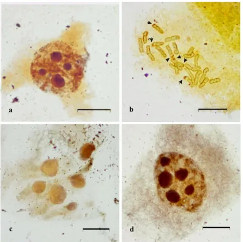
Figure 1 - Arrangement of chromosomes in metaphase plates and idiograms showing the location of 45S rDNA sites in (a) C. creticus, with one single 45S rDNA locus (b)
C. libanotis with two 45S rDNA loci (c) C. monspeliensis with three 45S rDNA loci and (d) C. grancanariae with four 45S rDNA loci, and in the related genera T. lignosa (e) and F. thymifolia (f) both with two 45S rDNA loci. The four chromosome landmarks based on the different position of 45S rDNA loci are indicated (g). Scale bars represent 10 μm.
CHAPTER 2
Figure 2 - Maximum number of silver-stained interphase nucleoli on non-pretreated root tips. (a) Cistus libanotis, (b) Cistus monspeliensis, and (c) Cistus grancanariae. Scale bar represents 10 μm.
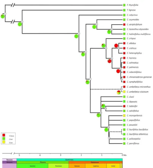
Evolutionary trends in rDNA site-number change
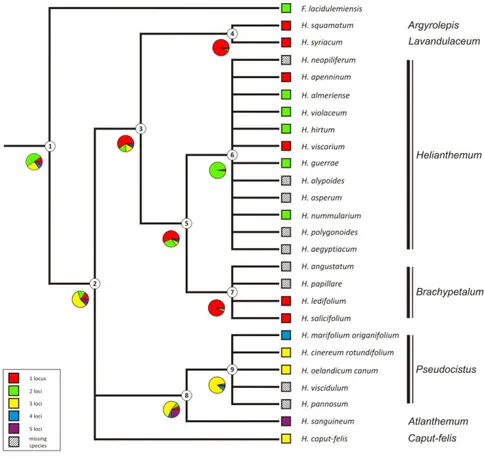
The results of the maximum likelihood and maximum parsimony reconstructions of the ancestral 45S rDNA site number state for each node are indicated in Table 3. Both the methods inferred on the backbone phylogenetic chronogram (Figure 3) yielded identical results with a single exception. Overall, the analysis showed multiple shifts between the number of the rDNA site of Cistus taxa as independent events occurred belatedly during the evolution of the group dated starting from the Pliocene-Pleistocene about 2 Ma (Guzmán et al., 2009). The most encountered states were two and one 45S rDNA sites. The two-loci state was unambiguously optimized as the ancestral condition and a stasis was inferred at the common ancestor of most of the main clades diversified during the Pleistocene. The first loss event of one rDNA locus has been inferred in the common ancestor of the lineage including the purple-flowered species - whose diversification took place recently in the Mid Pleistocene - after the splitting of the basal C. crispus (node 9). The loss event has been supported by a high likelihood score (0.93) and involved the whole clade without further changes. The one-site state appeared independently from the two-site ancestral state also at several terminal nodes. In contrast, rDNA loci amplification have evolved independently only twice at the C.
monspeliensis and C. umbellatus subsp. viscosum terminal nodes. The inferred ancestral state for the C. umbellatus subsp. micranthus and C. umbellatus subsp
viscosum clade (node 13) was ambiguous in the parsimony with all the recorded state numbers as equally parsimonious. However, the maximum likelihood method placed the one-locus state as the most likely.
28
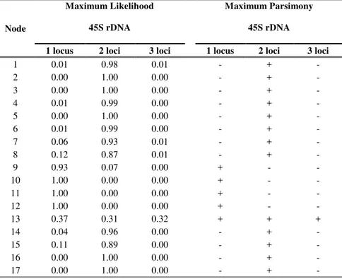
Table 3- Likelihood scores and Parsimony results of the three possible states of 45S rDNA loci number at each node indicated in Figure 3.
Node
Maximum Likelihood 45S rDNA
Maximum Parsimony 45S rDNA
1 locus 2 loci 3 loci 1 locus 2 loci 3 loci
1 0.01 0.98 0.01 - + - 2 0.00 1.00 0.00 - + - 3 0.00 1.00 0.00 - + - 4 0.01 0.99 0.00 - + - 5 0.00 1.00 0.00 - + - 6 0.01 0.99 0.00 - + - 7 0.06 0.93 0.01 - + - 8 0.12 0.87 0.01 - + - 9 0.93 0.07 0.00 + - - 10 1.00 0.00 0.00 + - - 11 1.00 0.00 0.00 + - - 12 1.00 0.00 0.00 + - - 13 0.37 0.31 0.32 + + + 14 0.04 0.96 0.00 - + - 15 0.11 0.89 0.00 - + - 16 0.00 1.00 0.00 - + - 17 0.00 1.00 0.00 - + -
CHAPTER 2
Figure 3 - Ancestral 45S rDNA site number reconstruction mapped on the backbone
Cistus chronogram. Pies illustrate relative likelihood estimates of the three possible states of 45S rDNA loci at each node. Numbers in the circles correspond to the node numbers indicated in Table 3. Dotted line indicates absence of age estimate.
30
DISCUSSION
Karyotype stasisThe present study confirms previous reports indicating that Cistus species are very uniform in chromosome number (2n=2x=18). Moreover, karyological uniformity is also detected in the karyotype patterns, mainly characterized by metacentric and sub-metacentric chromosome pairs, with little interspecific chromosome variability. Cistus grancanariae, not previously analyzed, has shown the same chromosome number and the karyotype was highly similar to that observed in the most related species C. monspeliensis. The uniformity in chromosome number and coarse karyotype features may be the result either of the two ways used to explain the karyotype evolution among related taxa: the karyotype orthoselection and the karyotype conservation (White, 1973). Karyotypic orthoselection is a process wherein the same type of chromosomal rearrangement is established repeatedly in the same lineage whereas karyotype conservation is the maintenance of similar chromosome morphology in different taxa through a lack of structural mutations. Similar conserved chromosome pattern has been reported for homoploid species like Helianthus (Lai et al., 2005) and Nemesia (Datson and Murray, 2006).
This karyotype stasis agrees with the young age of the genus inferred from molecular clock analyses (Guzmán & Vargas, 2009a, 2009b, 2010; Guzmán et
al. 2009; Fernández-Mazuecos & Vargas, 2010), by which the diversification of
Cistus species took place not earlier than the Pliocene, and mainly in the Pleistocene, largely driven by the Mediterranean climate changes occurred around 3.4-2.8 Ma ago (Suc, 1984; Palamarev, 1989; Fiz-Palacios and Valcárcel, 2013).
The karyotype conservatism displayed by Cistus and most of the paraphyletic Halimium group support the monophyletic origin of the assemblage Cistus-Halimium (Guzmán & Vargas, 2009a).
Changes in 45S rDNA sites in a genus characterized by conserved karyotype
When polyploidy is not taken into account and no chromosomal repatterning is evident, evolution of the number and location of the 45S rDNA is thought to proceed by gains and losses of sites. Different mechanisms have been postulated to account for the mobility and polymorphism of numbers, sizes and positions of rDNA sites, such as transposition, and chromosome rearrangements (translocation, inversion, duplication, deletion) caused by homologous or non-homologous unequal crossing-over (Raskina et al, 2008; Schubert and Lysak, 2011). These processes could act alone or in combination,