doi: 10.3389/fnmol.2020.578211
Edited by: Robert A. Nichols, University of Hawaii at Manoa, United States Reviewed by: Orly Lazarov, University of Illinois at Chicago, United States Juan Song, University of North Carolina at Chapel Hill, United States *Correspondence: Roberto Piacentini [email protected] Received: 30 June 2020 Accepted: 15 December 2020 Published: 22 January 2021 Citation: Li Puma DD, Piacentini R and Grassi C (2021) Does Impairment of Adult Neurogenesis Contribute to Pathophysiology of Alzheimer’s Disease? A Still Open Question. Front. Mol. Neurosci. 13:578211. doi: 10.3389/fnmol.2020.578211
Does Impairment of Adult
Neurogenesis Contribute to
Pathophysiology of Alzheimer’s
Disease? A Still Open Question
Domenica Donatella Li Puma1,2, Roberto Piacentini1,2* and Claudio Grassi1,2
1Department of Neuroscience, Università Cattolica del Sacro Cuore, Rome, Italy,2Fondazione Policlinico Universitario
A. Gemelli IRCCS, Rome, Italy
Adult hippocampal neurogenesis is a physiological mechanism contributing to hippocampal memory formation. Several studies associated altered hippocampal neurogenesis with aging and Alzheimer’s disease (AD). However, whether amyloid-β protein (Aβ)/tau accumulation impairs adult hippocampal neurogenesis and, consequently, the hippocampal circuitry, involved in memory formation, or altered neurogenesis is an epiphenomenon of AD neuropathology contributing negligibly to the AD phenotype, is, especially in humans, still debated. The detrimental effects of Aβ/tau on synaptic function and neuronal viability have been clearly addressed both in in vitroand in vivo experimental models. Until some years ago, studies carried out on in vitromodels investigating the action of Aβ/tau on proliferation and differentiation of hippocampal neural stem cells led to contrasting results, mainly due to discrepancies arising from different experimental conditions (e.g., different cellular/animal models, different Aβ and/or tau isoforms, concentrations, and/or aggregation profiles). To date, studies investigating in situ adult hippocampal neurogenesis indicate severe impairment in most of transgenic AD mice; this impairment precedes by several months cognitive dysfunction. Using experimental tools, which only became available in the last few years, research in humans indicated that hippocampal neurogenesis is altered in cognitive declined individuals affected by either mild cognitive impairment or AD as well as in normal cognitive elderly with a significant inverse relationship between the number of newly formed neurons and cognitive impairment. However, despite that such information is available, the question whether impaired neurogenesis contributes to AD pathogenesis or is a mere consequence of Aβ/pTau accumulation is not definitively answered. Herein, we attempted to shed light on this complex and very intriguing topic by reviewing relevant literature on impairment of adult neurogenesis in mouse models of AD and in AD patients analyzing the temporal relationship between the occurrence of altered neurogenesis and the appearance of AD hallmarks and cognitive dysfunctions.
Keywords: neural stem cells, adult neurogenesis, amyloid-beta protein, tau, Alzheimer’s disease, herpes simplex virus type 1
HIPPOCAMPAL NEUROGENESIS AND
MEMORY
The hippocampus is recognized as a brain area primarily involved in memory formation, e.g., pattern separation, emotional memory, and cognitive flexibility (Lazarov and Hollands, 2016; Anacker and Hen, 2017; Hainmueller and Bartos, 2020). The hippocampal circuitry consists of a unidirectional, trisynaptic excitatory pathway in which the dentate gyrus (DG) of the hippocampus receives inputs from the entorhinal cortex (EC), which are then delivered to the CA3 area of the hippocampus and from there to CA1. In turn, CA1 projects to the subiculum and hippocampal outputs are sent back to the deep layers of EC. The classical experimental paradigm used to investigate hippocampal plasticity, underlying memory formation, is the long-term potentiation (LTP) at the CA3-CA1 synapse not involving the DG (Malenka and Nicoll, 1999), and the EC-DG-CA3-CA1 axis could be strongly affected if this pathway is corrupted at the DG. In fact, DG supports various mnemonic functions including contextual discrimination, pattern separation, novelty detection, and integration of individual episodes into a framework of experiences (Hainmueller and Bartos, 2020). Interestingly, the subgranular zone (SGZ) of the DG in the hippocampus is known to be one of the two neurogenic niches in the adult brain, the other being the subventricular zone of the lateral ventricles (Gage, 2019). Adult neurogenesis occurring at the SGZ allows integration of newly formed neurons in the DG circuits (Kitabatake et al., 2007), thus providing this brain area with marked plasticity. As such, adult hippocampal neurogenesis at the DG has been proposed to strongly participate in formation of hippocampal-dependent memory (Gonçalves et al., 2016; Toda et al., 2019; Hainmueller and Bartos, 2020).
Additionally, the hippocampus is one of the brain areas primarily affected by aging and Alzheimer’s disease (AD) (Selkoe, 2011). Alzheimer’s disease is the most common cause of dementia in the elderly and is characterized by memory loss and cognitive dysfunction. The majority of AD cases are sporadic, and the remaining cases are associated with genetic factors [i.e., familial AD (FAD)]. Mutations in genes, encoding either the amyloid precursor protein (APP) or enzymes catalyzing its proteolytic cleavage (presenilin 1 and 2—PSEN1 and 2—subunits of γ-secretase responsible for amyloid-β peptide—Aβ–generation), along with microtubule-associated protein tau (MAPT) encoding the tau protein, are responsible for FAD. Nowadays, it is widely recognized that memory failure in AD is due to synaptic alterations caused by intra- and extracellular accumulation of Aβ and hyperphosphorylated tau oligomers (Crews et al., 2010; Puzzo et al., 2017). Experimental evidence also suggests that early alterations of DG neurogenesis may concur to the pathogenesis of this neurological disorder (Mu and Gage, 2011;
Unger et al., 2016).
AD AND NEUROGENESIS: IN VITRO
STUDIES
In the last 15 years, many studies investigated hippocampal neurogenesis [i.e., proliferation and
neuronal differentiation of hippocampal neural stem cells (NSCs)] in in vitro and in vivo experimental AD models.
In vitro experimental paradigms usually consisted of NSC incubation with Aβ. However, researches carried out in various laboratories led to contrasting results about the effects of proliferation/differentiation of hippocampal NSCs, mainly because of different (i) Aβ preparations (monomeric, oligomeric, or fibrillar); (ii) peptide lengths (40 vs. 42 amino acids); and (iii) working concentrations, which are often irrelevant from a physiological point of view. For example, in 2004 López-Toledano and Shelanski reported that in vitro treatment of hippocampal NSCs with micromolar concentrations of Aβ42 oligomers dose-dependently increased their neuronal differentiation (López-Toledano and Shelanski, 2004). Conversely, more recent results obtained either with lower Aβ concentrations or in cultured NSCs isolated from mouse models harboring the most common genetic alterations observed in FAD indicated impaired proliferation and reduced neuronal differentiation of hippocampus-residing NSCs. In particular, Lee et al. (2013) found that exposure of human NSCs to Aβ-containing conditioned medium from SK-N-MC cells expressing APP Swedish mutation reduced NSC proliferation, impaired neurogenesis, and promoted gliogenesis via glycogen synthase kinase 3β (GSK-3β) activation. Moreover, the exposure of human neural stem cell line hNS1 to nanomolar concentrations of Aβ42 significantly promoted cell proliferation and glial cell specification by increasing the pool of proliferating glial precursors without affecting neuronal differentiation (Bernabeu-Zornoza et al., 2019). A recent study from our lab demonstrated that treatment of cultured murine hippocampal NSCs with Aβ42 oligomers (200 nM)—able to cross plasma membrane, to accumulate intracellularly, and to induce GSK-3 activation (Ripoli et al., 2013, 2014,Scala et al., 2015)—negatively affected their proliferation and neuronal differentiation (Li Puma et al., 2019). Similar results were obtained in NSCs infected with HSV-1, which triggered APP phosphorylation and cleavage with consequent accumulation of several APP fragments including Aβ in several cell types (De Chiara et al., 2010; Piacentini et al., 2011, 2015). The HSV-1-induced hyperproduction of Aβ was correlated with the antimicrobic activity of Aβ42 and interpreted as a defensive response of the infected cell (Soscia et al., 2010; Kumar et al., 2016). Strategies aimed at limiting the production and accumulation of Aβ inside cells (as the use of a γ-secretase inhibitor or the 4G8 antibody raised against Aβ oligomers able to be intracellularly uploaded;Tampellini et al., 2007) counteracted the effects of Aβ42 on in vitro neurogenesis (Li Puma et al., 2019). Altered proliferation and neuronal differentiation were also observed in NSCs isolated from 3 × Tg-AD mouse embryos (Leone et al., 2019). These cells exhibited high levels of Aβ oligomers compared with NSCs isolated from wild-type (WT) mouse. Emerging evidence also suggests that downregulated expression of the nucleoporin Nup153 negatively affects the neurogenic niche of 3 × Tg AD mice. Accordingly, restoration of Nup153 levels in hippocampal 3 × Tg-AD NSCs promoted their proliferation, migration, and neuronal maturation (Leone et al., 2019).
HIPPOCAMPAL NEUROGENESIS IN FAD
MOUSE MODELS
More robust results have been obtained from studies performed in in vivo FAD mouse models often exhibiting impaired neurogenesis correlated with accumulation of molecular AD hallmarks (e.g.,Taniuchi et al., 2007; Hollands et al., 2016, 2017; Baglietto-Vargas et al., 2017). However, depending on the FAD model used, this impairment may rely on reduced neurogenesis (lower NSC proliferation, decreased neuronal differentiation, and/or reduced survival of newly formed neurons) or increased gliogenesis (normal of even increased NSC proliferation, followed by differentiation toward the glial rather than neuronal phenotype). Nevertheless, despite the consensus about altered neurogenesis in these mouse models, a clear understanding of whether and how much this impairment contributes to memory/cognitive dysfunction in AD is still missing.
Various FAD mouse models have been developed resembling peculiar features of the disease, which are based on one or more gene-coding mutations in proteins critically involved in AD (Unger et al., 2016). The most applied models use Tg2576 mice (Hsiao, 1998), which overexpress APP harboring the Swedish double mutation—KM670/671NL; PDAPP mice (Games et al., 1995), which overexpress APP harboring the Indiana (V717F) mutation; and J20 mice (Mucke et al., 2000), which overexpress APP harboring both Swedish and Indiana mutations. Other models associate APP mutations to other mutations accounting for PSEN1 and 2 genes encoding for presenilin 1 and 2, as for example the double transgenic “APP Swedish PS11E9” (APPswe/PS11E9) mouse model (Jankowsky et al., 2001) or APP/PS1 mice harboring APP Swedish and London (V717I) along with the PS1 M146L mutation (Baglietto-Vargas et al., 2017). Finally, the 5 × FAD mouse model, which is a more complex model harboring all five AD-linked mutations accounting for Aβ formation, has also been developed (Oakley et al., 2006). All these models do not consider familial mutations involving the tau protein, which is the other key protein in AD. In this regard, a mouse model representative of the full AD pathology has been developed, associating mutation in the MAPT gene, encoding for tau protein, with those of the other key proteins in AD (APP and PS1). This 3 × Tg-AD model contains three mutations associated with FAD: APP Swedish, PSEN1 M146V, and MAPT P301L (Oddo et al., 2003).
What about neurogenesis in these FAD mouse models?
Demars et al. (2010) reported a drastic reduction of NSC proliferation [identified through 5′-bromo-deoxyuridine
(BrdU) incorporation] in the SGZ of the hippocampus of APPswe/PS11E9 mice, at 2 months of age, with respect to the age-matched WT animals. These alterations, taking place before the formation of amyloid deposits, were followed by a significant reduction in the number of cells acquiring a neuronal (doublecortin+ -DCX) phenotype (i.e., BrdU+/DCX+
cells) with respect to age-matched WT mice. In agreement with Demars’s results, in the same experimental model Unger et al. (2016) found a reduced number of BrdU-positive cells evaluated at 3 months of age, 30 days after BrdU injection,
but an increased numbers of PCNA+ cells. PCNA is a protein
expressed by proliferating cells in the late G1 and S phases of mitosis, and this difference, observed with these two methods of analysis, may suggest alteration in the cell cycle. However, most of these new cells (positive for either BrdU or PCNA) did not survive during maturation resulting in a reduced number of BrdU+/DCX+ and PCNA+/DCX+ cells, thus
indicating impaired adult hippocampal neurogenesis. Similar findings were also obtained in Tg2576 mice, which showed increased NSC proliferation in the SGZ of the hippocampus DG but reduced integration of newly formed neurons in the DG at an age at which these mice exhibited neither amyloid extracellular deposits nor major cognitive impairment (Unger et al., 2016). Unger’s data in Tg2576 mice were in agreement with those obtained by Krezymon et al. (2013) in the same mouse model. In APP/PS1 mice, Baglietto-Vargas et al. found a reduced number of SGZ precursor cells along with reduced numbers of BrdU+/DCX+ cells at 4–6 months, i.e., slightly
before the onset of cognitive dysfunction (Baglietto-Vargas et al., 2017). Also, 5 × FAD mice exhibited an early impairment of neurogenesis with significantly reduced DCX+ cells in the DG
starting from 2 months of age (Moon et al., 2014). In 2008, Rodrìguez et al. demonstrated impaired neurogenesis in terms of NSC proliferation and neuronal differentiation/integration of the DG even in 3 × Tg-AD mice. In this mouse, a significant reduction of neurogenesis was evident in females at 4 months of age with respect to age-matched controls, while male mice exhibited these alterations later. Findings about early alteration of hippocampal neurogenesis in 3 × Tg-AD mice were also confirmed byHamilton et al. (2010). Interestingly,Zheng et al. (2017) demonstrated that intrahippocampal injection of Aβ42 in ovariectomized mice inhibited neurogenesis, which were recovered by 17β-estradiol (E2) treatment; this finding further supported the impact of estrogens in regulating neurogenesis and their potential role in AD pathogenesis. Of note, several conflicting results have been reported on the J20 mouse model, which was found to exhibit either increased neurogenesis with increasing age (López-Toledano and Shelanski, 2007) or impaired neurogenesis independently on Aβ (Pan et al., 2016) and additionally on the APP/PS1 mice, which exhibited increased neurogenesis at later age (Yu et al., 2009), thus indicating that effects on neurogenesis may also depend on a combination of mutations.
A detailed description of how neurogenesis is altered in different AD mouse models was reviewed byChuang (2010)and
Wirths (2017). In Table 1, we summarized how neurogenesis is altered in various AD models, highlighting the age at which neurogenesis was impaired and the age at which cognitive dysfunction started.
The finding that impairment of hippocampal neurogenesis in FAD mice occurs before (i) AD hallmarks accumulation and (ii) appearance of learning and memory dysfunction suggests that the former might have a causal role in cognitive decline characterizing prodromal AD.
In support of this hypothesis, experimental evidence indicates that ablation of hippocampal neurogenesis in APPswe/PS11E9
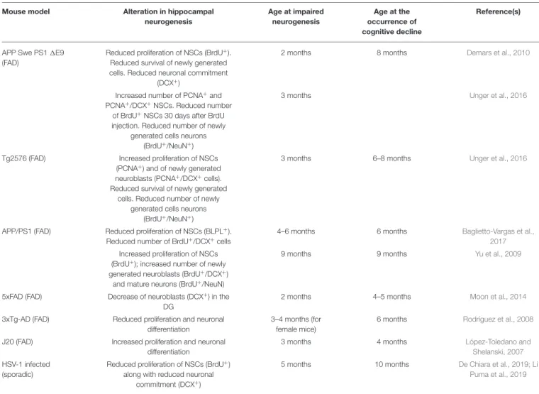
TABLE 1 | Alteration of adult hippocampal neurogenesis in FAD and sporadic AD mouse models.
Mouse model Alteration in hippocampal
neurogenesis Age at impaired neurogenesis Age at the occurrence of cognitive decline Reference(s)
APP Swe PS1 1E9 (FAD)
Reduced proliferation of NSCs (BrdU+). Reduced survival of newly generated cells. Reduced neuronal commitment
(DCX+)
2 months 8 months Demars et al., 2010
Increased number of PCNA+and PCNA+/DCX+NSCs. Reduced number
of BrdU+NSCs 30 days after BrdU injection. Reduced number of newly
generated cells neurons (BrdU+/NeuN+)
3 months Unger et al., 2016
Tg2576 (FAD) Increased proliferation of NSCs (PCNA+) and of newly generated neuroblasts (PCNA+/DCX+cells). Reduced survival of newly generated
cells. Reduced number of newly generated cells neurons
(BrdU+/NeuN+)
3 months 6–8 months Unger et al., 2016
APP/PS1 (FAD) Reduced proliferation of NSCs (BLPL+). Reduced number of BrdU+/DCX+cells
4–6 months 6 months Baglietto-Vargas et al.,
2017
Increased proliferation of NSCs (BrdU+); increased number of newly generated neuroblasts (BrdU+/DCX+)
and mature neurons (BrdU+/NeuN)
9 months 9 months Yu et al., 2009
5xFAD (FAD) Decrease of neuroblasts (DCX+) in the DG
2 months 4–5 months Moon et al., 2014
3xTg-AD (FAD) Reduced proliferation and neuronal differentiation
3–4 months (for female mice)
6 months Rodríguez et al., 2008
J20 (FAD) Increased proliferation and neuronal differentiation
3 months 4 months López-Toledano and
Shelanski, 2007
HSV-1 infected (sporadic)
Reduced proliferation of NSCs (BrdU+) along with reduced neuronal
commitment (DCX+)
5 months 10 months De Chiara et al., 2019; Li
Puma et al., 2019
mice alters hippocampal circuitry and excitability exacerbating performance deficits with respect to age-matched non-ablated animals (Hollands et al., 2017). Similar results were also observed in 5 × FAD mice, exhibiting worsened cognitive abilities after ablation of adult hippocampal neurogenesis (Choi et al., 2018). On the contrary, stimulation of neurogenesis with drugs (P7C3), Wnt3-expressing lentivirus, or physical exercise ameliorates cognitive deficits in transgenic 5 × FAD mice and reduces amyloid burden (Choi et al., 2018). These authors suggested that neither exercise nor stimulation of adult hippocampal neurogenesis alone had beneficial effects but only the association of the two stimuli was effective in this AD mouse model. In slight contrast with this study, physical exercise was sufficient to reduce Aβ plaque burden in 3 × Tg-AD mice, to increase neurogenesis at the DG, and to improve cognitive functions (Kim et al., 2019). Moreover, the experimental paradigm of “enriched environment” was effective in ameliorating cognitive functions in APPswe/PS11E9 mice along with rescue of neural progenitor cell proliferation in the hippocampus, survival and incorporation of newly born cells in preexisting hippocampal circuits, and reduction of Aβ load and tau phosphorylation
in the hippocampus of this FAD model (Lazarov et al., 2005; Hu et al., 2010). In any case, all these studies highlighted a positive correlation between hippocampal neurogenesis and cognitive functions in AD experimental models, even if it is known that physical exercise does not selectively improve neurogenesis but it acts on several targets (e.g., BDNF and other factors; Saraulli et al., 2017; Liu and Nusslock, 2018), which may support cognitive functions independently on neurogenesis. Finally,Yan et al. (2014)showed that adult bone marrow-derived mesenchymal stem cell transplantation improves memory and cognitive functions of APP/PS1 mice by enhancing endogenous neurogenesis in hippocampal SGZ. Another recent study (Micci et al., 2019) demonstrated that exosomes (containing miR-322, miR-17, and miR-485 miRNAs acting at the synaptic level), released from NSCs, significantly decrease Aβ oligomer binding at synapses and protect the hippocampus from Aβ oligomer-induced impairment of LTP and memory deficits. Therefore, NSCs might significantly contribute to fight the progression of the disease, independently on the replacement of lost neurons. Conversely, a partial rescue of the impairment of adult hippocampal neurogenesis was observed following reductions
of Aβ load in double transgenic APP/PS1 mice by passive Aβ immunotherapy (Biscaro et al., 2009).
Notably, all these studies demonstrated that stimuli ameliorating/increasing neurogenesis reduce the appearance of AD hallmarks; this suggests the possibility that not only Aβ/pTau affect neurogenesis, but also molecular mechanisms controlling neurogenesis influence Aβ clearance/degradation and/or tau phosphorylation.
Even if aging does not represent a pathological condition, it is a main risk factor for AD and aged people and experimental models often exhibit a decline of cognitive abilities. Dentate gyrus is one of the primary initial targets of normal aging (reviewed in Lazarov and Hollands, 2016), and hippocampal neurogenesis is negatively affected by this process, resulting in reduced NSC proliferation rates, neuroblast numbers, and immature neurons as well as differentiated granule cells in the DG (Lazarov and Hollands, 2016; Toda et al., 2019). These aging-dependent effects on neurogenesis could impact on structural and functional plasticity of the hippocampus, likely contributing to cognitive deficits in the elderly. Indeed, strategies aimed to increase adult neurogenesis in the hippocampus of aged mice [e.g., by transient overexpression of a negative regulator of dendritic spines, Kruppel-like factor 9 (McAvoy et al., 2016) or by attenuating bone morphogenetic protein signaling (Yousef et al., 2015)] improved their cognitive abilities and long-term memory (Toda et al., 2019). Notably, the above-cited FAD models also showed an age-dependent decrease in neurogenesis associated with an increase in the number of Aβ-containing neurons in the hippocampus and the presence of Aβ plaques.
HIPPOCAMPAL NEUROGENESIS IN
HSV-1-INFECTED MOUSE MODEL
All the mouse models discussed above are genetically modified to develop AD hallmarks/pathology. This does not resemble what occurs in sporadic AD, in which a “normal” subject not carrying familial AD mutations starts exhibiting signs of impaired memory and learning because of undefined triggering factors. From this point of view, data obtained in a mouse model of HSV-1 infection and the recurrent reactivation that we recently set up are worth mentioning (De Chiara et al., 2019). This mouse model is reminiscent of sporadic AD phenotype. In fact, after infection and multiple cycles of virus reactivation promoting its spreading within the brain, the infected mice exhibited accumulation of amyloid-β and hyperphosphorylated tau proteins in several brain areas including the hippocampus. These molecular changes were accompanied by memory deficits that were very marked after 7 cycles of viral reactivation in mice and not found in age-matched mock-infected mice not exhibiting Aβ or pTau accumulation (De Chiara et al., 2019).
Infected mice also exhibited impaired adult hippocampal neurogenesis consisting in (i) reduced proliferation of NSCs residing in the SGZ and (ii) decreased differentiation toward the neuronal phenotype. These changes were statistically significant before the onset of memory deficits, i.e., after 2 cycles of thermal stress. Specifically, in the brain of infected mice the number of
proliferating NSCs (identified through BrdU and Ki67 labeling) in the SGZ of the DG was significantly reduced with respect to mock-infected mice, and the percentage of cells acquiring glial phenotype [i.e., immunoreactive for the glial fibrillary acidic protein (GFAP)] vs. neuronal one [doublecortin (DCX)-positive] was significantly higher than in mock-infected cells (Li Puma et al., 2019). These findings suggested us that in this experimental model alteration of hippocampal neurogenesis precedes memory impairment, strongly supporting the contention that altered neurogenesis contributes to memory deficits also in sporadic AD.
HIPPOCAMPAL NEUROGENESIS, APP,
AND APP CLEAVAGE PRODUCTS
Interestingly, mice lacking amyloid precursor protein (APP KO) did not exhibit alterations in neurogenesis following HSV-1 infection and recurrent virus reactivations (Li Puma et al., 2019) despite the presence of viral particles in the hippocampus. Rather, mice lacking APP exhibited a higher number of BrdU-positive cells in the DG (Coronel et al., 2019a; Li Puma et al., 2019). These results suggest that virus per se does not have direct effects on neurogenesis and that APP cleavage products (e.g., Aβ) may play a major role in modulating adult hippocampal neurogenesis. As extensively reviewed inLazarov and Demars (2012), Aβ is not the only APP product affecting neurogenesis in the hippocampus of adult mice. Other APP metabolites, derived from proteolytic processing by specific secretases, such as the secreted N-terminal-soluble fragments of APP cleaved by α- (sAPPα) or β-secretase (sAPPβ), and the APP intracellular C-terminal domain (AICD), cleaved by γ-secretase, reportedly modulate various NSC functions including proliferation, neuronal vs. glial differentiation, and death (Coronel et al., 2019b). In particular, sAPPα, obtained by the physiological non-amyloidogenic cleavage of APP, has neuroprotective and neurotrophic functions by promoting proliferation of NSCs (Demars et al., 2011, 2013). In contrast, sAPPβ derived by the Aβ-producing amyloidogenic pathway of APP has a lower efficacy than α counterpart (Demars et al., 2013). However, both sAPPα and sAPPβ were found to increase proliferation of neural precursor cells derived from the SGZ of adult rats in vitro and to promote glial differentiation (Baratchi et al., 2012). Finally, in human embryonic teratocarcinoma cells (NT-2/D1), often used as experimental model to investigate neural differentiation, sAPPα promotes glial fate (Kwak et al., 2006) by stimulating bone morphogenic protein signaling (Kwak et al., 2014), which is involved in aged-associated cognitive decline.
Unlike sAPPα and sAPPβ, AICD seems to negatively impact on neurogenesis. In fact, as reviewed inCoronel et al. (2019b)
and Lazarov and Demars (2012), AICD strongly inhibited proliferation of NSCs acting as transcriptional regulator. Indeed, in AICD transgenic mice proliferation and survival of progenitor cells were strongly reduced while neuronal differentiation was unaffected (Ghosal et al., 2010). Moreover, NSC differentiation reportedly depended on γ-secretase activity (Gadadhar et al., 2011).
Although HSV-1 infection in cultured hippocampal NSCs determined increased APP processing with the consequent intracellular accumulation of Aβ peptides and intranuclear accumulation of AICD, we found that the 4G8 antibody (recognizing the 17–24 sequence of Aβ) was able to completely revert the effects of HSV-1 on in vitro neurogenesis, suggesting that the contribution of other APP fragments (e.g., AICD) to HSV-1-induced impaired neurogenesis is negligible. However, we cannot exclude, in vivo, that other APP metabolites generated after HSV-1-induced APP amyloidogenic cleavage may participate in the alteration of neurogenesis.
Finally, although most of the functions exerted by APP and its proteolytic fragments have been described, the molecular mechanisms and the signaling pathways involved in these effects remain mostly unknown. APP belongs to a superfamily including the homologs APLP1 and APLP2, which are expressed in APP KO mice. While mouse KO for APP/APLP1 is viable, APP/APLP2, and APLP1/APLP2 KO mice do not survive, which stresses the importance of APLP2. Interestingly, the in vivo silencing of APLP2 in an APP/APLP1 double knockout mouse keeps cortical progenitors much longer in their undifferentiated state, which is consistent with the view that APLP2 plays a key role in the commitment of neuronal progenitors to neuronal differentiation (Shariati et al., 2013).
ALZHEIMER’S DISEASE, MEMORY LOSS,
AND ALTERED NEUROGENESIS: THE
ROLE OF TAU
Although this review primarily deals with the impact of APP fragments on neurogenesis, experimental evidence suggests that tau is the real bad player in AD. Specifically, tau oligomers target neurons and astrocytes involved in tripartite synapses, which affect synaptic transmission and synaptic plasticity ( Guerrero-Muñoz et al., 2015; Fá et al., 2016; Piacentini et al., 2017; Puzzo et al., 2017; Li et al., 2018). Tau has also been reported to negatively affect hippocampal neurogenesis (see Fuster-Matanzo et al., 2012). In 2010, Demars et al. demonstrated that APPswe/PS11E9 mice exhibited a significant increase in tau phosphorylation in several brain areas including the hippocampus, likely contributing to the development of the AD phenotype. As discussed above, exposure of these mice to an enriched environment reduced, besides ameliorating cognitive functions and neurogenesis, also accumulation of phosphorylated tau in their hippocampus (Hu et al., 2010). In agreement with these results, more recent studies reported that human tau mice and FTDP-17 mutant tau mice exhibited a decrease in proliferation of neuronal precursors (Komuro et al., 2015; Houben et al., 2019). Pallas-Bazarra et al. (2016)
demonstrated a novel role played by tau in NSC survival after stressful stimuli; they demonstrated that tau is fundamental for morphological and functional maturation of newborn granule neurons using a tau KO mouse model. Tau−/− mice show
impairment in the maturation of newborn granule neurons, and they are insensitive to the modulation of adult hippocampal neurogenesis exerted by external stimuli. Tau protein also
facilitates DCX-positive cell migration from the SGZ to the granular layer of the hippocampus, a process that requires a dynamic microtubule network. Therefore, it is conceivable that the increased dynamics and destabilization of microtubules caused by hyperphosphorylation of tau protein may contribute to impaired hippocampal neurogenesis (Fuster-Matanzo et al., 2009, 2012). Very recent studies reported that pTau accumulation in DG interneurons impair adult hippocampal neurogenesis by suppressing GABAergic transmission (Zheng et al., 2020) and that tau KO mice exhibit increased neurogenesis in the DG at 14 months of age compared with WT mice matched for age ( Criado-Marrero et al., 2020). In support of a role for tau in neurogenesis,
Houben et al. (2019) demonstrated that deletion of tau in a transgenic mouse model of tauopathy (Tg30 mice harboring FTDP-17 mutant tau) rescued the alteration in hippocampal neurogenesis exhibited by these mice.
There is a strong interplay between Aβ and tau, and a common, although controversial, opinion in the field is that tau pathology would be triggered by Aβ (Bloom, 2014). For example, it was demonstrated that endogenously produced Aβ induces tau hyperphosphorylation in cell cultures (Wang et al., 2006) and recent in vivo PET-imaging studies suggested that Aβ is a prerequisite for tau pathology (Franzmeier et al., 2019;
Pontecorvo et al., 2019). Two main protein kinases have been shown to be involved in aberrant tau phosphorylation: the cyclin-dependent kinase (Cdk5) and GSK-3β. A deregulation of these kinases, induced by extracellular amyloid loading, results in tau hyperphosphorylation (Maccioni et al., 2001). As discussed above, tau hyperphosphorylation was found in FAD mice in which genetic alterations account for APP and its cleaving enzymes (APPswe/PS11E9;Demars et al., 2010), supporting the idea of a cross talk among Aβ and tau. Reduction of Aβ burden by scyllo-inositol in TgF344-AD rats reduced tau pathology and rescued adult hippocampal neurogenesis (Morrone et al., 2020). We cannot exclude that the effects of tau on adult neurogenesis are mediated by Aβ load, although another view has been recently proposed that Aβ and tau exert their detrimental action by acting in parallel, probably sharing common targets, rather than acting in series (Puzzo et al., 2020).
ALZHEIMER’S DISEASE, MEMORY LOSS,
AND ALTERED NEUROGENESIS: HUMAN
STUDIES
Despite the large number of studies about the relationship among altered neurogenesis and AD, some questions still remain unanswered: is this alteration disrupting the hippocampal circuitry involved in memory formation? Does it significantly contribute to memory loss in AD? Are the results obtained in in vitro and in vivo murine models translatable to humans? Is recovery/activation of neurogenesis a useful tool to prevent the onset and/or counteract the progression of AD? Some tentative answers to these questions can be found in recent reviews from
Kempermann et al. (2018)andCosacak et al. (2020), but more in-depth investigations are absolutely needed to address these issues.
What about neurogenesis in humans and its involvement in neurodegenerative diseases? Recently, some independent studies (Mathews et al., 2017; Boldrini et al., 2018; Moreno-Jiménez et al., 2019; Tobin et al., 2019) demonstrated that (i) adult hippocampal neurogenesis also occurs in human brain and it contributes to adding new granule cells to the DG throughout the lifespan even though the efficiency of this mechanism decreases with age and (ii) in AD patients, the number and maturation of newly generated neurons progressively decline as the disease proceeds. In particular, Tobin et al. found a significant inverse relationship between the number of newly formed neuroblasts and cognitive impairment, with MCI patients exhibiting fewer DCX+/PCNA+ cells than cognitive normal
subjects (Tobin et al., 2019). Another study correlated adult hippocampal neurogenesis with AD and major depressive disorder, which are known to interact reciprocally elevating the risk for one another (Berger et al., 2020). Although several previous researches unsuccessfully attempted to identify adult hippocampal neurogenesis in humans, and then questioned the existence of this process in the adult brain (Paredes et al., 2018; Sorrells et al., 2018), more recent studies demonstrated the existence of this process owing to the application of new methods of tissue sample preservation from postmortem brain, thus allowing a more precise recognition of NSCs, and additionally, the application of this approach to AD patient brains.
CONCLUSIONS
Collectively, literature discussed in this review adds new layers of knowledge on the link between impairment of adult hippocampal neurogenesis and cognitive dysfunction in AD. Specifically, it is reasonable to hypothesize that altered adult hippocampal neurogenesis, due to intracellular accumulation of Aβ and pTau, may have a significant impact on the hippocampal circuitry underlying memory formation, which actively contributes to
disease progression. Indeed, data obtained from murine models reminiscent of both FAD and sporadic AD clearly indicate that alterations of neurogenesis (in terms of reduced NSC proliferation, survival of neuroblasts, and functional integration of newly formed neurons) occur before the appearance of memory impairment and that stimuli, increasing hippocampal neurogenesis, ameliorate cognitive functions of AD mice. To date, a correlation between altered hippocampal neurogenesis and AD has been suggested in humans, although the cause– effect relationship between these two processes has not been ascertained yet. Therefore, strategies aimed at restoring and/or boosting adult hippocampal neurogenesis in both normal elderly people and subjects at high risk of AD (e.g., individuals with MCI) could emerge as effective strategies to prevent the onset and/or counteracting the progression of the disease. In this perspective, it is worth mentioning that in mouse models exposed to extremely low-frequency electromagnetic fields a significantly enhanced adult neurogenesis at both hippocampal DG and the subventricular zone has been reported along with memory improvement (Cuccurazzu et al., 2010; Leone et al., 2014; Podda et al., 2014; Mastrodonato et al., 2018).
AUTHOR CONTRIBUTIONS
Manuscript was written and revised by RP, DDLP, and CG. All authors approved the final version of the manuscript.
ACKNOWLEDGMENTS
Università Cattolica del Sacro Cuore contributed to funding this research project and its publication. We would like to thank Franziska M. Lohmeyer, Ph.D., Fondazione Policlinico Universitario A. Gemelli, for her support revising our manuscript.
REFERENCES
Anacker, C., and Hen, R. (2017). Adult hippocampal neurogenesis and cognitive flexibility — linking memory and mood. Nat. Rev. Neurosci. 18, 335–346. doi: 10.1038/nrn.2017.45
Baglietto-Vargas, D., Sánchez-Mejias, E., Navarro, V., Jimenez, S., Trujillo-Estrada, L., Gómez-Arboledas, A., et al. (2017). Dual roles of Aβ in proliferative processes in an amyloidogenic model of alzheimer’s disease. Sci. Rep. 7:10085. doi: 10.1038/s41598-017-10353-7
Baratchi, S., Evans, J., Tate, W. P., Abraham, W. C., and Connor, B. (2012). Secreted amyloid precursor proteins promote proliferation and glial differentiation of adult hippocampal neural progenitor cells. Hippocampus 22, 1517–1527. doi: 10.1002/hipo.20988
Berger, T., Lee, H., Young, A. H., Aarsland, D., and Thuret, S. (2020). Adult hippocampal neurogenesis in major depressive disorder and alzheimer’s disease. Trends Mol. Med. 26, 803–818. doi: 10.1016/j.molmed.2020.03.010 Bernabeu-Zornoza, A., Coronel, R., Palmer, C., Calero, M., Martínez-Serrano,
A., Cano, E., et al. (2019). Aβ42 peptide promotes proliferation and gliogenesis in human neural stem cells. Mol. Neurobiol. 56, 4023–4036. doi: 10.1007/s12035-018-1355-7
Biscaro, B., Lindvall, O., Hock, C., Ekdahl, C. T., and Nitsch, R. M. (2009). Aβ immunotherapy protects morphology and survival of adult-born
neurons in doubly transgenic APP/PS1 mice. J. Neurosci. 29, 14108–14119. doi: 10.1523/JNEUROSCI.2055-09.2009
Bloom, G. S. (2014). Amyloid-β and tau: the trigger and bullet in alzheimer disease pathogenesis. JAMA Neurol. 71, 505–508. doi: 10.1001/jamaneurol.2013.5847 Boldrini, M., Fulmore, C. A., Tartt, A. N., Simeon, L. R., Pavlova, I., Poposka, V.,
et al. (2018). Human hippocampal neurogenesis persists throughout aging. Cell Stem Cell. 22, 589–599.e5. doi: 10.1016/j.stem.2018.03.015
Choi, S. H., Bylykbashi, E., Chatila, Z. K., Lee, S. W., Pulli, B., Clemenson, G. D., et al. (2018). Combined adult neurogenesis and BDNF mimic exercise effects on cognition in an alzheimer’s mouse model. Science 361:eaan8821. doi: 10.1126/science.aan8821
Chuang, T. T. (2010). Neurogenesis in mouse models of alzheimer’s disease. Biochim. Biophys. Acta 1802, 872–880. doi: 10.1016/j.bbadis.2009.12.008 Coronel, R., Lachgar, M., Bernabeu-Zornoza, A., Palmer, C.,
Domínguez-Alvaro, M., Revilla, A., et al. (2019a). Neuronal and glial differentiation of human neural stem cells is regulated by amyloid precursor protein (APP) levels. Mol. Neurobiol. 56, 1248–1261. doi: 10.1007/s12035-018-1167-9
Coronel, R., Palmer, C., Bernabeu-Zornoza, A., Monteagudo, M., Rosca, A., Zambrano, A., et al. (2019b). Physiological effects of amyloid precursor protein and its derivatives on neural stem cell biology and signaling pathways involved. Neural Regen Res. 14, 1661–1671. doi: 10.4103/1673-5374.257511
Cosacak, M. I., Bhattarai, P., and Kizil, C. (2020). Alzheimer’s disease, neural stem cells and neurogenesis: cellular phase at single-cell level. Neural Regen Res. 15, 824–827. doi: 10.4103/1673-5374.268896
Crews, L., Rockenstein, E., and Masliah, E. (2010). APP transgenic modeling of alzheimer’s disease: mechanisms of neurodegeneration and aberrant neurogenesis. Brain Struct. Func. 214, 111–126. doi: 10.1007/s00429-009-0232-6
Criado-Marrero, M., Sabbagh, J. J., Jones, M. R., Chaput, D., Dickey, C. A., and Blair, L. J. (2020). Hippocampal neurogenesis is enhanced in adult tau deficient mice. Cells 9:210. doi: 10.3390/cells9010210
Cuccurazzu, B., Leone, L., Podda, M. V., Piacentini, R., Riccardi, E., Ripoli, C., et al. (2010). Exposure to extremely low-frequency (50 Hz) electromagnetic fields enhances adult hippocampal neurogenesis in C57BL/6 mice. Exp. Neurol. 226, 173–182. doi: 10.1016/j.expneurol.2010.08.022
De Chiara, G., Marcocci, M. E., Civitelli, L., Argnani, R., Piacentini, R., Ripoli, C., et al. (2010). APP processing induced by herpes simplex virus type 1 (HSV-1) yields several APP fragments in human and rat neuronal cells. PLoS ONE 5:e13989. doi: 10.1371/journal.pone.0013989
De Chiara, G., Piacentini, R., Fabiani, M., Mastrodonato, A., Marcocci, M. E., Limongi, D., et al. (2019). Recurrent herpes simplex virus-1 infection induces hallmarks of neurodegeneration and cognitive deficits in mice. PLoS Pathog. 15:e1007617. doi: 10.1371/journal.ppat.1007617
Demars, M., Hu, Y. S., Gadadhar, A., and Lazarov, O. (2010). Impaired neurogenesis is an early event in the etiology of familial alzheimer’s disease in transgenic mice. J. Neurosci. Res. 88, 2103–2117. doi: 10.1002/jnr.22387 Demars, M. P., Bartholomew, A., Strakova, Z., and Lazarov, O. (2011). Soluble
amyloid precursor protein: a novel proliferation factor of adult progenitor cells of ectodermal and mesodermal origin. Stem Cell Res. Ther. 2:36. doi: 10.1186/scrt77
Demars, M. P., Hollands, C., Zhao, K., and Lazarov, O. (2013). Soluble amyloid precursor protein-α rescues age-linked decline in neural progenitor cell proliferation. Neurobiol. Aging 34, 2431–2440. doi: 10.1016/j.neurobiolaging.2013.04.016
Fá, M., Puzzo, D., Piacentini, R., Staniszewski, A., Zhang, H., Baltrons, M. A., et al. (2016). Extracellular tau oligomers produce an immediate impairment of LTP and memory. Sci. Rep. 6:19393. doi: 10.1038/srep19393
Franzmeier, N., Rubinski, A., Neitzel, J., Ewers, M., and Alzheimer’s Disease Neuroimaging Initiative (ADNI) (2019). The BIN1 rs744373 SNP is associated with increased tau-PET levels and impaired memory. Nat. Commun. 10:1766. doi: 10.1038/s41467-019-09564-5
Fuster-Matanzo, A., de Barreda, E. G., Dawson, H. N., Vitek, M. P., Avila, J., and Hernández, F. (2009). Function of tau protein in adult newborn neurons. FEBS Lett. 583, 3063–3068. doi: 10.1016/j.febslet.2009. 08.017
Fuster-Matanzo, A., Llorens-Martín, M., Jurado-Arjona, J., Avila, J., and Hernández, F. (2012). Tau protein and adult hippocampal neurogenesis. Front. Neurosci. 6:104. doi: 10.3389/fnins.2012.00104
Gadadhar, A., Marr, R., and Lazarov, O. (2011). Presenilin-1 regulates neural progenitor cell differentiation in the adult brain. J. Neurosci. 31, 2615–2623. doi: 10.1523/JNEUROSCI.4767-10.2011
Gage, F. H. (2019). Adult neurogenesis in mammals. Science 364, 827–828. doi: 10.1126/science.aav6885
Games, D., Adams, D., Alessandrini, R., Barbour, R., Berthelette, P., Blackwell, C., et al. (1995). Alzheimer-type neuropathology in transgenic mice overexpressing V717F beta-amyloid precursor protein. Nature 373, 523–527. doi: 10.1038/373523a0
Ghosal, K., Stathopoulos, A., and Pimplikar, S. W. (2010). APP intracellular domain impairs adult neurogenesis in transgenic mice by inducing neuroinflammation. PLoS ONE 5:e11866. doi: 10.1371/journal.pone.0011866 Gonçalves, J. T., Schafer, S. T., and Gage, F. H. (2016). Adult neurogenesis
in the hippocampus: from stem cells to behavior. Cell 167, 897–914. doi: 10.1016/j.cell.2016.10.021
Guerrero-Muñoz, M. J., Gerson, J., and Castillo-Carranza, D. L. (2015). Tau oligomers: the toxic player at synapses in alzheimer’s disease. Front. Cell. Neurosci. 9:464. doi: 10.3389/fncel.2015.00464
Hainmueller, T., and Bartos, M. (2020). Dentate gyrus circuits for encoding, retrieval and discrimination of episodic memories. Nat. Rev. Neurosci. 21, 153–168. doi: 10.1038/s41583-019-0260-z
Hamilton, L. K., Aumont, A., Julien, C., Vadnais, A., Calon, F., and Fernandes, K. J. (2010). Widespread deficits in adult neurogenesis precede plaque and tangle formation in the 3xTg mouse model of alzheimer’s disease. Eur. J. Neurosci. 32, 905–920. doi: 10.1111/j.1460-9568.2010.07379.x
Hollands, C., Bartolotti, N., and Lazarov, O. (2016). Alzheimer’s disease and hippocampal adult neurogenesis; exploring shared mechanisms. Front. Neurosci. 10:178. doi: 10.3389/fnins.2016.00178
Hollands, C., Tobin, M. K., Hsu, M., Musaraca, K., Yu, T. S., Mishra, R., et al. (2017). Depletion of adult neurogenesis exacerbates cognitive deficits in alzheimer’s disease by compromising hippocampal inhibition. Mol. Neurodegener. 12:64. doi: 10.1186/s13024-017-0207-7
Houben, S., Leroy, K., Ando, K., Yilmaz, Z., Widomski, C., Buée, L., et al. (2019). Genetic ablation of tau in postnatal neurons rescues decreased adult hippocampal neurogenesis in a tauopathy model. Neurobiol. Dis. 127, 131–141. doi: 10.1016/j.nbd.2019.02.021
Hsiao, K. (1998). Transgenic mice expressing alzheimer amyloid precursor proteins. Exp. Gerontol. 33, 883–889. doi: 10.1016/S0531-5565(98)00045-X Hu, Y. S., Xu, P., Pigino, G., Brady, S. T., Larson, J., and Lazarov, O.
(2010). Complex environment experience rescues impaired neurogenesis, enhances synaptic plasticity, and attenuates neuropathology in familial alzheimer’s disease-linked APPswe/PS1DeltaE9 mice. FASEB J. 24, 1667–1681. doi: 10.1096/fj.09-136945
Jankowsky, J. L., Slunt, H. H., Ratovitski, T., Jenkins, N. A., Copeland, N. G., and Borchelt, D. R. (2001). Co-expression of multiple transgenes in mouse CNS: a comparison of strategies. Biomol. Eng. 17, 157–165. doi: 10.1016/S1389-0344(01)00067-3
Kempermann, G., Gage, F. H., Aigner, L., Song, H., Curtis, M. A., Thuret, S., et al. (2018). Human adult neurogenesis: evidence and remaining questions. Cell Stem Cell 23, 25–30. doi: 10.1016/j.stem.2018.04.004
Kim, D., Cho, J., and Kang, H. (2019). Protective effect of exercise training against the progression of alzheimer’s disease in 3xTg-AD mice. Behav. Brain Res. 374:112105. doi: 10.1016/j.bbr.2019.112105
Kitabatake, Y., Sailor, K. A., Ming, G., and Song, H. (2007). Adult neurogenesis and hippocampal memory function: new cells, more plasticity, new memories? Neurosurg. Clin. N. Am. 18, 105–113. doi: 10.1016/j.nec.2006.10.008 Komuro, Y., Xu, G., Bhaskar, K., and Lamb, B. T. (2015). Human tau expression
reduces adult neurogenesis in a mouse model of tauopathy. Neurobiol. Aging 36, 2034–2042. doi: 10.1016/j.neurobiolaging.2015.03.002
Krezymon, A., Richetin, K., Halley, H., Roybon, L., Lassalle, J. M., Francès, B., et al. (2013). Modifications of hippocampal circuits and early disruption of adult neurogenesis in the tg2576 mouse model of alzheimer’s disease. PLoS ONE 8:e76497. doi: 10.1371/journal.pone.0076497
Kumar, D. K., Choi, S. H., Washicosky, K. J., Eimer, W. A., Tucker, S., Ghofrani, J., et al. (2016). Amyloid-β peptide protects against microbial infection in mouse and worm models of alzheimer’s disease. Sci. Trans. Med. 8:340ra72. doi: 10.1126/scitranslmed.aaf1059
Kwak, Y. D., Brannen, C. L., Qu, T., Kim, H. M., Dong, X., Soba, P., et al. (2006). Amyloid precursor protein regulates differentiation of human neural stem cells. Stem Cells Dev. 15, 381–389. doi: 10.1089/scd.2006.15.381
Kwak, Y. D., Hendrix, B. J., and Sugaya, K. (2014). Secreted type of amyloid precursor protein induces glial differentiation by stimulating the BMP/Smad signaling pathway. Biochem. Biophys. Res. Commun. 447, 394–399. doi: 10.1016/j.bbrc.2014.03.139
Lazarov, O., and Demars, M. P. (2012). All in the family: how the APPs regulate neurogenesis. Front. Neurosci. 6:81. doi: 10.3389/fnins.2012.00081
Lazarov, O., and Hollands, C. (2016). Hippocampal neurogenesis: learning to remember. Prog. Neurobiol. 138–140, 1–18. doi: 10.1016/j.pneurobio.2015.12.006
Lazarov, O., Robinson, J., Tang, Y. P., Hairston, I. S., Korade-Mirnics, Z., Lee, V. M., et al. (2005). Environmental enrichment reduces Abeta levels and amyloid deposition in transgenic mice. Cell 120, 701–713. doi: 10.1016/j.cell.2005.01.015 Lee, I. S., Jung, K., Kim, I. S., and Park, K. I. (2013). Amyloid-β oligomers regulate the properties of human neural stem cells through GSK-3β signaling. Exp. Mol. Med. 45:e60. doi: 10.1038/emm.2013.125
Leone, L., Colussi, C., Gironi, K., Longo, V., Fusco, S., Li Puma, D. D., et al. (2019). Altered Nup153 expression impairs the function of cultured hippocampal neural stem cells isolated from a mouse model of alzheimer’s disease. Mol. Neurobiol. 56, 5934–5949. doi: 10.1007/s12035-018-1466-1
Leone, L., Fusco, S., Mastrodonato, A., Piacentini, R., Barbati, S. A., Zaffina, S., et al. (2014). Epigenetic modulation of adult hippocampal neurogenesis by extremely low-frequency electromagnetic fields. Mol. Neurobiol. 49, 1472–1486. doi: 10.1007/s12035-014-8650-8
Li Puma, D. D., Piacentini, R., Leone, L., Gironi, K., Marcocci, M. E., De Chiara, G., et al. (2019). Herpes simplex virus type-1 infection impairs adult hippocampal neurogenesis via amyloid-β protein accumulation. Stem Cells 37, 1467–1480. doi: 10.1002/stem.3072
Li, K., Wei, Q., Liu, F. F., Hu, F., Xie, A. J., Zhu, L. Q., et al. (2018). Synaptic dysfunction in Alzheimer’s disease: Aβ, tau, and epigenetic alterations. Mol. Neurobiol. 55, 3021–3032. doi: 10.1007/s12035-017-0533-3
Liu, P. Z., and Nusslock, R. (2018). Exercise-mediated neurogenesis in the hippocampus via BDNF. Front. Neurosci. 12:52. doi: 10.3389/fnins.2018.00052 López-Toledano, M. A., and Shelanski, M. L. (2004). Neurogenic effect of beta-amyloid peptide in the development of neural stem cells. J. Neurosci. 24, 5439–5444. doi: 10.1523/JNEUROSCI.0974-04.2004
López-Toledano, M. A., and Shelanski, M. L. (2007). Increased neurogenesis in young transgenic mice overexpressing human APP(Sw, Ind). J. Alzheimers. Dis. 12, 229–240. doi: 10.3233/JAD-2007-12304
Maccioni, R. B., Muñoz, J. P., and Barbeito, L. (2001). The molecular bases of alzheimer’s disease and other neurodegenerative disorders. Arch. Med. Res. 32, 367–381. doi: 10.1016/S0188-4409(01)00316-2
Malenka, R. C., and Nicoll, R. A. (1999). Long-term potentiation–a decade of progress? Science 285, 1870–1874. doi: 10.1126/science.285.5435.1870 Mastrodonato, A., Barbati, S. A., Leone, L., Colussi, C., Gironi, K., Rinaudo,
M., et al. (2018). Olfactory memory is enhanced in mice exposed to extremely low-frequency electromagnetic fields via Wnt/?-catenin dependent modulation of subventricular zone neurogenesis. Sci. Rep. 8:262. doi: 10.1038/s41598-017-18676-1
Mathews, K. J., Allen, K. M., Boerrigter, D., Ball, H., Shannon Weickert, C., and Double, K. L. (2017). Evidence for reduced neurogenesis in the aging human hippocampus despite stable stem cell markers. Aging Cell 16, 1195–1199. doi: 10.1111/acel.12641
McAvoy, K. M., Scobie, K. N., Berger, S., Russo, C., Guo, N., Decharatanachart, P., et al. (2016). Modulating neuronal competition dynamics in the dentate gyrus to rejuvenate aging memory circuits. Neuron 91, 1356–1373. doi: 10.1016/j.neuron.2016.08.009
Micci, M. A., Krishnan, B., Bishop, E., Zhang, W. R., Guptarak, J., Grant, A., et al. (2019). Hippocampal stem cells promote synaptic resistance to the dysfunctional impact of amyloid beta oligomers via secreted exosomes. Mol. Neurodegener. 14:25. doi: 10.1186/s13024-019-0322-8
Moon, M., Cha, M. Y., and Mook-Jung, I. (2014). Impaired hippocampal neurogenesis and its enhancement with ghrelin in 5XFAD mice. J. Alzheimer’s Dis. 41, 233–241. doi: 10.3233/JAD-132417
Moreno-Jiménez, E. P., Flor-García, M., Terreros-Roncal, J., Rábano, A., Cafini, F., Pallas-Bazarra, N., et al. (2019). Adult hippocampal neurogenesis is abundant in neurologically healthy subjects and drops sharply in patients with alzheimer’s disease. Nat. Med. 25, 554–560. doi: 10.1038/s41591-019-0375-9
Morrone, C. D., Bazzigaluppi, P., Beckett, T. L., Hill, M. E., Koletar, M. M., Stefanovic, B., et al. (2020). Regional differences in alzheimer’s disease pathology confound behavioural rescue after amyloid-β attenuation. Brain 143, 359–373. doi: 10.1093/brain/awz371
Mu, Y., and Gage, F. H. (2011). Adult hippocampal neurogenesis and its role in alzheimer’s disease. Mol. Neurodegener. 6:85. doi: 10.1186/1750-1326-6-85 Mucke, L., Masliah, E., Yu, G. Q., Mallory, M., Rockenstein, E. M.,
Tatsuno, G., et al. (2000). High-level neuronal expression of abeta 1-42 in wild-type human amyloid protein precursor transgenic mice: synaptotoxicity without plaque formation. J. Neurosci. 20, 4050–4058. doi: 10.1523/JNEUROSCI.20-11-04050.2000
Oakley, H., Cole, S. L., Logan, S., Maus, E., Shao, P., Craft, J., et al. (2006). Intraneuronal beta-amyloid aggregates, neurodegeneration, and neuron loss in transgenic mice with five familial alzheimer’s disease mutations: potential factors in amyloid plaque formation. J. Neurosci. 26, 10129–10140. doi: 10.1523/JNEUROSCI.1202-06.2006
Oddo, S., Caccamo, A., Shepherd, J. D., Murphy, M. P., Golde, T. E., Kayed, R., et al. (2003). Triple-transgenic model of alzheimer’s disease with plaques and tangles: intracellular Abeta and synaptic dysfunction. Neuron 39, 409–421. doi: 10.1016/S0896-6273(03)00434-3
Pallas-Bazarra, N., Jurado-Arjona, J., Navarrete, M., Esteban, J. A., Hernández, F., Ávila, J., et al. (2016). Novel function of Tau in regulating the effects of external stimuli on adult hippocampal neurogenesis. EMBO J. 35, 1417–1436. doi: 10.15252/embj.201593518
Pan, H., Wang, D., Zhang, X., Zhou, D., Zhang, H., Qian, Q., et al. (2016). Amyloid β is not the major factor accounting for impaired adult hippocampal neurogenesis in mice overexpressing amyloid precursor protein. Stem Cell Rep. 7, 707–718. doi: 10.1016/j.stemcr.2016.08.019
Paredes, M. F., Sorrells, S. F., Cebrian-Silla, A., Sandoval, K., Qi, D., Kelley, K. W., et al. (2018). Does adult neurogenesis persist in the human hippocampus? Cell Stem Cell 23, 780–781. doi: 10.1016/j.stem.2018.11.006
Piacentini, R., Civitelli, L., Ripoli, C., Marcocci, M. E., De Chiara, G., Garaci, E., et al. (2011). HSV-1 promotes Ca2+-mediated APP phosphorylation and Aβ accumulation in rat cortical neurons. Neurobiol. Aging 32, 2323.e13–e26. doi: 10.1016/j.neurobiolaging.2010.06.009
Piacentini, R., Li Puma, D. D., Mainardi, M., Lazzarino, G., Tavazzi, B., Arancio, O., et al. (2017). Reduced gliotransmitter release from astrocytes mediates tau-induced synaptic dysfunction in cultured hippocampal neurons. Glia 65, 1302–1316. doi: 10.1002/glia.23163
Piacentini, R., Li Puma, D. D., Ripoli, C., Marcocci, M. E., De Chiara, G., Garaci, E., et al. (2015). Herpes simplex virus type-1 infection induces synaptic dysfunction in cultured cortical neurons via GSK-3 activation and intraneuronal amyloid-β protein accumulation. Sci. Rep. 5:15444. doi: 10.1038/srep15444
Podda, M. V., Leone, L., Barbati, S. A., Mastrodonato, A., Li Puma, D. D., Piacentini, R., et al. (2014). Extremely low-frequency electromagnetic fields enhance the survival of newborn neurons in the mouse hippocampus. Eur. J. Neurosci. 39, 893–903. doi: 10.1111/ejn.12465
Pontecorvo, M. J., Devous, M. D., Kennedy, I., Navitsky, M., Lu, M., Galante, N., et al. (2019). A multicentre longitudinal study of flortaucipir (18F) in normal ageing, mild cognitive impairment and alzheimer’s disease dementia. Brain 142, 1723–1735. doi: 10.1093/brain/awz090
Puzzo, D., Argyrousi, E. K., Staniszewski, A., Zhang, H., Calcagno, E., Zuccarello, E., et al. (2020). Tau is not necessary for amyloid-beta-induced synaptic and memory impairments. J. Clin. Invest. 130, 4831–4844. doi: 10.1172/JCI137040 Puzzo, D., Piacentini, R., Fá, M., Gulisano, W., Li Puma, D. D., Staniszewski, A.,
et al. (2017). LTP and memory impairment caused by extracellular Aβ and Tau oligomers is APP-dependent. Elife 6:e26991. doi: 10.7554/eLife.26991 Ripoli, C., Cocco, S., Li Puma, D. D., Piacentini, R., Mastrodonato, A.,
Scala, F., et al. (2014). Intracellular accumulation of amyloid-β (Aβ) protein plays a major role in Aβ-induced alterations of glutamatergic synaptic transmission and plasticity. J. Neurosci. 34, 12893–12903. doi: 10.1523/JNEUROSCI.1201-14.2014
Ripoli, C., Piacentini, R., Riccardi, E., Leone, L., Li Puma, D. D., Bitan, G., et al. (2013). Effects of different amyloid β-protein analogues on synaptic function. Neurobiol. Aging 34, 1032–1044. doi: 10.1016/j.neurobiolaging.2012. 06.027
Rodríguez, J. J., Jones, V. C., Tabuchi, M., Allan, S. M., Knight, E. M., LaFerla, F. M., et al. (2008). Impaired adult neurogenesis in the dentate gyrus of a triple transgenic mouse model of alzheimer’s disease. PLoS ONE 3:e2935. doi: 10.1371/journal.pone.0002935
Saraulli, D., Costanzi, M., Mastrorilli, V., and Farioli-Vecchioli, S. (2017). The long run: neuroprotective effects of physical exercise on adult neurogenesis from youth to old age. Curr. Neuropharmacol. 15, 519–533. doi: 10.2174/1570159X14666160412150223
Scala, F., Fusco, S., Ripoli, C., Piacentini, R., Li Puma, D. D., Spinelli, M., et al. (2015). Intraneuronal Aβ accumulation induces hippocampal neuron hyperexcitability through A-type K+ current inhibition mediated by activation of caspases and GSK-3. Neurobiol. Aging 36, 886–900. doi: 10.1016/j.neurobiolaging.2014.10.034
Selkoe, D. J. (2011). Alzheimer’s disease. Cold Spring Harb. Perspect. Biol. 3:a004457. doi: 10.1101/cshperspect.a004457
Shariati, S. A., Lau, P., Hassan, B. A., Müller, U., Dotti, C. G., De Strooper, B., et al. (2013). APLP2 regulates neuronal stem cell differentiation during cortical development. J. Cell Sci. 126, 1268–1277. doi: 10.1242/jcs.122440
Sorrells, S. F., Paredes, M. F., Cebrian-Silla, A., Sandoval, K., Qi, D., Kelley, K. W., et al. (2018). Human hippocampal neurogenesis drops sharply in children to undetectable levels in adults. Nature 555, 377–381. doi: 10.1038/nature25975
Soscia, S. J., Kirby, J. E., Washicosky, K. J., Tucker, S. M., Ingelsson, M., Hyman, B., et al. (2010). The alzheimer’s disease-associated amyloid β-protein is an antimicrobial peptide. PLoS ONE 5:e9505. doi: 10.1371/journal.pone.0009505 Tampellini, D., Magrané, J., Takahashi, R. H., Li, F., Lin, M. T., Almeida, C. G., et al.
(2007). Internalized antibodies to the Abeta domain of APP reduce neuronal Abeta and protect against synaptic alterations. J. Biol. Chem. 282, 18895–18906. doi: 10.1074/jbc.M700373200
Taniuchi, N., Niidome, T., Goto, Y., Akaike, A., Kihara, T., and Sugimoto, H. (2007). Decreased proliferation of hippocampal progenitor cells in APPswe/PS1dE9 transgenic mice. Neuroreport 18, 1801–1805. doi: 10.1097/WNR.0b013e3282f1c9e9
Tobin, M. K., Musaraca, K., Disouky, A., Shetti, A., Bheri, A., Honer, W. G., et al. (2019). Human hippocampal neurogenesis persists in aged adults and alzheimer’s disease patients. Cell Stem Cell 24, 974–982.e3. doi: 10.1016/j.stem.2019.05.003
Toda, T., Parylak, S. L., Linker, S. B., and Gage, F. H. (2019). The role of adult hippocampal neurogenesis in brain health and disease. Mol. Psychiatry 24, 67–87. doi: 10.1038/s41380-018-0036-2
Unger, M. S., Marschallinger, J., Kaindl, J., Höfling, C., Rossner, S., Heneka, M. T., et al. (2016). Early changes in hippocampal neurogenesis in transgenic mouse models for alzheimer’s disease. Mol. Neurobiol. 53, 5796–5806. doi: 10.1007/s12035-016-0018-9
Wang, Z. F., Li, H. L., Li, X. C., Zhang, Q., Tian, Q., Wang, Q., et al. (2006). Effects of endogenous beta-amyloid overproduction on tau phosphorylation in cell culture. J. Neurochem. 98, 1167–1175. doi: 10.1111/j.1471-4159.2006. 03956.x
Wirths, O. (2017). Altered neurogenesis in mouse models of alzheimer disease. Neurogenesis 4:e1327002. doi: 10.1080/23262133.2017. 1327002
Yan, Y., Ma, T., Gong, K., Ao, Q., Zhang, X., and Gong, Y. (2014). Adipose-derived mesenchymal stem cell transplantation promotes adult neurogenesis
in the brains of alzheimer’s disease mice. Neural Regen Res. 9, 798–805. doi: 10.4103/1673-5374.131596
Yousef, H., Morgenthaler, A., Schlesinger, C., Bugaj, L., Conboy, I. M., and Schaffer, D. V. (2015). Age-associated increase in BMP signaling inhibits hippocampal neurogenesis. Stem Cells 33, 1577–1588. doi: 10.1002/stem. 1943
Yu, Y., He, J., Zhang, Y., Luo, H., Zhu, S., Yang, Y., et al. (2009). Increased hippocampal neurogenesis in the progressive stage of alzheimer’s disease phenotype in an APP/PS1 double transgenic mouse model. Hippocampus 19, 1247–1253. doi: 10.1002/hipo.20587
Zheng, J., Li, H. L., Tian, N., Liu, F., Wang, L., Yin, Y., et al. (2020). Interneuron accumulation of phosphorylated tau impairs adult hippocampal neurogenesis by suppressing GABAergic transmission. Cell Stem Cell 26, 331–345.e6. doi: 10.1016/j.stem.2019.12.015
Zheng, J. Y., Liang, K. S., Wang, X. J., Zhou, X. Y., Sun, J., and Zhou, S. N. (2017). Chronic estradiol administration during the early stage of alzheimer’s disease pathology rescues adult hippocampal neurogenesis and ameliorates cognitive deficits in Aβ1-42 mice. Mol. Neurobiol. 54, 7656–7669. doi: 10.1007/s12035-016-0181-z
Conflict of Interest:The authors declare that the research was conducted in the
absence of any commercial or financial relationships that could be construed as a potential conflict of interest.
Copyright © 2021 Li Puma, Piacentini and Grassi. This is an open-access article distributed under the terms of the Creative Commons Attribution License (CC BY). The use, distribution or reproduction in other forums is permitted, provided the original author(s) and the copyright owner(s) are credited and that the original publication in this journal is cited, in accordance with accepted academic practice. No use, distribution or reproduction is permitted which does not comply with these terms.