Doctoral School in Biology
Section ‘Biology Applied to Human Health’
XXVIII cycle
Ph.D Thesis
“A
LTERATIONI DELLE FUNZIONI PEROSSISOMIALIINDOTTE DALL
’
INSTABILITÀ GENOMICA E LALORO RILEVANZA NELL
’
INVECCHAIMENTO”
“A
LTERATIONS IN PEROXISOMAL FUNCTIONINDUCED BY GENOMIC INSTABILITY AND THEIR
RELEVANCE FOR AGING
”
Tutor:
Candidate:
Prof. Sandra Moreno
Dr Luana Barone
Co-Tutor:
Dr Pier Giorgio Mastroberardino
Abbreviations
Section I: Introduction and objectives
Chapter 1. Peroxisomes: an overview
1.1 Peroxisome biogenesis 2
1.2 Peroxisome metabolism 3
1.3 Peroxisome proliferator activated receptors (PPARs) 5
1.3.1 PPARα and its agonists 6
1.3.2 Fenofibrate and its actions 7
Chapter 2. DNA Damage and Genome Instability
2.1 DNA damage response 11
2.2 DNA repair systems: Nucleotide excision repair (NER) 12
2.2.1 GG-NER 13
2.2.2 TC-NER 15
2.3 DNA damage and ageing: NER deficiencies 16 2.4 Deficiency of XPF/Ercc1 complex in a mouse model 17
Chapter3. Aims of the research project 19
Section II: Results
Chapter 4. Expression of peroxisomal genes in Ercc1Δ/-mouse liver
4.1 Bioinformatic and qPCR analyses 22
4.2 Protein age-related expression 25 4.3 Morphological analyses: TEM and FIB/SEM 27
Chapter 5. Expression of peroxisomal markers in different brain areas of Ercc1Δ/- and Ercc1Δ/+
5.1 Peroxisomes in dopaminergic neurons of substantia
nigra (SNpc) 30
5.2 Peroxcisome in serotonergic neurons of locus coeruleus (lc) 32 5.3 Peroxisomes in Purkinje cells of cerebellar cortex (cb) 35
Chapter 6. Fenofibrate treatment of cellular models of genome instability
exposed to UV 38 6.3 Ultrastructural analyses of UV-exposed cells 40 6.4 Determination of the intracellular thiol redox state 42 6.5 Measures of unscheduled DNA synthesis and transcription
recovery from stress 46
Section III: Discussion and conclusion
Chapter 7. Discussion
7.1 Peroxisome involvement in mouse models of genomic
instability 54
7.1.1 Peroxisomal alterations in the liver of Ercc1 mutant
mice 55
7.1.2 Peroxisomal alterations in the brain of Ercc1 mutant
mice 57
7.2 Peroxisomal modulation affects cellular response to
genotoxic insult 59
7.2.1 Effect of FF on the expression of peroxisomal proteins
in cells under normal conditions 59
7.2.2 Effect of FF pretreatment on UVC-challenged cells 60
Chapter 8. Conclusions and Perspectives 62
i
playing a crucial role in numerous anabolic and catabolic functions, mainly related to lipid and reactive oxygen species (ROS) metabolism. Defects in either their biogenesis or metabolic aspects result in severe developmental disorders, involving multiple organs, markedly liver and brain. Peroxisomes are remarkably heterogeneous in the different cell types and are capable of modifying their own shape and functions, according to the cellular needs, to meet prevailing environmental conditions. Because of their essential role in cell homeostasis and preservation, peroxisomes' contribution to the ageing process is receiving increasing attention. Indeed, it has been shown that in normal ageing the inner organization of these organelles is compromised and upset. In addition, when catalase activity is chronically reduced, cells respond in a dramatic manner, displaying a cascade of accelerated ageing reactions.
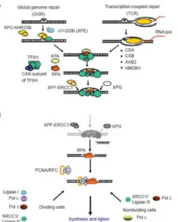
Ageing has been intrinsically associated with accumulation of macromolecular damage, particularly in DNA. Consistently, defects in DNA repair mechanisms result in accelerated ageing. Nucleotide excision repair (NER) is a versatile pathway responsible for repairing helix-distorting DNA lesions - including UV radiation-induced pyrimidine dimers - chemical adducts, and oxidative lesions. NER progresses along two sub-pathways: the global-genome NER (GG-NER) and the transcription-coupled NER (TC-NER). GG-NER removes lesions genomewide, while TC-NER repairs DNA damage that hampers the progression of the RNA polymerase II complex (RNAPIIo) and therefore concerns transcriptionally active genes. Both sub-pathways converge into a common mechanism that involves DNA unwinding, lesion verification and dual incision, followed by DNA re-synthesis and ligation.
Null and hypomorphic Ercc1 mutant mice display accelerated ageing phenotype accurately recapitulating that observed in progeroid human syndromes - which are caused by inherited defective NER - such as xeroderma pigmentosum and Cockayne syndrome. Particularly, previous investigations on hepatic tissue of Ercc1 mutant mice have revealed alterations in the expression of peroxisome proliferator-activated receptor (PPAR)-α and –γ. Changes in these genes, which are not only crucial
ii
Collectively, these findings emphasize the involvement of peroxisomes in ageing processes and provided the rationale for my PhD project. This aimed at exploring mutual relationship between peroxisomes and DNA damage, clarifying whether: (i) genomic instability affects peroxisomal function and/or biogenesis, and (ii) modulating peroxisomal function/biogenesis may influence cellular response to exogenous genotoxic insult.
The first part of my PhD project focussed on possible peroxisomal alterations in a progeroid condition generated by Ercc1 deficiency. To this end, we performed molecular and morphological experiments in the liver tissue of wild type and Ercc1Δ/- to evaluate the expression of peroxisomal membrane and matrix markers.
Relevantly, several important pathways seem to be dysregulated, in particular those related to ROS and lipid metabolism, and the variation of specific peroxisomal proteins emphasized that some alterations actually involved peroxisomes. Additionally, ultrastructural analyses highlighted abnormalities in the cellular organization of mutants in respect to wild type, especially concerning mitochondrial membrane system. This interesting finding is in line with the compelling evidence establishing a close evolutionary, functional, and biogenetic link between peroxisomes and mitochondria.
Furthermore, peroxisomal changes were investigated even in the brain, where the expression of peroxisomal proteins varies among specific neuronal populations. Defective DNA repair was demonstrated to also influence brain peroxisomes, in a non-linear fashion, since changes observed in mild vs. severe mutants can consist in either induction or repression of peroxisomal functions, also depending on the considered cerebral area.
As a conclusion for this part of my work, we may assert that defects in NER impact peroxisomal metabolism not only in the liver of mutants mice, but even in their brain, where their importance is still dimly understood and that we were able to shed light on the unexplored role of peroxisomes in accelerated ageing process.
iii
human dermal fibroblasts (CHDF) and neuroblastoma cells (SH-SY5Y). In view of a flipped experimental procedure, we have begun modulating the dynamic feature of peroxisomes, by pre-treating cells with fenofibrate (FF), a peroxisome proliferator mainly employed as a hypolipidemic drug. Subsequently, cells underwent exogenous DNA injury (UVC radiation). FF is able to influence the expression levels of peroxisomal proteins. This effect was observed in all cell types considered, and generally dose-dependent. Interestingly, UV radiation enhances the response to the drug, probably exacerbating microenvironmental stress conditions, which promote cellular response to the damaging insult.
More specifically, FF impacts NER capacity of the cells modulating both the unscheduled DNA synthesis (UDS) and transcription recovery. Noteworthy, cells, because of the combination of a double induced-stress condition, responded varying their own redox status according to the dose of the drug administered.
Definitively, our data provide a consistent depiction of how treatment with FF induces changes in damaged cells. Yet FF affects cellular metabolism depending on cell type considered and dose administered. Particularly, in non-neuronal, dividing, primary and secondary cells FF improves DNA repair; conversely in differentiated post-mitotic neuronal cells (retinoic acid treated SH-SY5Y) FF impairs DNA repair. The effect is dichotomous and may, or may not, involve PPARα activation, thus further studies will address the reasons of such differences.
In conclusion, data collected during my three-year PhD project strongly argue for a reciprocal modulation of the organelles and DNA damage repair mechanisms, particularly in accelerated ageing, either endogenously generated, or exogenously produced. These findings, shedding new light into the relationship linking cell senescence with peroxisomal functions and biogenesis, open the way to future studies aiming at investigating potential protective strategies against age-related processes.
iv
svolgono un ruolo cruciale in numerose funzioni anaboliche e cataboliche principalmente correlate al metabolismo lipidico e delle specie reattive dell’ossigeno (ROS). Anomalie perossisomiali che coinvolgono prevalentemente il processo di biogenesi e di alcuni funzioni metaboliche sono spesso causa dell’insorgenza di uno spettro di disordini caratterizzati da difetti nello sviluppo dell’organismo e di molti organi come fegato e cervello. Una ulteriore caratteristica dei perossisomi è quella di essere estremamente eterogenei a seconda del tipo cellulare considerato e sono, inoltre, versatili poiché capaci di adattare e modificare la loro forma, la dimensione e il contenuto enzimatico alle necessità richieste dalla cellula contribuendo in modo essenziale al mantenimento dell’omeostasi cellulare. Essendo, quindi, dei regolatori cellulari e preservando alcune funzioni delle cellule, i perossisomi sembrano essere coinvolti nel processo di invecchiamento. Infatti, è stato visto che durante l’invecchiamento fisiologico l’organizzazione interna di questi organelli è compromessa e danneggiata, e, inoltre, la ridotta attività della catalasi è associata con la manifestazione di eventi tipici di un processo di invecchiamento accelerato. Durante l’invecchiamento molte macromolecole, incluso il DNA, e diversi meccanismi, quali i sistemi di riparazione del DNA, sono sottoposti a continui insulti che ne danneggiano la loro funzionalità. Il sistema di riparazione per escissione di nucleotidi (NER) è un pathway molto versatile che viene attivato per riparare lesioni che distorcono la doppia elica del DNA e che sono causate da radiazioni UV, additivi chimici e da lesioni ossidative. Il NER è costituito da due subpathways global-genome NER (GG-NER) e il transcription-coupled NER (TC-NER) che convergono in unico meccanismo comune nella fase finale del processo riguardante lo svolgimento della molecola, il riconoscimento della lesione e la doppia incisione sul filamento, seguita poi dalla sintesi ex novo del DNA e infine richiede l’intervento di una ligasi. Attraverso il GG-NER vengono rimosse le lesioni che si estendono largamente sul genoma, mentre il TC-NER è coinvolto nella riparazione delle lesioni che ostacolano la progressione del complesso della RNA polimerasi II (RNAPIIo) and di conseguenza ha un effetto sul meccanismo di trascrizione dei geni.
I topi mutanti Ercc1, sia knockout che ipomorfici, sono fenotipicamente caratterizzati da un invecchiamento accelerato e presentano i tratti tipici identificati nelle sindromi progeroidi che colpiscono l’uomo – causate da un sistema NER difettivo – quali lo xeroderma pigmentosum e la malattia di Cockayne. In particolare, esperimenti condotti sul tessuto epatico dei topi
v
della regolazione della biogenesi e delle funzioni perossisomiali, ma più in generale del metabolismo cellulare, sono stati inseriti in un contesto di bioenergetica.
Collettivamente, questi dati enfatizzano il ruolo dei perossisomi nell’invecchiamento e forniscono un valido supporto per il mio progetto di ricerca che è finalizzato a esplorare il potenziale nesso tra i perossisomi e il danno al DNA. Abbiamo, quindi, voluto chiarire se (i) l’instabilità genomica influenza le funzioni perossisomiali e/o la loro biogenesi, e (ii) la modulazione della funzione/biogenesi perossisomiale può influenzare la risposta cellulare in seguito ad uno stimolo genotossico.
Nella prima parte del progetto di dottorato abbiamo valutato le possibili alterazioni perossisomiali, mediante tecniche morfologiche e molecolari esaminando l’espressione di markers perossisomiali, nei topi Ercc1 NER-deficienti.
Abbiamo notato che in questi topi diversi e importanti pathway, in particolare quelli relativi al metabolismo dei lipidi delle ROS, e la variazione di specifiche proteine perossisomiali hanno suggerito che effettivamente esistono delle alterazioni che interessano direttamente i perossisomi. I dati di microscopia elettronica hanno, inoltre, messo in evidenza anomalie a livello dell’organizzazione cellulare, particolarmente riconoscibili a livello del sistema di membrana dei mitocondri nei topi mutanti rispetto ai topi sani. Tuttavia, queste evidenze strutturali sono in linea con la letteratura che dimostra il stretto legame tra perossisomi e mitocondri da un punto di vista evoluzionistico, di biogenesi e funzionale. Successivamente, abbiamo rivolto la nostra attenzione al tessuto nervoso dello stesso modello murino analizzando diverse popolazioni neuronali dove l’espressione di proteine perossisomiali risulta cambiare notevolmente. Il NER difettoso sembra ugualmente influenzare i perossisomi presenti nel cervello, in modo non lineare, poiché l’espressione delle proteine varia a seconda della severità del fenotipo considerato e anche delle diverse aree cerebrali prese in esame.
Per questa prima parte del progetto possiamo supporre che il sistema NER difettoso può avere un effetto sul metabolismo perossisomale non solo a nel fegato ma anche nel cervello, dove l’importanza perossisomiale è ancora scarsamente conosciuta e abbiamo, inoltre, messo in evidenza un ruolo dei perossisomi non ancora esplicitato.
Nella seconda parte dello studio abbiamo ulteriormente esplorato il legame tra l’accumulo del danno al DNA e i perossisomi, utilizzando come modello
vi
perossisomiale principalmente utilizzato come un farmaco ipolipidemico. Successivamente, le cellule sono state sottoposte alle radiazioni UV inducendo un danno al DNA. Il FF influenza l’espressione delle proteine in tutti tipi cellulari considerati e più generalmente in modo dose dipendente. In modo interessante, abbiamo notato che apparentemente le radiazioni UV potenziano la risposta cellulare al farmaco, probabilmente incrementando una condizione di stress che promuove una risposta da parte delle cellule quando subiscono un danno.
Specificamente, il FF ha un effetto sulla capacità di attivazione del NER modulando sia la sintesi del DNA indipendente dalla fase S del ciclo cellulare e la trascrizione dell’RNA. Le cellule, inoltre, in una situazione di stress sembrano modificare anche il loro stato redox variandolo in base alla dose di FF somministrata.
I nostri dati dimostrano che il trattamento con il FF induce cambiamenti nelle cellule danneggiate. Ancora, il FF influenza il metabolismo cellulare in modo cellulo- e dose-dipendente. In particolare, nelle cellula in piena attività replicativa il FF influenza positivamente la capacità di riparare il DNA, al contrario, nelle cellule neuronali differenziate (SH-SY5Y trattate con acido retinoico) il FF sembra peggiorare la capacità di riparazione del DNA. L’effetto del FF è dicotomico e potrebbe essere o non essere dipendente dall’attivazione del PPARα, quindi successivi studi saranno necessari per chiarire la ragione di tali differenze.
In conclusione, i dati ottenuti nei tre anni di dottorato supportano fermamente una reciproca modulazione degli organelli e del meccanismo di riparazione di danno al DNA, particolarmente nell’invecchiamento accelerato. I nostri risultati, rilevanti il legame tra la senescenza e le funzioni perossisomiali, sono terreno fertile per studi futuri finalizzati ad identificare potenziali strategie terapeutiche per processi età-correlati.
BER (base-excision repair) Cb (Cerebellar cortex)
CHDF (human dermal fibroblasts)
COFS (Cerebro-oculo-facio-skeletal syndrome)
CS Cockayne syndrome ICL (DNA interstrand crosslink) DA (dopaminergic neurons)
FF (fenofibrate)
FIB-SEM (focusing ion-beam-scanning electron microscopy). GG-NER (global genome nucleotide excision repair)
GSEA (Gene Set Enrichment Analysis) Lc (locus coeruleus)
NER nucleotide-excision repair PD (Parkinson disease) Peroxin (PEX)
PMPs (peroxisomal matrix proteins)
PPAR (peroxisome proliferator-activated receptor) PUFAs (polyunsaturated fatty acids)
RA (retinoic acid)
ROS (reactive oxygen species)
RNAPIIo (RNA polymerase II complex) SH-SY5Y (neuroblastoma cells) SNpc (Substantia nigra pars compacta)
TC-NER (transcription-coupled nucleotide excision repair) TEM (transmission electron microscopy)
UDS (unscheduled DNA synthesis) VLCFAs (very long chain fatty acids) WB (western blot)
WT (wild type)
S
ECTION
I
Introduction
And
Objectives
2
Chapter 1
Peroxisomes: an overview
Peroxisomes are ubiquitous cytoplasmic organelles present in a wide variety of eukaryotic cells, from yeast to humans (Rucktäschel el al., 2010). They display roughly spherical shape (0.1-1µm diameter), and a single-limiting membrane surrounding a finely granular matrix. These organelles show a high heterogeneity with respect to morphology, protein content, and abundance in diverse tissues and during developmental processes, including ageing (Wanders et al., 2015).
Peroxisomes are also able to respond to environmental changes and extracellular stimuli by altering their enzyme content, morphology and abundance. The central role of peroxisomes in cell metabolism has emerged since their discovery, since they are involved in a wide range of anabolic and catabolic functions (De Duve and Baudhuin, 1966). Concerning the essential role of peroxisome in the maintenance of cell homeostasis, it is not negligible the contribution of peroxisome to ageing, indeed
d
uring normal ageing, peroxisomal functions are dramatically compromised and several enzymes show decreased activity and/or progressively mislocalize to the cytosol (Koepke et al., 2008; Fransen et al., 2013). In particular, decline in the peroxisomal protein catalase has been associated with both natural and accelerated ageing (Titorenko and Terlecky, 2011).1.1 Peroxisome biogenesis
Even though a large body of work has been done dealing with peroxisomes, their biogenesis results to be a matter of debate. Particularly albeit posttranslational targeting of peroxisomal matrix proteins (PMPs) to peroxisomes in vivo and in vitro is well documented, the mechanisms of insertion remains unclear (Ma et al., 2011).
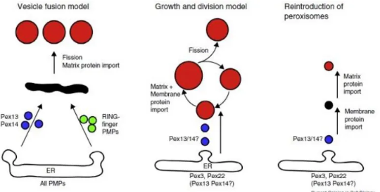
As peroxisomes neither contain DNA nor transcription/translation machineries, all peroxisomal proteins are encoded by the nuclear genome, synthesized on free polyribosomes in the cytosol and then imported post-translationally into preexisting peroxisomes (Lazarow and Fujiki, 1985; Rucktäschel el al., 2010). This concept, supporting the growth and division theory, asserts that peroxisomes are autonomous organelles as mitochondria and chloroplasts and that the endoplasmic reticulum (ER) is only a source of membrane lipids for enlargement of the organelles. This is however challenged by the de novo theory, emphasizing the crucial role of the ER in peroxisome generation (Hettema et al., 2014).
3
Peroxisome may receive newly synthesized membrane and matrix proteins and lipids from the ER via vesicular transport. Furthermore, recent evidence proposed that peroxisomal membranes derive from the ER via budding of vesicles containing PMPs (Fig. 1.1).
Moreover, differently from other organelles, peroxisomes can transport cargoes in a folded, cofactor-bound, and/or oligomeric state (Ma et al., 2011). The targeting of matrix proteins depends on two distinct peroxisomal targeting signals: PTS1 and PTS2. Peroxin 5 and 14 (Pex5, Pex14) controlling the assembly, inheritance and division of peroxisomes, can be considered component of the minimal translocon and both proteins have been proposed to form transient pores. Given their importance, mutations in Pex genes cause severe pathologies like the Zellweger Spectrum Syndrome (ZSS) (Ma el al., 2011).
1.2 Peroxisome metabolism
Peroxisomes are involved in a wide variety of anabolic and catabolic functions, including ROS metabolism (Schrader and Fahimi, 2006), β-oxidation of very long chain fatty acids (VLCFAs), biosynthesis of polyunsaturated fatty acids (PUFAs) and plasmalogens, cholesterol and dolichol biosynthesis (Fransen et al., 2014), and calcium homeostasis (Drago et al., 2008).
Although mitochondria have been described as the major source of endogenous ROS generation, peroxisomes have emerged as central
Figure 1.1. Schematic representation of models for peroxisome multiplication. (On
the left de novo model; in the middle the description of growth and division theory and on the right the reintroduction of peroxisomes) (Hettema et al., 2014).
4
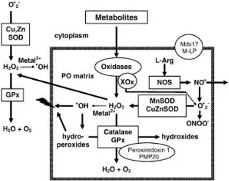
Figure 1.2. Overview of peroxisomal
ROS metabolism. H2O2 is produced
by several peroxisomal oxidases including XOx, and decomposed by
catalase and GPx, or converted to
hydroxyl radicals (-OH). Hydroxyl radicals can damage the peroxisomal membrane by lipid peroxidation of unsaturated fatty acids. Peroxisomal oxidases also generate superoxide anions (O2-) that are scavenged by
MnSOD and CuZnSOD. (Schrader and Fahimi, 2006)
organelles playing a key role in both production and scavenging of ROS (Fig 1.2) (Schrader and Fahimi, 2006).
This dual action derives from the fact that peroxisomes harbour both several H2O2-generating oxidases and antioxidant enzymes.
Peroxisomal ROS-scavenging enzymes include: (i) catalase (Cat), which converts H2O2 into water through either the catalitic or the peroxidatic
reaction; (ii) glutathione peroxidase (GPx), catalysing H2O2 removal with
concomitant conversion of reduced glutathione (GSH) to glutathione disulfide (GSSG); (iii) copper zinc superoxide dismutase (CuZnSOD, SOD1) and manganese superoxide dismutase (MnSOD, SOD2), catalysing superoxide anion conversion into H2O2. While Cat is bona fide peroxisomal,
all the other ROS-detoxifying enzymes are also present in other cell compartments. Oxidative balance needs to be tightly regulated; shifting this equilibrium via endo- or exogenous factors, ageing or disease conditions leads to a deregulation of the system, causing oxidative stress (Bonekamp et al., 2009).
The peroxisomal functions related to lipid metabolism have been extensively reviewed by Wanders and colleagues (2006) (Fig 1.3). The architecture of the peroxisomal β-oxidation system involves a set of four consecutive reactions where 2-carbon unit is split from each fatty acid in the form of an acetyl-CoA unit, which can then be degraded in the citric acid (Krebs) cycle to produce O2 and H2O (Wanders et al., 2015). Substrates for
peroxisomal β-oxidation, can enter the organelle through passive diffusion or by active transport mediated by different ATP-binding cassette (ABC) transporters belonging to subclass D. The ABCD subfamily contains four half-transporters of which three are localized in peroxisomes, including HsABCD1 (ALDP, adrenoleukodystrophy protein), HsABCD2 (ALDRP, adrenoleukodystrophy-related protein) and HsABCD3 (PMP70).
5
The ABCD transporter binds acyl-CoA, which is then hydrolyzed prior to or during transport. The free fatty acid is then delivered to the luminal side of the peroxisomal membrane where it is re-esterified by different peroxisomal acyl-CoA synthetases followed by oxidation of the resulting acyl- CoA by the peroxisomal β- oxidation machinery (van Roermund et al., 2014).
Interestingly, the transcriptional activation of genes involved in fatty acid oxidation in liver of rats and mice is regulated by α isotype of the peroxisome proliferator activated receptor (PPARα) (Reddy and Hashimoto, 2001).
1.3 Peroxisome Proliferator -Activated Receptors (PPARs)
Transcription factor families participate in metabolic regulation and contribute to the complex fine-tuning of gene activity required for the organism to adapt to changing conditions.
Peroxisome proliferator-activated receptors (PPARs), which were the first nuclear receptors identified as metabolic “sensors”, play a major role in energy homeostasis, lipid and lipoprotein metabolism, cell proliferation, death and differentiation. Through the years, the key role of PPARs as regulators of inflammatory and immune responses has been established, making them possible target for the treatment of chronic inflammatory diseases, diabetes, cancer and neurodegenerative disorders (Fidaleo et al., 2014). The three PPAR isotypes, α (NR1C1), β/δ (NR1C2) and γ (NR1C3), share the property of being lipid-activated transcription factors. Noteworthy, in addition to transcriptionally regulating genes involved in pivotal cellular functions, Aleshin and collegues (2013) introduced the “triad concept” demonstrating that PPARs control their own expression, by enhancing or repressing each other, so that the cellular response will depend on the
Figure 1.3 Schematic representation of
the peroxisomal β-oxidation pathway in humans. (Wanders and Waterham, 2006)
6
relative proportions of each PPAR isotype. This scenario has recently been synthesized by in the "triad" concept .As sensors, PPARs adapt gene expression to various lipid signals and other cues. The great diversity of functions in which they are
implicated parallels the large panel of ligands that can be accommodated in the PPAR ligand-binding pocket. The prevalent point of view is that PPARs translate modifications in the intracellular levels of these natural compounds into changes in metabolic activities, and other processes (Montagner et al., 2011).
1.3.1 PPARα and its agonists
PPARα acts primarily to regulate energy homoeostasis through its ability to stimulate the breakdown of fatty acids and cholesterol, driving gluconeogenesis and reducing serum triglyceride levels (Fidaleo et al., 2014).
Some of the key genes involved in fatty acid β-oxidation systems possess PPRE elements and are regulated by PPARα, though PPARβ/δ has also been shown to regulate some of these enzymes (Aleshin and Reiser, 2013). PPARα is known to be important for peroxisome proliferation; regulating their abundance in liver (Schrader et al., 2015). Its activation induces expression of Pex11, which is involved in peroxisomal biogenesis by promoting peroxisome division (Lodhi and Semenkovich, 2014). However, the transcriptional co-activator PGC1-α, promotes cold-induced peroxisome biogenesis in brown adipose tissue in a PPARα independent manner, implicating PPARγ or PPARβ in this effect (Bagattin et al., 2010). Recently, it was found that PPARγ promotes peroxisomal biogenesis in adipocytes. Collectively these data suggest that PPARs coordinate both peroxisome biogenesis and integrative lipid metabolism (Lodhi and Semenkovich, 2014).
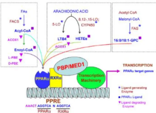
PPARα ligands fall into two general categories: (i) synthetic xenobiotics and (ii) biological molecules (Fig. 1.6). Synthetic ligands, referred to as peroxisome proliferators (PPs), include hypolipidemic drugs such as clofibrate, fenofibrate, gemfibrozil, bezafibrate, ciprofibrate, nafenopin,
Figure 1.4. Cellular and metabolic pathways
7
Figure 1.6. Biological ligands of
PPARα. Diagram illustrating different known biological ligands of PPARα. The PPARα and RXRα heterodimer are shown bound to a PPRE sequence in the promoter of a target gene with associated coactivator proteins forming a complex with the cellular transcription machinery (Pyper et al., 2010)
methyl clofenapate, tibric acid, and Wy-14,643 (pirinixic acid) used in the treatment of dyslipidemias, and industrial phthalate-monoester plasticizers, such as di-(2-ethylhexyl)-phthalate (DEHP), and di-(2-ethylhexyl) adipate (DEHA) used in the manufacture of polyvinyl chloride plastics. Proliferation of peroxisomes in liver parenchymal cells and dramatic transcriptional activation of fatty acid oxidation system genes are the hallmarks of PP-induced pleiotropic responses in the rat and mouse liver (Pyper et al., 2010).
Activation of PPARα by its ligands also appears to influence inflammatory response and to induce DNA replication and cellular proliferation in rodent liver (Peters et al., 2005). These carcinogenic-like effects probably result from inhibition of apoptosis, increased oxidative stress and regulation of genes controlling the cell cycle even though the direct transcriptional targets of PPARα mediating these effects still remain unknown (Feige et al., 2006).
1.3.2 Fenofibrate and its action
During the last decade, it has been shown that the pharmacological activation of PPARα by synthetic PPs (fibrates and NSAIDs) is an effective therapeutic approach to different conditions, such as hypertriglyceridemia, type 2 diabetes mellitus, cancer and neuro-pathologies (Fidaleo et al., 2014). Fenofibrate (FF) is a fibric acid derivative which, through the activation of PPARα, plays an important role in lowering the levels of serum cholesterol and tryglycerides and in elevating the levels of high density lipoproteins, and is therefore used for the treatment of dyslipidemia and to ameliorate insulin resistance as well as glucose intolerance (Brunmair et al, 2004). Importantly, in neuroinflammation-related disorders, treatment with PPARα synthetic agonists, including FF, decreases neurological deficits, suppressing NF-κB pathway and reducing inflammatory mediators, such as
8
tumor necrosis factor α (TNF-α), interleukin 1β (IL-1β), and cyclooxygenase-2 (COX-2) (Fidaleo et al., 2014).
In this regard it has been observed how, in experimental stroke or traumatic brain injury, FF or WY-14,643 administration reduced brain damage, by inhibiting inflammation pathways, and by upregulating brain antioxidant enzyme activities (Deplanque et al., 2003). Chronic treatment with FF or gemfibrozil was also shown to protect brain against ischemia in mice through an increase of mRNAs and activities of superoxide-dismutases (SODs) levels in brain microvessels (Wang et al., 2010).
Moreover, an in vitro study reports that FF attenuates NO-mediated neuronal and axonal damage and increases PPARα protein levels and catalase activity (Gray et al., 2011).
Noteworthy, PPARα modulates the activity of dopaminergic neurons, demonstrating to have direct antipsychotic effects in treating disorders in which dopamine dysfunction plays a prominent role, such as schizophrenia and nicotine addiction. Indeed, one recent study demonstrated on a preclinical level that FF can reverse some of the schizophrenia-like cognitive alterations induced by a neonatal lesion in rat (Rolland et al., 2012).
In recent in vivo and in vitro studies, various members of the fibrate family, which are all agonists of PPARα, demonstrate interesting anti-cancer effects (reviewed by Wilk et al., 2015). All of these studies encouraged the use of FF as a supplemental anticancer drug, a concept supported by recent clinical trials in which chronic administration of FF along with chemotherapeutic agents used at relatively low doses minimizes the toxicity and acute side effects of chemotherapy while maintaining efficacy for patients with recurrent brain malignancies and leukemia (Wilk et al., 2015).
The primary and conventional function of FF is the activation of PPARα transcriptional activity. In this process, FF must first be converted to fenofibric acid (FA), by blood and tissue esterases. FA then binds and activates PPARα, which triggers the expression of numerous metabolic enzymes involved in fatty acid β-oxidation (Grabacka et al., 2013). In addition, activated PPARα decreases glucose uptake by repressing the insulin-dependent glucose transporter GLUT4 (Ahmed et al., 2007) and elevated oxidation of the fatty acids and ketone bodies further suppresses the expression of glycolytic enzymes (Randle, 1998).
This metabolic switch could initiate a gradual decline in energy metabolism in tumor cells, which is consistent with the fundamental observation by Otto Warburg that tumor cells are distinctly dependent on glycolysis (Warburg 1956) for both energy production and biosynthesis of intermediate metabolites (Dell’Antone, 2012).
9
Even though the mechanism underlying FF anticancer properties is still unclear, several effects are however observed such as an apoptosis promoting process through the down-regulation of Bcl-xl and Survivin, the activation of caspase 3 and the up-regulation of Bad. Particularly, FF induces cell cycle arrest at G0/G1 phase in a time -and dose- dependent
manner through the down-regulation of cyclin D1 and Cdk4 and up-regulation of p21 and p27/Kip1; activation of NF-kB pathway accompanied by up-regulation of phosphor-IKα/β and IKKα, responsible for the phosphorylation of IKβα and its subsequent ubiquitination and degradation to release NF-kB in order to allow it to translocate to the nucleus and promote the transcription of target genes (Li et al., 2014).
To explain these unordinary activities of FF, it has been conjectured that the latter may also act in a PPARα-independent manner. In this regard, FF was shown to alter the expression of growth differentiation factor 15 (Araki et al., 2009); affect cell membrane fluidity in a manner similar to that of cholesterol (Gamerdinger et al., 2007); and interfere with the respiratory function of isolated liver and heart mitochondria (Nadanaciva et al., 2007). Noteworthy, Wilk and colleagues (2015) have been able to observe that FF, but not its PPARα-active metabolite fenofibric acid , accumulates in the mitochondrial fraction of human glioblastoma cells, resulting in a severe inhibition of mitochondrial respiration.
FF seems to affect mitochondrial respiration particularly by inhibiting the catalytic activity of complex I of the respiratory chain. This has been shown in isolated mitochondria that, when exposed to fenofibrate and provided with substrates for complex I, exhibited a markedly reduced rate of state 3 respiration (Brunmair et al, 2004).
In regards to its anti-cancer effects, Wilk and collaborators (2015) also noted that different concentrations of FF exert different actions on growth/viability of primary glial tumor cells and glioblastoma cell lines. Specifically, while low doses of the drug result in cell cycle arrest, not accompanied by substantial induction of apoptosis, high fibrate doses cause massive delayed apoptotic cell death, preceded by FoxO3A nuclear accumulation and expression of FoxO-dependent apoptotic protein, Bim. These properties of fenofibrate, coupled with its low systemic toxicity, make it a good candidate in support of conventional therapies against glial tumors.
The PPAR-α independent anti-proliferation and apoptosis-inducing effects of FF were reported in B-cell lymphoma, prostate cancer, hepatocellular carcinoma, mantle cell lymphoma, endometrial cancer and triple-negative breast cancer cells. However, the PPAR-α dependent mechanisms were used to explain the anti-cancer effects of FF in glioma, glioblastoma and
10
melanoma. This paradoxical phenomenon might be due to the differences in tumor types or experimental conditions (Li et al., 2014).
11
Chapter 2
DNA Damage and Genome Instability
2.1 DNA damage response
Within the complex chemical machinery of each cell, all biomolecules are subject to indiscriminate damage caused by spontaneous reactions and by numerous endogenous and exogenous reactive agents. Whereas all other molecules are renewable, nuclear DNA, which represents, with the exception of a few mitochondrial genes, the entire genetic information of a cell, is irreplaceable (Garinis et al., 2008).
Therefore, the long DNA molecules that compose this genetic material are not stable but constantly attacked by a large variety of genotoxic insults. It has indeed been estimated that, in each cell, tens of thousands of damaging events occur on a daily basis (Lindahl, 2013).
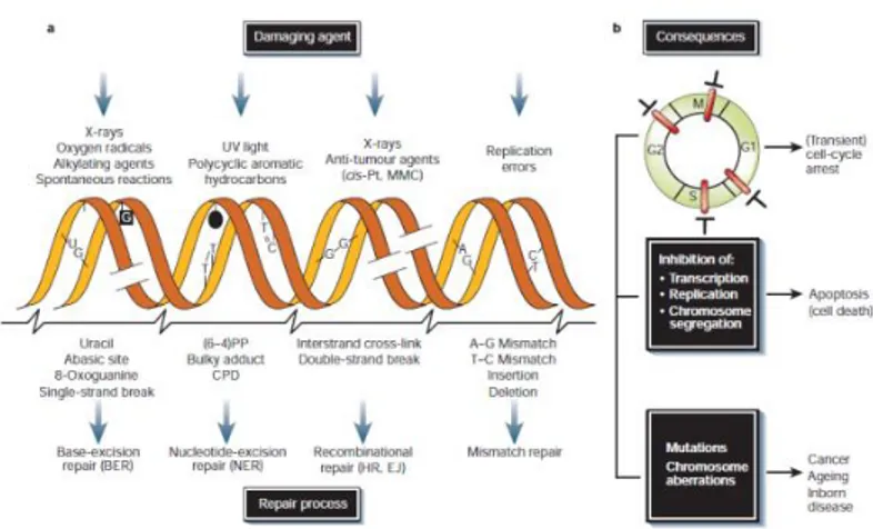
These genotoxic insults can stem from a large variety of both endogenous and exogenous sources such as ultraviolet (UV), ionizing radiations (IR), and genotoxic chemicals, whilst cellular metabolism, on the other hand, can produce ROS and their numerous subsequent reaction products like lipid peroxidation products (De Bont and van Larebeke, 2004). The consequences to DNA injury are generally unfavorable and determined by various parameters, the first of which is the type of damage. Some lesions are primarily mutagenic, greatly promoting cancer, whilst others are mainly cytotoxic or cytostatic, triggering cell death or senescence, causing degenerative changes such as those associated with ageing (Garinis et al., 2008).
Depending on the inflicted lesion cells have heavily invested in an intricate genome maintenance apparatus (Fig. 2.1), relying on the integrity of the somatic genome, which must be preserved during the entire lifetime of an organism (Hoeijmakers, 2001). Although the repair of different types of DNA lesions relies on different sets of proteins, the various forms of DNA damage nevertheless trigger common signal transduction pathways, which collectively go under the name of DNA damage response (DDR). On the basis of the severity of the damage a series of events are activated triggering the full DDR, the slowing or arrest of cell-cycle progression, as a result of those DNA damage checkpoints, which delay cell-cycle transitions until repair has occurred (Rouse and Jackson, 2002), through the action of sensors, transducers, and effectors, orchestrates the appropriate repair of DNA damage and resolution of DNA replication problems, coordinating these processes with ongoing cellular physiology.
12
2.2 DNA Repair systems: The Nucleotide Excision Repair (NER)
Because the problem of DNA damage has existed ab initio, DNA repair systems must have arisen early in evolution. This explains why all known repair pathways are highly conserved. Five main, partly overlapping, damage repair pathways operate in mammals: nucleotide-excision repair (NER), base-excision repair (BER), mismatch repair, homologous recombination and end joining (Hoeijmakers, 2001).
NER is a multistep ‘cut and patch’ process that recognizes and eliminates a wide spectrum of damage causing significant distortions in the DNA structure, such as UV-induced damage and bulky chemical adducts. The eukaryotic NER eliminates DNA damage by the excision of 24-32 nt single strand oligonucleotides from a damaged strand and the process involves the coordinated action of approximately 30 proteins (Volker, et al., 2001). NER consists of two pathways distinct in terms of initial damage recognition: global genome nucleotide excision repair (GG-NER) and transcription-coupled nucleotide excision repair (TC-NER) (Fig. 2.7).
13
2.2.1 GG-NER
GG-NER removes lesions genome wide, while TC-NER repairs DNA damage that hampers the progression of the RNA polymerase II complex (RNAPIIo) and therefore concerns transcriptionally active genes (Fousteri et al., 2008; Dijk et al., 2014). Both sub-pathways converge into a common mechanism that involves DNA unwinding, lesion verification, and dual incision, followed by DNA re-synthesis and ligation (Hoeijmakers, 2001). In GG-NER, XPC permanently scans the genome DNA in search of damage, in an 'association-dissociation' scanning mode, with the formation of a plethora of short-lived complexes. More stable XPC-DNA complexes are formed when XPC collides with damaged sites. Furthermore, XPC is usually exported from the nucleus and imported back; such an exchange, in the absence of damage, maintains the stationary level of its concentration, preventing redundant DNA probing that may interfere with other processes of nucleic metabolism. Under any effects on cells resulting in DNA damage, the rate of XPC transport to the cell decreases and XPC accumulates in the nucleus, which facilitates the rapid response of the repair system to genotoxic affection (Petruseva et al., 2014).
Within a cell, XPC exists as the heterotrimeric complex XPC-HR23B-Cen2. HR23B stabilizes the complex, protects it against proteasome degradation,
Figure 2.7. Model for mechanism of GG-NER and TC-NER. (Fousteri and
14
and stimulates the DNA-binding activity of XPC. Centrin-2, on the other hand, is believed to increase the stability, control affinity/selectivity of DNA binding by the XPC-HR23B dimer. Once the damage is recognized, XPC binds the repair/transcription factor TFIIH and facilitates its interaction with the lesioned DNA. TFIIH factor is a multisubunit complex composed of two helicases, XPB and XPD; enzymatic activity-free proteins, p62, p52, p44, p34 and p8; and the complex of CDK-activating kinase, CAK (cyclin H, Cdk7 and Mat1). Its core proteins form a slightly elongated ring-shaped structure with a hole of a diameter sufficient to enclose a double-stranded DNA helix (2.6-3.4 nm) (Schultz et al., 2000). XPC-dependent recruitment of TFIIH to the damage is mainly controlled by direct contact of XPC with XPB and p62 subunit. The TFIIH annular structure encompasses the dsDNA on the 5' side of the damage, releasing a kinase sub-complex.
Uncoiling of a DNA double helix around the damage catalyzed by two specialized helicases, XPB (3'-5') and XPD (5'-3'), is the most obvious result of TFIIH binding. This unwinding process separates the two DNA filaments and generates two short single strand stretches, which facilitate the recruitment of a further complex, composed by XPA, which has affinity for chemically altered DNA, and the ssDNA binding protein RPA1 which binds the non-damaged strand (Fanning et al., 2006). XPA, therefore protects DNA from illegitimate degradation and facilitates accurate positioning of XPG and Ercc1-XPF endonucleases (De Laat et al., 1998). Factor XPF is a structure-specific endonuclease that catalyzes incision of DNA at the site of the ssDNA/dsDNA junction on the 5' side of the damage and functions in NER within a heterodimer with the Ercc1 protein. The Ercc1-XPF heterodimer is involved into the complex through the ERCC1-XPA interaction and breaks the damaged strand on the 5' side of the damaged site. Both subunits contain a helix-hairpin-helix (HhH) motif required for the formation of a heterodimer near the C-ends (Sepe et al., 2013). The central domain of Ercc1, consisting of a groove containing the basic and aromatic residues, is the fragment that, through its interaction with XPA, connects Ercc1-XPF to other NER machineries (Tsodikov et al., 2007).
The nuclease domain of XPF comes into contact with the damaged DNA strand, while the XPF and Ercc1 HhH domains come into contact with the undamaged strand.
In the presence of catalytically inactive XPG, Ercc1-XPF catalyzes 5'-incision (15-25 nucleotides away from the damage) and forms an unbound 3' hydroxyl group required for the initiation of the repair synthesis and emergence of the mobile single-stranded fragment containing the damage. The changes in the structure of the protein-nucleic complex allow an XPG
15
to exhibit catalytic activity. 3'-incision of DNA (3-9 nucleotides from damage) completes the process of damaged site excision (Hoeijmakers, 2001).
Repair synthesis and DNA ligation are performed by the enzymes and protein factors that also participate in DNA replication.
The gap is filled by de novo synthesis of DNA by DNA-polymerase complexes that include polymerase δ, κ_, and _ε. These enzymes are recruited by the PCNA clamp in association with factors that are specific for the polymerase type. Pol_ is recruited by RPA, the clamp loader RCF, and p66, while pol_ requires the CTF18-RCF clamp loader. Pol_ is instead recruited by ubiquitinated PCNA and XRCC1. The final step is DNA ligation, which can be performed by two different enzymes. DNA ligase 1 operates exclusively during the S phase of the cell cycle, while DNA ligase IIIa XRCC1 complex operates throughout the whole cell cycle.
Most of NER substrates cannot cause as dramatic structural and thermodynamic alterations of dsDNA as double-strand breaks and interstrand crosslinks (ICLs); therefore, the detection of these damages is particularly challenging for a cell, and can be solved only through highly sensitive recognition. In the case of TC-NER, it is transcribing RNA polymerase II, stopped by damage, the sensor for the initial damage recognition; in GG-NER the same function is carried out by the complexes of the XPC factor (Petruseva et al., 2014).
2.2.2 TC-NER
The cytotoxic threat of blocked transcription is normally counteracted by TC-NER that allows accelerated repair of transcription-blocking damages and rapid resumption of transcription. A damage stalled RNAPII molecule is the initiator of TC-NER in which the CS factors (CSA and CSB) play an essential, though partly defined role (Hanawalt and Spivak, 2008).
It has been demonstrated that in human cells RNAPII remains on the damaged chromatin in close proximity to the TC-NER machinery (Fousteri et al., 2006). CSB is the key factor, essential for functional RNAPII/TC-NER complex assembly. Initially, a stable RNAPII/CSB complex is formed, followed by the assembly of NER pre-incision factors TFIIH, RPA, XPA and the two structure specific endonucleases XPG and XPF/ERCC1 to the damage site. This pre-incision TC-NER complex creates a stable open bubble structure surrounding the lesion and excises the damaged DNA fragment.
In, contrast to CSB, CSA is not required for the assembly of NER pre-incision factors TFIIH, XPA, RPA and XPF/ERCC1. It is generally
16
accepted that the same proteins involved in GG-NER perform the post-incision step in TC-NER as well (Lagerwerf et al., 2011).
2.3 DNA damage and ageing: NER deficiencies
The almost exclusive link between an extending class of syndromes with phenotypes resembling accelerated ageing in many, but not all, organs and tissues, and inborn defects in DNA metabolism, points to genomic damage as a major culprit in the ageing process (Garinis et al., 2008).
Additionally, the idea of a double-edged sword of DNA damage (damage-induced mutations causing cancer and damage-triggered cell death, senescence, malfunction contributing to degenerative forms of ageing), is consistent with the phenotypes of DNA repair and genome instability disorders and a growing list of mouse mutants deficient in DNA repair mechanisms (Campisi, 2005).
The overall picture emerging from these mutants is that genetic defects in DNA repair systems that mainly prevent mutagenesis are generally associated with a strong predisposition to specific types of cancer, with only minor symptoms of degenerative ageing phenotypes such as in xeroderma pigmentosum (XP) patients. On the other hand, deficiencies in repair and surveillance pathways that mainly protect from the cytotoxic and cytostatic effects of DNA damage tend to be characterized by a decrease in the incidence of cancer and premature appearance of some, but not all degenerative ageing phenotypes, such as that of Cockayne syndromes (CS) patients (Garinis et al., 2008).
Intriguingly, even though any mutation in NER genes confers cellular UV hypersensitivity, the pathological consequences of mutations affecting TC-NER or GG-TC-NER are fundamentally different (Hanawalt and Spivak, 2008). Impairment in GG-NER in humans causes XP, characterized by pigmentation abnormalities, photosensitivity, skin atrophy, and an increase of more than 1000-fold in the susceptibility to sun-induced skin cancer. This is explained by the fact that compromised GG-NER leads to accumulation of DNA lesions over the entire genome and with replication, which increases the risk of mutations (Di Giovanna and Kraemer, 2012).
On the other hand, defects in TC-NER genes CSA and CSB, are associated with the human progeroid disorder CS. This condition and associated mouse models show many symptoms of premature ageing, including progressive neurodevelopmental delay, ataxia, cachexia, kyphosis, retinal degeneration, deafness and photosensitivity, not developing skin cancer (Hoeijmakers, 2001). This is explained by the fact that TC-NER repairs only a small but vital part of the genome (the transcribed strand of active genes), and is
17
therefore not crucial for preventing mutations and thus cancer. Yet, it is crucial for promoting cell survival after DNA damage, as it enables resumption of the essential process of transcription. In a TC-NER mutant the balance between anti-ageing and anti-cancer genome maintenance responses is shifted to the latter, favoring cell death, thus protecting from cancer (Dolle et al., 2006).
Mutations in CSA and CSB can also cause the more severe CS type II as well as Cerebro-oculo-facio-skeletal syndrome (COFS) that is diagnosed at birth with craniofacial and skeletal abnormalities, severely reduced muscle tone, and impairment of reflexes (Wolters and Schumacher, 2013).
XFE is a distinct progeroid syndrome caused by a defect in XPF-ERCC1, the endonuclease required for NER as well as for DNA interstrand crosslink (ICL) repair. Failing defense against such spontaneous lesions triggers cell death and senescence, culminating in accelerated ageing (Niedernhofer et al., 2006).
Other repair systems, such as base excision repair, homologous recombination and end joining, probably perform both roles, they protect from cancer and ageing to different degrees. Therefore, most defects in distinct DNA repair systems can trigger cancer, ageing or both, revealing a fine-tuning among genome maintenance mechanisms that mainly protect from cancer, and those that predominantly prevent non-cancer, degenerative ageing phenotypes (Garinis et al., 2008).
2.4 Deficiency of Ercc1/XPF complex in a mouse model
Another critical piece of evidence indicating that Ercc1-XPF has functions distinct from NER is that Ercc1 and XPF knockout mice exhibit a much more severe phenotype than XPA null mice, which are completely deficient in NER. These mice die in the 4th week of life with ageing-like degenerative changes, including osteoporosis, neurodegeneration, bone marrow hypoplasia, epidermal atrophy, sarcopenia, and liver and kidney dysfunction (Gregg et al., 2011).
The knockout mice were generated by two different laboratories. interrupting different exons of the gene. The result was a truncation in the helix-hairpin-helix motif required for interaction with XPF.
Furthermore to probe the DNA repair function of Ercc1 in vivo, animals with mutant allele were made engineering a premature stop codon at position 292 of mErcc1. This results in a C-terminal deletion of 7 amino acids of the murine protein, including a phenylalanine residue at position 293, that does not alter the protein stability but solely modifies the affinity for binding of XPF. Homozygous Ercc1*292 (also referred to as Ercc1Δ/Δ)
18
mice live up to 6 months, which is 6X longer than Ercc1 null mice. In addition, the Ercc1Δ/- mice are healthy into adulthood (8 weeks) then begin to show numerous progressive symptoms associated with ageing. Ercc1Δ/+ mice, expressing a wild type allele besides the truncated one, do not display an overt phenotype, nonetheless present significant functional alterations, particularly in the brain.
Overall, thus far experiments on these animals have provided the strongest possible evidence that Ercc1 and XPF must function exclusively as a heterodimer. Ercc1 mutant mice led to the discovery of a new rare genetic disease (XFE progeroid syndrome) and contributed to the body of evidence that DNA damage is one type of cellular damage that promotes ageing related degenerative changes (e.g., neurodegeneration). Finally Ercc1 mouse models is unique amongst the NER-deficient mutant strains because they spontaneously develop neurodegeneration, which may be used to screen therapies for treating XP, CS and TTD patients (Gregg et al., 2011).
19
Chapter 3
Aims of the project
Peroxisomes are multipurpose organelles playing a crucial role in numerous anabolic and catabolic functions such as fatty acid α- and β-oxidation and ROS metabolism (Wanders et al., 2015; Smith and Aitchison, 2013). These organelles are remarkably heterogeneous in the different cell types and capable of modifying their own shapes and functions, depending on the cellular requests. Defects in either the biogenesis or the metabolic functions of peroxisomes result in developmental disorders, involving multiple organs (Wanders, 2015).
Because of peroxisome’s essential role for cellular homeostasis and preservation, their contribution to ageing process is receiving increasing attention (Titorenko and Terlecky, 2011). During normal ageing, peroxisomal functions are dramatically compromised and several enzymes show decreased activity and/or progressively mislocalize to the cytosol (Koepke et al 2008; Fransen et al. 2013). In particular, decline in Cat has been associated with both natural and accelerated ageing (Titorenko and Terlecky, 2011).
Ageing has been intrinsically associated with accumulation of macromolecular damage, particularly in DNA. Consistently, defects in DNA repair mechanisms result in accelerated ageing. NER is a versatile pathway responsible for correcting helix-distorting DNA lesions - including UV radiation-induced pyrimidine dimers, chemical adducts and oxidative lesions (Kamileri et al., 2012). NER progresses along two sub-pathways: the global-genome NER (GG-NER) and the transcription-coupled NER (TC-NER) (Hoeijmakers, 2001). GG-NER removes lesions genomewide, while TC-NER repairs DNA damage that hampers the progression of the RNA polymerase II complex (RNAPIIo) and therefore concerns transcriptionally active genes (Fousteri et al., 2008; Dijk et al., 2014). Both sub-pathways converge into a common mechanism that involves DNA unwinding, lesion verification and dual incision, followed by DNA re-synthesis and ligation.
The first part of the present work aims to clarify alterations in peroxisomes induced by DNA damage and their contribution to ageing, as studied in Ercc1 defective mouse models. We analyzed the accelerated ageing in Ercc1Δ/- animals which combine knockout of Ercc1 in one allele and a truncated allele. We also included in the study Ercc1Δ/+ mice, expressing a WT allele besides the truncated one (Gregg et al., 2011). Null and hypomorphic Ercc1 mutant mice display accelerated ageing phenotype accurately recapitulating that observed in progeroid human syndromes -
20
which are caused by inherited defective NER - such as xeroderma pigmentosum and Cockayne syndrome. These conditions parallel natural ageing in many respects and present systemic symptoms that include hepatic and neurological dysfunctions (Niedernhofer 2008; Niedernhofer et al., 2006; Sepe et al., 2013).
Particularly, previous investigations on hepatic tissue of Ercc1 mutant mice have revealed alterations to the expression of peroxisome proliferator-activated receptor (PPAR)-α and –γ (Niedernhofer et al., 2006). Changes in these transcription factors, which are not only crucial regulators of peroxisomal biogenesis and function, but more generally of cell metabolism, were framed in a context of bioenergetics (Niedernhofer et al., 2006). Collectively, these findings suggest that peroxisomal metabolism might be altered in progeria. Current knowledge, however, is confined to the enzyme Cat and evidence on other critical peroxisomal enzymes is not available thus far. Furthermore, peroxisomal changes in the brain, in progeria, are dimly understood.
This is why we first investigated alterations of expression in key peroxisomal genes in the liver of Ercc1∆/- progeroid mice compared to WT. We therefore performed morphological analysis also including Ercc1Δ/+ Secondly, we extended these observations to the nervous tissue, studying various brain regions, representative of different neuronal populations: locus coeruleus, substantia nigra pars compacta, and cerebellar cortex. Based on our preliminary results from this part of the study, suggesting alterations in peroxisome functions caused by genome instability, we addressed the issue of whether exogenous modulation of peroxisome population influences the cellular response to genotoxic damage. Specifically, as peroxisome proliferator we chose fenofibrate, a fibric acid derivative which, through the activation of PPARα, plays an important role in lowering the levels of serum cholesterol and tryglycerides and in elevating the levels of high density lipoproteins, and is therefore used for the treatment of dyslipidemia and to ameliorate insulin resistance as well as glucose intolerance (Brunmair et al, 2004).
To this aim, the cellular models chosen for this part of the project included commercial human dermal fibroblast cultures (CHDFs), and SH-SY5Y neuroblastoma cell - either in its undifferentiated state or treated with retinoic acid - which are largely used in Parkinson disease researches because they share many characteristics with dopaminergic neurons. pretreated with fenofibrate and submitted to UVC radiation, were analyzed for NER capacity. Also, we evaluated the redox state of the cells, in view of the involvement of reactive oxygen species in genotoxic damage.
S
ECTION
II
22
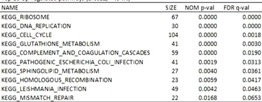
Table 4.1. List of pathways up-regulated and down-regulated in Ercc1Δ/- compared with WT mice as were calculated in GSEA. SIZE means the number of genes in each pathway, NOM p-value is the raw p value and FDR q-value is the false discovery rate adjusted for multiple comparisons.
Chapter 4
Expression of peroxisomal genes in Ercc1
Δ/-mouse liver
4.1 Bioinformatic and qPCR analyses
The connection between changes in peroxisomal metabolism and DNA damage accumulation emerged from bioinformatic studies on mutant mice harboring a mutation in NER gene ercc1(Niedernhofer et al., 2006). These knockout or Ercc1Δ/- mice display an accelerated ageing phenotype recapitulating that observed in human progeroid syndromes.
Here, Gene Set Enrichment Analysis (GSEA), performed on 20-week-old mouse liver, reveals dysregulation of essential pathways, some of which show upregulation, while others (markedly, peroxisome pathway) are downregulated (Table 4.1).
23
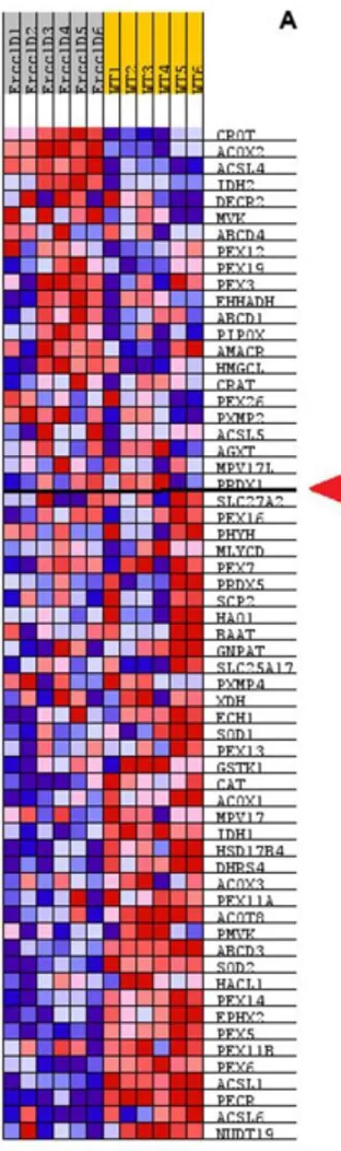
Alterations in the expression levels of specific peroxisomal genes in Ercc1Δ/- hepatic tissue, as compared to WT are reported in Fig. 4.1 A.
Figure 4.1. Heatmap of the level expression of genes in peroxisome pathway for
Ercc1Δ/- and WT samples. Values were z-score normalized, red means a higher level expression and blue means lower level expression. Red arrow shows the beginning of the enrichment for Ercc1 vs WT as was calculated using GSEA.
24
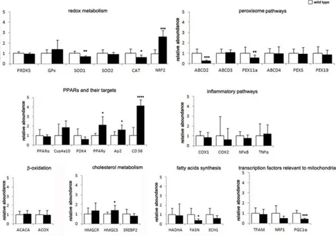
By quantitative Real Time PCR (qPCR), we could confirm alterations in the expression of multiple key players in peroxisomal functions in Ercc1Δ/-liver from 20-week-old mice (Fig. 4.2).
Transcription levels of the peroxisomal transporters of very long fatty acids (VLCFA), the ATP binding cassette transporter subfamily D (ABCD), display different trends. Expression of ABCD2, which participates to the transport of very long chain acyl-CoA (Morita and Imanaka, 2012), is significantly decreased, while ABCD3/PMP70, which contributes to the transport of long and branched chain acyl-CoA (Morita and Imanaka, 2012) and is the most abundant peroxisomal membrane protein in hepatocytes, is unaltered in the two genotypes.
Figure 4.2 Validation of genes expression in Ercc11Δ/- mouse liver, as compared to WT using qPCR. Note the strong overexpression of PPARγ pathway and of the redox sensor NRF2. By contrast, significant downregulation of antioxidant enzymes, namely SOD1 and Cat, is accompanied by lower expression levels of peroxisomal proteins ABCD2 and Pex11α, as well as the peroxisomal biogenesis regulator PGC1α. Disturbed lipid metabolism is indicated by altered levels of FASN (lower than normal) and HMGCS (higher than normal). *P<0.05; **= P<0.01; *** = P<0,001; **** = P<0.0001.
25
The principal peroxisomal gene, Cat, involved in ROS metabolism, shows significantly lower expression levels in Ercc1Δ/- mouse liver, which is paralleled by reduced expression of other antioxidant genes such as the cytosolic SOD1 and the mitochondrial SOD2. Surprisingly, the antioxidant transcription factor Nrf2 is expressed at higher levels in mutant mice. We also investigated mRNA levels of the nuclear receptors PPARα and PPARγ. While PPARγ levels are higher in mutant mice, PPARα mRNA is unchanged. Accordingly, expression levels of the PPARγ target genes CD36 and AP2, which are both major fatty acid transporters (Thompson et al., 2004; Wilson et al., 2015), are significantly increased. Genes directly involved in lipids metabolism and catabolism exhibit different trends. Those involved in de novo synthesis (e.g., fatty acid synthase, FASN) are significantly downregulated in mutant mice, while genes involved in fatty acid β-oxidation pathway, (e.g., ACOX) fail to show expression changes. Conversely, HMG-coA synthase, which is involved in cholesterol biosynthesis, is expressed at higher levels in Ercc1Δ/-.
Finally, it is worth noting that PGC1α - a cofactor of PPARγ which mainly modulates mitochondrial biogenesis and also orchestrates peroxisomal specialization and biogenesis (Villena, 2015) - shows a marked reduction in mRNA that could be consistent with downregulation of Pex11, that regulates peroxisome biogenesis in a PPARα-independent way (Bagattin et al., 2010).
4.2 Protein age-related expression
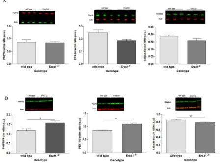
We next corroborated our findings on transcription by exploring possible peroxisomal dysregulation at protein expression level, in liver from 16- and 20-week-old Ercc1Δ/-mice.
While no significant variation is observed at 16 weeks of age (Fig. 4.3 A), pronounced alterations are detected at 20 weeks (Fig. 4.3 B). Specifically, peroxisomal membrane proteinsPMP70 and Pex14 display higher levels with respect to WT. By contrast and consistent with data collected at mRNA level, Cat shows strongly low expression at 20 weeks.
Protein levels of other antioxidant proteins indirectly related to peroxisome metabolism were also quantified on 20-week-old mouse liver.
26
Western blot analysis to quantify other proteins not directly involved in peroxisomal metabolism on 20-week-old mouse liver, for which we observed differences at mRNA level (i.e. SOD1, SOD2, and PGC-1a) confirmed the data on transcription (Fig. 4.4).
Figure 4.3.WB data concerning peroxisomal proteins of Ercc1Δ/- and WT mice 16- and 20-weeks-old. Data are expressed as mean ± SEM. *P < 0.05 **P < 0.01. (A) In younger mice, all markers do not display a considerable changes in expression levels. (B) In the older mice, the three proteins show a significant variation in older samples, particularly both PMP70 and Pex14 increase in mutants whilst Cat expression is lower than WT.
B A
27
Overall, expression data at the level of both mRNA and protein provide a consistent and broad depiction of peroxisomal alterations in Ercc1Δ/-.
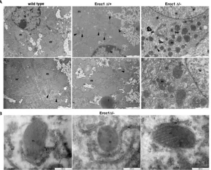
4.3 Morphological analyses: TEM and FIB/SEM
To investigate whether defective DNA repair is associated with morphological abnormalities to peroxisomes and/or other cytoplasmic organelles, we performed ultrastructural analyses using transmission electron microscopy (TEM) and focusing ion-beam-scanning electron .microscopy (FIB-SEM). EM sections were also processed for Cat cytochemistry, which allows unambiguous identification of the peroxisome population.
We decided to extend our field of observation also to Ercc1 mutants with milder defects than the Ercc1Δ/- (Gregg et al., 2011). We thus included Ercc1Δ/+ mice, which express one truncated and one WT allele, do not display an overt phenotype, nonetheless present significant functional alterations, particularly in the brain.
Hepatocytes from WT animals aging 20 weeks show regular ultrastructural features of cytoplasmic organelles, as expected. Livers from mutants instead
A
Figure 4.4.WB data concerning proteins
involved in antioxidant response of Ercc1Δ/- and WT mice 20-weeks-old. Data are expressed as mean ± SEM. *P < 0.05 **P < 0.01 ***P<0.001. (A, B and C) All protein expression show a significant decrease in mutant mice compare to WT depicting an alteration in redox state equilibrium and also the involvement of peroxisome as well as mitochondria.
B
C A
28
Figure 4.5. Ultrastructural analyses of WT and Ercc1Δ/- Ercc1Δ/-+ liver.
(A) WT have regular ultrastructural features of cytoplasmic organelles are observed at
20 weeks. Ercc1Δ/- hepatocytes show progressive damage including polymorphic mitochondria. Peroxisomes are indicated by arrowheads.
(B) The hepatic tissue of Ercc1Δ/- mice show disruption of mitochondrial membrane system with swelling of the outer mitochondrial membrane and cristae abnormalities. (m, mitochondrion; er, endoplasmic reticulum; N, nucleus; l, lipid droplet).
reveal morphological abnormalities, milder in Ercc1Δ/+ and more severe in Ercc1Δ/-. Indeed, progressive damage including polymorphic mitochondria and lipid droplets accumulation are observed (Fig. 4.5 A), while peroxisomes showed normal abundancy in Ercc1 mutants.
The tight nexus between peroxisomes and mitochondria (Delille et al., 2009) together with our data indicating reduced expression of PGC1α – an essential factor for mitochondria biogenesis - prompted us to focus on mitochondrial morphology (Fig. 4.5 B).
The organelles display disorganization of the membrane system. These anomalies concerned both the outer and inner membranes, which exhibited respectively swelling and cristae disruption in Ercc1Δ/- hepatocytes. FIB/SEM ultrastructural analysis confirmed the TEM data, further highlighting cellular damage - as evidenced by mitochondria and cytoplasmic abnormalities including lipid droplets - and cytological heterogeneity of the hepatic tissue in Ercc1Δ/- (Fig. 4.5 C).