To the people those have been closer to me: my family that support me during this path…
1 – INTRODUCTION_________________________________________________ 1 1.1 Cosmic Radiation______________________________________________ 1 1.1.1 Cosmic rays in the Earth atmosphere______________________ 2 1.1.2 Cosmic influence on chemistry of Earth atmosphere_________ 3 1.1.3 Effect of Radiation interactions with matter ________________ 5 1.1.3.1 Chemistry________________________________________ 5 1.1.3.2 Gamma-Ray interactions___________________________ 7 1.1.3.3 Neutron interactions_______________________________ 8 1.1.3.4 Effect on living cells_______________________________ 9 1.1.4 Radiochemistry of water_________________________________ 10 1.1.5 Interaction with organic matter___________________________ 11 1.1.6 Definitions of used quantities ______________________________ 13 1.1.7 Biological space studies___________________________________ 16 1.2 Photosynthesis and Photosystem II Complex_____________________ 18 1.2.1 The oxygenic photosynthesis_______________________________ 18 1.2.2 Photosystem II and Reaction Center________________________ 19 1.2.3 Light absorption - the antenna system_______________________ 21 1.2.4 The photosynthetic reaction center D1 protein ________________ 23 1.2.5 Carotenoids_____________________________________________ 23 1.2.6 Carotenoids and plastoquinone biosynthesis__________________ 25 1.2.7 Correlation between human health and photosynthetic compounds 28 1.3 Chlamydomonas reinhardtii____________________________________ 31 1.3.1 C. reinhardtii NPQ mutants_______________________________ 32 1.3.2 C. reinhardtii D1 mutants_________________________________ 33 1.4 The FOTON-BIOPAN Space missions__________________________ 35 1.5 STS-134 Space missions_______________________________________ 36 2 - AIM OF RESEARCH_____________________________________________ 38
3 - MATERIALS AND METHODS_____________________________________ 40
3.1 C. reinhardtii culture conditions_________________________________ 40 3.2 C. reinhardtii D1 mutants production_____________________________ 40 3.3 Site-directed PCR-based mutagenesis_____________________________ 40 3.4 Foton M3: flight and ground-control samples preparation____________ 44 3.5 The multicell fluorescence sensor: in-flight monitoring of chlorophyll a
temperature conditions____________________________________________ 46 3.7 The PHOTOEVOLUTION experiment: sample preparation and immobilization in the PHOTOI device _____________________________________________ 46 3.8 Chlorophyll a fluorescence induction _____________________________ 48 3.9 Functional characterization of the site-directed mutant strains under saturating light condition____________________________________________________ 48 3.10 RNA extraction and Real Time PCR_____________________________ 48 3.11 Quantitative and qualitative analyses of photosynthetic pigments_____ 51 3.11.1 Pigment standards and HPLC system________________________ 51 3.11.2 Carotenoids and chlorophylls extraction_____________________ 51 3.11.3 Pigments analyses________________________________________ 52 4 – RESULTS________________________________________________________ 53 4.1 The Foton M3 Space Mission: previous results from the Photo Project 53 4.1.1 Analysis of Photo-II fluorescence data of Foton M3 Space mission 54 4.1.2 Pigment content and composition____________________________ 59 4.1.3 q RT-PCR expression profiling______________________________ 60 4.2 STS-134 Space Mission Results__________________________________ 62
4.2.1 Production and characterization of Chlamydomonas D1 site-directed mutants_______________________________________________________ 62 4.2.2 Photosynthetic efficiency of site-directed mutants _______________ 65 4.2.3 The PHOTOEVOLUTION Project __________________________ 68 5 – DISCUSSION____________________________________________________ 70 6 – REFERENCES___________________________________________________ 74
1 - INTRODUCTION
1.1 Cosmic Radiation
The space environment is permeated by fluxes of complex radiation having variable energy and intensity. The Earth atmosphere is able to filter space radiation protecting living organisms from their deleterious effects deriving mainly from ionisation events.
According to its origin space radiation is classified as:
i) Galactic Cosmic Radiation, (GCR) composed by 87% protons, 12% alpha particles and 1% High Z Energetic particle - HZE with energies ranging from 1 and 103 GeV deriving by acceleration of stellar flares, supernova explosions, pulsar spin-offs, or from the explosions of nascent galactic nuclei within our galaxy. ii) Solar Particle Radiation, (SPE) consisting of charged particles in large clouds, mainly protons with an energy of about 1GeV. Solar radiation has lower energy compared to galactic cosmic radiation, but its production may be intense and is generally unpredicTable although it appears to reach a peak about every 11 years. There is a significant anti-correlation between solar activity and the intensity of the cosmic rays with energy values under approx. 10 GeV (O'Brien and Sauer, 2003). During the quiet sun period, no energetic particles in the solar wind can reach the sea level on Earth, because of the low energies of the particles. In the active sun period, the solar wind increases by factors of the order of 106, making it far denser than the galactic particle flux.
However, there is a more important aspect of the solar cycle than the increased particle flux. The active sun with its large solar wind creates a large distortion of the magnetic field about the Earth (the magnetosphere), which increases the Earth's shielding against intragalactic cosmic rays. This phenomenon leads to a net reduction of the sea-level cosmic rays during the period of the active sun.
iii) Geomagnetically Trapped Particle (GTP) radiation, generated by the interaction of space radiation with the Earth's geomagnetic field, comprising electrons with energies up to 7 MeV, protons with energies up to 600 MeV, and low energy heavy ions. Some of the particles from the Sun get trapped and collected by the Earth's magnetic field (Bert CW and Hyler,1962).
1.1.1 Cosmic rays in the Earth atmosphere
When space ionizing radiations particles collide with other atmospheric particles in our troposphere (e.g.: water molecules) disintegrates into smaller pions, muons and other particles forming the so called cosmic ray shower.
Except for protons and electrons near the top of the atmosphere, all particles are produced by interactions of the primary cosmic rays with the air. Muons and neutrinos are produced by decay of charged mesons, while electrons and photons originate by decays of neutral mesons.
The cosmic radiation incident at the top of the terrestrial atmosphere includes all sTable charged particles and nuclei with lifetimes of order 106 years or longer. Technically, "primary" cosmic rays are those particles accelerated at astrophysical sources and "secondary" are those particles produced in interaction of the primaries with interstellar gas. Thus electrons, protons and helium, as well as carbon, oxygen, iron, and other nuclei synthesized in stars, are primaries. Nuclei such as lithium, beryllium, and boron (which are not abundant end-products of stellar nucleosynthesis) are secondary. Antiprotons and positrons are partly, if not entirely, secondary, but the fraction of these particles that may be primary is a question of current interest (Miroshnichenko, 2003).
The incoming charged particles are modulated by the solar wind, the expanding magnetized plasma generated by the Sun, which decelerates and partially excludes the lower energy galactic cosmic rays from the inner solar system. In addition, lower-energy cosmic rays are deflected by the Earth's magnetic field although the protection is greater in the equatorial regions than at the poles. There is also some protection by the solar inter-planetary magnetic field and by the stratospheric absorption of low energy particles (Heinrich et al., 1999).
The Earth is well shielded by its atmosphere, but the dose may be significant in prolonged high altitude or space flight. Measurements of this type of radiation have been taken from high altitude aircraft. Fortunately the annual dose is relatively low in constantly exposed air crew (O'Brien et al., 1998).
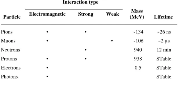
The incident particles, protons and neutrons, interact with atmospheric matter primarily with the strong interaction. The particles which are considered active in a cosmic ray cascade are reviewed in Table 1.
Particle
Interaction type
Mass
(MeV) Lifetime
Electromagnetic Strong Weak
Pions • • ~134 ~26 ns Muons • • ~106 ~2 µs Neutrons • 940 12 min Protons • • 938 STable Electrons • 0.5 STable Photons • STable
1.1.2 Cosmic influence on chemistry of Earth atmosphere
As we know the solar cycle drives solar irradiance variability in the UV region and also regulates the energetic particle fluxes, which are important for atmospheric chemistry and ozone (Crutzen et al., 1975; Heath et al., 1977; Fabian et al., 1979). While most of the atmospheric particle effects are in the thermosphere and mesosphere, some particle effects occur in the stratosphere and troposphere. The stratospheric odd nitrogen (NOy, where NOy =
N + NO + NO2 + NO3 + 2*N2O5 + HNO3 + HNO4 + ClONO2 + BrONO2) content is
influenced directly by GCR, which produces NO, in the lower stratosphere, and by solar proton events SPEs, which produce NOy in the upper stratosphere. Both SPEs and precipitating electrons produce NOy in the mesosphere and lower thermosphere, which could be transported downwards and possibly affect the upper stratospheric NOy balance. Jackman (Jackman et al., 1991; 1993; 2000) has reviewed the state of knowledge of the effects of charged particles on NOy and ozone in the middle atmosphere.
Rapid chemistry is initiated after N2 dissociation and most of the atomic nitrogen is
rapidly converted to NO and NO2. The precipitating particle-produced NO constituents can
then deplete ozone through the well-known catalytic reaction cycle:
NO + O3 --> NO2 + O2
NO2 + O --> NO + O2
A rich array of literature exists on the theory of charged particle production of HOx, which reacts with odd oxygen to cause a loss in ozone (Solomon et al., 1981) by a similar catalytic cycle:
OH + O H + O2
H + O3 OH + O2
Net: O + O3 2O2
It should be noticed that ozone change at 50 km and at higher altitudes regulates the ozone content practically at all altitudes due to its influence on the solar radiation flux penetration. Ozone observations, which occur during SPEs, support the idea of ozone destruction by solar protons (Krivolutsky et al., 2002).
Earth climate has been varying during all geologic time evolution. The origin of these variations has previously, and almost exclusively, been attributed to internal causes. For
example volcanic dust in the stratosphere can cause cooling of the order 0.5°C for a year or more. The same is assumed for the atmospheric/ocean oscillation in the Pacific called El Niño Southern Oscillation. It is expected that the annual variations in temperature will be a composite of several causes, of which only one is a solar influence. However, at time scales greater than 10 years it looks like the Sun has a significant influence on climate variations. This statement is based on the qualitative agreement between isotopes data about the Earth’s temperature over the last 1000 years. A remarkable correlation between cosmic ray flux and variations in the Earth’s cloud cover has been demonstrated (Svensmark, 2000).
1.1.3 Effect of Radiation interactions with matter
1.1.3.1 Chemistry
Living and not living matter can be affected by radiation coming from space. Interaction mechanisms are different depending on the type and the energy of the radiation as well as the characteristics of the penetrated matter.
Samples exposures with charged particles having high kinetic energies, result in ionization and excitation of the component, causing an electron loss from a molecule of a certain material. This interaction depends on the atomic make-up of the material and much less on the molecular structure. Also high energy photons of X- and gamma-rays knock electrons off atoms or molecules.
The particles able to ionize molecules are called ionizing radiation. Some examples are:
• Heavy charged particles such as protons, alpha particles, energetic nuclei, mesons;
• Light charged particles such as electrons and positrons;
• Electromagnetic radiation such as X-rays and gamma rays;
• High energy neutral particles such as neutrons;
The unique interaction characteristics of different particles and radiations will affect the severity of physiological damage during exposure of living systems to radiation.
Modes of interaction. There are five basic ways in which radiation interacts with
matter: ionization, kinetic energy transfer, molecular and atomic excitation, nuclear reactions, and radiative processes.
• Ionization is the removal of an atomic electron from an absorber atom to form an ion pair consisting of a negative electron and a more massive positive ion. Primary ionization is initiated directly by the incident radiation. Secondary ionization is produced subsequently by
the ions created in the primary ionization event. The amount of energy needed to form an ion pair varies with the type of absorbing medium.
• Kinetic energy transfers are interactions that impart kinetic energy to ion pairs above the amount required to form the pair. Kinetic energy transfers may also occur due to elastic collisions between the incoming radiation and the nuclei of the absorber.
• Molecular excitation and atomic electron excitation are modes of interaction that may occur even when the energy transferred is less than the absorber ionization energy. As the atomic electrons fall back to lower energy levels, x rays and Auger electrons will be emitted. Molecular excitation occurs through translational, rotational, and vibrational processes, and also through electronic excitation. The molecular excitation energy is eventually dissipated by bond rupture, luminescence, or evolution of heat.
• Nuclear reactions of incoming radiations with nuclei of absorber atoms can be important modes of interaction. This is especially true for high-energy charged particles and neutrons.
• Radiative processes are those in which electromagnetic energy is released by decelerating high-velocity particles.
Radiations can be divided into two groups: charged and uncharged. Charged particles include both unsTable atomic nucleus (α and β particles) and light varieties (photon). Uncharged radiations include γ-rays, x rays, neutrinos, and neutrons. These two groups of radiations have characteristic differences in their interactions with matter. Charged radiation interacts primarily with the electrons of the atoms in the absorbing medium through a series of many small energy-loss events. These events will result in the formation of ion pairs, kinetic energy transfers, and atomic or molecular excitation. Therefore, these radiations lose energy in an incremental and rather predicTable manner.
Uncharged radiations, in contrast, interact less frequently, or not at all, with the absorber electrons while dissipating their energy. Instead of a series of small sequential interactions, the uncharged radiations often undergo one or only a few major interaction events in which all their energy is lost. These interactions are not so predicTable as those for charged particles. It is possible for uncharged radiations to pass through a large amount of matter with a low probability of interaction. This is not the case for charged particles. Hence, uncharged radiations are much more penetrating than charged particles of the same energy (Turner, 1995).
Heavy Charged-Particle Interactions. Heavy charged particles may be considered to include those with masses greater than one dalton. In radioactive decay processes, these
particles have a positive charge. The heavy charged particles interact with matter primarily through ionization. For high-energy particles, nuclear reactions may also be important. The heavy charged particles are much more massive than the electrons with which they are interacting. Therefore, they are not significantly deflected by these interactions and the path of the particle through an absorber is relatively straight. Rarely, however, elastic collisions with the absorber nuclei will result in significant deviations of individual particles from a straight path (Rutherford scattering).
Alpha particles are the most comm
radiochemistry. The high likelihood of interaction of positively charged particles with absorber electrons combined with their straight interaction path suggest that the particles will have a fairly well-defined range in a given absorber. The range of the particle may be stated in a variety of units, all of which reflect the distance travelled through the absorber until the particle is "stopped" (i.e., reaches the ambient average kinetic energy of the absorber ato
The main reasons that gamma rays and neutrons are interesting in interaction radiation matter are described below:
Figure 1 Examples of radiation interactions with
In the first drawing of Figure
a fast neutron. It is a “collision event.” In the second drawing, a fast neutron bounces off of a target atom and slows down to a medium neutron. It is a “scatter event.” A gamma ray photon of a specific energy is given off. In the final drawing, a slow neutron is captured by and excites a target atom which then gives off a gamma ray photon of a specific energy. It is a “capture event”.
particles have a positive charge. The heavy charged particles interact with matter primarily energy particles, nuclear reactions may also be important. The heavy charged particles are much more massive than the electrons with which they are interacting. Therefore, they are not significantly deflected by these interactions and the path of particle through an absorber is relatively straight. Rarely, however, elastic collisions with the absorber nuclei will result in significant deviations of individual particles from a straight
Alpha particles are the most commonly encountered heavy charged particles in The high likelihood of interaction of positively charged particles with absorber electrons combined with their straight interaction path suggest that the particles will range in a given absorber. The range of the particle may be stated in a variety of units, all of which reflect the distance travelled through the absorber until the particle is "stopped" (i.e., reaches the ambient average kinetic energy of the absorber ato
The main reasons that gamma rays and neutrons are interesting in interaction radiation
interactions with matter.
1 a cosmic ray particle collides with a target atom and gives off a fast neutron. It is a “collision event.” In the second drawing, a fast neutron bounces off of a target atom and slows down to a medium neutron. It is a “scatter event.” A gamma ray photon a specific energy is given off. In the final drawing, a slow neutron is captured by and excites a target atom which then gives off a gamma ray photon of a specific energy. It is a particles have a positive charge. The heavy charged particles interact with matter primarily energy particles, nuclear reactions may also be important. The heavy charged particles are much more massive than the electrons with which they are interacting. Therefore, they are not significantly deflected by these interactions and the path of particle through an absorber is relatively straight. Rarely, however, elastic collisions with the absorber nuclei will result in significant deviations of individual particles from a straight
only encountered heavy charged particles in The high likelihood of interaction of positively charged particles with absorber electrons combined with their straight interaction path suggest that the particles will range in a given absorber. The range of the particle may be stated in a variety of units, all of which reflect the distance travelled through the absorber until the particle is "stopped" (i.e., reaches the ambient average kinetic energy of the absorber atoms).
The main reasons that gamma rays and neutrons are interesting in interaction
radiation-cosmic ray particle collides with a target atom and gives off a fast neutron. It is a “collision event.” In the second drawing, a fast neutron bounces off of a target atom and slows down to a medium neutron. It is a “scatter event.” A gamma ray photon a specific energy is given off. In the final drawing, a slow neutron is captured by and excites a target atom which then gives off a gamma ray photon of a specific energy. It is a
1.1.3.2 Gamma-Ray interactions
Because γ rays are electromagnetic radiation, they have no charge and no rest mass. The interactions of γ rays are, therefore, quite different from those experienced by charged particles. Gamma rays are not subject to Coulomb interactions, and so they do not lose energy continuously as do the charged particles.
Generally, the γ ray will experience only one, or perhaps a few, catastrophic interactions with the electrons or nuclei of atoms in the absorbing material. In these interactions, the γ ray either disappears completely or has its energy significantly altered. Ionization takes place, but most events are induced by secondary electrons and not by the primary γ ray itself. Gamma rays do not have discrete ranges. Instead, the intensity of a beam of γ rays is continuously reduced as it passes through matter in accordance with an exponential absorption law. Two major modes of interaction of gamma rays with matter are: i) the photoelectric effect (PE), when a γ ray interacts with an atom in a process that results in the ejection of an electron from the atom and the complete disappearance of the γ ray; ii) Compton scattering (CS), when the γ ray interacts with an atomic electron losing only part of its energy and it is deflected from its original path.
1.1.3.3 Neutron interactions
Neutron interactions with matter are quite different from those of the charged particles or electromagnetic radiation.
The uncharged neutrons interact almost exclusively with nuclei rather than with the atomic electrons. These nuclear interactions include elastic and inelastic scattering, and direct interactions. The capture of a neutron by a nucleus results in the subsequent emission of a charged particle or γ ray. This is used as the basis for detecting neutrons. Neutron ranges in matter longer than those of the charged particles with comparable or even greater energies.
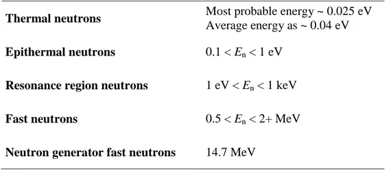
Table 2. Categories of Neutrons.
The type of interaction that a neutron will experience depends on its energy. Low-energy neutrons (with a mean Low-energy of about 0.04 eV) are called slow or thermal neutrons. They undergo both elastic scattering and direct interactions. The direct interactions experienced by thermal neutrons are most often of the radiative capture type. In this kind of reaction, the neutron is captured by a nucleus, resulting in an excited state that de-excites by emission of a γ ray. As the neutrons increase in energy from thermal to epithermal to resonance to fast, the probability of radioactive neutron capture generally decreases. Energies associated with these different categories of neutrons are given in Table 2.
1.1.3.4 Effect on living cells
When an electron passes through a cell, it releases its energy along its path (called a track) by interacting with the electrons of nearby molecules. The released energy is absorbed by atoms near the track, resulting in either excitation (a shift in the orbit of an electron to a higher energy level) or ionization (release of an electron from the atom). What differs from an ordinary chemical reaction is that when radiation donates energy to atoms or molecules, electrons other than those on the most outer orbit can be released, which makes the atoms very unsTable. Such unsTable atoms are called radicals and are chemically very reactive. Some radicals are so reactive that they exist only for as short a time as a microsecond.
Because the radiation causes ionization and excitation with a random process in the medium, the most abundant molecules have higher probability to undergo with the radiation effect. The biological matrix is composed for 70-90% by water and most of the absorbed energy is caught by water molecules. In order to understand the biological effect of radiation, it is important to describe the radiochemistry of water.
Thermal neutrons Most probable energy ~ 0.025 eV
Average energy as ~ 0.04 eV
Epithermal neutrons 0.1 < En < 1 eV Resonance region neutrons 1 eV < En < 1 keV
Fast neutrons 0.5 < En < 2+ MeV
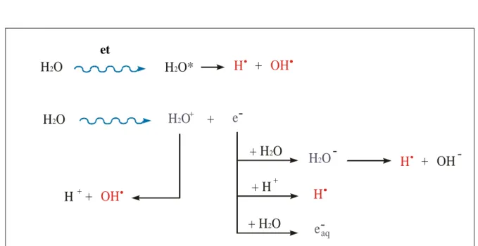
1.1.4 Radiochemistry of water
Bombarding liquid water with high energy photons or charged particles causes excitation and ionization of the water molecules (Figure 2).
The excitation of a molecule of water causes the prompt dissociation to the two radicals H OH :
O H2 H2O * H + OH
The ionization can give the water radiolysis producing an electron and a water molecule with a positive charge:
H2O H2O + + e
-H O*
2H O
2H O
2+ H O
2+ H O
2eccitazione
ionizzazione
H
H
H
H
+ H
H O
2H O
2+
+
+
+
+-
-+ +
-OH
OH
OH
e
e
aqFigure 2. Radiation interaction with molecules of water (Alpen ,1990).
The primary radical species formed in the radiolysis of liquid water are solvated electrons, hydroxyl radicals and hydrogen atoms. H2O2, H2 and H3O+ are also formed.
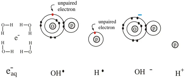
To some extent, presolvated electrons and H2O+ recombine in the tracks, producing an
excited water molecule, which can dissociated again to HO• and H• (Figure3). At very high water concentrations, some electrons can be scavenged in their presolvation state by the solute, resulting in a higher yield of scavenged electrons relative to the values from dilute solution. Yields of products such as HO•and H• are correspondingly reduced.
et molecule
e
9 cariche positive 9 cariche positive 9 cariche negative 10 cariche negative 1 elettrone spaiatoelettrone idrato radicale idrossilico radicale idrogeno ione idrossilico ione idrogeno
elettrone spaiato elettrone spaiato
e
aq
OH
H
OH
H
O O O O H H H H H H H H--
-
+
-O O p p p pFigure 3. Main products from interaction of the radiation with water molecules.
Inside the organic matter, free radicals produced by water radiolysis, can react with organic, biologically important molecules, causing damages. Moreover, the biological effectiveness of radiation can be enhanced by the oxygen. As matter of fact, if the matter hit by ionizing radiation contains dissolved oxygen, the chemical reactions of free radicals with oxygen produce superoxide, hydroperoxyl radicals and hydrogen peroxide:
• e-aq + O2 O2 (anion radical superoxide)
• H + O2 H2O ( hydroperoxyl radical)
• O2 + H + H2O ( hydroperoxyl radical)
• H2O H2O2 (hydrogen peroxide) + O2
Reactions of superoxide and hydroperoxyl radicals are studied because of their role in biological systems. The products of the reactions have a harmful power higher than primary radicals, because they show a big affinity with the molecules.
1.1.5 Interaction with organic matter
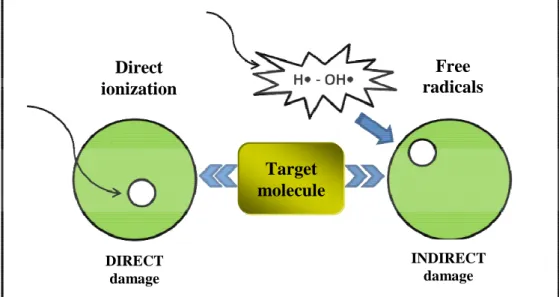
In general, radiation-induced ionization may act directly on the cellular component molecules or indirectly on water molecules, causing water-derived radicals (Figure 4).
unpaired electron
unpaired electron
Figure 4. Direct and indirect action of ionizing radiation.
Radicals react with nearby molecules in a very short time, resulting in breakage of chemical bonds or oxidation of the affected molecules. For examples free radicals can extract hydrogen from organic molecules (RH) forming secondary radicals R• (Coggle, 1998):
RH + OH R + H2O
RH + H R + H2
Secondary radicals, which can again react with other molecules, are formed also for direct action of ionizing radiation:
RH R H+ + e- R + H+
If a organic molecule is transformed directly or indirectly in a free radical it can react with oxygen in the following way:
R + O2 RO2
A chain reaction can involve several RH:
RO2 + RH RO2H + R Direct ionization Free radicals diffusion DIRECT damage INDIRECT damage Target molecule
Proteins and desossiribonucleic acid (DNA) are organic macromolecules (R) with a fundamental biological role inside living cells.
The major effect in cells is DNA breaks. Since DNA consists of a pair of complementary double strands, breaks of either a single strand or both strands can occur. However, the latter is believed to be much more important biologically. Most single-strand breaks can be repaired normally thanks to the double-stranded nature of the DNA molecule (the two strands complement each other, so that an intact strand can serve as a template for repair of its damaged, opposite strand). In the case of double-strand breaks, however, repair is more difficult and erroneous rejoining of broken ends may occur. These so-called misrepairs result in induction of mutations, chromosome aberrations, or cell death.
It is clear that exposure of biological material to ionising radiation could leads to a loss of function due to the modification of critical structures (Beauregard and Potier, 1985). One of the most important and discussed problems in radiation protection concerns the determination of the biological effectiveness of radiation on living organisms.In order to assess the radiation risk for living beings during long-term spaceflights, it is very important to clarify whether combination of cosmic radiation and other factors have a synergistic effect on the cell functions.
1.1.6 Definitions of used quantities
It is useful to outline some information about parameters utilized to determine Ionizing Radiation effect on living matter. Up to now, the human body is taken as reference (ICRP, Publication 74).
• Linear energy transfer (LET) is a measure of how, as a function of distance, energy is transferred from radiation to the exposed matter. A high value of LET indicates that energy is deposited within a small distance. It is also known as the restricted linear collision stopping power. High LET radiation: Radiation with high linear energy transfer, normally assumed to comprise protons, neutrons and alpha particles (or other particles of similar or greater mass).
Low LET radiation: Radiation with low linear energy transfer, normally assumed to comprise photons (including X rays and gamma radiation), electrons, positrons and muons.
• Absorbed dose (denoted as D) is the quotient of dε e by dm, where de is the mean energy imparted by ionizing radiation to matter of mass dm, thus
dm d D= ε
• Dose Equivalent (denoted as H) is the product of Q and D at a point in tissue, where D is the absorbed dose and Q is the quality factor for radiation at that point, thus
QD H =
The unit of Dose Equivalent is joule per kilogram (J ⋅ Kg –1 or sievert: Sv).
• Ambient Dose Equivalent (denoted as H*(d)), at a point in radiation field, is the dose equivalent that would be produced by the corresponding expanded and aligned field in the ICRU sphere at a depth d, on the radius opposing the direction of the aligned field. The unit of Ambient Dose Equivalent is joule per kilogram (J ⋅ Kg –1 or sievert Sv).
• Equivalent Dose in a Tissue T (denoted as HT,R) is the absorbed dose in an organ or tissue
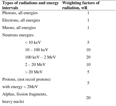
multiplied by the relevant radiation weighting factor (see Table 3), thus
R , T R R , T w D H =
where DT,R is the absorbed dose averaged over the tissue or organ T, due to radiation R, where
wR is the radiation weighting factor for radiation R.
When the radiation field is composed of radiations with different values of wR, the absorbed
dose is subdivided into blocks, each multiplied by its own value of wR and summed to
determine the total equivalent dose, i.e.
The unit of Equivalent Dose is joule per kilogram (J ⋅ Kg -1 or sievert (Sv).
• Effective Dose Equivalent (denoted as HE) is the weighted average of the dose equivalents,
each weighted by a tissue or organ weighted factor, thus:
∑
= T T T E w H Hwhere HT is the dose equivalent in tissue T, and wT is the is the weighting factor for tissue T,
as formerly recommended by ICRP.
∑
= R R T R T w D H ,Types of radiations and energy intervals
Weighting factors of radiation, wR
Photons, all energies 1
Electrons, all energies 1
Muons, all energies 1
Neutrons energies: < 10 keV 10 – 100 keV 100 keV – 2 MeV 2 – 20 MeV > 20 MeV 5 10 20 10 5 Protons, (not recoil protons)
with energy < 2MeV 5
Alphas, fission fragments,
heavy nuclei 20
Table 3. Values for radiation weighting factors (ICR, Publication 60).
Sanitary effects of ionizing radiation. Radiation exposure can determine risks of sanitary effects for living beings and requires adequate radio-protective precautions. Induced effects on men can be distinguished in somatic effects and genetic effects, distinguishing if they reveal their effect on the exposed individual or on his descendent. A lot of somatic effects are non – stochastic effects. Their seriousness is related to dose absorbed by the considered organ or tissue and each non-stochastic effect is characterized by a threshold value of absorbed dose. Only if the absorbed dose is higher than the threshold value, the effect appears. Threshold dose values, for different deterministic effects, are always very high and they are known with a good precision. All genetic effects and the most important somatic effects (as leukaemia, tumour) are stochastic effects. They are characterized by the absence of a threshold dose value and they occur with a probability that depends on the absorbed dose value.
1.1.7 Biological space studies
The main difficulties for the permanence of biological organisms in space (including man) are caused mainly by the presence of ionizing radiation, microgravity, high-temperature and high vacuum conditions. During the time, the enhanced science community interest on space biology was followed by the increased number of space missions aimed to understand space environmental effects on living organisms.
Permanence in space can result in a number of adverse effects such as bone loss, skeletal muscle atrophy, cardiovascular problems, immune system disregulation, alteration of sleep and circadian rhythms, cancer, cataracts and possible damage to the central nervous system which are mainly associated to the occurrence of microgravity and exposures to the GCR environment (Hellweg and Baumstark-Khan, 2007).
Plants are essential companion lifeforms for long-term space missions, where Human habitats must mimic the cycles of life on Earth to generate and recycle food, oxygen, and water. Photosynthetic organisms on the International Space Station (ISS) could be use as life supporting system, supplying oxygen and removing carbon dioxide from the station atmosphere. They also prevent gases like ammonia and acetone, which people emit in small amounts, from building up to dangerous levels.
The majority of biological studies on the physiological effects of space have been concerned with the gravitropic responses of higher plants. Nowadays, numerous are the scientific data available on this topic.
Microgravity is one of the space environmental condition affecting cell division, cell components and their distribution. Many studies focused on the effects of Space radiation and/or microgravity on the number, viability, kinetics of germination, growth rate and mutation frequency of spores formed in Space (Takahashi, 2001). Dormant seeds do not appear to be sensitive to gravity. However, during development, mitosis and cytokinesis, plants appear to be influenced by the absence of gravity. Mitosis is disturbed in the absence of gravity, as demonstrated by reduced or inhibited cell division, anomalies in the separation of chromosomes and the fragmentation of the chromatids (Kordyum, 1997). Not only Space
radiation can interfere with biological process, but also space flights appear to alter the process of differentiation particularly in root tissues, perhaps on account of an altered hormonal equilibrium. Chlorella vulgaris cells grown during a space flight differred from controls showing a reduction in the amount of polysaccharide reserves in chloroplasts. Increased concentrations of mono and disaccharides resulted in the increased activity of amylases. Rearrangements in chloroplast structure, such as the decrease of polysaccharide
reserves were probably due to an increase in hydrolytic processes by microgravity (Popova and Shniukova, 1995).
The absence of gravity seems to affect also the organelles membranes, as evidenced by the destruction of the grana and the detachment of the thylakoids in the chloroplast stroma in seedlings grown in space (Nedukha, et al., 1999).
Additional consequences include the deformation of the endoplasmic reticulum, the swelling of the mitochondria, aggregation of the chromatin and the presence of multiple nucleoli within the nucleus. These changes suggest a major influence of space condition on the growth and development of micro-organisms (Nedukha, 1997; Kordyum, 1997).
The relationship between gravity and the processes of growth and development of photosynthetic organisms can only be determined by eliminating gravity, as occurs in space.
Space environment affects almost all biological processes, in particular germination and flowering. Embryo lethality and lethal mutation frequency were observed in Arabidopsis thaliana seeds (Kranz, 1990) and in other organisms such as Escherichia coli and Bacillus subtilis (Horneck, 1995)
.
Despite the numerous study on plant response under microgravity condition, very little is still known about the effects of ionizing radiation on the photosynthetic apparatus (Sakashita, 2002; Saakov, 2003).
A limiting factor in this research field is that there are very appreciable temporal variations in both radiation composition and intensity (Internet site on Solar chart: http://hiraiso.crl.go.jp/). For these reasons, measuring and reproducing ground space radiation in all its components is a nearly impossible task. Up to date, the best approach to study radiation effect is utilizing single beam, produced on ground by properly equipped facilities.
Unicellular organisms were the first subjects in biological studies in Space. Rapid multiplication, tiny size and simple procedure to keep growth conditions are the advantages in using these simplest biological systems to estimate cosmic radiation effect (Nechitailo, 2001). In on ground studies, several investigations have been focused on microorganisms able to survive to high doses of ionizing radiation like Micrococcus radiodurans (Hansen, 1982), Deinococcus radiodurans (Venkateswaran, 2000), Chroococcidiopsis cells (Billi, 2000) and Archeon Pyrococcus abyssi (Jolivet, 2003).
1.2 Photosynthesis and Photosystem II Complex
1.2.1 The oxygenic photosynthesis
Solar radiation is an example of ionizing radiation exploited by photosynthetic organisms for photosynthesis, the physico-chemical process by which plants, algae and certain bacteria convert light energy into chemical energy of organic compounds, with concomitant use of these components in the bioenergetic processes. In plants, algae and cyanobacteria, the photosynthetic processes result in the fixation of carbon dioxide (CO2)
from the atmosphere that is used to synthesize carbohydrates and release of molecular oxygen to the atmosphere. This process is known as oxygenic photosynthesis.
The first step in photosynthesis is the absorption of photon by a pigment molecule of photosynthetic antenna resulting in conversion of the photon energy to an excited electronic state of pigment molecule. The antenna consists of hundreds of pigment molecules (chlorophylls, bacteriochlorophylls, carotenoids, etc.) that are bounded to proteins within the photosynthetic membrane. The excited electronic state is transferred over the antenna molecules as an exciton. Some excitons are converted back into photons and emitted as fluorescence, some converted to heat. For most of the excited pigment molecules the most useful decay pathway is “energy transfer” to a photochemical reaction centers, and it is of vital importance to photosynthesis. Excitons trapped by a reaction center provide the energy for the primary photochemical reaction of photosynthesis – the photoinduced transfer of an electron. Subsequent electron transfer reactions occur in the dark which results in accumulation of chemical bound energy. Electron transfer reactions in photosynthesis involve electron carriers or electron-transfer proteins, among others, (bacterio) chlorophylls, quinones, cytochromes, and iron-sulfur proteins that has been recently reviewed by Feyziyev (2010). Electron transport chain trough specialized protein, called photosystem two (PSII) and photosystem one (PSI) protein complex, leads to convert light energy into chemical energy and reducing equivalents, adenosine triposphate (ATP) and nicotinamide adenine dinucleotide phosphate (NADPH), which are used in the CO2 fixation to produce carbohydrates (CH2O)n
(Mitchell, 1961). Oxygenic photosynthesis is driven by visible light (wavelengths from 400 to 700 nm) that is absorbed by different pigment molecules, chlorophyll a and b, carotenoids and phycobilins. The pigment molecules are bound to polypeptides forming pigment-protein complexes. Two types of antenna complexes, the inner (core) antenna consisting of pigment-proteins that are an integral part of the reaction center complex, and outer (peripheral) antenna complexes are distinguished in oxygenic species. In plants and green algae the light absorbing
pigment molecules are chlorophyll a, chlorophyll b and carotenoids. The cyanobacteria and red algae have a different outer antenna system. In cyanobacteria and red algae, the phycobiliproteins are assembled into supromolecular aggregates called phycobilisomes, which are attached to the photosynthetic membrane surface and transfer electronic excitation energy (via chlorophyll) to the reaction centers to initiate photochemical reactions. Carotenoids and phycobiliproteins serve as accessory pigments, to absorb light energy not absorbed in the spectral region of chlorophyll absorption and transfer this energy to chlorophylls. The efficiency of singlet-singlet energy transfer from certain carotenoids to chlorophyll may be very high ranging from 70% to nearly 100%. Phycobilisomes can funnel the absorbed energy to the reaction center with more than 95% efficiency (Ke, 2001).
1.2.2 Photosystem II and Reaction Center
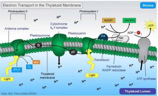
The multisubunit pigment-protein complex Photosystem II of higher plants, algae and cyanobacteria is located inside the thylakoid membranes (Figure 5).
Since the existence of oxygenic photosynthesis, it catalyses the light-induced transfer of electrons from water to plastoquinone, oxidizing water molecules to oxygen, sustaining an aerobic atmosphere on Earth. During these reactions reduced equivalents are produce resulting in carbon dioxide fixation, creating biomass, food and fuel.
Its unique properties require an elaborate arrangement of integral membrane proteins, specifically bound pigment moieties, extrinsic proteins and inorganic cofactors. PSII uses light energy to drive two chemical reactions - the oxidation of water and the reduction of plastoquinone. The PSII core is the minimal unit which is capable of catalysing full PSII function. It is composed of a reaction center, which consists of the D1 and D2 polypeptides (encoded by the chloroplast psbA and psbD genes, respectively), cytochrome b559 (Cyt b559),
the psbI protein and six chlorophyll (Chl) and two pheophytin (Pheo) molecules, an inner antenna of chlorophyll-binding proteins termed CP47 and CP43, and the extrinsic lumenally bound proteins of the OEC, 33 kDa protein (psbO), 23 kDa (psbP) and 17 kDa (psbQ), in higher plants and green algae, whereas in cyanobacteria psbP and psbQ are replaced by the 15 kDa psbV (cytochrome c550) and the 12 kDa psbU. These intrinsic and extrinsic proteins, together with a number of low-molecular weight subunits make up the core complex (Debus, 1992).
Light is funnelled from the light harvesting complexes to the photochemically active reaction center (RC) of PSII. The RC contains a special form of Chl a, P680 (the primary electron donor of PSII), which acts as an exciton trap and is converted to a strong reducing
agent after excitation (P680*), P680* is then photooxidized and donates an electron to the primary acceptor of PSII, a protein-bound pheophytin.
P680• P680 +Phe- 3Chl•
This charge separation is stabilized by the transfer of an electron to a plastoquinone molecule (QA), and subsequently to a second plastoquinone (QB). On the lumenal side, the
oxidized form of P680 is reduced by a redox active tyrosine residue of the D1 protein, TyrZ. The oxidized tyrosine radical TyrZ extracts an electron and a proton from a cluster of four manganese atoms that binds substrate water (Lydakis-Simantiris et al., 1997). The QA
plastoquinone is a one-electron acceptor, firmly bound to the D2 protein. In contrast, the QB
plastoquinone is a two-electron acceptor, bound to the D1 protein.
A second photochemical turnover leads to the accumulation of two reducing equivalents on QB, which is then released from PSII, as plastoquinol, to be subsequently oxidized by PSI
via the cytochrome b6f complex.
Figure 5. The electron transport pathway of oxygenic photosynthetic organisms.
The empty QB site is then occupied by another plastoquinone molecule. Two further
photochemical turnovers provide the oxygen-evolving complex with four oxidizing equivalents.
The accumulation of four oxidizing equivalents, in a manner consistent with the observed S-state transitions, leads to the release of molecular oxygen from the complex. During the accumulation of oxidizing equivalents, protons are released to the lumenal space of the thylakoid membrane (Mattoo et al., 1989).
1.2.3 Light absorption - the antenna system
The light-harvesting (or antenna) complex of plants is a protein-pigments complex embedded in the thylakoid membrane. It is formed predominantly by chlorophyll b, xanthophylls and carotenoids that transfer light energy to the central chlorophyll a, known as core pigment. In order to broad the range of light adsorption each antenna component has a different spectra ranging from 400 to 700 nm. The entirely light harvest complex is constitued by six different type of apoproteins, Lhcb 1–6.
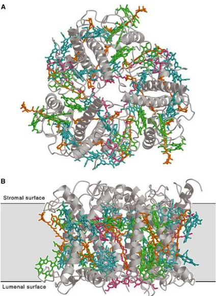
The native form of LHCII (Figure 6), accounting for about 60% of the total chlorophyll content of thylakoid membranes, is a trimer composed of three Lhcb proteins: Lhcb1, Lhcb2 and Lhcb3 (Lucinski and Jackowski, 2006). Lhcb4, Lhcb5 and Lhcb6 consitute, in turn, the polypeptide moiety of minor peripheral light-harvesting complexes, namely CP29, CP26 and CP24, respectively.
Lhcb1 is constituted by three tylakoid transmembrane α –helices, and also two short lumen-exposed amphipatic α-helices. This protein has the N-terminus of the molecule facing the stroma and the C-terminus penetrating the lumen. Each Lhcb1 binds eight Chl a , six Chl b and xanthophylls: one neoxanthin, one violaxanthin. The amino acid composition of Lhcb1 is 94 % identical to that of Lhcb2 (Standfuss et al., 2005). Lhcb2 occurs in vivo as a heterotrimer with Lhcb1 while the Lhcb3 constitutes the smallest fraction of LHCII proteins, not exceeding 11 % of the total apoprotein content of LHCII, and differs from Lhcb1 and Lhcb2 in its structure (Lucin´ski and Jackowski, 2006).
Each antenna system has between 250 and 400 pigment molecules and the adsorbed energy is transferred in a resonance way towards the reaction center of each photosystem, where complex chemical reactions capture light energy in the form of chemical bonds.
Under changing light conditions, the antenna complex is fundamental in balancing the excitation energy between photosystems I and II, by the reversible phophorylation of light harvesting chlorophyll a/b binding proteins (LHCII), helping in the avoidance of possible photo-oxydative damages when hight light condition occur (Kargul and Barber, 2008).
Carotenoids not only actively participate at the harvesting of light energy but they also play a crucial role in prevent the photo-oxidative damages of chlorophyll molecules (Cogdell 2006; van Oort et al., 2007; Barros et al., 2009).
Figure 6. The LHC-II trimer: (A) top view from stromal side; (B) side view. LHC-II protrudes from a 35A° lipid
bilayer (black lines) by 13A° on the stromal side and by 8A° on the lumenal side. Grey, polypeptide; cyan, Chl
1.2.4 The photosynthetic reaction center D1 protein
D1 is a 32 kDa protein encoded by the plastid psbA gene and translated on thylakoid membrane-bound ribosomes. Mature D1 has five transmembrane domains (TMs) with the N-terminus on the stromal side of the thylakoid membrane. Light has an important role in regulation of various stages of D1 synthesis from translation initiation to elongation, maturation and insertion into the thylakoids (Zhang and Aro, 2002).
D1 turnover is also a physiological highly regulated process in photosynthetic organisms: the degradation and synthesis of new D1 protein is genetically regulated and is a stress recovery mechanism (Mulo et al., 2012).
The detection of amino acid mutations specifically in the D1 reaction center subunit is greatly supporting the understanding of PSII structure and function. These discoveries identified the D1 subunit as the “herbicide binding protein“. Since the inhibitors tested were known to compete with the native plastoquinone QB for its binding site this protein was likely to be also
the QB binding protein. Furthermore, a specific region within D1 emerged which we now
address as herbicide binding. Abundant biochemical and genetic evidence has accumulated which places the binding niche between amino acids 211 and 275.
A number of mutations in the gene psbA have been found to confer herbicide-resistance. In algae mutants, mutations at positions 219, 251, 252, 255, 256, 264 and 275 are most common and result in resistance to several classes of herbicides (Oetteier, 1999). All these psbA mutations are located on helices IV and V helices and their connecting loop in D1 protein, which is a specific region of the polypeptide between amino acid residues 211 and 275 (Ohad, 1992).
1.2.5 Carotenoids
In oxygenic photosynthetic organisms, carotenoids are the most widespread natural pigments and fulfil a variety of functions. Carotenoids are polyisoprenoid compounds containing 40 carbon atoms. They possess a long chain of conjugated double bonds in the central part of the molecule, and variable end groups: different level of hydrogenation and introduction of oxygen-containing functional groups create a large family of over 600 natural compounds (Green and Durnford, 1996). In higher plants, two different classes are found into thylakoids: (i) carotenes (e.g. β-carotene), which are hydrocarbones with linear structure and with cyclic groups in one or both extremities, and (ii) xanthophylls (e.g. lutein, zeaxanthin), which are oxygenated derivatives of the first group. In higher plants, the carotenoids normally
associated with thylakoid membranes are
complex of both photosystems) (Green and Durnford
zeaxanthin, violaxanthin and neoxanthin (bound to antenna complexes) (Formaggio et al. 2001; Caffarri et al., 2001). Carotenoids ar
probably involving hydrophobic interactions (Gastaldelli et al.
Carotenoids have three main functions in photosynthesis: a) structure stabilisation and assembly of protein complexes in the thylakoid
state energy transfer to the chlorophylls (Green and Durnford photo-oxidative damages (Havaux and Niyogi
Figure 7. Biosynthetic pathway of
pigments denote enzymatic conversion caused by xanthophyll cycling. Enzymes involved are also reported. Carotenoids involved in the xanthophyll
al., 1999), not in the core complex and inner antenna of PSII. Upon de zeaxanthin is distributed among LHCII and minor antennae.
Carotenoids composition of thylakoid is not constant: it can undergo modificati during long-term acclimation of plants to stressing condition, as well as rapidly changes following fluctuations of solar light intensity (Green and Durnford
cycle (Figure 7) involves the three xanthophylls violaxanthin, anther and consists in a light-dependent, reversible de
the intermediate antheraxanthin; the former reaction is catalysed by violaxanthin deepoxidase (VDE), a lumenal enzyme activated by acidi
Durnford, 1996).
associated with thylakoid membranes are α- and β- carotene (bound especially to the core complex of both photosystems) (Green and Durnford, 1996) and the xanthophylls lutein, zeaxanthin, violaxanthin and neoxanthin (bound to antenna complexes) (Formaggio et al.
2001). Carotenoids are non-covalently bound to the protein complexes, probably involving hydrophobic interactions (Gastaldelli et al., 2003).
Carotenoids have three main functions in photosynthesis: a) structure stabilisation and assembly of protein complexes in the thylakoid membrane; b) light absorption and excited state energy transfer to the chlorophylls (Green and Durnford, 1996); c) protection against
oxidative damages (Havaux and Niyogi, 1999).
Biosynthetic pathway of β-carotene-derived xanthophylls in higher plants
pigments denote enzymatic conversion caused by xanthophyll cycling. Enzymes involved are also reported. Carotenoids involved in the xanthophylls cycle are localized in the peripheral antenna proteins of PSI
al., 1999), not in the core complex and inner antenna of PSII. Upon de-epoxidation, the newly synthesized zeaxanthin is distributed among LHCII and minor antennae.
Carotenoids composition of thylakoid is not constant: it can undergo modificati term acclimation of plants to stressing condition, as well as rapidly changes following fluctuations of solar light intensity (Green and Durnford, 1996). The xanthophyll
) involves the three xanthophylls violaxanthin, antheraxanthin and zeaxanthin, dependent, reversible de-epoxidation of violaxanthin to zeaxanthin the intermediate antheraxanthin; the former reaction is catalysed by violaxanthin deepoxidase (VDE), a lumenal enzyme activated by acidification of the luminal compartment (Green and arotene (bound especially to the core 1996) and the xanthophylls lutein, zeaxanthin, violaxanthin and neoxanthin (bound to antenna complexes) (Formaggio et al., covalently bound to the protein complexes,
Carotenoids have three main functions in photosynthesis: a) structure stabilisation and membrane; b) light absorption and excited 1996); c) protection against
xanthophylls in higher plants. The arrows between
pigments denote enzymatic conversion caused by xanthophyll cycling. Enzymes involved are also reported. cycle are localized in the peripheral antenna proteins of PSII (Ruban et epoxidation, the newly synthesized
Carotenoids composition of thylakoid is not constant: it can undergo modifications term acclimation of plants to stressing condition, as well as rapidly changes 1996). The xanthophylls axanthin and zeaxanthin, epoxidation of violaxanthin to zeaxanthin via the intermediate antheraxanthin; the former reaction is catalysed by violaxanthin deepoxidase fication of the luminal compartment (Green and
1.2.6 Carotenoids and plastoquinone biosynthesis
In vascular plants and algae carotenoids are usually synthesized in plastid, which is also the site of their are located. The majority of carotenoids are bound to membrane proteins associated with the photosynthetic apparatus, and to some extent with plastid envelope membranes (Derguini et al., 1991). Green algae are photosynthetic organisms able to accumulate extraplastidic carotenoids under unfavourable conditions. For example, astaxanthin of the green alga Haematococcus pluvialis is located in cytoplasmic lipid globules (Boussiba, 2000). The prominent carotenoids in green algae and vascular plants are β-carotene, lutein, 9-cisneoxanthin, and violaxanthin (Figure 8), being rapidly converted to antheraxanthin and zeaxanthin under high light conditions, as reported above. The carotenogenic enzymes are encoded by nuclear genes in Chlamydomonas reinhardtii. Biosynthesis of carotenoids (Figure 8) is initiated with the formation of isopentenyl-diphosphate (IPP), called “active isoprene.” The subsequent reaction sequences are often divided into four stages: (a) stepwise condensation of isoprene units to form the first carotenoid, phytoene, (b) extension of the π-electron system by sequential desaturation resulting in lycopene formation, (c) cyclization reactions that generate the carotenes, and (d) synthesis of xanthophylls by the introduction of the oxygen functions (Grossman et al., 2004). Sequential addition of three molecules of IPP, results in the formation of the C20-compound geranylgeranyl pyrophosphate (GGPP) (Figure 8). This chain elongation is catalyzed by the enzyme GGPP synthase. GGPP is not only an intermediate in carotenogenesis, but also a substrate for the formation of a variety of other cellular components, including the phytol moiety necessary for the synthesis of Chl, tocopherols and phylloquinone and, in the case of vascular plants but not algae or bacteria, the hormone gibberellin. In chloroplasts and mitochondria, polyprenyl transferases synthesize the long chain isoprenoid component of plastoquinone and ubiquinone, respectively, by adding isoprene units to GGPP (Figure 8).
Head-to-head condensation of two molecules of GGPP by phytoene synthase (PSY) results in formation of phytoene, the first carotenoid of the pathway. This reaction and all subsequent steps are linked to plastid membranes as the carotenoid substrates are hydrophobic and membrane associated (Schledz et al., 1996; Bonk et al., 1997). As PSY catalyzes the first committed step of carotenoid biosynthesis, it was expected to represent a key target for regulatory control. Experimental evidence supports this expectation. Over-expression of the psy gene in tomato plants resulted in dwarf plants because elevated phytoene production in this strain caused a severe reduction in gibberellin synthesis (Fray et al., 1995). Similarly, the
carotenoid content of tomato fruits increased substantially in concert with the fruit-specific expression of a bacterial psy gene (Fraser et al., 2002). In vascular plants, psy mRNA increases in red light in a phytochrome-dependent manner (Von Lintig et al., 1997). In H. pluvialis, expression of psy appears to be under photosynthetic redox control because increased transcript accumulation in high light could be prevented by the addition of the herbicide 3-(3,4-dichlorophenyl)-1,1-dimethylurea, which blocks electron transport at PSII (Steinbrenner and Linden, 2001). In C. reinhardtii, psy mRNA increases following exposure of cells to blue light, suggesting that the gene may be under the control of a blue light photoreceptor such as PHOT1 or CRY1 (Bohne and Linden, 2002).
Figure 8. Schematic representation of carotenoids (A) and plastoquinone (B) biosytetic pathways in C.
reinhardtii
Principal enzymes involved in carotenoids biosynthesis are reported in Figure 8. Phytoene contains only three conjugated double bonds and consequently shows no visible absorbance.
In vascular plants and green algae, two desaturases sequentially introduce additional conjugated double bonds into phytoene, thereby extending the π-electron system and shifting the absorbance to longer wavelengths. These enzymes are structurally and functionally related
geranylgeranyl-diphospate (GGPP)
O P O P OH
OH OH
ISOPENTENYL DIPHOSPHATE (IPP)
GGPS phytoene PSY ζζζζcarotene PDS lycopene ZDS α αα αcarotene ββββcarotene LYC β LYC ε lutein CHY β CHY β zeaxanthin antheraxanthin violaxanthin VDR1 VDR1 ZEP ZEP p-hydroxyphenilpyruvate 2-methyl-6-phytyl-1,4-benzoquinone HST 1 homogentisate Plastoquinone-9 MPBQ-MT 3 B A
and likely have evolved from a common ancestor by gene duplication (Sandmann, 2002). The initial desaturation by phytoene desaturase (PDS) introduces two double bonds into the molecule, resulting in the formation of ζ -carotene. Two additional desaturation steps catalyzed by ζ -carotene desaturase (ZDS) yield lycopene. Both PDS and ZDS contain an amino-terminal conserved dinucleotide binding motif and use FAD as a redox cofactor.
Electrons generated by the reaction are transferred from FADH2 to plastoquinone, and
from there can be funnelled either into the photosynthetic electron transfer chain or to O2 via a
plastid terminal oxidase (Carol and Kuntz, 2001). Similar to psy, expression of pds is under photosynthetic redox control in H. pluvialis (Steinbrenner and Linden, 2003), but is induced by blue light in C. reinhardtii (Bohne and Linden, 2002).
Following cyclization of lycopene the carotenogenesis pathway splits, forming the α- and β- branches of carotenes (Figure 8). Introduction of β-ionon rings at both ends of the linear lycopene molecule results in the formation of the symmetric β,carotene, termed β-carotene. This reaction is catalyzed by a single enzyme, lycopene β-cyclase (LCYB). The concerted action of LCYB on one side of the lycopene molecule and a second cyclase on the opposite end results in formation of β, ε-carotene, more commonly known as α-carotene. In most plants, the enzymatic capacity of the second cyclase, designated lycopene ε-cyclase (LCYE), is restricted to the introduction of a single ε-ionon ring per lycopene (Cunningham and Gantt, 1998). The α-carotene molecule is the precursor of lutein and loroxanthin, whereas violaxanthin, zeaxanthin, and neoxanthin are synthesized from β-carotene. The genes encoding both lyc-β and lyc-ε are nuclear and have been cloned from a number of vascular plants (Cunningham et al., 1996; Hirschberg, 1998). The encoded proteins share significant similarity and the genes probably evolved as a consequence of a duplication (Cunningham and Gantt 2001; Sandmann, 2002).
In photosynthetic tissues of vascular plants, the introduction of oxygen functions into carotenoids is limited to hydroxylations and epoxidations. Hydroxylation takes place at the C3-atoms of the ionon rings of both α- and β-carotene, resulting in the formation of lutein (β,ε-carotene-3,3-diol) or zeaxanthin (β,β-carotene-3,3-diol), respectively. The β-ionons can be oxidized by carotene β-hydroxylase (CHYB), a nonheme di-iron protein with four predicted trans-membrane helices (Bouvier et al., 1998, Sun et al., 1996). H. pluvialis is the only green alga from which a putative chy-β homolog was cloned, and the encoded protein was shown to be capable of hydroxylation of β-ionon rings as well as their 4-keto derivatives (Linden, 1999). Like the other carotenogenic genes, expression of chy-β from H. pluvialis is under redox control (Steinbrenner and Linden, 2001; Steinbrenner and Linden, 2003).
Violaxanthin is synthesized from zeaxanthin via the intermediate antheraxanthin by the stepwise introduction of epoxy-groups at the C5, C6 double bonds of the two ionon rings. Epoxidation by zep is restricted to β-ionon rings carrying a hydroxyl group at the C3 position (Bouvier et al., 1996). The zep gene from C. reinhardtii was identified by searching an EST library for clones encoding polypeptides with similarity to vascular plant zep; strains of this alga with point mutations in the zep gene were impaired in zeaxanthin epoxidation (Baroli et al., 2003). In vascular plants, violaxanthin de-epoxidase (VDE) catalyzes the reverse reaction of the ZEP enzyme, the de-epoxidation of violaxanthin to form zeaxanthin via antheraxanthin. Although not necessary for carotenoid biosynthesis, the reaction is part of the photoprotective xanthophylls cycle involved in the generation of hydroxy-carotenoids that function in the dissipation of excess absorbed light energy within the light-harvesting apparatus (Niyogi, 1999). The vde gene was identified in a number of vascular plants (Bugos et al., 1998, Bugos and Yamamoto, 1996; Emanuelsson, 2003; Zhang et al., 2003), and a VDE mutant of C. reinhardtii (npq1) was isolated by screening for mutants with reduced non-photochemical quenching (NPQ) (Niyogi et al., 1997). Loroxanthin, a xanthophyll present in C. reinhardtii but not in vascular plants, is derived from lutein by hydroxylation of the methyl group at C9 of the polyene chain (Britton, 1998). The mechanism of this reaction and the nature of the putative loroxanthin synthase (LSY) are not known. Surprisingly, although there are no reports on the presence of astaxanthin in C. reinhardtii, its genome contains an open reading frame with strong similarity to the carotene β-ketolase (BKT) from H. pluvialis; this enzyme catalyzes the introduction of keto groups at C4 and C4 of β-carotene as a step toward the biosynthesis of astaxanthin (Britton, 1998; Lotan and Hirschberg, 1995). This putative bkt gene is located next to chy-β and expressed since it is represented in cDNA libraries. A biochemical analysis of the protein product of this gene will establish whether it has ketolase activity or has acquired a different catalytic function.
1.2.7 Correlation between human health and photosynthetic compounds
Space ionizing radiations are one of the main limits to the permanence of biological organisms in space since they trigger the radical’s formation potentially dangerous for all biological macromolecules. A direct correlation was observed between exposure to radiations and occurrence of DNA damages, cancer formation, cells apoptosis, skin and aging problems (Hellweg and Baumstark-Khan, 2007).