DIPARTIMENTO DI MEDICINA VETERINARIA
CORSO DI DOTTORATO IN SCIENZE VETERINARIE
INDIRIZZO:PRODUZIONE,QUALITÀ E SICUREZZA ALIMENTARE
CICLO:XXXII
Coordinatore del Corso Prof.ssa Fiammetta Berlinguer
Cystic echinococcosis in cattle:
histological and proteomic features of inflammation
Tutor: Dottorando
Prof. Stefano Rocca Dott.ssa Ylenia Pilicchi
Co-Tutor:
Dott.ssa Tiziana Cubeddu Prof.ssa Maria Filippa Addis Prof. Antonio Scala
La presente tesi è stata prodotta durante la frequenza del corso di dottorato in Scienze Veterinarie, Indirizzo: Produzione, Qualità e Sicurezza Alimentare dell’Università degli Studi di Sassari, A.A. 2018/2019 – XXXII ciclo, con il sostegno di una borsa di studio finanziata con le risorse del P.O.R. SARDEGNA F.S.E. 2014-2020 Asse III - Istruzione e Formazione - Obiettivo Tematico 10 “Investire nell’istruzione, nella formazione e nella formazione professionale per le competenze e l’apprendimento permanente”.
Ylenia Pilicchi – Cystic echinococcosis in cattle: histological and proteomic features of inflammation – Corso di Dottorato di Ricerca in “Scienze Veterinarie” - Indirizzo “Produzione, Qualità e Sicurezza Alimentare” – XXXII Ciclo- Università degli Studi di Sassari
Abstract
Cystic echinococcosis (CE) is caused by the larval stage of Echinococcus granulosus and is the most widespread zoonotic disease in both developed and developing countries. The World Health Organization (WHO) included CE in the list of Neglected Tropical Diseases in order to eliminate the disease and support the affected countries. E. granulosus has a multistage life cycle that involves two mammalian hosts, dogs as definitive hosts and herbivores as intermediate hosts. The adult form of the tapeworm measures approximately 7 mm, is hermaphrodite, equipped with six segments and able to make contact with the intestine of the definitive hosts in which it develops, producing fully mature eggs. These, released in the external environment through the faeces, show resistance to a wide range of conditions. Once ingested by the intermediate host, the larval stage of the parasite, called metacestode, develops in the internal organs in which it produces cystic lesions.
The aim of the study was to compare fertile and infertile cysts from naturally infected cattle by means of histological and proteomic analysis in order to elucidate the molecular cross-talk between host and parasite and obtain further information on the bovine host immune response against E. granulosus.
This survey is organised into two main parts based on histologic and proteomic investigations, respectively.
In this study, a total of 70 hydatids, 55 from lungs and 15 from livers, were collected from 22 naturally infected cattle, slaughtered between 2015-2018. Each cyst was measured during macroscopic examination and processed by routine histology. Fertility was assessed by microscopic examination of protoscoleces (PSCs) presence in the hydatid fluid (HCF). The adventitia layer (AL) (collagen capsule and inflammatory infiltrate) of each cyst was measured, and the number and distribution of giant cells (GCs) were scored in four categories.
Ylenia Pilicchi – Cystic echinococcosis in cattle: histological and proteomic features of inflammation – Corso di Dottorato di Ricerca in “Scienze Veterinarie” - Indirizzo “Produzione, Qualità e Sicurezza Alimentare” – XXXII Ciclo- Università degli Studi di Sassari Germinal layer (GL) categories were determined and also scored in three different groups. The evaluation of the immune response was carried out by indirect immunohistochemistry (IHC) in order to identify T and B lymphocytes, macrophages and T-reg cells, while molecular identification of E. granulosus was carried out by polymerase chain reaction (PCR) and the sequencing of two target genes.
As a result, lungs and livers revealed multiple cysts of varying sizes from 0.5 to 5 cm. Microscopically HCF observation, revealed the presence of 2 pulmonary fertile cysts and 68 infertile ones. The cysts were classified into four categories of inflammation (Absent, Mild, Moderate, Severe) based on a score index assigned to the following parameters: GCs, GL, Cd3 and Cd79a, grading them with a score. Two pulmonary cysts were assigned to absent inflammation, 18 to mild, 33 to moderate and 17 to severe. Indirect immuno-histochemical analysis showed a significant prevalence of T vs B lymphocytes. In addition, statistical analysis showed that the number of Cd3 positive cells is inversely proportional to the thickness of the collagen capsule (p-value < 0.05). Conversely, the number of MAC387 and FoxP3 positive cells was completely negligible and not scored. Molecular screening demonstrated that all isolates belonged to the G1 genotype, i.e. E. granulosus sensu stricto (also called sheep strain).
In the second part of the study, a total of 27 pulmonary tissue and 10 hydatid fluid samples were subjected to proteomic analysis. Specifically, 20 lung tissues surrounding cysts and 10 hydatid fluid samples were chosen based on the previous histologic classification. In addition, 7 samples from healthy bovine lungs were used as negative controls. Samples were prepared for analysis with filter aided sample preparation (FASP) and analysed with a Tandem Mass Tag (TMT)-based quantitative approach.
As a result of this second part of the study, a total of 5047 proteins were identified in lung tissues surrounding the cysts, of which 137 with significantly differential abundances in the comparison of lung tissue with different inflammation degrees.
Ylenia Pilicchi – Cystic echinococcosis in cattle: histological and proteomic features of inflammation – Corso di Dottorato di Ricerca in “Scienze Veterinarie” - Indirizzo “Produzione, Qualità e Sicurezza Alimentare” – XXXII Ciclo- Università degli Studi di Sassari In the hydatid fluid samples, a total of 2772 proteins were identified. HCF was analysed according to two different schemes: fertile vs infertile cysts, that showed the presence of 94 parasite and host proteins with significantly differential abundances, and sterile with different inflammation degrees in which 6 proteins were differential. In order to understand the mechanisms employed by E. granulosus to ensure its survival and by the host to defend itself from the parasite, we identified a total of 12 interesting proteins based on their abundance in hydatid fluid. Moreover, the comparative analysis of lung tissues and hydatid fluids showed a total of 19 statistically significant proteins shared between the groups based on the inflammation degrees. The functional association at a systemic level of proteins, performed through the web resource STRINGdb v11.0, showed the differential proteins map in Biological Process, Reactome Pathways and UniProt Keywords. The STRING analysis showed the association of 6 proteins to immune processes.
Discussing our results we can say that from a histopatological point of view, E. granulosus induces in the intermediate host a granulomatous tissue reaction, whose hallmarks are epithelioid and multinucleated giant cells directed to eliminate the foreign body. The fertility rate detected in our study (2.86%) is higher compared to the 0.76% detected in other studies on CE in Sardinia. Nevertheless, in other geographical areas fertility rates in cattle were less than 30%, consistently with our results that show low fertility rates. Lymphocytes characterisation carried out through immuno-histochemical analysis showed a high number of Cd3 positive cells compared to Cd79a positive cells, therefore a prevalence of T lymphocytes vs B, in both livers and lungs. Lungs results are consistent with data reported for sheep. In part, we could compare our degrees of inflammation to the suggested development phases that divide the established phases of E. granulosus, into 4 subclasses. The first one, ‘established maturing phase’ in which the metacestode starts to grow and is still susceptible to immune killing by the host could be compared to absent inflammation.
Ylenia Pilicchi – Cystic echinococcosis in cattle: histological and proteomic features of inflammation – Corso di Dottorato di Ricerca in “Scienze Veterinarie” - Indirizzo “Produzione, Qualità e Sicurezza Alimentare” – XXXII Ciclo- Università degli Studi di Sassari The second, ‘established stable phase’ in which the parasite could induce a response that is permissive in its regards and protective to the host, could be compared to mild inflammation. The third ‘establishment unstable stage’, in which the parasite develops into death process or produces daughter cysts, could be compared to moderate inflammation. Finally, the fourth, ‘established degenerative phase’ in which immunological features may involve a large infiltrate of leucocytes combined with high antibody levels, could be compared to severe inflammation.
Moreover, an important evidence provided by our study is the identification of Echinococcus
granulosus sensu stricto (G1, sheep strain) in all examined samples, consistently with results
reported by other surveys in Italy.
Based on the constantly growing search for biomarkers and the need of new effective vaccines, proteomic analysis is considered a very promising study area. Our study on lung tissue surrounding hydatid cysts provided numerous indications. The comparison of the different inflammation groups (Healthy controls vs Absent + Mild inflammation; Healthy controls vs Moderate + Severe; Absent + Mild vs Moderate + Severe) showed the absence of unique proteins attributable to one combination. This may be due to the tissue of origin (lung), the same for all analysed groups. Furthermore, the highest number of differential proteins was related to the Moderate + Severe vs Healthy Controls comparison. According to Principal Component Analysis, tissue samples clustered into three groups: Healthy controls, Absent + Mild and Moderate + Severe. This confirmed the histological findings. Obviously, the differences between healthy tissue and affected tissues were evident. In contrast, the histological appearance of cysts with absent and mild inflammation was very similar. Similarly, the differences between cysts with moderate and severe inflammation were very slight. In agreement with previous studies, our results confirm that serum proteins, such as serotransferrin and immunoglobulin, are very abundant in hydatid fluid as well as immunogenic proteins produced by the parasite as antigen 5 and antigen B.
Ylenia Pilicchi – Cystic echinococcosis in cattle: histological and proteomic features of inflammation – Corso di Dottorato di Ricerca in “Scienze Veterinarie” - Indirizzo “Produzione, Qualità e Sicurezza Alimentare” – XXXII Ciclo- Università degli Studi di Sassari The similarity between the host proteins of both plasma and HCF has been demonstrated by several studies, suggesting that E. granulosus might be capable to adsorb host proteins through the GL. In our study some differential proteins were overlapped in tissues and HCF. Two proteins, in particular, deserve attention: beta-2-microglobulin and peroxiredoxin. The first one is involved in chronic inflammation, while the second one is involved in protecting cells from oxidative stress. In conclusion, the results of this thesis highlight the complex interaction between the bovine host and E. granulosus. Therefore, the combination of histological and proteomic approaches applied in this work provided a better understanding of the host-parasite interplay and of the bovine host immune response against E. granulosus.
Ylenia Pilicchi – Cystic echinococcosis in cattle: histological and proteomic features of inflammation – Corso di Dottorato di Ricerca in “Scienze Veterinarie” - Indirizzo “Produzione, Qualità e Sicurezza Alimentare” – XXXII Ciclo- Università degli Studi di Sassari
Index
Chapter I
Introduction and literature review 1
1.1 Global significance of Cystic Echinococcosis 2-6
1.2 General morphology of the genus Echinococcus 6
1.2.1 Adult 6
1.2.2 Eggs 6-7 1.2.3 Metacestode 7
1.3 Life-cycle and transmission of Echinococcus granulosus 7-8
1.3.1 Definitive hosts 8-10 1.3.2 Intermediate hosts 11-131.4 Taxonomy 14
1.4.1 Species of the genus Echinococcus 14
1.4.1.1 Subspecies 14-16 1.4.2 Echinococcus granulosus complex 17
1.4.2.1 Echinococcus granulosus sensu stricto 17
1.4.2.2 Echinococcus equinus 17 1.4.2.3 Echinococcus ortleppi 17 1.4.2.4 Echinococcus canadensis 18 1.4.2.5 Echinococcus felidis 18 1.4.2.6 Echinococcus shiquiqus 18 1.4.3 Echinococcus multilocularis 19 1.4.4 Echinococcus oligarthra 19 1.4.5 Echinococcus vogeli 19
Ylenia Pilicchi – Cystic echinococcosis in cattle: histological and proteomic features of inflammation – Corso di Dottorato di Ricerca in “Scienze Veterinarie” - Indirizzo “Produzione, Qualità e Sicurezza Alimentare” – XXXII Ciclo- Università degli Studi di Sassari
1.5 Geographical distribution of Echinococcus spp. and molecular
epidemiology CE in livestock and humans 20
1.5.1 America
20
1.5.1.1 North America and US 20
1.5.1.2 Central America 21
1.5.1.3 South America 21-22 1.5.2 Asia and Eastern Europe 22
1.5.2.1 Middle East: Iran, Iraq; Israel, Jordan Kuwait, Lebanon, Oman, Palestine, Qatar, Saudi Arabia, Turkey and Yemen 23
1.5.2.2. South Asia: Afghanistan, Pakistan, India, Bhutan, Nepal, Bangladesh, Sri Lanka, Maldives 24
1.5.2.3 East Asia: China, Mongolia, Korea, Japan 24
1.5.2.4 South East Asia: Indonesia, Vietnam, the Philippines, Malaysia, Thailand and the Lao People’s Democratic Republic 24-26 1.5.3 Australia and New Zealand 27
1.5.4 Africa 28
1.5.4.1 North Africa: Morocco, Algeria, Tunisia, Libya, Egypt 28
1.5.4.2 Sub-Saharan Africa 28-29 1.5.4.3. East Africa: South Sudan, Ethiopia, Eritrea, Somalia, Uganda, Kenya 29
1.5.4.4. West and Central Africa: Nigeria, Burkina Faso, Cameroon, the Central African Republic, Democratic Republic of Congo 30 1.5.4.5 Southern Africa: Angola, Zambia, Mozambique, Zimbabwe, Namibia and South Africa 30-32
Ylenia Pilicchi – Cystic echinococcosis in cattle: histological and proteomic features of inflammation – Corso di Dottorato di Ricerca in “Scienze Veterinarie” - Indirizzo “Produzione, Qualità e Sicurezza Alimentare” – XXXII Ciclo- Università degli Studi di Sassari 1.5.5 Europe 33-34
1.5.5.1 Western and Northern Europe: Iceland, Ireland, Great Britain, Norway, Sweden, Finland and Denmark 34 1.5.5.2 Central Europe: Belgium, The Netherlands, Luxembourg, Germany, Switzerland, Austria and Czech Republic 35 1.5.5.3 Eastern Central Europe: Poland and Baltic countries, Belarus, Ukraine, Moldova, Slovakia, Hungary 36 1.5.5.4 Southeastern Central Europe: Romania, Bulgaria, Serbia, Croatia, Slovenia, Bosnia and Herzegovina, Kosovo, FYROM and Albania 37 1.5.5.5 Southern Europe: Portugal, Spain, France, Italy, Greece 38-40
1.6 Special focus on Italian epidemiology 41-42
1.6.1 Special focus on Sardinia Island 43
1.7 Aetiology of echinococcosis in humans 44-45
1.7.1 Diagnosis of cystic echinococcosis 45-48 1.7.2 Treatments 48 1.7.2.1 Additional treatment 49 1.7.2.2 Percutaneous treatment 49-50 1.7.2.3 Chemotherapy 50-51 1.7.2.4 Watch and wait 51 1.7.2.5 Follow-up, prevention, and control 51
1.8 Echinococcosis in animals 52
1.8.1 Diagnosis and treatment in definitive hosts 52-53 1.8.2 Diagnosis in intermediate hosts 53
Ylenia Pilicchi – Cystic echinococcosis in cattle: histological and proteomic features of inflammation – Corso di Dottorato di Ricerca in “Scienze Veterinarie” - Indirizzo “Produzione, Qualità e Sicurezza Alimentare” – XXXII Ciclo- Università degli Studi di Sassari
Aim of the thesis 73-74
Chapter II 75
2.1 Introduction 76
2.2 Immunology of CE 77-79
2.3 Materials and Methods 80
2.3.1 Animals and cysts 80
2.3.2 Histology 80
2.3.3 Collagen capsule thickness measurement 81
2.3.4 Evaluation of giant cells (GCs) and germinal layer (GL) 81
2.3.5 Immunohistochemistry 82
2.3.6 Biomolecular analysis 82
2.3.7 Statistical analysis 83
2.4 Results 84-85
2.4.1 Histology 85-92 2.4.2 Immunohistochemistry and lymphocytes score 93-94 2.4.3 Biomolecular analysis 952.5 Discussion 96-100
2.6 Supplementary tables 101-108
2.7 References 109-115
Chapter III 116
3.1 Introduction 117-118
Ylenia Pilicchi – Cystic echinococcosis in cattle: histological and proteomic features of inflammation – Corso di Dottorato di Ricerca in “Scienze Veterinarie” - Indirizzo “Produzione, Qualità e Sicurezza Alimentare” – XXXII Ciclo- Università degli Studi di Sassari 3.2.1 Tissue and hydatid fluid treatment 120 3.2.2 Protein identification and quantification using Tandem Mass Tags (TMT) approach 120-122 3.2.3 Data analysis 122-124
3.3 Results 124
3.3.1 Lung tissues 124-131 3.3.2 Hydatid fluid 132-138 3.3.3 Proteins involved in the immune interplay between host and parasite 139-140 3.3.4 Gene Ontology (GO) 141
3.4 Discussion 142-147
3.5 Conclusion 148-149
3.6 References 150-154
Appendix 1 155
Ylenia Pilicchi – Cystic echinococcosis in cattle: histological and proteomic features of inflammation – Corso di Dottorato di Ricerca in “Scienze Veterinarie” - Indirizzo “Produzione, Qualità e Sicurezza Alimentare” – XXXII Ciclo- Università degli Studi di Sassari
Chapter I
Ylenia Pilicchi – Cystic echinococcosis in cattle: histological and proteomic features of inflammation – Corso di Dottorato di Ricerca in “Scienze Veterinarie” - Indirizzo “Produzione, Qualità e Sicurezza Alimentare” – XXXII Ciclo- Università degli Studi di Sassari
1.1 Global significance of Cystic Echinococcosis
The term cystic echinococcosis (CE) describes a widespread zoonotic infection caused by the larval stage of the cestode belonging to the genus Echinococcus (McManus D. P., 2012).
Echinococcus granulosus is a Platyhelminth with a multistage life cycle that causes a
cyst-forming disease in a wide range of mammalian hosts, mostly sheep and cattle. Humans are considered aberrant hosts, incidentally affected by the ingestion of parasite eggs shed by the dog (definitive host). The impact of E. granulosus in livestock is around $ 125.000 billion (US dollars) per year (Budke, 2006). These are probably underestimated data due to the lack of annual global reports both in humans and in animals.
The World Health Organization (WHO) included CE in the list of Neglected Tropical Diseases (NZDs) asking the various stakeholders to cooperate in order to eliminate the disease and support the affected countries (WHO, 2018).
NZDs are frequent in poor countries whose economy is dependent on livestock and wildlife, increasing the risk of transmission to humans (Mableson H.E., 2014).
Echinococcus granulosus causes significant health and economic problems in intermediate
and aberrant hosts (Torgerson P.R., 2003) and has been recognized as part of foodborne diseases (FBDs) (Togerson P.R., 2015).
The FBDs are pathologies derived from the consumption of unsafe food. FBDs include diseases due to microbial pathogens, parasites, chemical contaminant and biotoxins (WHO, 2007). The issue of food security has become increasingly important due to the high mortality and morbidity caused by FBDs in the world population.
Ylenia Pilicchi – Cystic echinococcosis in cattle: histological and proteomic features of inflammation – Corso di Dottorato di Ricerca in “Scienze Veterinarie” - Indirizzo “Produzione, Qualità e Sicurezza Alimentare” – XXXII Ciclo- Università degli Studi di Sassari The Foodborne Disease Burden Epidemiology Reference Group (FERG), established by the World Health Organization (WHO) in 2006, includes six task forces:
Enteric Diseases; Parasitic Diseases;
Chemical and Toxin Task Forces; The Source Attribution Taskforce; Country Studies Task Force; Computational Task Force.
The former three study the FDBs from a qualitative point of view in order to establish the burden in terms of prevalence or incidence.
The Source Attribution Taskforce estimates the contribution to the burden of food and non-food origin, indicating which of the non-food can represent a health problem. Developing local investigation tools to analyse the weight of FBDs country by country, is a function of the Country Studies Task Force and finally, the Computational Task Force deals with data analysis.
CE has been included into the FERG in 2007 (Kuchenmüller T., 2009) together with other pathologies caused by Protozoa (Giardia, Entamoeba, Cryptosporidium spp., Toxoplasma
gondii); Platyhelminthes, Trematoda (Fasciola spp., Opisthorchis spp., Clonorchis spp.) and
Cestoda (Taenia solium, Echinococcus spp.); Nematoda (Anisakis simplex, Ascaris
lumbricoides).
CE has been analysed by the Parasitic Diseases Task Force (PDTF) constituted by experienced FERG members in the field of parasitology in order to estimate the number of infections, sequelae, deaths and Disability Adjusted Life Years (DALYs) (Togerson P.R., 2015).
Ylenia Pilicchi – Cystic echinococcosis in cattle: histological and proteomic features of inflammation – Corso di Dottorato di Ricerca in “Scienze Veterinarie” - Indirizzo “Produzione, Qualità e Sicurezza Alimentare” – XXXII Ciclo- Università degli Studi di Sassari FBDs are important causes of morbidity and mortality but the quantification of consequences attributable to these diseases is still impossible, due to the lack of data concerning developing countries. Thirty-one FBDs have been estimated by the FERG with subsequent over 600 million illnesses and 420,000 deaths in the world in 2010 (Havelaar A.H., 2015) (Hoffmann S., 2017).
The impact on human health caused by echinococcosis has been estimated through the DALYs system. DALY is an internationally applied metric established by WHO, calculated by adding the number of years of life lost to mortality (YLL) and the number of years lived with disability due to morbidity (YLD), DALY = YLL + YDL.
DALYs can be calculated using two approaches, using the prevalence to estimates the current burden in a population considering the previous cases or using, the latter used more frequently, the incidence in which both current and future health cases are included. (WHO, 2015). Every year, 188.000 cases of CE, resulting in 184.000 DALYs (0.98 DALYs per case) are estimated (Togerson P.R., 2015).
Ylenia Pilicchi – Cystic echinococcosis in cattle: histological and proteomic features of inflammation – Corso di Dottorato di Ricerca in “Scienze Veterinarie” - Indirizzo “Produzione, Qualità e Sicurezza Alimentare” – XXXII Ciclo- Università degli Studi di Sassari
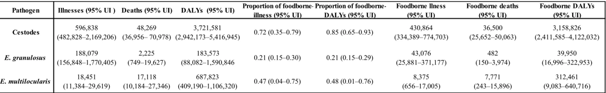
Table 1 Median number of CE foodborne illnesses, deaths, and Disability Adjusted Life Years (DALYs) with 95% uncertainty intervals, 2010
Modified table from (Togerson P.R., 2015) Illnesses are defined as the numbers of new cases in 2010.
Pathogen Illnesses (95% UI ) Deaths (95% UI) DALYs (95% UI) Proportion of
foodborne-illness (95% UI) Proportion of foodborne-DALYs (95% UI) Foodborne llness (95% UI) Foodborne deaths (95% UI) Foodborne DALYs (95% UI) Cestodes 596,838 (482,828–2,169,206) 48,269 (36,956– 70,978) 3,721,581 (2,942,173–5,416,945) 0.72 (0.35–0.79) 0.85 (0.65–0.93) 430,864 (334,389–774,703) 36,500 (25,652–50,063) 3,158,826 (2,411,585–4,122,032) E. granulosus 188,079 (156,848–1,770,405) 2,225 (749–19,627) 183,573 (88,082–1,590,846 0.21 (0.15–0.30) 0.21 (0.15–0.29) 43,076 (25,881–371,177) 482 (150–3,974) 39,950 (16,996–322,953) E. multilocularis 18,451 (11,384–29,619) 17,118 (10,184–27,346) 687,823 (409,190–1,106,320) 0.47 (0.04–0.75) 0.48 (0.01–0.76) 8,375 (656–17,005) 7,771 (243–15,896) 312,461 (9,083–640,716)
Ylenia Pilicchi – Cystic echinococcosis in cattle: histological and proteomic features of inflammation – Corso di Dottorato di Ricerca in “Scienze Veterinarie” - Indirizzo “Produzione, Qualità e Sicurezza Alimentare” – XXXII Ciclo- Università degli Studi di Sassari Combining data of Cystic Echinococcosis and Alveolar Echinococcosis, the total burden is approximately of 871,000 DALYs of which:
- CE 184,000, 95% [UI 88,100–1.59 million] DALYs; - AE 688,000, 95% [UI 409,000–1.1 million] DALYs.
Global costs for CE treatment has been estimated in 4.1 billion dollars per year, of which 46% destinated to human therapy and 54% associated with animal treatment (WHO, 2016 ) (WHO, 2017).
1.2 General morphology of the genus Echinococcus
1.2.1 Adult
The adult tapeworm measures approximately 7 mm (rarely exceeds this lenght), constituted by no more than six segments. The anterior end is characterized by a specialised attachment structure, called scolex, able to make contact with the intestine of the definitive host, by the presence of four muscular suckers on the rostrellum and two rows of hooks.
The body of the tapeworm is called strobila, and reproductive units, named proglottids, (2-6 segments), give it a peculiar segmented appearance.
The adult is hermaphrodite, it shows reproductive ducts in the lateral genital pore. The cirrus sac is prominent, horizontal or tilted anteriorly, while the vitellarium is globular.
The last proglottid could be almost completely filled by the uterus that dilates after fertilisation when eggs are fully developed.
1.2.2 Eggs
The eggs, produced in the terminal proglottid (definitive host), measure 30 to 40 µm in diameter. Eggs are composed by the oncosphere, or hexacanth embryo, that is the first larval stage surrounded by different envelops. The keratinised layer is responsible of the dark striated appearance and gives high resistance to a wide range of environmental temperatures
Ylenia Pilicchi – Cystic echinococcosis in cattle: histological and proteomic features of inflammation – Corso di Dottorato di Ricerca in “Scienze Veterinarie” - Indirizzo “Produzione, Qualità e Sicurezza Alimentare” – XXXII Ciclo- Università degli Studi di Sassari and physiochemical conditions, while the other layers disappear once released from the definitive hosts.
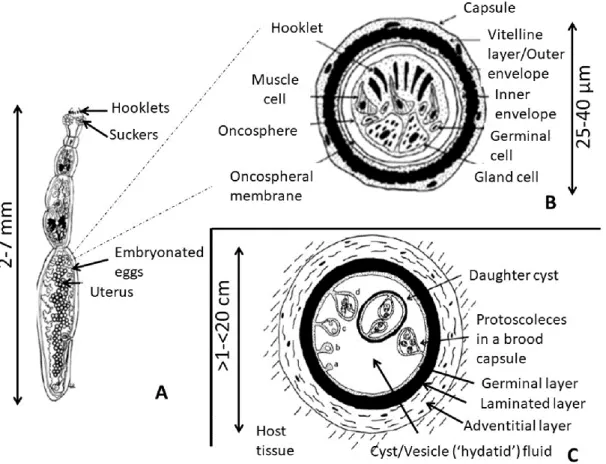
1.2.3 Metacestode
The metacestode is the larval stage of the parasite and develops in the intermediate hosts. It consists of a bladder surrounded by two layers, the acellular and outer one, called laminated layer, and the nucleated inner one, called germinal layer (GL). The latter is responsible for the asexual budding to brood capsules and protoscoleces develop from the inner wall of the brood capsules. The last component of cysts is the hydatid fluid (HF), formed by a complex of parasite-derived proteins, whose production is mostly due to GL. Some of its components are highly immunogenic (Manzano-Román R., 2015).Rupture of the cysts may result in the spillage of hydatid material and could cause an immediate anaphylactic reaction or a development of secondary cysts in contaminated sites (Katz A.M., 1958).
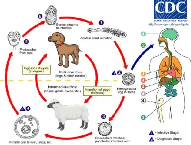
1.3 Life-cycle and transmission of Echinococcus granulosus
Figure 1 Life cycle of E. granulosus Image from the Centers for Disease Control and Prevention
Ylenia Pilicchi – Cystic echinococcosis in cattle: histological and proteomic features of inflammation – Corso di Dottorato di Ricerca in “Scienze Veterinarie” - Indirizzo “Produzione, Qualità e Sicurezza Alimentare” – XXXII Ciclo- Università degli Studi di Sassari Echinococcus needs two mammalian hosts for completion of its life cycle, a carnivore
definitive host, in which the adult develops in the intestine, and an herbivore intermediate host in which metacestode grows-up in an internal organ.
Intermediate hosts become infected through the ingestion of eggs, released from the definitive hosts with the feces and spread on the grass and water.
In suitable environmental conditions, the eggs acquire an extreme resistance, and remain infective for several months in a wide range of temperatures, from 4°C to 15°C. However, they are susceptible to high temperature, UV exposition and desiccation (Gemmell M.A., 1968). Nevertheless, eggs are extremely resistant to low temperatures depending on the timing of exposure.
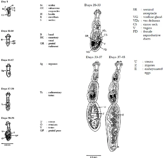
1.3.1 Definitive hosts
The definitive host becomes infected by the ingestion of viable protoscoleces. If the latter are contained in the cyst, the excystment is favourite by the masticatory process and furthermore by the pepsin action in the stomach. Protoscoleces undergo a change in their apical part constituted by suckers, rostrellum and hooks, invaginating within the mucopolysaccharide-coated basal region of the protoscolex tegument.
Evagination is first stage in the development of the adult form of the parasite in the definitive host . Protoscoleces evaginate in response to several stimuli as temperature, osmotic pressure and agitation. Evagination time is changeable, but generally starts after 6 hours and takes approximately three days (Thompson R.C.A., 1977). Aerobiosis is the essential condition for the evagination, whereas some enzimes and bile, even if not essential, can favour theprocess (Smyth J.D., 1967).
In the first phases after evagination, protoscoleces use their energy to quickly locate and attach to the mucosal surface of Lieberkühn's crypts. Some protoscoleces reach the crypts in six hours after infections, using glycogen as a reserve of energy (Smyth J.D., 1967).
Ylenia Pilicchi – Cystic echinococcosis in cattle: histological and proteomic features of inflammation – Corso di Dottorato di Ricerca in “Scienze Veterinarie” - Indirizzo “Produzione, Qualità e Sicurezza Alimentare” – XXXII Ciclo- Università degli Studi di Sassari Once in the small intestine, protoscoleces start the differentiation and divide into four phases: proglottisation, maturation, growth, and segmentation. Proglottisation and maturation lead to the germinal differentiation, while the others two to somatic differentiation (Thompson R.C.A., 1995). The proglottisation of protoscoleces is characterized by a high number of calcareus corpuscles, that disappeared at day 11 to 14. In this second stage, lateral excretory canals become evident and the first proglottid starts to be visible, due to a constriction that marks the area of the first segment. From the 14th to 17th days the genital rudiment divides into two parts, with the full development of a first segment. The formation of the second proglottid is anticipated by the formation of rudimentary tests in the first one. Male genitalia is completed at 20-28 days, when female’s one is still developing. Starting from day 28 both male and female genitalia are fully mature, penultimate proglottid has developed, and a band or the third segment appears.
Finally, from day 33 to 37, ovulation and fertilisation of the last proglottid take place concurrently, the uterus is dilated and contains zygotes in division, and both genitalia degenerate in the terminal proglottid. In the penultimate genitalia are mature and developin the ante-penultimate one. The body is divided into three or four segments.
From 37 to 58 days, terminal proglottid appears gravid with several eggs, the penultimate contains zygotes and the strobila is divided in three to five segments.
The adult parasite starts the senescence after 6-20 months, even if it is reported that worms are capable to survive for 2 years or longer in the definitive hosts (Schantz P.M., 1988). The eggs, from spherical to ellipsoid in shape, are constituted by several layers and measure 30 to 40 µm in diameter.
Ylenia Pilicchi – Cystic echinococcosis in cattle: histological and proteomic features of inflammation – Corso di Dottorato di Ricerca in “Scienze Veterinarie” - Indirizzo “Produzione, Qualità e Sicurezza Alimentare” – XXXII Ciclo- Università degli Studi di Sassari The first envelope called embryophore gives protection to the embryo (oncosphere). Thickness and impermeability of eggs are ensured by protection, due to some properties of embryophore, as keratin-like layers held together by a glue substance (Sakamoto T., 1981).
Ylenia Pilicchi – Cystic echinococcosis in cattle: histological and proteomic features of inflammation – Corso di Dottorato di Ricerca in “Scienze Veterinarie” - Indirizzo “Produzione, Qualità e Sicurezza Alimentare” – XXXII Ciclo- Università degli Studi di Sassari 1.3.2 Intermediate hosts
Once ingested by the intermediate host, eggs undergo to the hatching enabled by the passive disaggregation of embryophoric blocks in the gastric and in the intestinal envinroment, exerted by proteolytic enzymes, such as pepsin and prancreatin (Thompson R.C.A. and Lymbery A.J., 1995).
The proper activation of the oncosphere begins after it’s liberation from the envelope (Lethbridge R., 1980), by the modification in the membrane permeability exerted by the bile salts (Smyth J. D., 1969). The oncosphere in now enabled to penetrate the small intestine through hooks movements in the apical portion of the villi and reaches the lamina propria in 3 to 120 minutes after the hatching (Lethbridge R., 1980). Finally, oncosphere is passively carried through the vessels to the liver and other organs such as kidneys, spleen, muscles, brain and many others (Thompson R.C.A. and Lymbery A.J., 1995). Once arrived in the final location of the intermediate hosts, the oncosphere develops in metacestode larval stage, able to continue the parasite life cycle.
The full development in metacestode (post-oncospheral process) occurs in two weeks. During this time the oncosphere reorganizes itself in terms of cellular proliferation, hooks degeneration, muscular atrophy, vesciculization, and central cavity formation and finally development of both germinal and laminated layers (Heath D. D. and Lawrence S. B., 1976). The formation of protoscoleces begins within the hatching capsules, through an asexual asynchronous reproduction process, which leads to the simultaneous presence of different stages of protoscolic maturation. (Thompson R.C.A., 1995).
Germinal layer (GL) plays a role in the secretion of molecules probably involved in the evasion mechanism from host immune response, such as myo-inositol hexakiphosphate IP6, able to inhibit the complement activation, allowing cyst’s establishment and survival (Breijo M., 2008).
Ylenia Pilicchi – Cystic echinococcosis in cattle: histological and proteomic features of inflammation – Corso di Dottorato di Ricerca in “Scienze Veterinarie” - Indirizzo “Produzione, Qualità e Sicurezza Alimentare” – XXXII Ciclo- Università degli Studi di Sassari GL is surrounded by the laminated layer (LL), a carbohydrate-protein complex of highly glycosylated mucin and glycoprotein (Kilejian A. and Schwabe C. W., 1971) which gives intracystic tension and works as a physiochemical barrier.
LL is a network of microfibrils with aggregates of electron-dense material. It has been demonstrated that LL is entirely of parasiteic orgine, produced by the GL and plays a role in the defence of metacestode favouring the protection from immunological attack. Immunoglobulin can pass through LL, an inert barrier, whereas the GL can regulate the penetration of molecules inside the cyst cavity. It has been recently demonstrated that LL protects the metacestode by preventing the nitric oxide production by the host , through the induction of an increase in the arginase activity of macrophages (Amri M. and Touil-Boukoffa C., 2015).
After the post-oncospheral development, the host produces the adventitial layer (AL), a fibrous capsule constituted by several layers of inflammatory cells. Depending on the strenght of the host immune response two possibilities can take place. A strong inflammatory reaction causes the degeneration and death of the parasite, whereas, as a second option, a favourable envinroment for the oncosphere can lead to the production of a fibrous capsule. Several months may be necessary before protoscoleces production (fertile stage of metacestode). Although E. granulosus usually produces unilocular cavity cysts, the formation of secondary chambers, starting from the cyst wall and in communication with the central cavity, is not a rare event. Central cavity and chambers can be in communication by incomplete septa, or alternatively cysts may adhere to each other and produce clusters of small cysts of different size.
Each cyst may contain thousands of protoscoleces, each of which able to develops into an adult worm in the definitive host, even if some cysts are unable to produce protoscoleces (sterile metacestode).
Ylenia Pilicchi – Cystic echinococcosis in cattle: histological and proteomic features of inflammation – Corso di Dottorato di Ricerca in “Scienze Veterinarie” - Indirizzo “Produzione, Qualità e Sicurezza Alimentare” – XXXII Ciclo- Università degli Studi di Sassari Once produced, protocoleces are potentially ready for the ingestion from the definitive hosts, reach the upper duodenum where, following several signals as pH change, bile production and temperature increasing can evaginate e progress the cycle.evaginate . From four to six weeks are required to allow the protoscoleces to develop in adult and sexually mature worm.
Ylenia Pilicchi – Cystic echinococcosis in cattle: histological and proteomic features of inflammation – Corso di Dottorato di Ricerca in “Scienze Veterinarie” - Indirizzo “Produzione, Qualità e Sicurezza Alimentare” – XXXII Ciclo- Università degli Studi di Sassari
1.4 Taxonomy
Humans and livestock echinococcosis are parasitic zoonotic diseases long known, with several names, since the ancient age, due to the larval or adult stage of the cestode belonging to the species Echinococcus spp.
Until the end of the 19th century different names, based on the morphology of the parasite or the host origin, have been used (Abuladze K. I., 1970). The first valid name was Hydatigena
granulosa, given by Batsch in 1786 and based on a fertile cyst of sheep observed in
Germany. In 1801, Rudolphi assigned the name Echinococcus, based on the aspect of protoscoleces. Hence from the union of these two names ‘granulosa’ and ‘Echinococcus’ originated the term Echinococcus granulosus still in use today (Romig T., 2015). From the end of 19th century, although there was still confusion about the names, the term was used to identify both the larval and adult stage of the parasite. Echinococcus multilocularis, causative agent of alveolar echinococcosis (AE) has been described by Leuckart in 1863. 1.4.1 Species of the genus Echinococcus
Based on the morphology of the adult worm, several species of Echinococcus have been described, E. multilocularis by Leuckart (1863) and E. oligarthra (Diesing K.M., 1863), E.
vogeli in 1972 (Rausch R.L., 1972) and finally E. shiquicus in 2005 (Xiao N., 2005).
1.4.1.1 Subspecies
In order to clarify the relationships between species and subspecies of E. granulosus, an informal system of intraspecific strains has been proposed, that included eleven strains called: sheep, Tasmanian sheep, buffalo, horse, cattle, camel, pig, variant pig (or humas-pig), American cervid, Fennoscandian cervid, and lion strain.
Ylenia Pilicchi – Cystic echinococcosis in cattle: histological and proteomic features of inflammation – Corso di Dottorato di Ricerca in “Scienze Veterinarie” - Indirizzo “Produzione, Qualità e Sicurezza Alimentare” – XXXII Ciclo- Università degli Studi di Sassari Although the strains characterization was conducted on the basis of host-specificity, geographical distribution, morphology and stages of development, starting from 1990s molecular biology techniques start to become more useful for the strains identification and definition. Identification of Echinoccocus and Taenia species, subspecies and strains were conducted through the use of several molecular methods by specific nuclear or mitochondrial genomes. The publication of the partial sequences of two mitochondrial genes, cox1 and nad1, for seven strains of the species E. granulosus and for the other three species E.
multilocularis, E. vogeli and E. oligarthra, was a decisive turning point in the determination
of relationships and taxonomy of Echinococcus species and strains (Bowles J., 1992) (Bowles J. a. M., 1993). The nomenclature system was upgraded throughout the introduction of the term genotype instead of strains, even if after the terms were used as synonymous. To the seven strains/genotypes of E. granulosus, three new of them were added: the American cervid strain (G8) (Bowles J. B. D., 1994), the variant-pig or human-pig strain (G9) (Scott J.C., 1997) and the Fennoscandian cervid strain (G10) (Lavikainen A., 2003 ).
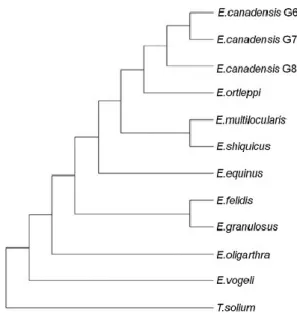
Figure 4 Cladogram on phylogenetic relationships, based on four mitochondrial genes (cox1, nad1,
Ylenia Pilicchi – Cystic echinococcosis in cattle: histological and proteomic features of inflammation – Corso di Dottorato di Ricerca in “Scienze Veterinarie” - Indirizzo “Produzione, Qualità e Sicurezza Alimentare” – XXXII Ciclo- Università degli Studi di Sassari Currently, several species within the genus Echinococcus are recognised, showing differences in definitive and intermediate/aberrant hosts and in the geographical distribution. In humans, different species of the parasite are recognised as the causative agents of well characterised pathologic entities such as Echinococcus granulosus in the cystic echinococcosis (CE), Echinococcus multilocularis in the alveolar echinococcosis (AE) and
E. oligarthra and E. vogeli in the polycystic echinococcosis (PE). The genus Echinococcus
presents a high variability in terms of hosts specificity, morphology, antigenicity, development rate and transmission cycle.
Table 2 Taxonomy of the genus Echinococcus
Species Subspecies Strains Genotypes
Echinococcus granulosus E. granulosus s.s. Sheep G1
(Batsch 1796) Tasmanian sheep G2
Buffalo G3 E. equinus Horse G4 E. ortleppi Cattle G5 E. canadensis Camel G6 Pig G7 Intermedius (camel/pig) G6/7 American Cervid G8 Fennoscandian G10
Pig-human or pig variant G9
E. felidis Lion
E. shiquicus E. multilocularis
E. oligarthra E. vogeli
Ylenia Pilicchi – Cystic echinococcosis in cattle: histological and proteomic features of inflammation – Corso di Dottorato di Ricerca in “Scienze Veterinarie” - Indirizzo “Produzione, Qualità e Sicurezza Alimentare” – XXXII Ciclo- Università degli Studi di Sassari 1.4.2 Echinococcus granulosus complex
Molecular studies, based on mitochondrial DNA analysis, demonstrated the genetic variability of the complex Echinococcus granulosus. Ten distinct genotypes have been identified within this complex, including two sheep strains (G1 and G2), two bovid strains (G3 and G5), one horse strain (G4), one camel strain (G6), two pig strain (G7 and G9) and two cervid strains (G8 and G10).
For the parasite E. granulosus complex both wild-life and domestic cycle have been reported. The first one is characterised in the ancestral form by a wild cycle involving wolves and cervids (moose and reindeer), maintained by a predator-prey relationship, while the domestic cycle seems to have evolved from the cervid cycle adapting in ungulates due to the human intervention in the breeding process (Eckert J., 2001).
1.4.2.1 Echinococcus granulosus sensu stricto: G1, G2 and G3 strains have been
considered part of the same taxonomic group due to the phylogenetic relationship based on analyses on mitochondrial genome (Nakao M., 2006). E. granulosus s.s is cosmopolitan (worldwide distributed) and its definitive hosts are represented by domestic dog, wolf, dingo, jackal and other canids. The intermediates hosts involved in its cycle are sheep, goat, cattle, pig, camel, buffalo, horse, wild ungulates, marsupials, etc. Human cases have been reported.
1.4.2.2 Echinococcus equinus (G4) is present in Eurasia and Africa, involves dog as
definitive host and horse, cervids and other equids as intermediate . Nohuman cases have been reported reported.
1.4.2.3 Echinococcus ortleppi (G5) is present in Eurasia and Africa, involving the
domestic dog as definitive host and cattle as intermediates. Human diseases has been reported.
Ylenia Pilicchi – Cystic echinococcosis in cattle: histological and proteomic features of inflammation – Corso di Dottorato di Ricerca in “Scienze Veterinarie” - Indirizzo “Produzione, Qualità e Sicurezza Alimentare” – XXXII Ciclo- Università degli Studi di Sassari
1.4.2.4 Echinococcus canadensis (G6, G7, G8, G10), distributed in Eurasia, Africa, North
and South America, involves domestic dog and wolf as definitive hosts and pig, camel and cervids as intermediates, with some cases in humans . Based on an evolutionary species concept, some Authors suggested that E. canadensis should be split into three species: Echinococcus intermedius for G6/G7 (as mentioned below in 1.5 Geographical distribution of Echinococcus spp. and molecular epidemiology CE in livestock and humans as reported by Deplazes et al., (2017)), Echinococcus borealis for G8, and E. canadensis for G10.
1.4.2.5 Echinococcus felidis, typical of Africa, involves lion as the definitive host and
several intermediate hosts as hyena, warthog, zebra, wildebeest, bush pig, buffalo, antelopes, giraffe, hippopotamous. Human cases are not reported.
1.4.2.6 Echinococcus shiquiqus, distributed in the Tibetan Plateau, involves Tibetan fox
as intermediate hosts and Ochotona curzioniae (named Tibetan plateau pika) as intermediates host. No human cases have been reported.
Ylenia Pilicchi – Cystic echinococcosis in cattle: histological and proteomic features of inflammation – Corso di Dottorato di Ricerca in “Scienze Veterinarie” - Indirizzo “Produzione, Qualità e Sicurezza Alimentare” – XXXII Ciclo- Università degli Studi di Sassari
1.4.3 Echinococcus multilocularis
E. multilocularis is characterised by a sylvatic cycle involving foxes and rodents. Predation
of wild animals and capture of rodents can infect domestic cat and dog as intermediate hosts. The cycle is reported in Europe, Japan and other regions. Another way of transmission could be perpetuated by domestic cats and rodents in some areas, although this chance is considered less important. E. multilocularis has been reported in Eurasia and North America affecting all foxes species wolf, racoon, dog, domestic dog and cat as definitive hosts, and arvicoline and microtine rodents and small herbivorous mammals including lagomorphs (e.g. pika); pigs, boars, horses, cattle, nutrias, primates and dog as intermediate hosts, even if the last seven species are considered as accidental hosts. Infection in humans is occasionally reported.
1.4.4 Echinococcus oligarthra
E. oligarthra, present in Central and South America affects wild felids (cougar, jaguar,
ocelot, jaguarondi and Geoffroyi’s cat) as definitive hosts, while agouti and opossum are the intermediates. Human cases have been reported.
1.4.5 Echinococcus vogeli
E. vogeli is distributed in Central and South America (Sousa O.E. and Thatcher V.E., 1969),
withbush dog (Speothos venaticus) and domestic dog as definitive hosts and paca (Cuniculus) as intermediate. It involves also humans. The transmission of this strain is mostly guaranteed by a sylvatic predator/prey cycle, whereas the domestic cycle that involves the dog of some rural area of South America, seems to be responsible of the human’s infection.
Ylenia Pilicchi – Cystic echinococcosis in cattle: histological and proteomic features of inflammation – Corso di Dottorato di Ricerca in “Scienze Veterinarie” - Indirizzo “Produzione, Qualità e Sicurezza Alimentare” – XXXII Ciclo- Università degli Studi di Sassari
1.5 Geographical distribution of Echinococcus spp. and molecular
epidemiology CE in livestock and humans
Echinococcus granulosus is widely distributed throughout the world, with the exception of
few areas such as Iceland, Ireland and Greenland that are considered human CE free (Budke, 2006).
1.5.1 America
1.5.1.1 North America and US
In North America, livestock CE is mostly associated with Echinococcus granlosus sensu
lato that involves sheep, swine and cattle as intermediate hosts.
Canada, Alaska and the northern part of US are characterised by the presence of E.
Canadensis (G8-G10) that involves wolves (including C. lupus) and cervids (moose, Alces alces; caribou, Rangifer tarandus; and elk, or wapiti, Cervus canadensis).
A non-genotyped strain of E. granulosus is involved in the dog-sheep cycle in the western part of the US. G7 strain (Echinococcus intermedius) is responsible for the dog-swine cycle in Mexico, where E. granulosus G1 and E. ortleppi G5 are also present (Deplazes P., Global Distribution of Alveolar and Cystic Echinococcosis, 2017).
A formal surveillance for CE in wildlife species (canids and ungulates) doesn’t exist , while examination at the slaughterhouse is a common practice for domestic livestock in US, Canada and México.
Human CE in North America is primarily due to E. Canadensis (G8), that has cervids as intermediates hosts. Data on CE in human in the North America territory lack due to absence of national register, however two studies demonstrated the presence of G8 strain in Alaska. G5 strain has been reported in Mexico (Maravilla P., 2004) (McManus D.P., 2002).
Ylenia Pilicchi – Cystic echinococcosis in cattle: histological and proteomic features of inflammation – Corso di Dottorato di Ricerca in “Scienze Veterinarie” - Indirizzo “Produzione, Qualità e Sicurezza Alimentare” – XXXII Ciclo- Università degli Studi di Sassari 1.5.1.2 Central America
Cases of human CE have been occasionally reported in different countries of Central America as Costa Rica (Brenes Madrigal R.R., 1977), Guatemala, El Salvador, Honduras, Cuba, Panama (Sánchez G.A., 1992) (Sousa O.E. and Lombardo Ayala J.D., 1965) but information on molecular data and local transmission have not been described. Nevertheless, Cuba is the only country able to shows updates data on human CE (Deplazes P., Global Distribution of Alveolar and Cystic Echinococcosis, 2017).
1.5.1.3 South America
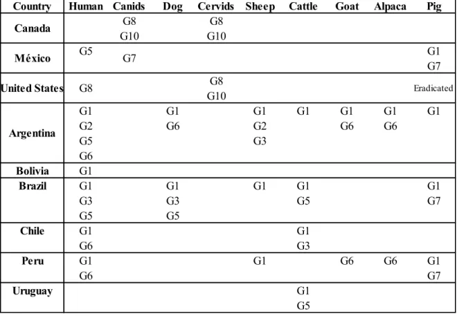
In South America, CE has an important socio-economic impact due to a high prevalence in livestock. It shows higher prevalence rates in Argentina (in particular Patagonia, Pampas, Coast), southwest of Bolivia, south of Brazil, South of Chile, southern and central Peru, and Uruguay. All species of Echinococcus responsible of CE in South America involve dogs as definitive host, and several mammalian species as intermediate hosts. The intermediate hosts of E granulosus s.s (G1-G3) are sheep, cattle, goats, alpaca and swine, for E. ortleppi (G5) is the cattle, E. intermedius G6 involves goats (Soriano S.V., 2010) and the swine is the intermediate host for G7 (Deplazes P., Global Distribution of Alveolar and Cystic Echinococcosis, 2017). The Pan American Health Organization (PAHO) applies a control for the CE in South America through post mortem examination, providing good information about the situation in livestock. On the other hand, since there is no systematic surveillance of infection in dogs, the real prevalence of the disease could be underestimated due to the limited data available.Similarly, due to several notification systems belonging to different countries of South America, the situation regarding human CE is usually underreported (PAHO, 2015), even if studies conducted between 2009 and 2015 reported 29,556 cases, most of which in Peru, 20,785.
Ylenia Pilicchi – Cystic echinococcosis in cattle: histological and proteomic features of inflammation – Corso di Dottorato di Ricerca in “Scienze Veterinarie” - Indirizzo “Produzione, Qualità e Sicurezza Alimentare” – XXXII Ciclo- Università degli Studi di Sassari Table 3 Genotypes and related geographical distribution of Echinococcus granulosus in America (Deplazes P., 2017)
1.5.2 Asia and Eastern Europe
Echinoccocus species are widely distributed in the northern part of Asia (Russian
Federation), involving humans, dogs, farmed animals and wildlife.
Several genotypes are involved in the infection as E. granulosus (G1-G3), E. Canadensis (G8, G10) and E. intermedius (G6/G7).
In Russia, the Federal Centre of Hygiene and Epidemiology reports 573 cases of human CE
per annum (2006 – 2010). However, the data crossing between individual Russian district,
regional centre and local scientific reports showed a higher number of cases per year, equal to 950 (Deplazes P., 2017), confirming CE as major health issue in Russia.
Country Human Canids Dog Cervids Sheep Cattle Goat Alpaca Pig
G8 G8 G10 G10 G5 G1 G7 G8 G10 G1 G1 G1 G1 G1 G1 G1 G2 G6 G2 G6 G6 G5 G3 G6 Bolivia G1 Brazil G1 G1 G1 G1 G1 G3 G3 G5 G7 G5 G5 Chile G1 G1 G6 G3 Peru G1 G1 G6 G6 G1 G6 G7 Uruguay G1 G5 Eradicated Argentina United States G8 México G7 Canada
Ylenia Pilicchi – Cystic echinococcosis in cattle: histological and proteomic features of inflammation – Corso di Dottorato di Ricerca in “Scienze Veterinarie” - Indirizzo “Produzione, Qualità e Sicurezza Alimentare” – XXXII Ciclo- Università degli Studi di Sassari 1.5.2.1 Middle East: Iran, Iraq; Israel, Jordan Kuwait, Lebanon, Oman, Palestine, Qatar, Saudi Arabia, Turkey and Yemen
The Middle East is considered one of most affected area by CE in the world (Cardona G. A. and Carmena D., 2013) (Dakkak A., 2010). Several factors are involved in local CE endemicity, such as a limited knowledge by the population regarding the transmission of the disease, a high number of stray dogs involved in the transmission, poor hygienic conditions in slaughterhouses in addition to home slaughtering; the rural/nomadic lifestyle accompanied by the use of sheep dogs on farms and the consumption of raw vegetables. However, some other factors works as opposing fources acing against the CE transmission, such as the arid / semi-arid climatic conditions that prevent the survival of Echinococcus eggs, religious traditions that do not allow pig breeding and the habit of avoiding dogs in Muslim communities. (Dakkak A., 2010) (Harandi .M.F., 2011) (Rokni M. B., 2009).
E. granlosus s.s (G1-3) is the main responsible for both human and animal CE in the Middle
East (Harandi .M.F., 2011) (Sharbatkhori M., 2009) (Utuk A. E., 2008), followed by E.
intermedius (G6) with an increasing prevalence of the infection in humans (Rostami, 2015)
(Al Kitani F. A., 2015). E. intermedius (G7) have been reported in Turkey and Iran (Fadakar B., 2015) (Eryıldız C. and Şakru N., 2012).
Even if sheep and goats are the most affected intermediate hosts, G1-G3 involves also cattle, buffalo, one- and two-humped camels, horse, donkey, pig, wild sheep (Ovis orientalis), goitered gazelle (Gazalla subgutturosa) and free-ranging Baboon (Papio hamadryas). Human CE is often considered a disease of rural areas, but reports showed an increase in the number of the cases also in urban areas (Ok U.Z., 2007). Data available on human CE in the Middle East come from hospital reports, because some region as Iran, Palestine and Turkey recently took part to the European Register of CE (Rossi P., 2016).
Ylenia Pilicchi – Cystic echinococcosis in cattle: histological and proteomic features of inflammation – Corso di Dottorato di Ricerca in “Scienze Veterinarie” - Indirizzo “Produzione, Qualità e Sicurezza Alimentare” – XXXII Ciclo- Università degli Studi di Sassari 1.5.2.2. South Asia: Afghanistan, Pakistan, India, Bhutan, Nepal, Bangladesh, Sri Lanka, Maldives
E. granulosus. genotypes (G1 and G3) are the most represented in the South of Asia,
affecting mostly sheep and buffaloes. G1-G3 is considered the genotype with highest zoonotic potential, even if also G5 and G6 can infect humans. Prevalence based studies on intermediate hosts reported that CE is endemic in the majority of South Asia.
1.5.2.3 East Asia: China, Mongolia, Korea, Japan
Several genotypes have been reported in East Asia. E. granulosus s.s (G1-3) has been commonly isolated from humans, dogs, sheep, cattle and yaks. E. intermedius (G6) in humans, dogs, cattle and camels. G7, G10, G4 and G5 have been occasionally described. 1.5.2.4 South East Asia: Indonesia, Vietnam, the Philippines, Malaysia, Thailand and the Lao People’s Democratic Republic
CE cycle in South East Asia is not maintained (McManus D.P., 2010), and the existence of CE is questioned, with the hypothesis thet infection could be restricted to wild animals. Infection in animals has been reported in 1974 in Indonesia in a dog (Carney W.P., 1974), bu further studies on 63 dogs in dogs did not demonstrate the presence of E. granulosus. Data are not available in livestock.
Ylenia Pilicchi – Cystic echinococcosis in cattle: histological and proteomic features of inflammation – Corso di Dottorato di Ricerca in “Scienze Veterinarie” - Indirizzo “Produzione, Qualità e Sicurezza Alimentare” – XXXII Ciclo- Università degli Studi di Sassari
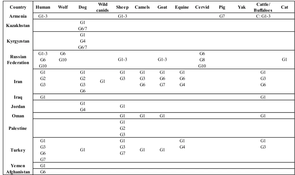
Table 4 Genotypes and related distribution of Echinococcus granulosus in Asia and European part of Russia (Deplazes P., 2017)
Country Human Wolf Dog Wild
canids Sheep Camels Goat Equine Cervid Pig Yak
Cattle/ Buffaloes Cat Armenia G1-3 G1-3 G7 C: G1-3 G1 G6/7 G1 G4 G6/7 G1-3 G6 G6 G6 G10 G8 G10 G10 G1 G1 G1 G1 G1 G1 G1 G2 G2 G3 G3 G6 G6 G3 G3 G3 G6 G7 G4 G6 G6 Iraq G1 G1 G1 G4 Oman G1 G1 G1 G1 G1 G2 G3 G1 G1 G1 G1 G3 G3 G4 G3 G6 G7 G7 Yemen G1 Afghanistan G6 Turkey G1 G1 G1 Palestine Jordan G1 G1 Iran G1 Russian Federation G1-3 G1-3 Kyrgyzstan Kazakhstan
Ylenia Pilicchi – Cystic echinococcosis in cattle: histological and proteomic features of inflammation – Corso di Dottorato di Ricerca in “Scienze Veterinarie” - Indirizzo “Produzione, Qualità e Sicurezza Alimentare” – XXXII Ciclo- Università degli Studi di Sassari
Ylenia Pilicchi – Cystic echinococcosis in cattle: histological and proteomic features of inflammation – Corso di Dottorato di Ricerca in “Scienze Veterinarie” - Indirizzo “Produzione, Qualità e Sicurezza Alimentare” – XXXII Ciclo- Università degli Studi di Sassari 1.5.3 Australia and New Zealand
E. granulosus was introduced in both countries during the European settlement mostly
through sheep livestock (Gemmell M.A., 1990), parasite’s cycle was perpetuated between sheep and dog, or cattle and dog, causing a high rate of infection in humans. Furthermore, the maintenance of the parasite cycle is allowed by wild species suche as dingoes and macropods.
CE in animal decreased during the last 30 years and rarely affected sheep that came into contact with wild animals. The parasite is also infrequent in dog, and generally limited to rural areas where the infection is still present with a low prevalence, in the dog-cattle cycle. Dingoes and macropods marsupial in the eastern and southwest of Australia are susceptible to the infection (Jenkins D.J., 2014).
New Zealand and Australia have implemented successful control programs and the Ministry of Agriculture and Forestry declared New Zealand provisionally CE free in 2002 (Anonymous, 2012).
Human CE cases on the mainland of Australia are rarely reported. Some new cases have been identified, but most of these are attributable to immigrants infected before joining Australia (Thompson R.C.A and Jenkins D.J., 2014). In Tasmania no new cases have been reported (O’Hern J. A. and Cooley L., 2013).
Ylenia Pilicchi – Cystic echinococcosis in cattle: histological and proteomic features of inflammation – Corso di Dottorato di Ricerca in “Scienze Veterinarie” - Indirizzo “Produzione, Qualità e Sicurezza Alimentare” – XXXII Ciclo- Università degli Studi di Sassari 1.5.4 Africa
1.5.4.1 North Africa: Morocco, Algeria, Tunisia, Libya, Egypt
CE is considered one of the most important diseases in North Africa, causing economic losses and human health issues.
The maintenance of the parasite cycle is mainly due to rural domestic transmission, involving dogs as definitive hosts and several intermediate hosts such as sheep, cattle, goats, camels, dromedaries, and donkeys. CE in wild animals involves golden wolves as definitive hosts and wild boars and antelope as intermediate hosts.
The main causes of CE persistence are epidemiological factors such as contacts between human population and livestock, scarce knowledge of the CE disease, high dog to human ratio, poor hygienic conditions in slaughterhouses or the practice of home slaughtering (El Berbri I., 2015). Moreover, the cycle is maintained due to the habit of feeding dogs with offal discarded (Kouidri M., 2012). Incidence rates in human are around 5-10 cases per 100.000 inhabitants (Torgerson P.R. and Macpherson C.N., 2011). CE endemicity is high in all North Africa’s countries (Dakkak A., 2010).
1.5.4.2 Sub-Saharan Africa
CE is widespread in sub-Saharan Africa except in two arid zones in the northern part of the region (Sahara and Sahel). As well as North Africa, in sub-Saharan Africa, CE is highly endemic (Romig T. O. R., 2011) (Magambo J., 2006).
Several genotypes of Echinococcus are present in this area and, among these, E. felidis, seems to be widespread in Eastern and Southern Africa in wild animals with a cycle that involve lion, hyenas and warthogs. There are noreports in domestic animals or human species. Similarly to the previous one, E. equinus is widespread in South Africa, in a wildlife cycle that involve lions, wild dogs and zebras, with no reports in humans.
Ylenia Pilicchi – Cystic echinococcosis in cattle: histological and proteomic features of inflammation – Corso di Dottorato di Ricerca in “Scienze Veterinarie” - Indirizzo “Produzione, Qualità e Sicurezza Alimentare” – XXXII Ciclo- Università degli Studi di Sassari From Sudan to South Africa, E. ortleppi is widespread in the livestock cattle.
E.intermedius is typical of the arid region on the North where camels are the key points for
the transmission of the parasite. G6 is the predominant genotype of the region, perpetuated by goats and wildlife animals. The number of human cases is lower if compared to other regions, leading to suppose that this genotype could be less infective for humans.
1.5.4.3. East Africa: South Sudan, Ethiopia, Eritrea, Somalia, Uganda, Kenya
Eastern part of Africa is mostly affected by E. granulosus s.s. caused by the sheep-dog cycle. Ethiopia is the area of the region with the highest prevalence in sheep, probably because this genotype is responsible for the all fertile cysts (Hailemariam Z., 2012) (Maillard S., 2007). In Kenya the prevalence is moderate even if G1-3 is the major responsible of the infection and cause of all fertile cysts in Maasailand (Addy F., 2012) (Romig T., 2011).
Both countries show a low prevalence in goats and all the fertile cysts are due to E.
intermedius and E. ortleppi.
Cattle, affected by G1-3, are characterised mostly by infertile cysts, whereas infection due to E. ortleppi and G6 strain are less frequent but resulting in a higher rate of fertility (Mbaya H., 2014). South Sudan and Somalia show low rates of infection in sheep due to E.
intermedius G6/7 (Omer R.A., 2010).
Ethiopia, Somalia and North-East of Kenia are characterised by low prevalence in camel, in contrast to North-West part that reported an extremely high prevalence in this species. Most of them are infected by G6/7 and in case of fertility by G1-3 (Dinkel A., 2004).
Wild animals are infected by E. felidis and G1-3 strains affect lions, hyenas and warthog. Cases of human CE has been mostly found in eastern South Sudan, southwest of Ethiopia and northwest of Kenya (Romig T., 2011) , but also in Uganda, Rwanda and Somalia (Babady N.E., 2009).
Ylenia Pilicchi – Cystic echinococcosis in cattle: histological and proteomic features of inflammation – Corso di Dottorato di Ricerca in “Scienze Veterinarie” - Indirizzo “Produzione, Qualità e Sicurezza Alimentare” – XXXII Ciclo- Università degli Studi di Sassari 1.5.4.4. West and Central Africa: Nigeria, Burkina Faso, Cameroon, the Central African Republic, Democratic Republic of Congo
Poor information are available for these countries, however high rates of infection of camels are reported in the northern Nigeria while there is a low prevalence in sheep, goats and cattle. Scarce or old data are described in Burkina Faso, Cameroon, Central African Republic and the Democratic Republic of Congo, suggesting that these countries are poorly affected by CE.
1.5.4.5 Southern Africa: Angola, Zambia, Mozambique, Zimbabwe, Namibia and South Africa
Surveys conducted in livestock in 60’ years demonstrated that the parasite is widespread in South Africa. These information were confirmed by molecular surveys that showed the presence of the genotypes G1-3, G4, G5, G6 and E. felidis. However, accurate data about the impact of the parasite in livestock, wild animals and humans still lack. Human CE is rarely reported due to the absence of surveys, but some case reports exist and indicate that CE is widespread in West and Central Africa.
Ylenia Pilicchi – Cystic echinococcosis in cattle: histological and proteomic features of inflammation – Corso di Dottorato di Ricerca in “Scienze Veterinarie” - Indirizzo “Produzione, Qualità e Sicurezza Alimentare” – XXXII Ciclo- Università degli Studi di Sassari
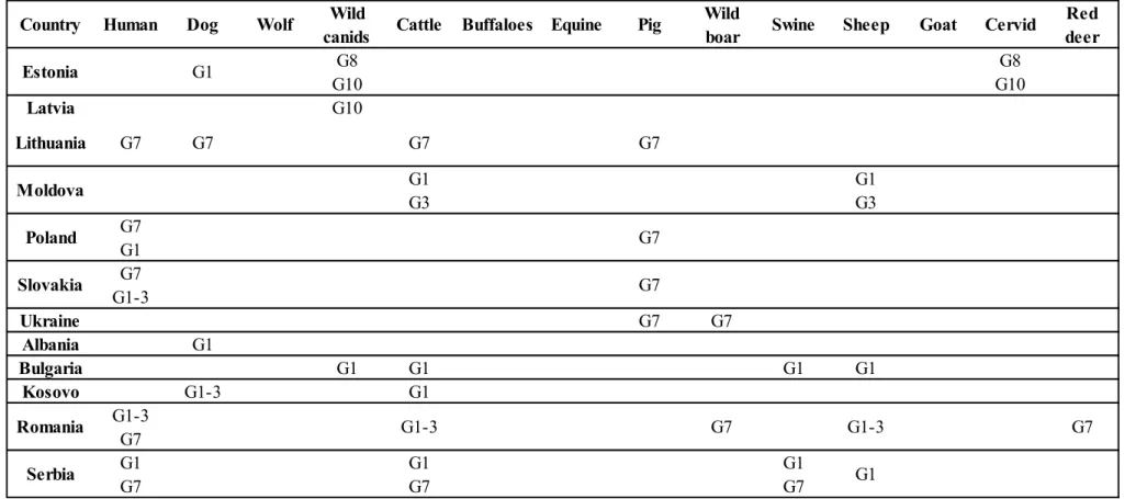
Table 5 Genotypes and related geographical distribution of Echinococcus granulosus in Africa (Deplazes P., 2017)
Cattle/ Wild Buffalo boar Algeria G1 G1 G1 G2 G2 G2 G1 G1 G1 G6 G6 G6 G5 G7 G7 G6 Lybia G1 G1 G1 G1 G1 Morocco G1 G1 G1 G1 G1 G1 G1 G1 G1 G3 G6 G4 G6 G1-3 C: G1-3 G1 G1 G6/7 C: G5 G5 G6/7 C: G6/7 Ghana G6 G1-3 G1-3 G1-3 G1-3 C: G1-3 G1 G1 G6 E. felidis G5 G6/7 C: G5 G5 G5 C: G6/7 G6 G6/7 Mali G6 Kenya G1 G1-3 G1 G1 Ethiopia G1-3 Tunisia G1 G1 G1 G1 G1 G4 Egypt G6 Antelope Swine G1
Camel Pig Warthog
Equine/ Wild Equine
Goat Wild
ruminants
Country Human Dog Wild
canids
Wild
Ylenia Pilicchi – Cystic echinococcosis in cattle: histological and proteomic features of inflammation – Corso di Dottorato di Ricerca in “Scienze Veterinarie” - Indirizzo “Produzione, Qualità e Sicurezza Alimentare” – XXXII Ciclo- Università degli Studi di Sassari
Cattle/ Wild Buffalo boar Mauritania G6 G6 C: G6 G1-3 We: G4 G6/7 G4 We: G5 G4 G5 Somalia G6 G1-3 G5 G6/7 C: G5 C: G6/7 G5 C: G5 G6 C: G6 G1 E. felidis Zambia C: G5 Uganda G6 Sudan G6 G6 G6/7 South Sudan G1-3 G6 South Africa E. felidis E. felidis Namibia G6/7 C: G5 Antelope Swine
Camel Pig Warthog
Equine/ Wild Equine
Goat Wild
ruminants
Country Human Dog Wild
canids
Wild