0
S
iJ
ATLANTO-AXIAL SUBLUXATION WITH SPONDYLOSCHISIS IN DOWN SYNDROME 1417
VOL. 67-A, NO. 9. DECEMBER 985
9. SEMINE, A. A.: ERTEL, A. N.; GOLDBERG, M. J.: and BULL. M. J.: Cervical-Spine Instability in Children with Down Syndrome (Trisomy 21).
J. Bone and Joint Surg. .60-A: 649-652, July 1978.
10. SPITZER. R.: RAB1NOWITCH, J. Y.: and WYBAR. K. C.: A Study of the Abnormalities of the Skull. Teeth and Lenses in Mongolism. Canadian
Med. Assn. J.. 84: 567-572. 1961.
I I . TISHE.ER. JACK, and MARTEL, WIt.LIAM: Dislocation of the Atlas in Mongolism. Preliminary Reprt. Radiology. 84: 9()4-906. 1965.
Coprighi 955 h T1i JouruiI ‘I Bone itid Joitti .5itren .Iieor,s’rattd
Pathology
of Infantile
Cortical
Hyperostosis
(Caffey’s
Disease)
REPORT OF A CASE*t1
BY UGO E. PAZZAGLIA, M.D.*. PAUL D. BYERS, F.R.C.P.#, GIAMPIERO BELUFFI. M.D.. GIUSEPPE CHIRICO, M.D.,
GIORGIO RONDINI, M.D.. AND LUCIANO CECILIANI, M.D., PAVIA, ITALY
F,o,,z the !Sti11t() di Ortopedia e Traiinatologia (I(’I/Univ(’rsit(i di Patio. arid I.R.C.C.S. Polieliiiio San Maueo. P(01a
A few prenatal cases of infantile cortical hyperostosis.
or Caffey’s disease, have been reported’2’#{176}’3. Two fetuses
were dead at the thirty-first and twenty-fourth weeks of
gestation2. In two siblings, reported on by Barba and
Frer-iks, the condition was demonstrated radiographically in
utero during the last month of gestation. One child survived
an unusually severe and protracted course, and the other
recovered in a few weeks.
As a rule the disease appears in the first months of life;
the clinical course is highly variable, with eventual
recov-ery455’5’92. Only five patients are reported to have died9’3
5.24; in none was the cause of death related to Caffey’s
disease. Knowledge of the pathology has come from biopsy
in nine children7”’92, radical amputation in two7, and
nec-ropsy in four9’4’5.
In this paper we report a fatal case, with classic features
apparent at birth, in a child who survived for five and a half
months. A postmortem study was carried out in which the
humerus, tibia, fibula, ribs, vertebrae, and skull were
ex-amined by light and electron microscopy.
Case Report
This girl was born in Pavia. Italy. on February 22, 1982. In neither branch of the family was infantile cortical hyperostosis recognized. The mother was healthy throughout this first pregnancy. which was normal until the fifth month, when polyhydramnios was observed. The mother had been taking Tavor (lorazepam) and Valium (diazepam) until she be-came aware of the pregnancy. Placental detachment at the twenty-ninth
* This article was accepted for publication prior to July 1. 1985. No conflict-of-interest statement was requested from the authors.
1-Read in part at the Congress of the European Society of Paediatric Radiology. Paris. France. May 5. 1983.
1:Supported by Grant CT8l00l2604 from the Italian National Re-search Council and by the Institute of Orthopaedics. University of Pavia.
§Istituto di Ortopedia e Traumatologia dell’Universit#{224} di Pavia, via Taramelli. 27100 Pavia. Italy. Please address reprint requests to Dr. Paz-zaglia.
# Morbid Anatomy Department. Institute of Orthopaedics. London University. Brockley Hill, Stanmore. Middlesex HA7 4LP. United King-dom.
week led to a cesarean delivery. The child. who had anoxia. was maintained by mechanical ventilation for the first five days of life. She was hyptonic. with a weak cry. The skin of all of the limbs was pigmented. and the bones were enlarged and bowed. The skull and thoracic cage were
cx-FIG. I
Radiographic survey of the skeleton made at birth. There are extensive lesions of the long bones. ribs. left scapula. and mandible.
,.
I
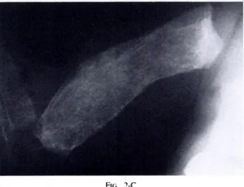
FIG. 2-C
At the age of five months. cortical hone is no longer observable.
1418 U. E. PAZZAGI.IA ET AL.
tilE JOtRNAE. g: BONE AND JOINT SURGERY
FIG. 2-A FI;. 2-B
Figs. 2-A, 2-B, and 2-C: Radiographs of the right humerus. Fig. 2-A: At birth.
Fig. 2-B: At the age of two and one-half months. New tissue is laid down around the original cortical bone. which is progressively remodeled and
resorbed.
trenely soft. The cranial sutures and fontanelles were normal. hut the ribs were expanded. The illness was characterized by anemia. requiring frequent transfusions: difficult fieding. the birth weight being regained only on the forty-fifth day: episodes of urinary tract infection: respiratory difficulty: and progressively niore severe bronchopulmonary infections. leading to death at the age of five and a half months.
Radiographic Observations
At birth, all of the major long hones on both sides, as
well as the mandible, left scapula, and all ribs. were affected
by the same abnormality: an increase in diameter through
periosteal apposition (Fig. I). In the less alfected hones
much of the original structure, normal in dimensions, was
enclosed by the new tissue. Serial radiographs showed that
the abnormal bone increased in amount and the original
bone was progressively remodeled out of existence (Figs.
2-A. 2-B, and 2-C).
Both femora, tibiae, and fibulae were bowed (Fig. 3).
The skull, metacarpals, phalanges of the hands, right
scap-ula, both clavicles, vertebrae, pelvis. metatarsals, and
pha-langes of the feet were normal at birth. However. by the
age of five months the vertebrae had collapsed.
Laboratory Findings
The following measurements and tests were carried out
on one or more occasions and had nortiial results: Coonibs’
test; coagulation tests; polymorphonuclear leukocyte count;
glucose; nitrogen; creatine; hilirubin; serum
glutaniic-ox-aloacetic transaminase; serum glutamic-pyruvic
transami-nase; sodiuni chloride: potassium; calcium; niagnesium;
inorganic phosphate; alkaline phosphatase; parathyroid and
thyroid hormones ; Venereal Disease Research Laboratory
test: antibodies against toxoplasma. mononucleosis , and
ru-bella; viral cultures; bacterial blood cultures; and a
karyo-grani.
The abnormal laboratory-test results were for anemia
(despite repeated blood transfusions): immunoglobulins in
the first weeks o1 life (lgG, 961 milligrams per 100
Fio. 3
PATHOLOGY OF INFANTILE CORTICAl. HYPEROSTOSIS 1419
VOL. 67-A, NO. 9. DECEMBER 1955
TABLE I
LEVEI.S OF HYDROXtYStNE GtvcosiDEs
Diglycoside
Age Diglycoside Monoglycoside Hydroxylysine
Monoglycoside Ratio
Per Cent Glycosylation
(Days) (ing/l(X) sil) (,ngJO() ??lI) (,n,, 1(W) izI)
24 305 164 137 1.85 77
75 282 221 365 1.27 58
1gM, seventy-two milligrams per 100 milliliters); and
un-nary hydroxylysine glycosides (Table 1).
Necropsv
Necropsy was performed twenty hours after death. The
following bones, with the peniosteum. were removed: right
humerus, right tibia and fibula, second through sixth
tho-tenial were fixed in glutaraldehyde, buffered at pH 7.4,
post-fixed in osmium tetroxide, embedded in Epon 812 resin.
sectioned, and then stained with uranyl acetate and lead
citrate.
Observations
Bilateral bronchopneurnonia and suppurative
bronchi-Lateral radiographs of the right leg. made at birth (a) and at the age of five months (b). The tihial and fihular diaphyses show 90 degrees of bowing.
racic vertebrae, second through sixth ribs from both sides.
sternum, and right parietal bone. Radiographs ofthese bones
were made and specimens were selected for light and
dcc-tron microscopy. Those intended for study by light
mi-croscopy were fixed in neutral forrnalin. Some were
decalcified in EDTA, embedded in paraffin. and sectioned.
The stains that were used were hematoxylin and eosin,
pe-riodic acid-Schiff, and alcian blue in 0.05, 0.3, and
0.8-molar solutions of magnesium chloride, some being
pre-treated with bovine testicular hyaluronidase.
The undecalcified specimens were embedded in
meth-ylmethacrylate, sectioned, and stained with solochrome. For
electron microscopy, specimens of epiphyseal cartilage,
growth plate, periosteum. and subperiosteal calcified
ma-tis were confirmed at autopsy, and signs of terminal heart
failure were found.
All of the bones that were studied showed the same
features. with only minor differences. The condition of the
humerus will he described fully as an example,
supple-mented by comments on the other bones.
No cortical bone was detectable either by the naked
eye or radiographically. The epiphyses were entirely
car-tilaginous. with a normal vascular pattern. The growth-plate
organization and calcification were normal, but some
col-umns of chondrocytes persisted into the primary ossification
zone. introducing an abnormal element in the development
of primary metaphyseal bone trabeculae. Nevertheless,
FIi;. 4 1420 U. E. PAZZAGI.IA ET At.. p m’
F:.:E
,
t , ‘.‘.p&4,:.i,;i
mfT(#{149};i
-: : .--:. . - :-,, I #{149} . %#{149} # ::
.-:
#{149}
‘ ,.- b;3
:‘..- -dI::#{149}#{149}.
.T:
:,t:;::I t:
“1,
;:Y. , .Decalcified section of the right humeral growth plate and metaphysis. There is persistence of hypertrophic chondrocyte columns in primary me-taphyseal trabeculae. Remodeling is carried out by osteoclasts. A richly
vascular fibrous tissue with many small round lymphoid cells is present
between trabeculae (hematoxylin and eosin. x 120).
trabeculae of the usual woven collagen structure. They were
f’ully calcified. with osteoid seams ofnonrnal width. Between
the trabeculae was a richly vascular fibrous tissue, infiltrated
by lymphocytes (Fig. 4). Hernatopoietic cells were few and
aggregated in foci of active heniatopoiesis.
All Of the diaphyseal hone was abnormal; it was
con-stituted by partially calcified trabeculae between which was
fibrous tissue. continuous with that in the metaphysis.
Scat-tered hematopoietic cells were present. hut no other
inflam-matorycells. There was a marrow cavity in the middle of
the diiphysis. approximately corresponding to the
diaph-yseal channel of a normal bone, which was filled with
he-matopoietic marrow (Fig. 5).
The diaphyseal trabeculae had an immature structure,
with irregularly distributed large, round lacunae. There was
vigorous remodeling; the extensive osteoid was mostly
coy-ered by active osteoblasts, and osteoclasts were readily
iden-tified on the resorption surfaces. Inert surfaces were few.
The replacetiient of the original bone had been so extensive
that it was only with great difficulty that definitely lamellar
bone was found. It was incorporated in the new tissue (Figs.
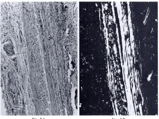
6-A and 6-B). The fibrous peniosteum was thick, with
sev-eral layers of large collagen fibers running in a longitudinal,
transverse. or oblique direction (Figs. 7-A and 7-B). Many
vessels were present in the outer layers.
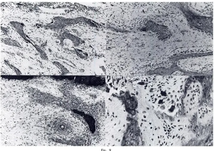
Focally. the inner surface of the peniosteum was
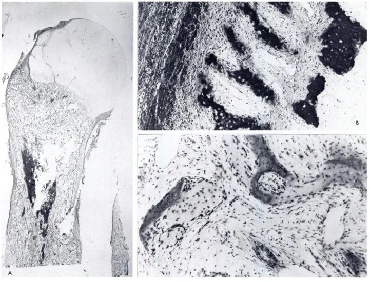
in-tensely cellular, with frequent mitotic sites (Figs. 8-A,
8-B, and 8-C) where slender trabeculae were being formed
perpendicular to the long axis of the bone. These trabeculae
are destined to develop into tiiore substantial structures as
they come to lie beneath later additions. Elsewhere,
sub-peniosteal bone resorption was active (Fig. 9). The
distri-bution pattern of periosteal activities was clearly
deter-mining the shape of the bone.
In the tibia (Fig. 10) there was no trace at all of the
diaphyseal channel. The costochondral cartilage had the
same irregularity as did the humeral growth plate. No trace
of diploe was observed in the calvaniurn. whose structure,
uniform throughout, was of the same stout immature
tra-beculae as elsewhere. The vertebrae were reduced in height
and, again, had the same irregularities in the growth plate.
The marrow space was niainly fibrous, but there were
re-sidual areas of hematopoiesis.
The periodic acid-Schiff and alcian-blue preparations
showed a normal matrix in the epiphyseal and growth-plate
cartilage . Ultrastructurally non-spec ific degenerative
changes were present in epiphyseal cells. However, the cells
of the physis were better preserved and a normal pattern of
maturation was present. with mucopolysaccharide
precur-sons in large cisternae in cells of the proliferative layer. An
unusual feature was the presence of a paranuclear lipidic
droplet in many cells (Fig. 1 1). Hydroxyapatite crystal
dep-osition was normal.
The outer peniosteal layer was composed almost
en-tirely of densely packed, normal collagen fibnils. The
re-maining fibers. rather less than I per cent. were abnormal,
irregularly distributed, large. aperiodic, and composed of
-:
. - : .; \. ::::‘-. ,) 1 ‘.
. ..,x
#{149}‘ .: ,;
.; #{149}?Z . . I #{149} .-,, 1. -1A‘fl . . . 1,.. ‘- . . #{149}‘#{149}‘% ., J -, ,j.:
‘ .. A 4.,#{149} ‘ $ ‘. #{149}:‘-..4 . . i..t#{149}, .FIi. 6-A FIG. 6-B
VOL. 67-A, NO. 9. [)ECEMBER 1955
.fVA .., . - .. ., t..
.,
.? : , ,, !t;
ijy (1PATHOLOGY OF INFANTILE CORTICAL HYPEROSTOSIS
Fi;. 5
1421
The right humeral diaphysis. .4. Decalcified longitudinal section of the humerus. No cortical hone is observed. and the diaphysis is entirely constituted
of cancellous hone. Bone-marrow tissue is present in the middle of the diaphysis (hematoxylin and eosin. x 2.5). II. Undecalcified section showing
subpenosteal formation of partially calcified bone trabeculae (solochrome. X lOt)). C. Decalcilied section showing cancellous hone of the diaphysis
and fibrous tissue s ith lytiiphoid cells between traheculae (hematoxvlin and eosin. X 1(K)).
they were elastic fibrils. but this could not be confirmed.
An incomplete initial phase of calcification of the
per-iosteal osteoid conformed with the high rate of
bone-re-modeling, as did the large osteocyte lacunae with their
irregular perilacunar uncalcified matrix.
Discussion
The diagnosis in this case lies between infantile cortical
Figs. 6-A and 6-B: Decalcified section of the right humerus (hematoxylin and eosin. X 4(K)). Fig. 6-A: Residual lamellar bone is incorporated in the new immature bone.
. . ,. . - .I ., . ,%,:.
‘:;:;.
\‘
.FIG. 7-A Fi;. 7-B
Ftc;. 8-A
FIG. 8-B Fto. 8-C
THE JOURNAE. OF BONE ANt) JOINT SURGERY
I 422 .‘, - ,..-‘; - ‘ , ..:-. -,. . ‘ s.’.’ I l_,; ,
;s
_.) . .-.. . S .‘ -. . 5S5 - - .,. . . S . . .. U. E. PAZZAGLIA ET AL.U-1 - c,*S ... .4 4 ‘;S
‘,
_c
II I. ‘S . ‘U 14 /h.
VOl.. 67-A, NO. 9. t)ECEMBER 955
PATHOLOGY OF INFANTILE CORTICAl. HYPEROSTOSIS 1423
hyperostosis and osteogenesis imperfecta. Factors that argue
against the latter are the involvement of the mandible, the
absence of a hereditary component, the lack of other signs
such as blue sclerae and delicate skin. the failure of the
original cortical tissue to persist. and, most important of all.
the total absence of fractures. From the time of recognition
of the infant’s illness at birth, this feature was looked for.
Although bowing of long bones was present, it was clearly
tense proliferation of subperiosteal cells, which, in two
pa-tients was misdiagnosed as malignant disease and led to
amputation7; subperiosteal new-bone formation; and fibrosis
of bone marrow. Nevertheless, some unusual features were
present: onset in utero (only two cases previously reported);
generalized bone lesions, including the skull and the
ver-tebrae (features not previously reported); and a fatal outcome
(only five cases previously reported). The etiology is
un--
.T
T
:r::4.
4 5’ .,T.’
..,. -‘ 4’:
:
‘ 5- -ft’ t_-:
--I 1 . ..
: .: - -- -. . .. .,.w ... . - -.
. . ..-...,-
? . ,. ‘.:‘-.- :Y: - .. .--:---. ; . . #{149}. _&__.S., . .- .1 ... .-.-.
(..
. ... C #{149} - . . - - - .-C
Fio. 9Remodeling of imiiture bone trabeculae in the right humerus. A. Osteoclasts are resorhing immature bone traheculae (decalcified section, hematoxylin and eosin. X (Xi). B. Osteoblasts line the surface of traheculae. and medullary fibrosis is also evident (decalcified section, hematoxylin and eosin.
x 1(X)). C. Note the wide osteoid border of immature bone traheculae (undecalcified section, solochrome, x 100). D. Immature bone trabeculae and osteoclasts (decalcified section . hematoxylin and eosin . X 400).
a manifestation of periosteal remodeling. and could not be
satisfactorily imputed to intrauterine fracture. The child was
sufficiently active, and had to be handled to a degree that
would certainly have resulted in fractures in a patient with
osteogenesis imperfecta of this severity. The
histopatholog-ical observations were the same as those in previous biopsy
and necropsy reports. and may be summarized as follows:
thickening of the periosteum; absence of cortical bone;
in-known but. given the pathogenetic mechanism of increased
periosteal growth and diffuse diaphyseal remodeling of
af-fected bones, the possibilities can be narrowed down to
genetic, inflammatory, and metabolic causes. The last of
these is not supported by the extensive evidence from
re-ported cases or by the many investigations of this patient.
and will not be considered further. The first is suggested
by the appearance of disease in siblings’3224 and in
Figs. 7-A and 7-B: Decalcified section of the right humerus (hematoxylin and eositi. X 100).
Fig. 7-A: The periosteum is thickened. with multiple layers of collagen fibers and vessels on the outer surface. Fig. 7-B: The same field under polarized light.
Figs. 8-A. 8-B. and 8-C: Decalcified section of the right humerus.
Fig. 8-A: There is intense suhperiosteal cellular proliferation (heniatoxylin and eostn. x 100). Fig. 8-B: The same field as Fig. 8-A. tinder polarized light.
1,
C
FIG. 10
1424 U. E. PAZZAGI.IA ET Al..
THE JOURNAL OF BONE AND JOINT SURGERY
families’2’52”22. Opposed to this is the natural course of the
disease in the majority of’ patients - onset. progression,
and recovery without sequelae - which is best accounted
for by an inflammatory pathogenesis. Moreover. several
features of the case of our patient do not fit in with the
genetic hypothesis: epiphyseal and metaphyseal
develop-ment was found to be normal; the main site of pathogenetic
activity was diaphyseal periosteal activity and cortical
re-modeling; and the initial development of the long bones
A
does not refute the hypothesis. The example of Paget’s
disease o1 bone usefully illustrates this. In addition, the
disease started in utero, when the infant was not under
observation. Moreover, in biopsy specimens taken early in
the course of the disease in other patients. the presence of
inflammation has been established7. Support for the
hy-pothesis of an inflammatory etiology due to infection in this
patient rests on the increased concentrations of plasma IgA
and 1gM. which were determined in the first week of life.
Decalcified sections of the right tibia. A. Section in the sagittal plane (hentatoxylin and eosin. x 2.5). B. Suhperiosteal osteoclastic resorption on the COflVCX side (hematoxylin and eosin. X 100). C. Pronounced osteoblastic activity on the concave side (hematoxylin and eosin. x 100).
must have been normal, as evidenced by the presence on
radiographs. made at birth, of cortex in the humerus and
by residual lamellar bone identifiable post mortem.
Bone is capable only of a limited type of response.
which consists of turnover and remodeling. The rate and
duration of its two components. resorption and formation.
can vary widely. so that end results may differ in degree,
but not in kind. The absence of any sign of osseous
inflam-mation, either clinically or pathologically. in this patient.
They point to stimulation of immunity before birth. Raised
levels of inimunoglobulins have been reported in two other
cases’3. The inimunoglobulin determinations were not
re-peated because of the many blood and plasma transfusions,
necessitated by the persistent anemia. which in turn was
caused by widespread myelotibrosis. Although many clues
observed in this and other reported cases suggest an
inflam-matory pathogenesis and infective etiology. no definitive
FIG. 12
VOL. 67-A, NO. 9. DECEMBER 1985
PATHOLOGY OF INFANTILE CORTICAL HYPEROSTOSIS 1425
. , . .. . .. 7
. ...
- ..
v... #{149} - ..,.
. :. . __._t.#{248}._t -I :r” ,,
#{149}: ‘- :“..#{149} ‘ . ‘,‘;:‘‘ ,.. . .,t,. ...S:,::
..
. .-: ,, -. .-.
&: ;“* , ? ‘....c.cI
:cr-:; ‘ ‘ - s--, . . ‘...,-..
A :‘ : ,. .. . . . (, .; .5.. . . . . . . . .. .; . - , , -: ,..“. . _5 . . ... - ...
.,.. ....- . S. . -I ..,-.,..
4. . . . . . . . . . . ‘ . .,. . .. ,. , . . .-. . . . 1 7’ ‘-#{149}‘ - -. . . . . .... ..,-.-. . . . .‘,. t.-,
. ; ::. ...
. Fi;. I IUndecalcified section of the humeral growth plate. The cytoplasm of a hypertrophic chondrocyte contains a lipid droplet and clusters of hydroxyapatite crystals ( x 12,600).
Decalcified section of the diaphyseal periosteum of the humerus. The periosteum is formed by densely packed bundles of collagen fibrils, among them large fibrils without periodicity ( x 21,000).
we are studying only the expression of the bone’s reaction will reveal the etiology of this disease.
to the primary injury, which occurs in utero. The dictum Non: The auihor-. hank Mr... G. Bodini or hcr it.-chnicaI a..sisiancc: Professor G. Cciia or
hydroxystnc gscosidc dcierminaiions: md Professor C. Dcll’Orfx’ and Dr. D. Quacci for the
1426 u. E. PAZZAGLIA ET AL.
References
I. BARBA, W. P., II, and FRERIKS, D. J.: The Familial Occurrence of Infantile Cortical Hyperostosis in Utero. J. Pediat., 42: 141-150, 1953.
2. BENNETT, H. S., and NELSON, T. R.: Case Reports. Prenatal Cortical Hyperostosis. British J. Radiol. , 26: 47-49, 1953.
3. Boyrs, J. G., and DEMY, N. 0.: Infantile Cortical Hyperostosis: A Familial Disease? Am. J. Roentgenol., 65: 924-930, 1951. 4. CAFFEY, JOHN: On Some Late Skeletal Changes in Chronic Infantile Cortical Hyperostosis. Radiology, 59: 651-657, 1952.
5. CAFFEY, JOHN: Infantile Cortical Hyperostosis: A Review of the Clinical and Radiographic Features. Proc. Roy. Soc. Med. , 50: 347-354, 1957.
6. CAFFEY, JOHN, and SILVERMAN, W. A.: Infantile Cortical Hyperostoses. Preliminary Report on a New Syndrome. Am. J. Roentgenol. , 54:
1-16, 1945.
7. EVERSOLE, S. L. , JR.; HOLMAN, 0. H.; and ROBINSON, R. A.: Hitherto Undescribed Characteristics of the Pathology of Infantile Cortical
Hyperostosis. Caffey’s Disease. Bull. Johns Hopkins Hosp. , 101: 80-99. 1957.
8. FINSTERBUSH, A. , and RANG, M.: Infantile Cortical Hyperostosis. Follow-up of 29 Cases. Acta Orthop. Scandinavica, 46: 726-736, 1975.
9. JAFFE, H. L.: Metabolic, Degenerative, and Inflammatory Diseases of Bones and Joints, pp. 281-297. Munich, Urban and Schwarzenberg, 1972.
10. JENKINSON, E. L.; PFISTERER, W. H. ; LATTEIER, K. K.;and MARTIN, MARY: A Prenatal Diagnosis of Osteopetrosis. Am. J. Roentgenol. , 49:
455-462, 1943.
11. KATZ, J. M.; KIRKPATRICK, J. A.; PAPANICOLAOU, N.; and DESAI, P.: Case Report 139. Skel. Radiol., 6: 77-80, 1981.
12. KITCHIN, I. D.: An Atypical Case of Infantile Cortical Hyperostoses. J. Bone and Joint Surg. , 33-B(2): 248-250, 1951.
13. LABRUNE, M.; GUEDJ, G.; VIAL, M.; BESSIS, R.; ROSET, M.; and KERBRAT, V.: Maladie de Caffey adebut ant#{233}natal. Arch. francaise pediat.,
40: 39-43, 1983.
14. MATHESON, W. J., and MARKHAM, MARY: Infantile Cortical Hyperostosis. British Med. J., 1: 742-744, 1952.
15. MOSSBERGER, J. I.: Infantile Cortical Hyperostosis. Report of a Case with Observations at Autopsy. Am. J. Dis. Child., 80: 610-620, 1950.
16. PAJEWSKI, M., and VuR, E.: Late Manifestations of Infantile Cortical Hyperostosis (Caffey’s Disease). British J. Radiol. , 40: 90-95, 1967.
17. RACHMANDER, V., and RAMKISSOON, R.: Infantile Cortical Hyperostosis with Raised Immunoglobulins. Arch. Dis. Child., 53: 426-428, 1978.
18. SAUL, R. A.; LEE, W. H.; and STEVENSON, R. E.: Caffey’s Disease Revisited. Further Evidence for Autosomal Dominant Inheritance with
Incomplete Penetrance. Am. J. Dis. Child. . 136: 56-60, 1982.
19. SHERMAN, M. S. , and HELLYER, D. T.: Infantile Cortical Hyperostosis. Review of the Literature and Report of Five Cases. Am. J. Roentgenol.,
63: 212-222, 1950.
20. SIDBURY, J. B. ,JR. , and SIDBURY, J. B.: Infantile Cortical Hyperostosis. An Inquiry into the Etiology and Pathogenesis. New England J. Med., 250: 309-314, 1954.
21 . STAHELI, L. T.;CHURCH, C. C. ;and WARD, B. H. : Infantile Cortical Hyperostosis (Caffey’s Disease). Sixteen Cases with a Late Follow-up of
Eight. J. Am. Med. Assn. , 203: 384-388, 1968.
22. TAMPAS, J. P.; VAN BUSKIRK, F. W.; PETERSON, 0. S., JR.; and SOULE, A. B.: Infantile Cortical Hyperostosis. J. Am. Med. Assn., 175:
491-493, 1961.
23. TEMPERLEY, I. J.;DOUGLAS, S. J. ; and REES, J. P. R. : Raised Immunoglobulin Levels and Thrombocytosis in Infantile Cortical Hyperostosis.
Arch. Dis. Child. ,47: 982-983, 1972.
24. VAN ZEBEN, W.: Infantile Cortical Hyperostoses. Acta Paediat. ,35: 10-20, 1948.