UNIVERSITA' DEGLI STUDI DI SALERNO
FACOLTA' DI SCIENZE MATEMATICHE FISICHE E NATURALI
Dottorato di ricerca in Chimica
Synthesis and properties of
linear and cyclic peptoids
-X Cycle- Nuova serie (2008-2011)
Tutor: Prof. Francesco De Riccardis PhD candidate: Chiara De Cola
Co-tutor: Prof. Irene Izzo
1
INDEX
CHAPTER 1: INTRODUCTION,3
1.1 PEPTIDOMIMETICS,5
1.2 PEPTOIDS: A PROMISING CLASS OF PEPTIDOMIMETICS,9 1.3 CONFORMATIONAL STUDIES OF PEPTOIDS,11
1.4 PEPTOIDS’ APPLICATIONS,14 1.5 PEPTOID SINTHESYS,39
1.6 SYNTHESYS OF PNA MONOMERS AND OLIGOMERS,41
1.7 AIMS OF THE WORK,49
CHAPTER 2: CARBOXYALKYL PEPTOID PNAS: SYNTHESIS AND HYBRIDIZATION PROPERTIES,51 2.1 INTRODUCTION,51
2.2. RESULTS AND DISCUSSION,55 2.3. CONCLUSIONS,60
2.4 EXPERIMENTAL SECTION,60
CHAPTER 3: STRUCTURAL ANALYSIS OF CYCLOPEPTOIDS AND THEIR COMPLEXES,80 3.1 INTRODUCTION,80
3.2. RESULTS AND DISCUSSION,85 3.3. CONCLUSIONS,102
3.4 EXPERIMENTAL SECTION,103
CHAPTER 4: CATIONIC CYCLOPEPTOIDS AS POTENTIAL MACROCYCLIC NONVIRAL VECTORS,115
4.1 INTRODUCTION,115
4.2. RESULTS AND DISCUSSION,122 4.3. CONCLUSIONS,125
4.4 EXPERIMENTAL SECTION,125
CHAPTER 5: COMPLEXATION WITH GD(III) OF CARBOXYETHYL CYCLOPEPTOIDS AS POSSIBLE CONTRAST AGENTS IN MRI,132
5.1 INTRODUCTION,132
5.2 LARIAT ETHER AND CLICK CHEMISTRY,135 5.3 RESULTS AND DISCUSSION,141
5.4 EXPERIMENTAL SECTION,145
CHAPTER 6: CYCLOPEPTOIDS AS MIMETIC OF NATURAL DEFENSINS,157
6.1 INTRODUCTION,157
6.2 RESULTS AND DISCUSSION,162 6.3 CONCLUSIONS,167
2
List of abbreviations Cbz Benzyl chloroformate DCC N,N’-Dicyclohexylcarbodiimide DCM Dichloromethane DIPEA Diisopropylethylamine DMF N, N’-dimethylformamideFmoc Fluorenylmethyloxycarbonyl chloride
HBTU O-Benzotriazole-N,N,N',N'-tetramethyl-uronium-hexafluorophosphate
HATU O-(7-Azabenzotriazol-1-yl)-N,N,N',N'-tetramethyluronium hexafluorophosphate
PyBOP benzotriazol-1-yl-oxytripyrrolidinophosphonium hexafluorophosphate
PNA Peptide nucleic acid
t-Bu terz-Butyl
3
Chapter 1
1. Introduction“Giunto a questo punto della vita, quale chimico, davanti alla tabella del Sistema Periodico, o agli indici monumentali del Beilstein o del Landolt, non vi ravvisa sparsi i tristi brandelli, o i trofei, del proprio passato professionale? Non ha che da sfogliare un qualsiasi trattato, e le memorie sorgono a grappoli: c’è fra noi chi ha legato il suo destino, indelebilmente, al bromo o al propilene o al gruppo –NCO o all’acido glutammico; ed ogni studente in chimica, davanti ad un qualsiasi trattato, dovrebbe essere consapevole che in una di quelle pagine, forse in una sola riga o formula o parola, sta scritto il suo avvenire, in caratteri indecifrabili, ma che diventeranno chiari <<POI>>: dopo il successo o l’errore o la colpa, la vittoria o la disfatta.
Ogni chimico non più giovane, riaprendo alla pagina << verhangnisvoll >> quel medesimo trattato, è percosso da amore o disgusto, si rallegra o si dispera.”.
Da “Il Sistema Periodico”, Primo Levi.
Proteins are vital for essentially every known organism. The development of a deeper understanding of protein–protein interactions and the design of novel peptides, which selectively interact with proteins are fields of active research.
One way how nature controls the protein functions within living cells is by regulating protein– protein interactions. These interactions exist on nearly every level of cellular function which means they are of key importance for virtually every process in a living organism. Regulation of the protein-protein interactions plays a crucial role in unicellular and multicellular organisms, including man, and represents the perfect example of molecular recognition1.
Synthetic methods like the solid-phase peptide synthesis (SPPS) developed by B. Merrifield2 made it possible to synthesize polypeptides for pharmacological and clinical testing as well as for use as drugs or in diagnostics.
As a result, different new peptide-based drugs are at present accessible for the treatment of prostate and breast cancer, as HIV protease inhibitors or as ACE inhibitors to treat hypertension and congestive heart failures, to mention only few examples1.
Unfortunately, these small peptides typically show high conformational flexibility and a low in-vivo stability which hampers their application as tools in medicinal diagnostics or molecular biology. A major difficulty in these studies is the conformational flexibility of most peptides and the high dependence of their conformations on the surrounding environment which often leads to a conformational equilibrium. The high flexibility of natural polypeptides is due to the multiple conformations that are energetically possible for each residue of the incorporated amino acids. Every amino acid has two degrees of conformational freedom, N–Cα (Φ) and Cα–CO (Ψ) resulting in approximately 9 stable local conformations1. For a small peptide with only 40 amino acids in length the
1
A. Grauer, B. König Eur. J. Org. Chem. 2009, 5099–5111.
4
number of possible conformations which need to be considered escalates to nearly 10403. This extraordinary high flexibility of natural amino acids leads to the fact that short polypeptides consisting of the 20 proteinogenic amino acids rarely form any stable 3D structures in solution1. There are only few examples reported in the literature where short to medium-sized peptides (<30–50 amino acids) were able to form stable structures. In most cases they exist in aqueous solution in numerous dynamically interconverting conformations. Moreover, the number of stable short peptide structures, which are available is very limited, because of the need to use amino acids having a strong structure inducing effect like for example helix-inducing amino acids as leucine, glutamic acid or lysine. In addition, it is dubious whether the solid state conformations determined by X-ray analysis are identical to those occurring in solution or during the interactions of proteins with each other1. Despite their wide range of important bioactivities, polypeptides are generally poor drugs. Typically, they are rapidly degraded by proteases in vivo, and are frequently immunogenic.
This fact has inspired prevalent efforts to develop peptide mimics for biomedical applications, a task that presents formidable challenges in molecular design.
1.1 Peptidomimetics
One very versatile strategy to overcome such drawbacks is the use of peptidomimetics4. These are small molecules, which mimic natural peptides or proteins and thus produce the same biological effects as their natural role models.
They also often show a decreased activity in comparison to the protein from which they are derived. These mimetics should have the ability to bind to their natural targets in the same way as the natural peptide sequences, from which their structure was derived, do and should produce the same biological effects. It is possible to design these molecules in such a way that they show the same biological effects as their peptide role models but with enhanced properties like a higher proteolytic stability, higher bioavailability and also often with improved selectivity or potency. This makes them interesting targets for the discovery of new drug candidates.
For the progress of potent peptidomimetics, it is required to understand the forces that lead to protein–protein interactions with nanomolar or often even higher affinities.
These strong interactions between peptides and their corresponding proteins are mainly based on side chain interactions indicating that the peptide backbone itself is not an absolute requirement for high affinities.
This allows chemists to design peptidomimetics basically from any scaffold known in chemistry by replacing the amide backbone partially or completely by other structures. Peptidomimetics,furthermore, can have some peculiar qualities, such as a good solubility in aqueous solutions, access to facile sequences-specific assembly of monomers containing chemically diverse side chains and the capacity to form stable, biomimetic folded structures5.
Most important is that the backbone is able to place the amino acid side chains in a defined 3D-position to allow interactions with the target protein, too. Therefore, it is necessary to develop an idea of the required structure of the peptidomimetic to show a high activity against its biological target.
3J. Venkatraman, S. C. Shankaramma, P. Balaram, Chem. Rev. 2001, 101, 3131–3152. 4
J. A. Patch, K. Kirshenbaum, S. L. Seurynck, R. N. Zuckermann and A. E. Barron, in Pseudo-peptides in Drug Development, ed. P. E. Nielsen, Wiley-VCH, Weinheim, Germany, 2004, 1–31.
5
The most significant parameters for an optimal peptidomimetics are: stereochemistry, charge and hydrophobicity, and these parameters can be examined by systematic exchange of single amino acids with modified amino acid. As a result, the key residues, which are essential for the biological activity, can be identified. As next step the 3D arrangement of these key residues needs to be analyzed by the use of compounds with rigid conformations to identify the most active structure1. In general, the development of peptidomimetics is based mainly on the knowledge of the electronic, conformational and topochemical properties of the native peptide to its target.
Two structural factors are particularly important for the synthesis of peptidomimetics with high biological activity: firstly the mimetic has to have a convenient fit to the binding site and secondly the functional groups, polar and hydrophobic regions of the mimetic need to be placed in defined positions to allow the useful interactions to take place1.
One very successful approach to overcome these drawbacks is the introduction of conformational constraints into the peptide sequence. This can be done for example by the incorporation of amino acids, which can only adopt a very limited number of different conformations, or by cyclisation (main chain to main chain; side chain to main chain or side chain to side chain).5
Peptidomimetics, furthermore, can contain two different modifications: amino acid modifications or peptides‘ backbone modifications.
Figure 1.1 reports the most important ways to modify the backbone of peptides at different positions.
Figure 1.1. Some of the more common modifications to the peptide backbone (adapted from
literature).6
5
a) C. Toniolo, M. Goodman, Introduction to the Synthesis of Peptidomimetics, in: Methods of Organic Chemistry:
Synthesis of Peptides and Peptidomimetics (Ed.: M. Goodman), Thieme, Stuttgart, New York, 2003, vol. E22c, p.
1–2; b) D. J. Hill, M. J. Mio, R. B. Prince, T. S. Hughes, J. S. Moore, Chem. Rev. 2001, 101, 3893–4012. 6J. Gante, Angew. Chem. Int. Ed. Engl. 1994, 33, 1699–1720.
6
Backbone peptides modifications are a method for synthesize optimal peptidomimetics, in particular is possible:
the replacement of the α-CH group by nitrogen to form azapeptides, the change from amide to ester bond to get depsipeptides,
the exchange of the carbonyl function by a CH2 group,
the extension of the backbone (β-amino acids and γ-amino acids), the amide bond inversion (a retro-inverse peptidomimetic),
The carba, alkene or hydroxyethylene groups are used in exchange for the amide bond.
The shift of the alkyl group from α-CH group to α-N group.
Most of these modifications do not guide to a higher restriction of the global conformations, but they have influence on the secondary structure due to the altered intramolecular interactions like different hydrogen bonding. Additionally, the length of the backbone can be different and a higher proteolytic stability occurs in most cases 1.
1.2 Peptoids: A Promising Class of Peptidomimetics.
If we shift the chain of α-CH group by one position on the peptide backbone, we produced the disappearance of all the intra-chain stereogenic centers and the formation of a sequence of variously substituted N-alkylglycines (figure 1.2).
Figure 1.2. Comparison of a portion of a peptide chain with a portion of a peptoid chain.
Oligomers of N-substituted glycine, or peptoids, were developed by Zuckermann and co-workers in the early 1990‘s7
. They were initially proposed as an accessible class of molecules from which lead compounds could be identified for drug discovery.
Peptoids can be described as mimics of α-peptides in which the side chain is attached to the backbone amide nitrogen instead of the α-carbon (figure 1.2). These oligomers are an attractive scaffold for biological applications because they can be generated using a straightforward, modular synthesis that allows the incorporation of a wide variety of functionalities8. Peptoids have been evaluated as tools to
7
R. J. Simon, R. S. Kania, R. N. Zuckermann, V. D. Huebner, D. A. Jewell, S. Banville, S. Ng, L.Wang, S. Rosenberg, C. K. Marlowe, D. C. Spellmeyer, R. Tan, A. D. Frankel, D. V. Santi, F. E. Cohen and P. A. Bartlett, Proc. Natl. Acad. Sci. U. S. A., 1992, 89, 9367–9371.
7
study biomolecular interactions,8 and also hold significant promise for therapeutic applications due to
their enhanced proteolytic stabilities8 and increased cellular permeabilities9 relative to α-peptides. Biologically active peptoids have also been discovered by rational design (i.e., using molecular modeling), and were synthesized either individually or in parallel focused libraries10. For some applications, a well-defined structure is also necessary for peptoid function to display the functionality in a particular orientation, or to adopt a conformation that promotes interaction with other molecules. However, in other biological applications, peptoids lacking defined structures appear to possess superior activities over structured peptoids.
This introduction will focus primarily on the relationship between peptoid structure and function. A comprehensive review of peptoids in drug discovery, detailing peptoid synthesis, biological applications, and structural studies, was published by Barron, Kirshenbaum, Zuckermann, and co-workers in 20044.Since then, significant advances have been made in these areas, and new applications for peptoids have emerged. In addition, new peptoid secondary structural motifs have been reported, as well as strategies to stabilize those structures. Lastly, the emergence of peptoid with tertiary structures has driven chemists towards new structures with peculiar properties and side chains. Peptoid monomers
are linked through polyimide bonds, in contrast to the amide bonds of peptides. Unfortunately, peptoids do not have the hydrogen of the peptide secondary amide, and are consequently incapable of forming the same types of hydrogen bond networks that stabilize peptide helices and β-sheets.
The peptoids oligomers backbone is achiral; however stereogenic centers can be included in the side chains to obtain secondary structures with a preferred handedness4. In addition, peptoids carrying N-substituted versions of the proteinogenic side chains are highly resistant to degradation by proteases, which is an important attribute of a pharmacologically useful peptide mimic4.
1.3 Conformational studies of peptoids
The fact that peptoids are able to form a variety of secondary structural elements, including helices and hairpin turns, suggests a range of possible conformations that can allow the generation of functional folds.11
Some studies of molecular mechanics, have demonstrated that peptoid oligomers bearing bulky chiral (S)-N-(1-phenylethyl) side chains would adopt a polyproline type I helical conformation, in agreement with subsequent experimental findings12.
Kirshenbaum at al.12 has shown agreement between theoretical models and the trans amide of N-aryl peptoids, and suggested that they may form polyproline type II helices. Combined, these studies suggest that the backbone conformational propensities evident at the local level may be readily translated into the conformations of larger oligomers chains.
N-α-chiral side chains were shown to promote the folding of these structures in both solution and the solid state, despite the lack of main chain chirality and secondary amide hydrogen bond donors crucial to the formation of many α-peptide secondary structures.
8
S. M. Miller, R. J. Simon, S. Ng, R. N. Zuckermann, J. M. Kerr, W. H. Moos, Bioorg. Med. Chem. Lett., 1994, 4, 2657–2662.
9
Y. U.Kwon and T. Kodadek, J. Am. Chem. Soc., 2007, 129, 1508–1509.
10 T. Hara, S. R. Durell, M. C. Myers and D. H. Appella, J. Am. Chem. Soc., 2006, 128, 1995–2004. 11
G. L. Butterfoss, P. D. Renfrew, B. Kuhlman, K. Kirshenbaum, R. Bonneau, J. Am. Chem. Soc., 2009, 131, 16798–16807.
8
While computational studies initially suggested that steric interactions between N-α-chiral aromatic side chains and the peptoid backbone primarily dictated helix formation,both intra- and intermolecular aromatic stacking interactions12 have also been proposed to participate in stabilizing such helices13.
In addition to this consideration, Gorske et al.14 selected side chain functionalities to look at the effects of four key types of noncovalent interactions on peptoid amide cis/trans equilibrium: (1) n→π* interactions between an amide and an aromatic ring (n→π*Ar), (2) n→π* interactions between two carbonyls (n→π* C=O), (3) side chain-backbone steric interactions, and (4) side chain-backbone hydrogen bonding interactions. In figure 1.3 are reported, as example, only n→π*Ar and n→π*C=O interactions.
A B
Figure 1.3. A: (Left) n→π*Ar interaction (indicated by the red arrow) proposed to increase of
Kcis/trans (equilibrium constant between cis and trans conformation) for peptoid backbone amides. (Right)
Newman projection depicting the n→π*Ar interaction. B: (Left) n→π*C=O interaction (indicated by the red arrow) proposed to reduce Kcis/trans for the donating amide in peptoids. (Right) Newman
projection depicting the n→π*C=O interaction.
Other classes of peptoid side chains have been designed to introduce dipole-dipole, hydrogen bonding, and electrostatic interactions stabilizing the peptoid helix.
In addition, such constraints may further rigidify peptoid structure, potentially increasing the ability of peptoid sequences for selective molecular recognition.
In a relatively recent contribution Kirshenbaum15 reported that peptoids undergo to a very efficient head-to-tail cyclisation using standard coupling agents. The introduction of the covalent constraint enforces conformational ordering, thus facilitating the crystallization of a cyclic peptoid hexamer and a cyclic peptoid octamer.
Peptoids can form well-defined three-dimensional folds in solution, too. In fact, peptoid oligomers with α-chiral side chains were shown to adopt helical structures 16
; a threaded loop structure was formed
12 C. W. Wu, T. J. Sanborn, R. N. Zuckermann, A. E. Barron, J. Am. Chem. Soc. 2001, 123, 2958–2963. 13
T. J. Sanborn, C. W. Wu, R. N. Zuckermann, A. E. Barron, Biopolymers, 2002, 63, 12–20. 14
B. C. Gorske, J. R. Stringer, B. L. Bastian, S. A. Fowler, H. E. Blackwell , J. Am. Chem. Soc., 2009, 131, 16555–16567.
15 S. B. Y. Shin, B. Yoo, L. J. Todaro, K. Kirshenbaum, J. Am. Chem. Soc., 2007, 129, 3218-3225. 16
(a) K. Kirshenbaum, A. E. Barron, R. A. Goldsmith, P. Armand, E. K. Bradley, K. T. V. Truong, K. A. Dill, F. E. Cohen, R. N. Zuckermann, Proc. Natl. Acad. Sci. U.S.A. 1998, 95, 4303–4308. (b) P. Armand, K. Kirshenbaum, R. A. Goldsmith, S. Farr-Jones, A. E. Barron, K. T. V. Truong, K. A. Dill, D. F. Mierke, F. E. Cohen, R. N. Zuckermann, E. K. Bradley, Proc. Natl. Acad. Sci. U.S.A. 1998, 95, 4309–4314. (c) C. W. Wu, K. Kirshenbaum, T. J. Sanborn, J. A. Patch, K. Huang, K. A. Dill, R. N. Zuckermann, A. E. Barron, J. Am. Chem. Soc. 2003, 125, 13525–13530.
9
by intramolecular hydrogen bonds in peptoid nonamers20; head-to-tail macrocyclizations provided conformationally restricted cyclic peptoids.
These studies demonstrate the importance of (1) access to chemically diverse monomer units and (2) precise control of secondary structures to expand applications of peptoid helices.
The degree of helical structure increases as chain length grows, and for these oligomers becomes fully developed at length of approximately 13 residues. Aromatic side chain-containing peptoid helices generally give rise to CD spectra that are strongly reminiscent of that of a peptide α-helix, while peptoid helices based on aliphatic groups give rise to a CD spectrum that resembles the polyproline type-I helical.
1.4 Peptoids’ Applications.
The well-defined helical structure associated with appropriately substituted peptoid oligomers can be employed to construct compounds that closely mimic the structures and functions of certain bioactive peptides. In this paragraph, are shown some examples of peptoids that have antibacterial and antimicrobial properties, molecular recognition properties, of metal complexing peptoids, of catalytic peptoids, and of peptoids tagged with nucleobases.
1.4.1 Antibacterial and antimicrobial properties
The antibiotic activities of structurally diverse sets of peptides/peptoids derive from their action on microbial cytoplasmic membranes. The model proposed by Shai–Matsuzaki–Huan17 (SMH) presumes alteration and permeabilization of the phospholipid bilayer with irreversible damage of the critical membrane functions. Cyclization of linear peptide/peptoid precursors (as a mean to obtain conformational order), has been often neglected18,despite the fact that nature offers a vast assortment of powerful cyclic antimicrobial peptides19. However, macrocyclization of N-substituted glycines gives
17
(a) Matsuzaki, K. Biochim. Biophys. Acta 1999, 1462, 1; (b) Yang, L.; Weiss, T. M.; Lehrer, R. I.; Huang, H. W. Biophys. J. 2000, 79, 2002; (c) Shai, Y. Biochim.Biophys. Acta 1999, 1462, 55.
18
Chongsiriwatana, N. P.; Patch, J. A.; Czyzewski, A. M.; Dohm, M. T.; Ivankin, A.; Gidalevitz, D.; Zuckermann, R. N.; Barron, A. E. Proc. Natl. Acad. Sci. U.S.A. 2008, 105, 2794.
19
Interesting examples are: (a) Motiei, L.; Rahimipour, S.; Thayer, D. A.; Wong, C. H.; Ghadiri, M. R. Chem. Commun. 2009, 3693; (b) Fletcher, J. T.; Finlay, J. A.; Callow, J. A.; Ghadiri, M. R. Chem. Eur. J. 2007, 13, 4008; (c) Au, V. S.; Bremner, J. B.; Coates, J.; Keller, P. A.; Pyne, S. G. Tetrahedron 2006, 62, 9373; (d) Fernandez-Lopez, S.; Kim, H.-S.; Choi, E. C.; Delgado, M.; Granja, J. R.; Khasanov, A.; Kraehenbuehl, K.; Long, G.; Weinberger, D. A.; Wilcoxen, K. M.; Ghadiri, M. R. Nature 2001, 412, 452; (e) Casnati, A.; Fabbi, M.; Pellizzi, N.; Pochini, A.; Sansone, F.; Ungaro, R.; Di Modugno, E.; Tarzia, G. Bioorg. Med. Chem. Lett. 1996, 6, 2699; (f) Robinson, J. A.; Shankaramma, C. S.; Jetter, P.; Kienzl, U.; Schwendener, R. A.; Vrijbloed, J. W.; Obrecht, D. Bioorg. Med. Chem. 2005, 13, 2055.
10
circular peptoids20, showing reduced conformational freedom21and excellent membrane-permeabilizing activity22.
Antimicrobial peptides (AMPs) are found in myriad organisms and are highly effective against bacterial infections23. The mechanism of action for most AMPs is permeabilization of the bacterial cytoplasmic membrane, which is facilitated by their amphipathic structure24.
The cationic region of AMPs confers a degree of selectivity for the membranes of bacterial cells over mammalian cells, which have negatively charged and neutral membranes, respectively. The hydrophobic portions of AMPs are supposed to mediate insertion into the bacterial cell membrane. Although AMPs possess many positive attributes, they have not been developed as drugs due to the poor pharmacokinetics of α-peptides. This problem creates an opportunity to develop peptoid mimics of AMPs as antibiotics and has sparked considerable research in this area25.
De Riccardis26 et al. investigated the antimicrobial activities of five new cyclic cationic hexameric α-peptoids comparing their efficacy with the linear cationic and the cyclic neutral counterparts (figure
1.4).
20
(a) Craik, D. J.; Cemazar, M.; Daly, N. L. Curr. Opin. Drug Discovery Dev. 2007, 10, 176; (b) Trabi, M.; Craik, D. J. Trend Biochem. Sci. 2002, 27, 132.
21
(a) Maulucci, N.; Izzo, I.; Bifulco, G.; Aliberti, A.; De Cola, C.; Comegna, D.; Gaeta, C.; Napolitano, A.; Pizza, C.; Tedesco, C.; Flot, D.; De Riccardis, F. Chem. Commun. 2008, 3927; (b) Kwon, Y.-U.; Kodadek, T. Chem. Commun. 2008, 5704; (c) Vercillo, O. E.; Andrade, C. K. Z.; Wessjohann, L. A. Org. Lett. 2008, 10, 205; (d) Vaz, B.; Brunsveld, L. Org. Biomol. Chem. 2008, 6, 2988; (e) Wessjohann, L. A.; Andrade, C. K. Z.; Vercillo, O. E.; Rivera, D. G.. In Targets in Heterocyclic Systems; Attanasi, O. A., Spinelli, D., Eds.; Italian Society of Chemistry, 2007; Vol. 10, pp 24–53; (f) Shin, S. B. Y.; Yoo, B.; Todaro, L. J.; Kirshenbaum, K. J. Am. Chem. Soc. 2007, 129, 3218; (g) Hioki, H.; Kinami, H.; Yoshida, A.; Kojima, A.; Kodama, M.; Taraoka, S.; Ueda, K.; Katsu, T. Tetrahedron Lett. 2004, 45, 1091.
22
(a) Chatterjee, J.; Mierke, D.; Kessler, H. Chem. Eur. J. 2008, 14, 1508; (b) Chatterjee, J.; Mierke, D.; Kessler, H. J. Am. Chem. Soc. 2006, 128, 15164; (c) Nnanabu, E.; Burgess, K. Org. Lett. 2006, 8, 1259; (d) Sutton, P. W.; Bradley, A.; Farràs, J.; Romea, P.; Urpì, F.; Vilarrasa, J. Tetrahedron 2000, 56, 7947; (e) Sutton, P. W.; Bradley, A.; Elsegood, M. R.; Farràs, J.; Jackson, R. F. W.; Romea, P.; Urpì, F.; Vilarrasa, J. Tetrahedron Lett. 1999, 40, 2629.
23
A. Peschel and H.-G. Sahl, Nat. Rev. Microbiol., 2006, 4, 529–536. 24
R. E. W. Hancock and H.-G. Sahl, Nat. Biotechnol., 2006, 24, 1551–1557.
25 For a review of antimicrobial peptoids, see: I. Masip, E. Pèrez Payà, A. Messeguer, Comb. Chem. High Throughput Screen., 2005, 8, 235–239.
11
Figure 1.4. Structures of synthesized linear and cyclic peptoids described by De Riccardis at al. Bn
= benzyl group; Boc= t-butoxycarbonyl group.
The synthesized peptoids have been assayed against clinically relevant bacteria and fungi, including Escherichia coli, Staphylococcus aureus, amphotericin β-resistant Candida albicans, and Cryptococcus neoformans27.
The purpose of this study was to explore the biological effects of the cyclisation on positively charged oligomeric N-alkylglycines, with the idea to mimic the natural amphiphilic peptide antibiotics. The long-term aim of the effort was to find a key for the rational design of novel antimicrobial compounds using the finely tunable peptoid backbone.
The exploration for possible biological activities of linear and cyclic α-peptoids, was started with the assessment of the antimicrobial activity of the known21a N-benzyloxyethyl cyclohomohexamer (Figure
1.4, Block I). This neutral cyclic peptoid was considered a promising candidate in the antimicrobial
27
M. Benincasa, M. Scocchi, S. Pacor, A. Tossi, D. Nobili, G. Basaglia, M. Busetti, R. J. Gennaro, Antimicrob. Chemother. 2006, 58, 950.
12
assays for its high affinity to the first group alkali metals (Ka ~ 106 for Na+, Li+ and K+)21aand its ability to promote Na+/H+ transmembrane exchange through ion-carrier mechanism28,a behavior similar to that observed for valinomycin, a well known K+-carrier with powerful antibiotic activity29. However, determination of the MIC values showed that neutral chains did not exert any antimicrobial activity against a group of selected pathogenic fungi, and of Gram-negative and Gram-positive bacterial strains even at concentrations up to 1 mM.
Detailed structure–activity relationship (SAR) studies30have revealed that the amphiphilicity of the peptides/peptidomimetics and the total number of positively charged residues, impact significantly on the antimicrobial activity. Therefore, cationic versions of the neutral cyclic α-peptoids were planned (Figure 1.4, block I and block II compounds). In this study were also included the linear cationic precursors to evaluate the effect of macrocyclization on the antimicrobial activity. Cationic peptoids were tested against four pathogenic fungi and three clinically relevant bacterial strains. The tests showed a marked increase of the antibacterial and antifungal activities with cyclization. The presence of charged amino groups also influenced the antimicrobial efficacy, as shown by the activity of the bi- and tricationic compounds, when compared with the ineffective neutral peptoid. These results are the first indication that cyclic peptoids can represent new motifs on which to base artificial antibiotics.
In 2003, Barron and Patch31 reported peptoid mimics of the helical antimicrobial peptide magainin-2 that had low micromolar activity against Escherichia coli (MIC = 5–20 mM) and Bacillus subtilis (MIC = 1–5 mM).
The magainins exhibit highly selective and potent antimicrobial activity against a broad spectrum of organisms5. As these peptides are facially amphipathic, the magainins have a cationic helical face mostly composed of lysine residues, as well as hydrophobic aromatic (phenylalanine) and hydrophobic aliphatic (valine, leucine and isoleucine) helical faces. This structure is responsible for their activity4.
Peptoids have been shown to form remarkably stable helices, with physical characteristics similar to those of peptide polyproline type-I helices. In fact, a series of peptoid magainin mimics with this type of three-residue periodic sequences has been synthesized4 and tested against E. coli JM109 and B. subtulis BR151. In all cases, peptoids are individually more active against the Gram-positive species. The amount of hemolysis induced by these peptoids correlated well with their hydrophobicity. In summary, these recently obtain results demonstrate that certain amphipathic peptoid sequences are also capable of antibacterial activity.
1.4.2 Molecular Recognition
Peptoids are currently being studied for their potential to serve as pharmaceutical agents and as chemical tools to study complex biomolecular interactions. Peptoid–protein interactions were first demonstrated in a 1994 report by Zuckermann and co-workers,8 where the authors examined the high-affinity binding of peptoid dimers and trimers to G-protein-coupled receptors.These groundbreaking studies have led to the identification of several peptoids with moderate to good affinity and, more
28
C. De Cola, S. Licen, D. Comegna, E. Cafaro, G. Bifulco, I. Izzo, P. Tecilla, F. De Riccardis, Org. Biomol. Chem. 2009, 7, 2851.
29
N. R. Clement, J. M. Gould, Biochemistry, 1981, 20, 1539.
30 J. I. Kourie, A. A. Shorthouse, Am. J. Physiol. Cell. Physiol. 2000, 278, C1063. 31
13
importantly, excellent selectivity for protein targets that implicated in a range of human diseases. There are many different interactions between peptoid and protein, and these interactions can induce a certain inhibition, cellular uptake and delivery. Synthetic molecules capable of activating the expression of specific genes would be valuable for the study of biological phenomena and could be therapeutically useful. From a library of ~100000 peptoid hexamers, Kodadek and co-workers recently identified three peptoids (24-26) with low micromolar binding affinities for the coactivator CREB-binding protein (CBP) in vitro (Figure 1.5)9.This coactivator protein is involved in the transcription of a large number of mammalian genes, and served as a target for the isolation of peptoid activation domain mimics. Of the three peptoids, only 24 was selective for CBP, while peptoids 25 and 26 showed higher affinities for bovine serum albumin. The authors concluded that the promiscuous binding of 25 and 26 could be attributed to their relatively ―sticky‖ natures (i.e., aromatic, hydrophobic amide side chains).
Inhibitors of proteasome function that can intercept proteins targeted for degradation would be valuable as both research tools and therapeutic agents. In 2007, Kodadek and co-workers32 identified the first chemical modulator of the proteasome 19S regulatory particle (which is part of the 26S proteasome, an approximately 2.5 MDa multi-catalytic protease complex responsible for most non-lysosomal protein degradation in eukaryotic cells). A ―one bead one compound‖ peptoid library was constructed by split and pool synthesis.
Figure 1.5. Peptoid hexamers 24, 25, and 26 reported by Kodadek and co-workers and their
dissociation constants (KD) for coactivator CBP33.Peptoid 24 was able to function as a transcriptional activation domain mimic (EC50 = 8 mM).
14
Each peptoid molecule was capped with a purine analogue in hope of biasing the library toward targeting one of the ATPases, which are part of the 19S regulatory particle. Approximately 100 000 beads were used in the screen and a purine-capped peptoid heptamer (27, Figure 1.6) was identified as the first chemical modulator of the 19S regulatory particle. In an effort to evidence the pharmacophore of 2733(by performing a ―glycine scan‖, similar to the ―alanine scan‖ in peptides) it was shown that just the core tetrapeptoid was necessary for the activity.
Interestingly, the synthesis of the shorter peptoid 27 gave, in the experiments made on cells, a 3- to 5-fold increase in activity relative to 28. The higher activity in the cell-based essay was likely due to increased cellular uptake, as 27 does not contain charged residues.
Figure 1.6. Purine capped peptoid heptamer (28) and tetramer (27) reported by Kodadek preventing
protein degradation
1.4.3 Metal Complexing Peptoids
A desirable attribute for biomimetic peptoids is the ability to show binding towards receptor sites. This property can be evoked by proper backbone folding due to:
1) local side-chain stereoelectronic influences, 2) coordination with metallic species,
3) presence of hydrogen-bond donor/acceptor patterns.
Those three factors can strongly influence the peptoids‘ secondary structure, which is difficult to observe due to the lack of the intra-chain C=O···H–N bonds, present in the parent peptides.
Most peptoids‘ activities derive by relatively unstructured oligomers. If we want to mimic the sophisticated functions of proteins, we need to be able to form defined peptoid tertiary structure folds and introduce functional side chains at defined locations. Peptoid oligomers can be already folded into helical secondary structures. They can be readily generated by incorporating bulky chiral side chains
15
into the oligomer22,34-35. Such helical secondary structures are extremely stable to chemical denaturants and temperature13.The unusual stability of the helical structure may be a consequence of the steric hindrance of backbone φ angle by the bulky chiral side chains36.
Zuckermann and co-workers synthesized biomimetic peptoids with zinc-binding sites8, since zinc-binding motifs in protein are well known. Zinc typically stabilizes native protein structures or acts as a cofactor for enzyme catalysis37-38. Zinc also binds to cellular cysteine-rich metallothioneins solely for storage and distribution39.The binding of zinc is typically mediated by cysteines and histidines50-51. In order to create a zinc-binding site, they incorporated thiol and imidazole side chains into a peptoid two-helix bundle.
Classic zinc-binding motifs, present in proteins and including thiol and imidazole moieties, were aligned in two helical peptoid sequences, in a way that they could form a binding site. Fluorescence resonance energy transfer (FRET) reporter groups were located at the edge of this biomimetic structure in order to measure the distance between the two helical segments and probe and, at the same time, the zinc binding propensity (29, Figure 1.7).
29
Figure 1.7. Chemical structure of 29, one of the twelve folded peptoids synthesized by Zuckermann,
able to form a Zn2+ complex.
Folding of the two helix bundles was allowed by a Gly-Gly-Pro-Gly middle region. The study demonstrated that certain peptoids were selective zinc binders at nanomolar concentration.
The formation of the tertiary structure in these peptoids is governed by the docking of preorganized peptoid helices as shown in these studies40.
A survey of the structurally diverse ionophores demonstrated that the cyclic arrangement represents a common archetype equally promoted by chemical design22f and evolutionary pressure.Stereoelectronic effects caused by N- (and C-) substitution22f and/or by cyclisation, dictate the conformational ordering of peptoids‘ achiral polyimide backbone. In particular, the prediction and the assessment of the covalent
34
Wu, C. W.; Kirshenbaum, K.; Sanborn, T. J.; Patch, J. A.; Huang, K.; Dill, K. A.; Zuckermann, R. N.; Barron, A. E. J. Am. Chem. Soc. 2003, 125, 13525–13530.
35 Armand, P.; Kirshenbaum, K.; Falicov, A.; Dunbrack, R. L., Jr.; Dill,K. A.; Zuckermann, R. N.; Cohen, F. E. Folding Des. 1997, 2, 369–375.
36
K. Kirshenbaum, R. N. Zuckermann, K. A. Dill, Curr. Opin. Struct. Biol. 1999, 9, 530–535. 37
Coleman, J. E. Annu. ReV. Biochem. 1992, 61, 897–946.
38 Berg, J. M.; Godwin, H. A. Annu. ReV. Biophys. Biomol. Struct. 1997, 26, 357–371. 39
Cousins, R. J.; Liuzzi, J. P.; Lichten, L. A. J. Biol. Chem. 2006, 281, 24085–24089. 40 B. C. Lee, R. N. Zuckermann, K. A. Dill, J. Am. Chem. Soc. 2005, 127, 10999–11009.
16
constraints induced by macrolactamization appears crucial for the design of conformationally restricted peptoid templates as preorganized synthetic scaffolds or receptors.In 2008 were reported the synthesis and the conformational features of cyclic tri-, tetra-, hexa-, octa and deca- N-benzyloxyethyl glycines (30-34, figure 1.8)21a.
Figure 1.8. Structure of cyclic tri-, tetra-, hexa-, octa and deca- N-benzyloxyethyl glycines.
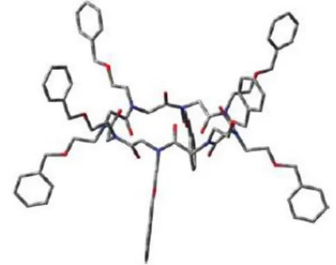
It was found, for the flexible eighteen-membered N-benzyloxyethyl cyclic peptoid 32, high binding constants with the first group alkali metals (Ka ~ 106 for Na+, Li+ and K+), while, for the rigid cis– trans–cis–trans cyclic tetrapeptoid 31, there was no evidence of alkali metals complexation. The conformational disorder in solution was seen as a propitious auspice for the complexation studies. In fact, the stepwise addition of sodium picrate to 32, induced the formation of a new chemical species, whose concentration increased with the gradual addition of the guest. The conformational equilibrium between the free host and the sodium complex, resulted in being slower than the NMR-time scale, giving, with an excess of guest, a remarkably simplified 1H NMR spectrum, reflecting the formation of a 6-fold symmetric species (Figure 1.9).
Figure 1.9. Picture of the predicted lowest energy conformation for the complex 32 with sodium.
A conformational search on 32 as a sodium complex suggested the presence of an S6-symmetry axis
17
this conformation, hampering the ring inversion up to 425 K. The complexity of the r.t. 1H NMR spectrum recorded for the cyclic 33, demonstrated the slow exchange of multiple conformations on the NMR time scale. Stepwise addition of sodium picrate to 33, induced the formation of a complex with a remarkably simplified 1H NMR spectrum. With an excess of guest, we observed the formation of an 8-fold symmetric species (Figure 1.10) was observed.
Figure 1.10. Picture of the predicted lowest energy conformations for 33 without sodium cations.
Differently from the twenty-four-membered 33, the N-benzyloxyethyl cyclic homologue 34 did not yield any ordered conformation in the presence of cationic guests. The association constants (Ka) for the complexation of 32, 33 and 34 to the first group alkali metals and ammonium, were evaluated in H2O–
CHCl3 following Cram‘s method (Table 1.1) 41
. The results presented in Table 1.1 show a good degree of selectivity for the smaller cations.
Table 1.1 R, Ka, and G◦ for cyclic peptoid hosts 32, 33 and 34 complexing picrate salt guests in CHCl3 at 25 ◦C; figures within ±10% in multiple experiments, guest/host stoichiometry for extractions was assumed as 1:1.
18
The ability of cyclic peptoids to extract cations from bulk water to an organic phase prompted us to verify their transport properties across a phospholipid membrane.
The two processes were clearly correlated although the latter is more complex implying, after complexation and diffusion across the membrane, a decomplexation step.42-43 In the presence of NaCl as added salt, only compound 32 showed ionophoric activity while the other cyclopeptoids are almost inactive. Cyclic peptoids have different cation binding preferences and, consequently, they may exert selective cation transport. These results are the first indication that cyclic peptoids can represent new motifs on which to base artificial ionophoric antibiotics.
1.4.5 Catalytic Peptoids
An interesting example of the imaginative use of reactive heterocycles in the peptoid field, can be found in the ―foldamers‖ mimics. ―Foldamers‖ mimics are synthetic oligomers displaying conformational ordering. Peptoids have never been explored as platform for asymmetric catalysis. Kirshenbaumreported the synthesis of a library of helical ―peptoid‖ oligomers enabling the oxidative kinetic resolution (OKR) of 1-phenylethanol induced by the catalyst TEMPO (2,2,6,6-tetramethylpiperidine-1-oxyl) (figure 1.14)44.
Figure 1.14. Oxidative kinetic resolution of enantiomeric phenylethanols 35 and 36.
The TEMPO residue was covalently integrated in properly designed chiral peptoid backbones, which were used as asymmetric components in the oxidative resolution.
The study demonstrated that the enantioselectivity of the catalytic peptoids (built using the chiral (S)- and (R)-phenylethyl amines) depended on three factors: 1) the handedness of the asymmetric environment derived from the helical scaffold, 2) the position of the catalytic centre along the peptoid backbone, and 3) the degree of conformational ordering of the peptoid scaffold. The highest activity in the OKR (e.e. > 99%) was observed for the catalytic peptoids with the TEMPO group linked at the N-terminus, as evidenced in the peptoid backbones 39 (39 is also mentioned in figure 1.14) and 40 (reported in figure 1.15). These results revealed that the selectivity of the OKR was governed by the global structure of the catalyst and not solely from the local chirality at sites neighboring the catalytic centre.
42 R. Ditchfield, J. Chem. Phys., 1972, 56, 5688. 43
K. Wolinski, J. F. Hinton and P. Pulay, J. Am. Chem. Soc., 1990, 112, 8251.
19
Figure 1.15. Catalytic biomimetic oligomers 39 and 40
1.4.6 PNA and Peptoids Tagged With Nucleobases.
Nature has selected nucleic acids for storage (DNA primarily) and transfer of genetic information (RNA) in living cells, whereas proteins fulfill the role of carrying out the instructions stored in the genes in the form of enzymes in metabolism and structural scaffolds of the cells. However, no examples of protein as carriers of genetic information have yet been identified.
Self-recognition by nucleic acids is a fundamental process of life. Although, in nature, proteins are not carriers of genetic information, pseudo peptides bearing nucleobases, denominate ―peptide nucleic acids‖ (PNA, 41, figure 1.16),4
can mimic the biological functions of DNA and RNA (42 and 43, figure
1.16).
Figure 1.16. Chemical structure of PNA (19), DNA (20), RNA (21). B = nucleobase
The development of the aminoethylglycine polyamide (peptide) backbone oligomer with pendant nucleobases linked to the glycine nitrogen via an acetyl bridge now often referred to PNA, was inspired by triple helix targeting of duplex DNA in an effort to combine the recognition power of nucleobases with the versatility and chemical flexibility of peptide chemistry4. PNAs were extremely good structural mimics of nucleic acids with a range of interesting properties:
DNA recognition,
20
1. RNA targeting 2. DNA targeting 3. Protein targeting 4. Cellular delivery 5. Pharmacology Nucleic acid detection and analysis,
Nanotechnology,
Pre-RNA world.
The very simple PNA platform has inspired many chemists to explore analogs and derivatives in order to understand and/or improve the properties of this class DNA mimics. As the PNA backbone is more flexible (has more degrees of freedom) than the phosphodiester ribose backbone, one could hope that adequate restriction of flexibility would yield higher affinity PNA derivates.
The success of PNAs made it clear that oligonucleotide analogues could be obtained with drastic changes from the natural model, provided that some important structural features were preserved.
The PNA scaffold has served as a model for the design of new compounds able to perform DNA recognition. One important aspect of this type of research is that the design of new molecules and the study of their performances are strictly interconnected, inducing organic chemists to collaborate with biologists, physicians and biophysicists.
An interesting property of PNAs, which is useful in biological applications, is their stability to both nucleases and peptidases, since the ―unnatural‖ skeleton prevents recognition by natural enzymes, making them more persistent in biological fluids.45 The PNA backbone, which is composed by repeating N-(2 aminoethyl)glycine units, is constituted by six atoms for each repeating unit and by a two atom spacer between the backbone and the nucleobase, similarly to the natural DNA. However, the PNA skeleton is neutral, allowing the binding to complementary polyanionic DNA to occur without repulsive electrostatic interactions, which are present in the DNA:DNA duplex. As a result, the thermal stability of the PNA:DNA duplexes (measured by their melting temperature) is higher than that of the natural DNA:DNA double helix of the same length.
In DNA:DNA duplexes the two strands are always in an antiparallel orientation (with the 5‘-end of one strand opposed to the 3‘- end of the other), while PNA:DNA adducts can be formed in two different orientations, arbitrarily termed parallel and antiparallel (figure 1.17), both adducts being formed at room temperature, with the antiparallel orientation showing higher stability.
Figure 1.17. Parallel and antiparallel orientation of the PNA:DNA duplexes
PNA can generate triplexes PAN-DNA-PNA, the base pairing in triplexes occurs via Watson-Crick and Hoogsteen hydrogen bonds (figure 1.18).
45
Demidov V.A., Potaman V.N., Frank-Kamenetskii M. D., Egholm M., Buchardt O., Sonnichsen S. H., Nielsen P.E., Biochem. Pharmscol. 1994, 48, 1310.
21
Figure 1.18. Hydrogen bonding in triplex PNA2/DNA: C+GC (a) and TAT (b)
In the case of triplex formation, the stability of these type of structures is very high: if the target sequence is present in a long dsDNA tract, the PNA can displace the opposite strand by opening the double helix in order to form a triplex with the other, thus inducing the formation of a structure defined as ―P-loop‖, in a process which has been defined as ―strand invasion‖ (figure 1.19).46
Figure 1.19. Mechanism of strand invasion of double stranded DNA by triplex formation
However, despite the excellent attributes, PNA has two serious limitations: low water solubility47and poor cellular uptake48.
Many modifications of the basic PNA structure have been proposed in order to improve their performances in term of affinity and specificity towards complementary oligonucleotide sequences. A modification introduced in the PNA structure can improve its properties generally in three different ways:
i) Improving DNA binding affinity;
ii) Improving sequence specificity, in particular for directional preference (antiparallel vs parallel) and mismatch recognition;
46 Egholm M., Buchardt O., Nielsen P.E., Berg R.H., J. Am. Chem. Soc., 1992, 114,1895. 47
(a) U. Koppelhus and P. E. Nielsen, Adv. Drug. Delivery Rev., 2003, 55, 267; (b) P. Wittung, J. Kajanus, K. Edwards, P. E. Nielsen, B. Nordén, and B. G. Malmstrom, FEBS Lett., 1995, 365, 27.
48
(a) E. A. Englund, D. H. Appella, Angew. Chem. Int. Ed., 2007, 46, 1414; (b) A. Dragulescu-Andrasi, S. Rapireddy, G. He, B. Bhattacharya, J. J. Hyldig-Nielsen, G. Zon, and D. H. Ly, J. Am. Chem. Soc., 2006, 128, 16104; (c) P. E. Nielsen, Q. Rev. Biophys., 2006, 39, 1; (d) A. Abibi, E. Protozanova, V. V. Demidov, and M. D. Frank-Kamenetskii, Biophys. J., 2004, 86, 3070.
22
iii) Improving bioavailability, (cell internalization, pharmacokinetics, etc.).
Structure activity relationships showed that the original design containing a 6-atom repeating unit and a 2-atom spacer between backbone and the nucleobase was optimal for DNA recognition. Introduction of different functional groups with different charges/polarity/flexibility have been described and are extensively reviewed in several papers49,50,51.These studies showed that a ―constrained flexibility‖ was necessary to have good DNA binding (figure 1.20).
Figure 1.20. Strategies for inducing preorganization in the PNA monomers59. The first example of ―peptoid nucleic acid‖ was reported by Almarsson and Zuckermann52
.The shift of the amide carbonyl groups away from the nucleobase (towards thebackbone) and their replacement with methylenes, resulted in a nucleosidated peptoid skeleton (44, figure 1.21). Theoretical calculations showed that the modification of the backbone had the effect of abolishing the ―strong hydrogen bond between the side chain carbonyl oxygen (α to the methylene carrying the base) and the backbone amide of the next residue‖, which was supposed to be present on the PNA and considered essential for the DNA hybridization.
Figure 1.21. Peptoid nucleic acid
49
a) Kumar, V. A., Eur. J. Org. Chem., 2002, 2021-2032. b) Corradini R.; Sforza S.; Tedeschi T.; Marchelli R.; Seminar in Organic Synthesis, Società Chimica Italiana, 2003, 41-70.
50 Sforza, S.; Haaima, G.; Marchelli, R.; Nielsen, P.E.. Eur. J. Org. Chem. 1999, 197-204. 51
Sforza, S.; Galaverna, G.; Dossena, A.; Corradini, R.; Marchelli, R. Chirality, 2002, 14, 591-598. 52 O. Almarsson, T. C. Bruice, J. Kerr, and R. N. Zuckermann, Proc. Natl. Acad. Sci. USA, 1993, 90, 7518.
23
Another interesting report, demonstrating that the peptoid backbone is compatible with hybridization, came from the Eschenmoser laboratory in 200753.This finding was part of an exploratory work on the pairing properties of triazine heterocycles (as recognition elements) linked to peptide and peptoid oligomeric systems. In particular, when the backbone of the oligomers was constituted by condensation of iminodiacetic acid (45 and 46, Figure 1.22), the hybridization experiments, conducted with oligomer 45 and d(T)12, showed a Tm = 22.7 °C.
Figure 1.22. Triazine-tagged oligomeric sequences derived from an iminodiacetic acid peptoid backbone.
This interesting result, apart from the implications in the field of prebiotic chemistry, suggested the preparation of a similar peptoid oligomers (made by iminodiacetic acid) incorporating the classic nucleobase thymine (47 and 48, figure 1.23)54.
Figure 1.23. Thymine-tagged oligomeric sequences derived from an iminodiacetic acid backbone
The peptoid oligomers 47 and 48 showed thymine residues separated by the backbone by the same number of bonds found in nucleic acids (figure 1.24, bolded black bonds). In addition, the spacing between the recognition units on the peptoid framework was similar to that present in the DNA (bolded grey bonds).
Figure 1.24. Backbone thymines positioning in the peptoid oligomer (47) and in the A-type DNA.
53
G. K. Mittapalli, R. R. Kondireddi, H. Xiong, O. Munoz, B. Han, F. De Riccardis, R. Krishnamurthy, and A. Eschenmoser, Angew. Chem. Int. Ed., 2007, 46, 2470.
54
R. Zarra, D. Montesarchio, C. Coppola, G. Bifulco, S. Di Micco, I. Izzo, and F. De Riccardis, Eur. J. Org. Chem., 2009, 6113.
24
However, annealing experiments demonstrated that peptoid oligomers 47 and 48 do not hybridize complementary strands of d(A)16 or poly-r(A). It was claimed that possible explanations for those results resided in the conformational restrictions imposed by the charged oligoglycine backbone and in the high conformational freedom of the nucleobases (separated by two methylenes from the backbone).
Small backbone variations may also have large and unpredictable effects on the nucleosidated peptoid conformation and on the binding to nucleic acids as recently evidenced by Liu and co-workers55,with their synthesis and incorporation (in a PNA backbone) of N-ε-aminoalkyl residues (49, Figure 1.25). N H N N N N H N O O O B B B X n X= NH2 (or other functional group) 49 O O O
Figure 1.25. Modification on the N- in an unaltered PNA backbone
Modification on the γ-nitrogen preserves the achiral nature of PNA and therefore causes no stereochemistry complications synthetically.
Introducing such a side chain may also bring about some of the beneficial effects observed of a similar side chain extended from the R- or γ-C. In addition, the functional headgroup could also serve as a suitable anchor point to attach various structural moieties of biophysical and biochemical interest.
Furthermore, given the ease in choosing the length of the peptoid side chain and the nature of the functional headgroup, the electrosteric effects of such a side chain can be examined systematically. Interestingly, they found that the length of the peptoid-like side chain plays a critical role in determining the hybridization affinity of the modified PNA. In the Liu systematic study, it was found that short polar side chains (protruding from the γ-nitrogen of peptoid-based PNAs) negatively influence the hybridization properties of modified PNAs, while longer polar side chains positively modulate the nucleic acids binding. The reported data did not clarify the reason of this effect, but it was speculated that factors different from electrostatic interaction are at play in the hybridization.
1.5 Peptoid synthesis
The relative ease of peptoid synthesis has enabled their study for a broad range of applications. Peptoids are routinely synthesized on linker-derivatized solid supports using the monomeric or submonomer synthesis method. Monomeric method was developed by Merrifield2 and its synthetic procedures commonly used for peptides, mainly are based on solid phase methodologies (e.g. scheme
1.1).
The most common strategies used in peptide synthesis involve the Boc and the Fmoc protecting groups.
25
Cl HO N R O Fmoc O N R O Fmoc Pyperidine, 20% in DMF O H N R O HATU or PyBOP repeatScheme 1.1. monomer synthesis of peptoids
Peptoids can be constructed by coupling N-substituted glycines using standard α-peptide synthesis methods, but this requires the synthesis of individual monomers4, this is based by a two-step monomer addition cycle. First, a protected monomer unit is coupled to a terminus of the resin-bound growing chain, and then the protecting group is removed to regenerate the active terminus. Each side chain requires a separate Nα-protected monomer.
Peptoid oligomers can be thought of as condensation homopolymers of N-substituted glycine. There are several advantages to this method, but the extensive synthetic effort required to prepare a suitable set of chemically diverse monomers is a significant disadvantage of this approach. Additionally, the secondary N-terminal amine in peptoid oligomers is more sterically hindered than primary amine of an amino acid, for this reason coupling reactions are slower.
Sub-monomeric method, instead, was developed by Zuckermann et al. (Scheme 1.2)56.
Cl HO Br O O Br O R-NH2 O H N R O DIC repeat
Scheme 1.2. Sub-monomeric synthesis of peptoids
Sub-monomeric method consists in the construction of peptoid monomer from C- to N-terminus using N,N-diisopropylcarbodiimide (DIC)-mediated acylation with bromoacetic acid, followed by amination with a primary amine. This two-step sequence is repeated iteratively to obtain the desired oligomer. Thereafter, the oligomer is cleaved using trifluoroacetic acid (TFA) or by hexafluorisopropanol, scheme 1.2. Interestingly no protecting groups are necessary for this procedure.
The availability of a wide variety of primary amines facilitates the preparation of chemically and structurally divergent peptoids.
1.6 Synthesis of PNA monomers and oligomers
The first step for the synthesis of PNA, is the building of PNA‘s monomer. The monomeric unit is constituted by an N-(2-aminoethyl)glycine protected at the terminal amino group, which is essentially a pseudopeptide with a reduced amide bond. The monomeric unit can be synthesized following several methods and synthetic routes, but the key steps is the coupling of a modified nucleobase with the secondary amino group of the backbone by using standard peptide coupling reagents (N,N'-dicyclohexylcarbodiimide, DCC, in the presence of 1-hydroxybenzotriazole, HOBt). Temporary masking the carboxylic group as alkyl or allyl ester is also necessary during the coupling reactions. The
26
protected monomer is then selectively deprotected at the carboxyl group to produce the monomer ready for oligomerization. The choice of the protecting groups on the amino group and on the nucleobases depends on the strategy used for the oligomers synthesis. The similarity of the PNA monomers with the amino acids allows the synthesis of the PNA oligomer with the same synthetic procedures commonly used for peptides, mainly based on solid phase methodologies. The most common strategies used in peptide synthesis involve the Boc and the Fmoc protecting groups. Some ―tactics‖, on the other hand, are necessary in order to circumvent particularly difficult steps during the synthesis (i.e. difficult sequences, side reactions, epimerization, etc.). In scheme 1.3, a general scheme for the synthesis of PNA oligomers on solid-phase is described.
N H N OH O O NH2 First monomer loading N H N N H O O Deprotection H2N N N H O O N H N OH O O Coupling N H N N H O O N H N O O Repeat deprotection and coupling First cleavage NH2 H N H N O O B n PNA B-PGs B-PGs B-PGs B-PGs B-PGs B-PGs PGt PGt PGt PGt
PGs: Semi-permanent protecting group PGt: Temporary protecting group
Scheme 1.3. Typical scheme for solid phase PNA synthesis.
The elongation takes place by deprotecting the N-terminus of the anchored monomer and by coupling the following N-protected monomer. Coupling reactions are carried out with HBTU or, better, its 7-aza analogue HATU57 which gives rise to yields above 99%. Exocyclic amino groups present on cytosine, adenine and guanine may interfere with the synthesis and therefore need to be protected with semi-permanent groups orthogonal to the main N-terminal protecting group.
In the Boc strategy the amino groups on nucleobases are protected as benzyloxycarbonyl derivatives (Cbz) and actually this protecting group combination is often referred to as the Boc/Cbz strategy. The Boc group is deprotected with trifluoroacetic acid (TFA) and the final cleavage of PNA from the resin, with simultaneous deprotection of exocyclic amino groups in the nucleobases, is carried out with HF or with a mixture of trifluoroacetic and trifluoromethanesulphonic acids (TFA/TFMSA). In the Fmoc strategy, the Fmoc protecting group is cleaved under mild basic conditions with piperidine, and is
27
therefore compatible with resin linkers, such as MBHA-Rink amide or chlorotrityl groups, which can be cleaved under less acidic conditions (TFA) or hexafluoisopropanol. Commercial available Fmoc monomers are currently protected on nucleobases with the benzhydryloxycarbonyl (Bhoc) groups, also easily removed by TFA. Both strategies, with the right set of protecting group and the proper cleavage condition, allow an optimal synthesis of different type of classic PNA or modified PNA.
1.7 Aims of the work
The objective of this research, is to gain new insights in the use of peptoids as tools for structural studies and biological applications. Five are the themes developed in the present thesis:
1. Carboxyalkyl Peptoid PNAs. Nγ-carboxyalkyl modified peptide nucleic acids (PNAs), containing the four canonical nucleobases, were prepared via solid-phase oligomerization. The inserted modified peptoid monomers (figures 1.26: 50 and 51) were constructed through simple synthetic procedures, utilizing proper glycidol and iodoalkyl electrophiles.
Figure 1.26. Modified peptoid monomers
Synthesis of PNA oligomers was realized by inserting modified peptoid monomers into a canonical PNA, by this way four different modified PNA oligomers were obtained (figure 1.27).
Figure 1.27. Modified PNA.
Thermal denaturation studies performed, in collaboration with Prof. R. Corradini from the University of Parma, with complementary antiparallel DNA strands, demonstrated that the length of the Nγ-side chain strongly influences the modified PNAs hybridization properties. Moreover, multiple negative
GTAGAT*50CACT–Gly–NH2, 52
G T*50AGAT*50CAC T*50–Gly–NH2, 53
GTAGAT*51CACT–Gly–NH2, 54
G T*51AGAT*51CAC T*51–Gly–NH2, 55
N H N N N O Base O O O N H N Base O O N NH O O O HO 50 N H N N N O Base O O O N H N Base O O N NH O O O HO 51 Fmoc N N OH N NH O t-BuO O O O O n 50, n = 1 51, n = 5
28
charges on the oligoamide backbone, when present on γ-nitrogen C6 side chains, proved to be beneficial
for the oligomers water solubility and DNA hybridization specificity.
2. Structural analysis of cyclopeptoids and their complexes. The aim of this work was the
studies of structural properties of cyclopeptoids in their free and complexed form (figure 1.27: 56, 57 and 58). N N N O O O N O N N O O 56 N N N OO O N O 57 N N N O O O O N O O N N O O O O O O 58
Figure 1.27. N-Benzyl-cyclohexapeptoid 56, N-benzyl-cyclotetrapeptoid 57 and
N-metoxyethyl-cyclohexapeptoid 58.
The synthesis of hexa- and tetra- N-benzyl glycine linear oligomers and of hexa- N-metoxyethyl glycine linear oligomer (59, 60 and 61, figure 1.28), was accomplished on solid-phase (2-chlorotrityl resin) using the ―sub-monomer‖ approach.58
HO N H O HO N H O O n=6,61 n=6,59 n=4,60 n n
Figure 1.28. linear Benzyl-hexapeptoid 59, linear benzyl tetrapeptoid 60 and linear
N-metoxyethyl-hexapeptoid 61.
29
All cycles obtained were crystallized and caractherizated by X-ray analysis in collaboration with Dott. Consiglia Tedesco from the University of Salerno and Dott. Loredana Erra from European Synchrotron Radiation Facility (ESRF), Grenoble, France.
3. Cationic cyclopeptoids as potential macrocyclic nonviral vectors.
The aim of this work was the synthesis of three different cationic cyclopeptoids (figure 1.28: 62, 63 and 64) to assess their efficiency in DNA cell transfection, in collaboration with Prof. G. Donofrio of the University of Parma.
N N N N N N O O O O O O H3N H3N 2X CF3COO -N N N N N N O O O O O O H3N NH3 NH3 H3N 4X CF3COO -62 63 N N N N N N O O O O O O H3N NH3 H3N H3N NH3 NH3 6X CF3COO -64
Figure 1.29. Di-cationic cyclohexapeptoid 62, Tetra-cationic cyclohexapeptoid 63, Hexa-cationic
cyclohexapeptoid 64.
4. Complexation with Gd3+ of carboxyethyl cyclopeptoids as possible contrast agents in MRI. Three cyclopeptoids 65, 66 and 67 (figure 1.30) containing polar side chains, were synthesized
and, in collaboration with Prof. S. Aime, of the University of Torino, the complexation properties with Gd3+ were evaluated.