Università degli Studi del Piemonte Orientale
“Amedeo Avogadro”
Dipartimento di Scienze Chimiche, Alimentari, Farmaceutiche e Farmacologiche
Dottorato di Ricerca in Biotecnologie Farmaceutiche ed Alimentari
X ciclo a.a. 2011-2014
“Exploring carbohydrates antigens conjugated to
pathogen-related protein carriers”
Alberto Nilo
Supervised by Dr. Roberto Adamo
Alberto Nilo was the recipient of a Ph. D. fellowship by
Novartis Vaccines S.r.l., Siena, Italy, that is deeply acknowledged.
Contents
Chapter 1
Introduction 7
Chapter 2
Outline of the thesis 29
Chapter 3
“Recombinant Clostridium difficile toxin fragments as carrier protein for PSII surface polysaccharide preserve their neutralizing activity” 36
Chapter 4
“Tyrosine-directed conjugation of large glycans to proteins via copper-free click chemistry” 51
Chapter 5
“Anti-Group B Streptococcus carbohydrate-based vaccines using pilus proteins as carriers: Comparing lysine and tyrosine-directed conjugation” 65
Chapter 6
Conclusions
83
List of publications
86Introduction
General principles of vaccination
The immune responses of mammals can be divided into innate and adaptive immunity. Innate immunity fights intruding pathogens in a fast, yet non-specific way and can be considered a first line of defense. Onset of adaptive immunity instead is delayed, but all responses are directed specifically against the respective intruder. A multitude of pathogen-specific cells are created by clonal expansion of few, “fitting” pre-cursor cells. And, once infection is resolved, some of these cells survive in the body to form immunologic memory ready to fight the same pathogen faster and more efficiently, should a second encounter occur. Vaccination takes advantage of this potential of adaptive immunity. The principle of vaccination is that exposure to a small sample of a disease-causing microorganism - or to a part or portion of it - teaches the immune system to rapidly recognize the menace and to create memory that enables the body to fight the real pathogen efficiently during a later encounter. Vaccination basically mimics a natural infection without causing disease. Several are the components of host immune responses that are set in motion following vaccination. Important vaccine-induced immune effectors are antibodies produced by B lymphocytes. These antibodies are capable to recognize and specifically bind to a toxin or a pathogen (or to a portion representative of it)1. Antibody binding interferes with pathogen entry into host cells or other important functions. Moreover bound antibodies facilitate uptake and elimination of the pathogen by other immune cells. Other important immune effectors are cytotoxic CD8+ T lymphocytes (CTL) that may limit the spread of infectious agents by recognizing and killing infected cells or by secreting specific antiviral cytokines. The generation and maintenance of both B and CD8+ T cell responses is supported by growth factors and signals provided by CD4+ T helper (Th) lymphocytes, which are historically subdivided into T helper 1 (Th1) and T helper 2 (Th2) subtypes, and more recently also in T helper 17 (Th17). These effector cells are controlled by regulatory T cells (Treg) that are involved in maintaining immune tolerance.2 Most of the antigens and vaccines trigger both B and T cell responses, and the two responses benefit of their interconnection: CD4+ T cells are required for most antibody responses, while antibodies exert significant influences on T cell responses to intracellular pathogens3
Early protective efficacy of a vaccine is often conferred by microorganism-specific antibodies. But just triggering a specific antibody response is not sufficient. The quality of such antibody responses, e.g., antibody avidity, has been identified as a determining factor of efficacy. Although B lymphocytes
represent the specialized lineage in antibody production once differentiated in plasma cells, T cells can largely contribute to effective and long-lasting immune responses. In fact, these cells are essential to immune memory, and novel vaccine targets have been identified against which T cells are likely to be the prime effectors.
The stimulation of antigen-specific T cell responses requires their activation by specific antigen presenting cells (APCs), essentially dendritic cells (DCs), which reside in tissues or patrol through the body and are recruited by inflammatory signals to the site of infection. When exposed to pathogens, immature DCs will phagocytose and process them proteolytically, so that some pathogen components can be loaded on MHC class II molecules. DCs then undergo maturation, modulate specific surface receptors and migrate towards secondary lymph nodes, where the trigger of T and B cell responses occurs.
The central role for mature DCs in the activation of vaccine responses reflects their unique ability to provide both antigen-specific and co-stimulation signals to T cells. In fact, T cells require at least two signals to become fully activated: i) a first antigen-specific signal, is provided through the T cell receptor which interacts with the antigen peptide-loaded MHC II molecules on the surface of the APCs; ii) a second antigen-unspecific signal, is a co-stimulatory incentive and is provided by the interaction between co-stimulatory molecules expressed on the membrane of the APC upon maturation with its counterparts on the T cell. APCs can also secrete cytokines that are sensed by the interacting T cell and determine its further phenotype (Th1, Th2, Th17).
The very first requirement to elicit vaccine responses is thus to provide sufficient stimuli through vaccine antigens and eventually adjuvants, to trigger an inflammatory reaction that is mediated by cells of the innate immune system4. In addition, the antibody response to bacterial polysaccharides is poorly affected by adjuvants, IgM represents the major class of antibodies induced and since their immune response does not induce memory, it is not boosted by subsequent immunizations.
These characteristics are due to the fact that, unlike proteins that are T-cell dependent (TD) antigens, polysaccharides are T-cell independent (TI) antigens in that they do not require T-cell activation for the induction of specific B-cell (antibody) responses. Polysaccharide antigens directly activate polysaccharide- specific B cells which differentiate then into plasma cells to produce antibodies, but memory B cells are not formed; moreover a pre-existing memory B-cell pool can be depleted by immunization with unconjugated polysaccharide, with risk of hypo-responsiveness on subsequent immunizations5.
Differently from polysaccharides, proteins are TD antigens; following interaction with antigen-presenting cells (APC) like dendritic cells, macrophages and B-cells, protein antigens are internalized and processed into small peptides which are then re-exposed and presented to T lymphocytes in association with the major histocompatibility complex (MHC) class II molecules. Interaction with T cells induces B cells to differentiate into plasma cells and memory B cells, thus initiating downstream adaptive immune responses. Unlike TI antigens, TD antigens are immunogenic early in infancy, the immune response induced can be boosted, enhanced by adjuvants, and is characterized by antibody class switch and production of antigen-specific IgG.
Polysaccharide Vaccines
Polysaccharides are important virulence factor especially for encapsulated bacteria that present on their surface complex carbohydrate structures. Surface polysaccharides have several functions: a) in some cases they protect microorganisms from desiccation when they are exposed to the external environment -an example is the hyaluronic capsule of group A Streptococcus, whose adhesive properties help the pathogen to invade the host; b) other capsular polysaccharides prevent the activation of the alternate complement pathway; c) sometimes they mimic molecules produced by human cells so that the pathogen is not recognized as foreign by our immune system (serogroup B meningococcal capsular polysaccharide, hyaluronic acid).
Around 1930s the protective role of antibodies (Abs) induced by pneumococcal polysaccharide started to be investigated and in 1945 the first vaccine composed by purified polysaccharide from selected pneumococcal serotypes was tested in humans6.
The research on vaccines development was subsequently slowed down by the introduction of antibiotics, however with the emergence of drug resistant strains the development of polysaccharide vaccines started again and a number of them have been studied in large clinical studies. Polysaccharide vaccines against meningococcus serogroup ACWY, Streptococcus pneumoniae and Haemophilus
influenzae type be were licensed between the seventies and the eighties.7,8 These bacteria possess
polysaccharides which are polymers formed by one monosaccharide unit (homopolymers) or more complex oligosaccharide repeats (heteropolymers), that can be charged or neutral.
Polysaccharide vaccines however did not completely solve the problem of bacterial diseases caused by encapsulated microorganisms. Being T-cell independent antigens, one of their main features which emerged from clinical trials, was their scarce immunogenicity in children less than two years of age.9,10 As a consequence, polysaccharide vaccines can be used in adults, but not in infancy and elderly which are the most sensitive target populations.
Glycoconjugate Vaccines
Glycoconjugate vaccines are among the safest and most efficacious vaccines developed during the last 30 years. They are a potent tool for prevention of life-threating bacterial infectious disease like meningitis and pneumonia.
The limitation of polysaccharides vaccines has been overcome by covalent conjugation to a carrier protein as source of T-cell epitopes. Since 1929 it has been demonstrated by Avery and Goebel that non immunogenic sugars after conjugation to a carrier protein become able to induce antibodies in animal model.11 However the first application of this concept to a vaccine for human use started only in 1980
with the development of the first conjugate vaccine against Haemophilus influenzae type b (Hib) that was later on licensed vaccine between 1987 and 1990.12,13
Many other glycoconjugate vaccines have been developed against bacterial pathogens such as Neisseriae meningitidis, Streptococcus pneumoniae and group B Streptococcus. Today glycoconjugate vaccines are among the safest and most efficacious vaccines developed during the last 30 years and they are currently used in the immunization schedules of different countries like for example the UniteStates (US).14-20
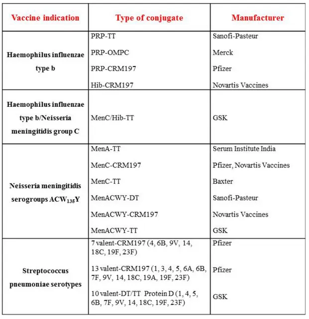
Table 1: Licensed glycoconjugate vaccines.
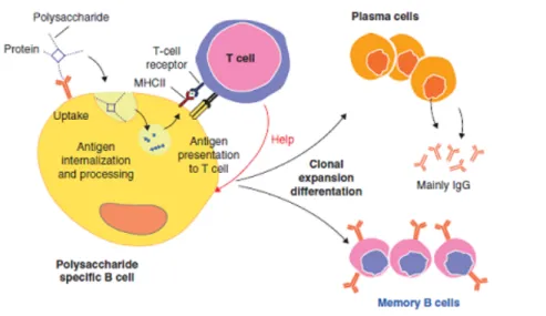
Chemical conjugation of polysaccharides to protein carriers allows processing of the protein carrier by polysaccharide-specific B cells and presentation, on their surface, of the resulting peptides or in association with MHC class II. Further interaction with carrier-specific cells then induces a TD response already early in life which leads to immunological memory and boosting of the response by further doses of the vaccine (Fig. 1). Recently it has been proposed a novel mechanism according to which internalization of glycoconjugate and following proteolytic digestions generate glycopeptides which are re-exposed by MHCII (Fig. 2) . This model was confirmed by isolation of specific T-cell
clones directed to the sugar. This observation proves that although glycoconjugates are safe and effective, their mechanism of function is still not totally understood.
Fig. 1 Mechanism of action of a glycoconjugate vaccine
Fig. 2 Recent working model of action of a glycoconjugate vaccine
Chemistry of glycoconjugate vaccines
In glycoconjugate vaccines, a covalent linkage between the carbohydrate moiety and the carrier protein has to be installed. Approaches based on different chemistry of conjugation, have been so far used. One is based on the random chemical activation of the hydroxyl or carboxyl groups of saccharide chain followed by covalent binding with Lys residues, onto glutamic or aspartic residues of the carrier protein. These are usually the most abundant amino acid residues on protein surface, and the regioselectivity of conjugation is hard to achieve. As a consequence these vaccines are effective, but two limitations can be found: a) certain degree of batch-to-batch variability in the immunological properties can be present. b) it has not been possible to apply precise structure-immunogenicity relationships and use the classic medicinal chemistry approach to investigate vaccine candidates.
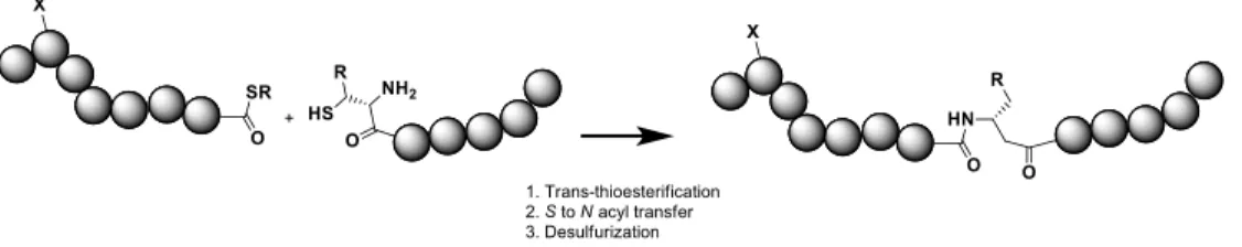
Recently different methods to target specifically some residues for bioconjugation are emerging. In general, three main approaches for site-selective incorporation of carbohydrates onto proteins can be described21 (1) chemical ligation-based strategies, which rely on the reaction of two unprotected
peptide moieties bearing a C-terminal thioester and a N-terminal cysteine; (2) chemoenzymatic transformations (“remodeling”) of glycoproteins with endoglycosidases or other glycan processing enzymes; (3) site selective conjugation to natural and unnatural amino acid residues, such as homoallylglycine, S-allylcysteine or Se-allylselenocysteine, azidoalanine presenting chemical groups, which can react under the mild conditions present in biological systems (aqueous media, mild pH, and temperature). Combination of oligosaccharide synthesis and novel site specific methods may enable an accurate antigen design and a more precise correlation between the glycoconjugate structure and the immunological properties.22
Among these methods, targeting tyrosine residues by chemical manipulations has appeared very attractive. Proteins usually present much less tyrosine than lysine, glutamate or aspartate residues, and they are also often buried. The accessible tyrosine residues on protein surface are usually few, thus the regioselectivity of conjugation can be expected. Recently a reliable tyrosine-selective conjugation method via triazolidinone derivatives in tris buffer has been developed.22 The method enabled the
insertion of alkyne-containing bifunctional linkers onto the tyrosine residues of the genetically detoxified diphtheria toxin CRM197. The subsequent condensation of a synthetic β-glucan by Cu(I)
catalyzed azide-alkyne [3+2] cycloaddition (CuAAC)23,24 enabled the creation of an anti-candidiasis vaccine.25
This strategy was proven a powerful method to obtain a robust structure-immunogenicity relationship from glycoconjugates with defined sugars at predetermined sites. In addition, it has been shown that the triazole generated by the cycloaddition is either moderately or non-immunogenic and does not affect the anti-carbohydrate response. Therefore, this conjugation approach appears a robust approach for site-selective incorporation of sugars at predetermined sites of the protein.
Fig. 3 Chemical ligation based preparation of glycoconjugates
Fig. 5 Glycan biosynthesis and enzymatic conjugation in E.coli and site selective ligation
GBS
Streptococcus agalactiae, also known as group B Streptococcus (GBS), is one of the major causes of sepsis, pneumonia, and meningitis in neonatal and infants in the first three months after birth.26
It is a Gram positive beta-hemolytic coccus which appears in pairs (Fig.6) or chains27
, that colonizes the urogenital and gastrointestinal tracts of more than 30% of the healthy population and, in particular, the vagina of 25-40% of healthy women.28-30
Fig.6 Immunogold Electron Microscopy of GBS strain CJB111(Rosini, Rinaudo et al. 2006).
It was classified for the first time in 1930 by R. Lancefield and R. Hare during a serological differentiation study on human isolates and other groups of hemolytic streptococci.31
the group B of “Lancefield System”, where Streptococci are identified with alphabetical letters from A to O, based on capsular polysaccharide antigens and groups A, B and D are the most dangerous32
. GBS clinical isolates are also classified into ten serotypes, according to the chemical nature of capsular polysaccharides (PS): Ia, Ib, II, III, IV, V, VI, VII, VIII and IX.33-35
However around 8-14% of the clinical isolates in the Europe and USA are non-typeable strains because cannot be distinguished on the basis of PS antigenicity. All GBS serotypes contain, in different combinations, these carbohydrates: galactose, glucose, rhamnose, N-acetylglucosammine and sialic acid (N-acetylneuraminic acid) (Fig. 7) .
Fig. 7 Chemical structures of isolated GBS serotypes
Streptococcus agalactiae is known as the most frequent cause of sepsis, pneumonia and meningitis in neonates within first months from birth.36, 37 It is an etiologic agent also in adults, in particular elderly
persons, immunocompromised, diabetics, and patients affected by others chronic pathology of liver and kidney. In the cellular hyper-trophism of vaginal epithelium, where it can adheres despite low values of pH38
, it forms a permanent colonization in more than 40% of women. During pregnancy, the uro-genital tract colonization is cause of severe infections/year, leading to chorioamnionitis, endometrial tenderness, tachycardia, cystitis and fever which make necessary antibiotic therapies also after delivery. GBS can also be cause of intrauterine death, aborts, prelabor rupture of the membranes, with preterm labor/delivery, causing the exposure of 90% of premature neonates to infection. In most cases infection
is transferred vertically from asymptomatic mothers and, every year, only for US, 8000-12000 infected babies and 2000 deaths are recorded. Infection appears in uterus or at delivery for contact of throat, ear, nostril, umbilicus and respiratory ways with amniotic liquid or infected vaginal fluids. After the aspiration/ingestion of bacteria, neonatal lungs become the starting focus of infection. From here it can rapidly arrive to flow of blood, continue towards others tissues and organs like blood-brain barrier (Fig.8).
Fig.8 Stages of neonatal group B streptococcal (GBS) infection (Doran and Nizet 2004).
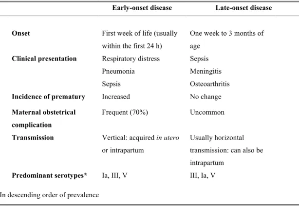
Neonatal disease can occur in two different forms: the Early-Onset Disease (EOD) and the Late-Onset Disease (LOD).
EOD occurs during the first seven days of life, with the vast majority of cases (approximately 90%) present during the first 24 hours of life.39
Neonates with EOD present respiratory disease (54%), sepsis without focus (27%) and meningitis (15%).40 Risk factors include: GBS bacteriuria during pregnancy;
gestational age less than 37 weeks; previous infant with invasive GBS disease; preterm labor/delivery; prelabor rupture of membranes; birth weight less than 2500g; black race and teenage mothers.
LOD occurs beyond seven days of life and can develop up to three months of age. In 50% of the cases it has a maternal origin. Neonates with LOD present sepsis (46%), meningitis (37%), urinary infection (7%), osteoarthritis (6%), respiratory disease (4%) and cellulitis (4%). Over 20% of survivors of GBS meningitis have permanent sequelae, including hearing loss, mental retardation, cortical blindness and seizures (Tab. 2).
Tab. 2 Neonatal manifestation of group B streptococcal disease (Shet and Ferrieri 2004). Early-onset disease Late-onset disease
Onset First week of life (usually within the first 24 h)
One week to 3 months of age
Clinical presentation Respiratory distress Pneumonia
Sepsis
Sepsis Meningitis Osteoarthritis Incidence of prematury Increased No change Maternal obstetrical
complication
Frequent (70%) Uncommon Transmission Vertical: acquired in utero
or intrapartum
Usually horizontal transmission: can also be intrapartum
Predominant serotypes* Ia, III, V III, Ia, V *In descending order of prevalence
Risk factors include non-white race and preterm birth, but most of time it is an horizontal transmission, after contact with sanitary staff or colonized mothers. LOD is basically due to serotype III strain, with an incidence until 60% depending on the geographic area. Studies performed on non-pregnant adults with GBS associated invasive disease revealed that GBS serotypes Ia, III and V accounted for more than two-third of cases. More than 25% of the subjects had invasive GBS disease caused by type V strains (Tab. 3).
Table 3 Serotype distribution of group B streptococcal isolates from non-pregnant adults with invasive GBS infection, 1992-1999 (Edwards and Baker 2005).
The clinical manifestations of GBS infection in elderly adults are: skin and soft-tissue infections and in these cases, cellulites is the most frequent clinical manifestation, urinary tract infections (bacteremic urinary tract infections account for 33.4% of the cases in adults >70 years of age), pneumoniae (only in older debilitated adults), bacteremia with no identified focus (in the 15% of non-pregnant adults affected by GBS invasive diseases), arthritis, osteomyelitis, meningitis (in only 2% of non-pregnant adults with GBS invasive disease) and endocarditis.
GBS Vaccine
Early in 1930s Rebecca Lancefield et al. demonstrated that polyclonal antibodies from rabbit sera, able to recognize PS epitopes, conferred protection against GBS infection in animal models. During the last two decades, plain GBS polysaccharides have been extensively studied as vaccines in preclinical and human clinical studies. However, the first human clinical trials conducted in the 1980s showed that the purified native PS from serotype III was not sufficient to induce an robust IgG response in adults and insignificant in neonates.
Subsequently, conjugation of PSs to immunogenic proteins, such as tetanus toxoid (TT) and mutated diphtheria toxoid (CRM197),
41, 42
was shown to dramatically increase the immune response in children, GBS serotype N° (%) of subjects (n=589) Ia 143 (24.3) Ib 72 (12.2) II 70 (11.9) III 97 (16.5) IV 2 (0.3) V 162 (27.5) VI 0 VII 0 VIII 1 (0.2) Non-typeable 42 (7.1)
eliciting the differentiation of memory cells associated to a long term protection. Following these findings, PS-TT conjugate vaccines based on nine GBS serotypes were produced and tested pre-clinically.41
These studies, carried out in animal models, showed that conjugate antigens were able to induce functional PS-specific IgG that, in presence of complement, stimulate in vitro the opsonization and killing of GBS by human peripheral blood leukocytes. This success constituted the rationale to proceed with the clinical studies in human. Further studies demonstrated that glycoconjugate vaccines constituted by serotypes Ia, Ib, II, III, and V PS linked to TT were safe, well-tolerated and highly immunogenic in human adults,42
but cross-protection between serotypes was still lacking. Therefore a multivalent vaccine was required in order to obtain a broad coverage of the vaccine against the prevalent circulating GBS serotypes. A tetravalent combination of PS –TT conjugates (serotypes Ia, Ib, II, and III) was successfully tested in a mouse model and further human trials were performed using the combination of two PS TT-conjugates (serotypes II and III). Results showed that the combination had the same immunogenicity and reactogenicity of each monovalent PS vaccine.42
However, although recent epidemiological studies43
suggest that a tetravalent combination of serotypes Ia, Ib, III and V would be sufficient to achieve a coverage against the majority of GBS strains circulating in Europe and North America, there are other geographical areas where such combination would be not be effective owing to a different serotypes distribution (i.e. VI and VIII, predominant in Japan). Moreover, the PS-conjugate vaccine would not protect against all the non-typeable isolates. An additional obstacle to the licensure of vaccine against GBS is the difficulty of conducting clinical efficacy trials in human: large sample size would be required, but the use of IAP reduces the incidence of neonatal disease. A possible solution to overcome this difficulty came from the studies of Feng-Ying C. Lin and coworkers who carried out a prospective way to estimate the maternal GBS-PSs antibody levels needed to give protection to neonates from EOD. The amount of maternal antibodies against GBS-PSIa was measured by ELISA in 45 case patients (mothers whose neonates developed EOD) and 319 controls (mothers of neonates colonized by GBS-PSIa but without EOD). Distribution of maternal antibody concentrations showed that the probability of developing EOD declined with increasing maternal levels of anti-PSIa IgG.44
This work demonstrated that it is possible to define thresholds of anti-GBS PSIa specific IgG levels in the mothers which are predictive for the protection of newborns. More recently, thresholds have also been set for the levels of maternal anti GBS-PSIII IgGs required to protect newborns from EOD caused by GBS serotype III.45
The results of Feng-Ying C. Lin suggested that complex clinical trials required for a GBS vaccine registration, might be replaced with an in vitro correlate of protection based on the quantification of vaccine induced antibodies.
Pilus protein-based vaccine
The protection induced by a polysaccharide based is limited to strains expressing the same CPSs included in the vaccine formulation. For this reason the development of a cross-protective protein vaccine is another relevant option. In order to develop a serotype independent universal GBS vaccine, several efforts have been focused on the identification of highly protective protein antigens. Unlike CPS antigens, proteins antigens are able to induce protective T-cell-dependent antibody responses and long-lasting immunity and conjugation to carrier molecules (i.e. TT or CRM197) is not necessary. Since
antibodies directed against surface antigens can interfere with bacterial virulence factors and can promote complement dependent opsonophagocytosis, they are considered excellent vaccine candidates. Before 2005, only a few GBS protein antigens have been identified as potential vaccine candidates; these include Rib, the alpha and beta subunits of the C protein, Sip and the C5a peptidase proteins. However, these proteins, with the exception of C5a peptidase and Sip, are either not expressed on all strains or are highly variable in different isolates.46
The C-protein complex was able to induce passive protection against GBS infections in an animal model.47
Further studies also showed that the C-protein complex could be one of the factors that confer resistance to opsonization.48 Rib is a surface protein
with a similar structure and sequence to the alpha subunit of the C protein and it is also able to induce protective immunity.49
Unfortunately, the alpha subunit of the C protein is present in the genome of only approximately 50% of clinical, while Rib is present in the genome of all serotype III strains, but not in other serotypes. Moreover, both these proteins (the alpha subunit of the C protein and Rib) contain repeated sequences that show strain-to-strain variations and that can affect their immunogenicity. Sip (surface immunogenic protein) is a surface GBS protein that was identified after immunological screening of a genomic library. Sip has been identified in GBS strains of every serotype and the sip gene is highly conserved among GBS isolates.50
The protection conferred by the Sip protein was determined by a mouse neonatal infection model. Newborn mice were protected against infections of GBS strains of serotypes Ia, Ib, II, III, and V.50
Furthermore, it has been observed that sera collected from pregnant women and their healthy newborns have Sip antibodies. The C5a peptidase is a serine-protease localized on the surface that inactivates the human C5a a factor produced during complement activation. C5a is highly conserved surface-bound protein that is expressed on the surface of all serotypes of both group A (GAS) and group B streptococcus.51
The GBS C5a peptidase (ScpB) is 98% identical in sequence to that expressed by GAS. It has been shown that C5a peptidase is a protective
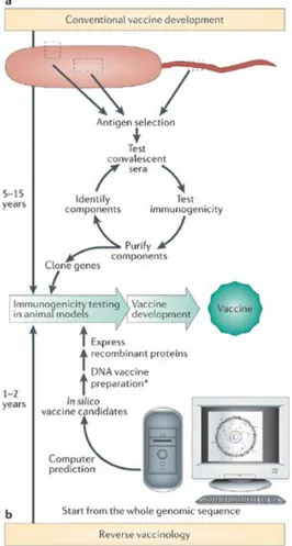
antigen, and could be also used as a carrier protein for type III polysaccharide vaccine. Up to 1995, when the first complete microbial genome sequences became available, vaccines has been classically developed by isolating, inactivating and injecting the cause of the infection. These traditional approaches are time-consuming and expensive. In 1995, a new era named “the genomic era”, began chancing completely the approach for vaccine development. The microbial genome sequencing,52
provided a new impulse to the vaccinology field. A new approach named Reverse Vaccinology, based on integration of several techniques such as genomics, bioinformatics, and molecular biology.53
Fig. 9 Comparison between conventional approach and Reverse Vaccinology
(Johri, Paoletti et al. 2006).
Unlike the conventional vaccine approaches, the reverse vaccinology, permits the identification of less common, low expressed and/or not expressed in vitro antigens and can be applied also on non-cultivable microorganisms. On the other hand, it can be applied only on the discovery of proteins antigens but not to others antigens such as lipopolysaccharides and glycolipids.54
Application of the reverse vaccinology approach to the development of vaccine against GBS commenced from the
sequencing of the complete genome of a virulent GBS strain (2603v/r, serotype V). However, a Comparative Genome Hybridization (CGH) analysis showed that the genetic variability within the GBS isolates was too high and represented a limit for the identification of vaccine candidates. From this analysis it was evident the need to include genome sequences of more serotypes for the selection of protein antigens. In order to study the genome variability in GBS, Tettelin and coworkers sequenced the genome of 6 GBS strains which represent the most disease-causing serotypes (serotype Ia strains A909 and 515, type Ib strain H36B, type II strain 18RS21, type III strain COH1 and type V strain CJB111). By a comparative analysis of all available genomes, the new sequenced genomes plus the two already published genomes, it was possible to identify two subgenomes: the “core genome” and the “variable genome”, together defined as “pan genome”.55
The “core genome” includes genes present in all the strains and is around the 80% of each genome and contains all genes necessary for the basic biology of the bacteria, on the other hand, the “variable genome” is responsible for strain diversity and represents the part of genes that is dispensable and unique to each strain.
The surface availability of the antigens to antibody recognition is a prerequisite for a good protective immune response. Maione and coworkers searched within the GBS pangenome for the genes coding for putative surface-associated and secreted proteins. Using this approach around 589 putative surface proteins were selected, among them 396 belonged to the core genome and 193 were variable genes. The proteins containing more than three trans-membrane domains were excluded because of the difficulties predicted for their production in E. Coli. By using a high-throughput cloning and expression approach, 312 of the selected GBS genes were successfully expressed in E. coli. Each of the genes was cloned with sequences coding for either an N-terminal 6XHistidine Tag or for a C-terminal glutathione S-transferasetag and the expressed proteins were purified by affinity chromatography. All the 312 purified recombinant GBS antigens were tested by an active maternal immunization/neonatal pup challenge mouse model of GBS infection. Briefly, the antigens were used to immunize female mice with a three dose immunization schedule. After the last immunization, mice were mated and their pups were challenged, within 48 h after birth, with a lethal dose of GBS. The survival of the neonates was monitored for 3 days and immune sera were collected for in vitro analysis. Immunoblot assays were used for the identification of the natural protein in GBS total protein extracts, while flow cytometry assays were carried out to confirm the surface exposure of the antigens. From this first systematic screening four antigens were identified as capable of significantly increasing the survival rate among challenged infant mice. When the four antigens were mixed and administered simultaneously, an almost universal protection was achieved against challenge model using a panel of
strains comprehensive of the most pathogenic GBS serotypes. In particular, the levels of protection reached were similar to those achieved using the polysaccharides-based vaccines. Only one (SAG0032) of these four antigens was part of the “core genome”, and this protein was the already described Sip protein. The other three antigens named GBS67 (SAG1408), GBS80 (SAG0645) and GBS104 (SAG0649) were present in the variable portion of the subgenome. The major outcome of this study was that the combination of these antigens, each effective against overlapping populations of isolates, was able to confer broad serotype-independent protection. Characterization studies revealed that three of these antigens (GBS67, GBS80 and GBS104) were component of pilus-like structure (Fig. 10). 56
Fig.10 Examples of pilus-like structures in Gram-negative and Gram-positive bacteria. Electron micrographs of pili in Gram-negative organisms: E. coli (A) and Salmonella enterica (B). Electron microscopy of two different types of pili in Gram-positive bacteria: fimbrie in Streptococcus salivarius (C) and pili in Streptococcus agalactiae (D) (Rosini, Rinaudo et
al. 2006).
In the 1970s Brinton et al had already showed that pilus-based vaccines induced protection in humans against gonococcus and against enterotoxigenic E.coli.57
Genome sequence analysis of GBS revealed three independent loci named Pilus Island 1 (PI-1) and Pilus Island 2a and 2b (PI-2a, PI-2b) which encode structurally distinct pilus types. In fact, Pilus Island 2b (PI-2b) is an allelic variant of PI-2a that, has a similar genetic organization to PI-1 and PI-2a, but varies substantially in gene sequence (Fig. 11).
Fig.11 Schematic representation of GBS pilus island regions (A. PilusIsland 1; B. PilusIsland 2)
(Rosini, Rinaudo et al. 2006).
The overall gene corresponds to the major pilus subunit (backbone protein [BP]) and the two ancillary proteins (AP1 and AP2). BP and AP1 were shown to elicit opsonophagocytic antibodies able to protect mice in the active maternal immunization model. From the studies of Maione et al. and later of Rosini et al., it was demonstrated that at least 2 of the 3 pilus structural components, the BP and the AP1, encoded by the PI-1 (respectively GBS80 and GBS104) and by the PI-2a (respectively GBS59 and GBS67) were able to induce protective immunity against GBS infection in mice. More recently, Margarit and coworkers showed that all GBS strains carry at least one of the three Pilus Islands, thus a vaccine containing one component of each pilus is capable of providing a high level of protection against different strains (Fig.12).58
Fig.12 Distribution of pilus islands among 289 GBS isolates grouped by disease type (A) and capsular polysaccharide serotype (B). NT = non-typeable(Margarit, Rinaudo et al. 2009).
Two backbone proteins of PI-1 (GBS80) and PI-2b (GBS1523) and the ancillary protein of PI-2a (GBS67) were identified as potential antigens to include in a final GBS vaccine formulation, in order to increase the coverage of a polysaccharide-based vaccine.58
Clostridium difficile
Clostridium difficile is a gram-positive anaerobic bacterium able to infect either humans or animals and commonly found in the environment. It was isolated for the first time in 1935 from the intestinal flora of neonates and was initially considered a normal non-pathogenic resident of the gut. Only in 1970s was C. difficile identified as one of the agents responsible for antibiotic- related diarrhea and pseudomembranous colitis.59-63
C. difficile infection (CDI) has grown tremendously since 1978, and over the last decade, the incidence and severity of CDI has increased significantly and affected new patient groups. Today, the disease represents a major social and economic burden. Since 2005, CDI has been increasingly reported among young, healthy individuals residing in the community. An estimated 20% to 28% of CDI is community
associated with an incidence of 20 to 50 cases per 100,000 populations in the United States, Sweden and England. At the moment, there is no vaccine against C. difficile, despite the increase in the incidence of the disease observed in the last decades. The efficacy of anti-CDI vaccines based on the administration of formalin inactivated toxoids A and B has been described for over 3 decades. This vaccine consists of formalin-detoxified toxins A and B obtained by purification from culture of VPI 10463, which is a hyper-productive strain for both toxins. The use of toxoid-based vaccines in humans has been limited for a long time, despite several studies in animal models having demonstrated the importance of toxin immunity in preventing the lethal outcome of CDI. To overcome the safety issues associated with the large-scale production of toxoids, such as exposure to toxins and spores, Donald and colleagues have recently proposed a novel recombinant toxoid-based candidate vaccine consisting of genetically modified TcdA and B produced in a non-sporulating strain of C. difficile lacking the genes for the native toxins. Although site-directed mutations abrogate cytotoxicity linked to the glucosyl- transferase activity of the toxins, a residual toxicity was observed which has been prevented by formalin treatment. This genetically and chemically detoxified recombinant vaccine induced functional antibodies in the hamster model and conferred a partial protection to lethality. A phase I clinical trial of this vaccine is currently ongoing. Polysaccharides coating the surface of bacterial pathogens represent an optimal target for eliciting carbohydrate specific antibodies. Glycans are T cell independent antigens, but they can be turned into molecules able to evoke a T cell memory response following conjugation to a carrier protein. This strategy has found application in the prevention of many deadly infectious diseases. Consequently, great attention has been directed in the recent years to the structural analysis of polysaccharides on the surface of C. difficile with the result of identifying 3 glycan structures, named PSI, PSII, and PSIII. Following the discovery of PSII, it was not clear whether PSII was part of a capsule or a surface glycoprotein, or released to the external surface of the bacterium. Antibodies against the conjugated PSII detected the polysaccharide at the surface of the bacterial vegetative cells, thus confirming this molecule as a target for a carbohydrate based vaccine. However the sugar coating was not as thick and uniformly distributed as expected for a capsule. Therefore, it can be hypothesized that PSII is expressed by the bacterium either as cell wall-linked polysaccharide not bound to peptidoglycan or as a conjugate with lipoteichoic acids.
Outline of the thesis
In the last ten years a considerable effort in developing more effective vaccines for the prevention of bacterial infectious diseases has been made. Conjugation to protein carrier is a well-established approach to trigger a T-cell-dependent response against poorly immunogenic microbial carbohydrates. During my PhD I investigated the use of proteins, which are antigens per se, as carrier for carbohydrate antigens. We have explored the possibilities of using protein which are 1) bacterial surface antigens or 2) toxins.
In this first case, we have employed GBS pili proteins, which by the “reverse vaccinology” approach have been demonstrated important structures for bacterial adhesion and invasion, and also powerful antigen against GBS infections. In this context we exploited GBS80 pilus protein as antigen and carrier for GBS type II polysaccharide (PSII) in order to broaden the vaccine coverage. Site-selective bioconjugation methods represent potent tools to generate novel therapeutic proteins, chemical biology probes, or to engineer targeted delivery systems and bionanomaterials.While classic procedures for conjugation relies on the random reaction of the with the carrier protein, new efficient methods for site selective conjugation of glycans are emerging. These methods may represent important tools to investigate the effect of the conjugation sites on the immunological properties of glycoconjugate vaccines. Hence, the impact of coupling site on the protective epitopes of GBS80 has been evaluated. Accordingly, as first step we developed a method for efficient tyrosine-directed coupling of small glycans and large polysaccharides to proteins via copper-free click chemistry. Then glycoconjugates from GBS pili proteins and polysaccharides were prepared using a classic random conjugation strategy and the novel method for tyrosine-directed ligation. The capability to elicit carbohydrate and anti-protein antibody titers that induce opsonophagocytic killing of strains expressing exclusively PSII, or GBS80 was evaluated. In addition, survival of new born mice against GBS infection following vaccination of the mothers with the different glycoconjugates was compared.
In another study of my PhD we evaluated the efficacy, in naive mice model, of PSII glycoconjugates where recombinant toxins A and B fragments (TcdA_B2 and TcdB_GT respectively) were used as carriers. Both glycoconjugates were evaluated for the proficiency at inducing anti-PSII IgG titers and retain the functionality of TcdA_B2 and TcdB_GT toxin fragments.
References
1. Cooper NR, Nemerow GR. The role of antibody and complement in the control of viral infections. J Invest Dermatol 83:121s–127s, 1984
2. Bacchetta R, Gregori S, Roncarolo MG. CD4+ regulatory T cells: mechanisms of induction and effector function. Autoimmun Rev 4:491–496, 2005
3. Igietseme JU, Eko FO, He Q, et al. Antibody regulation of T cell immunity: implications for vaccine strategies against intracellular pathogens. Expert Rev Vaccines 3:23–34, 2004.
4. Hoebe K, Janssen E, Beutler B. The interface between innate and adaptive immunity. Nat Immunol 5:971–974, 2004
5. Costantino,P.; Rappuoli, R.; Berti, F. The design of semi-synthetic and synthetic glycoconjugate vaccines Expert Opin. Drug Discov.2011, 6 (10), 1045-1066.
6. Macleod, C.M.; Hodges, R.G.; Heidelberg, M.; Bernhard, W.G. Prevention of pneumococcal pneumonia by immunization with specific capsular polysaccharides, J. Exp. Med, 1945, 82, 445-465.
7. Artenstein, M.S.; Gold, R.; Zimmerly, J.G. Prevention of meningococcal disease by group C polysaccharide vaccine. N Engl J Med, 1970, 282, 417-20.
8. Gold R.; Artenstein, M.S.; Meningococcal infections. 2. Field trial of group C meningococcal polysaccharide vaccine in 1969-70. Bull World Health Organ 1971, 45, 279-82.
9. Peltola, H.; Makela, H.; Kayhty, H.; Clinical efficacy of meningococcus group A capsular polysaccharide vaccine in children three months to five years of age. N Engl J Med, 1977,297, 686-91.
10. Peltola, H.; Kayhty, H.; Sivonen, A.; Makela, H. Haemophilus influenzae type b capsular polysaccharide vaccine in children: a double-blind field study of 100,000 vaccinees 3 months to 5 years of age in Finland. Pediatrics, 1977, 60, 730-7
11. Avery,O.T.; Goebel, W.F. Chemo-immunological studies on conjugated carbohydrate-proteins : ii. Immunological specificity of synthetic sugar-protein antigens. J Exp Med, 1929, 50, 533-50. 12. Schneerson, R.; Barrera, O.; Sutton, A.; Robbins, J.B. Preparation, characterization, and
immunogenicity of Haemophilus influenzae type b polysaccharide-protein conjugates. J Exp Med, 1980, 152, 361-76.
13. Anderson, P.W.; Pichichero, M.E.; Insel, R.A.; Vaccines consisting of periodate-cleaved oligosaccharides from the capsule of Haemophilus influenzae type b coupled to a protein carrier:structural and temporal requirements for priming in the human infant. J Immunol, 1986, 137, 1181-6.
14. Costantino, P.; Viti, S.; Podda, A.; Development and phase 1 clinical testing of a conjugate vaccine against meningococcus A and C. Vaccine, 1992, 10, 691-814.
15. Eby, R. Pneumococcal conjugate vaccines. Pharm Biotechnol, 1995, 6, 695-718.
16. Ravenscroft, N.; Averani, G.; Bartoloni, A. Size determination of bacterial capsular oligosaccharides used to prepare conjugate vaccines. Vaccine 1999, 17, 2802-16.
17. Lakshman, R.; Finn, A. Meningococcal serogroup C conjugate vaccine. Expert Opin Biol Ther , 2002, 2, 87-96.
18. Paoletti, L.C.; Kasper, D.L. Glycoconjugate vaccines to prevent group B streptococcal infections. Expert Opin Biol Ther, 2003, 3, 975-84.
19. Snape, M.D.; Perrett, K.P.; Ford, K.J. Immunogenicity of a tetravalent meningococcal glycoconjugate vaccine in infants: a randomized controlled trial. JAMA, 2008, 299,173-84. 20. Bardotti, A.; Averani, G.; Berti, F. Physicochemical characterisation of glycoconjugate vaccines
for prevention of meningococcal diseases. Vaccine, 2008, 26, 2284-96.
21. Adamo, R., Nilo, A., Castagner, B., Boutureira, O., Berti, F., and Bernardes, G. J. L.; 2013 Synthetically defined glycoprotein vaccines: Current status and future directions. Chem. Sci. 4, 2995−3008.
22. Hu, Q.-Y., Allan, M., Adamo, R., Quinn, D., Zhai, H., Wu, G., Clark, K., Zhou, J., Ortiz, S., Wang, B., Danieli, E., Crotti, S., Tontini, M., Brogioni, G., and Berti, F. Synthesis of a well-defined glycoconjugate vaccine by a tyrosine-selective conjugation strategy. Chem. Sci., DOI: 10.1039/C3SC51694F
23. Rostovtsev, V. V.; Green, L. G.; Fokin, V. V.; Sharpless, K. B., A Stepwise Huisgen Cycloaddition Process: Copper(I)-Catalyzed Regioselective Ligation of Azides and Terminal Alkynes. Angew. Chem., Int. Ed. 2002, 41, 2596-2599.
24. Tørnoe, C. W.; Christensen, C.; Meldal, M., Peptidotriazoles on Solid Phase: [1,2,3]-Triazoles by Regiospecific Copper(I)-Catalyzed 1,3-Dipolar Cycloadditions of Terminal Alkynes to Azides. J. Org. Chem 2002, 67, 3057-3064
25. Adamo, R.; Martin, A.; Berti, B.; Danieli, E.; Hu, Q.-Y., Tyrosine ligation process and crosslinking agents used to form tyrosine-contg. polypeptide conjugates for raising an immune response in a mammal 2013, PCT Int. Appl. (2013), WO 2013009564 A1 20130117.
26. Lauer P, Rinaudo CD, Soriani M, Margarit I, Maione D, Rosini R, Taddei AR, Mora M, Rappuoli R, Grandi G, Telford JL: Genome analysis reveals pili in Group B Streptococcus. Science. 2005 Jul 1; 309(5731):105.
27. Facklam, R: What Happened to Streptococci: Overview of Taxonomic and Nomenclature Changes ;CLINICAL MICROBIOLOGY REVIEWS, Oct. 2002, p. 613–630 Vol. 15, No. 4
28. Dillon, HC; Gray, E; Pass, MA and Gray, BM Anorectal and Vaginal Carriage of Group B Streptococci During Pregnancy; THE JOURNAL OF INFECTIOUS DISEASES; VOL. 145, NO.6. 1982
29. Schuchat, A ;Epidemiology of Group B Streptococcal Disease in the United States: Shifting Paradigms; Clinical Microbiology Reviews,July 1998; 497-513
30. Hansen,S ; Uldbjerg, N ; Kilians, M ; Sorensen,UBS ;Dynamics of Streptococcus agalactiae Colonization in Women during and after Pregnancy and in Their Infants; JOURNAL OF CLINICAL MICROBIOLOGY, 2004, p. 83–89
31. Lancefield, R. C. 1934 A serological differentiation of specific types of Bovine Hemolytic Streptococci (Group B), J. Exp. Med. 59, 441-458
32. Lancefield, R.C. and Hare , R.:The Serological Differentiation of Pathogenic and Non-Pathogenic Strains of Hemolytic Streptococci from Parturient Women
33. Wagner, M; Wagner, B; Kubin, V:Immunoelectron Microscopic Study of the Location of Group-specific and Type-specificPolysaccharide Antigens on Isolated Walls of Group B Streptococci; Journal of General Microbiology 1980, 120, 369-376.
34. Kong, Gowan et al. 2002: Serotype Identification of Group B Streptococci by PCR and Sequencing; Journal Of Clinical MIrobiology; p.216-226
35. Slotved, Kong et al. 2007 : Serotype IX, a Proposed New Streptococcus agalactiae Serotype ; JOURNAL OF CLINICAL MICROBIOLOGY, Sept. 2007, p. 2929–2936
36. McCracken 1973: Group B streptococci: The new challenge in neonatal infections
37. Schuchat, A.:Epidemiology of Group B Streptococcal Disease in the United States: Shifting Paradigms; Clinical microbiology reviews July 1998 p.497-513
38. Tamura, Kuypers et al. 1994: Adherence of Group B Streptococci to CulturedEpithelial Cells: Roles of Environmental Factors and Bacterial Surface Components; INFECTION AND IMMUNITY, June 1994, p. 2450-2458
39. Garland, S. 1991:Early Onset Neonatal Group B Streptococcus Infection: Associated Obstetric Risk Factors
40. Yagupsky, Menegus et al. 1991; The changing spectrum of Group B Streptococcal disease in infants : an eleven-year experience in a tertiary care hospital
41. Paoletti, L. C., Wessels, M. R., Michon, F., Difabio, J., Jennings, H. J., and Kasper, D. L. 1992 Group-B Streptococcus Type-II polysaccharide-Tetanus Toxoid conjugate vaccine, Infect. Immun. 60, 4009-4014.
42. Paoletti, L. C., Peterson, D. L., Legmann, R., and Collier, R. J. 2001 Preclinical evaluation of group B streptococcal polysaccharide conjugate vaccines prepared with a modified diphtheria toxin and a recombinant duck hepatitis B core antigen; Vaccine 20, 370-376.
43. Harrison, Elliott et al. 1998; Serotype Distribution of Invasive Group B Streptococcal Isolates in Maryland: Implications for Vaccine Formulation; The Journal of Infectious Diseases 1998;177:998–1002
44. Lin FY, Philips JB 3rd, Azimi PH, Weisman LE, Clark P, Rhoads GG, Regan J, Concepcion NF, Frasch CE, Troendle J, Brenner RA, Gray BM, Bhushan R, Fitzgerald G, Moyer P, Clemens JD; Level of maternal antibody required to protect neonates against early-onset disease caused by group B Streptococcus type Ia: a multicenter, seroepidemiology study.
45. Lin FY1, Weisman LE, Azimi PH, Philips JB 3rd, Clark P, Regan J, Rhoads GG, Frasch CE, Gray BM, Troendle J, Brenner RA, Moyer P, Clemens JD; Level of maternal IgG anti-group B streptococcus type III antibody correlated with protection of neonates against early-onset disease caused by this pathogen.
46. Madoff LC1, Michel JL, Gong EW, Rodewald AK, Kasper DL: Protection of neonatal mice from group B streptococcal infection by maternal immunization with beta C protein. Infect Immun. 1992 Dec ;60 (12):4989-94.
47. Yang HH1, Mascuch SJ, Madoff LC, Paoletti LC: Recombinant group B Streptococcus alpha-like protein 3 is an effective immunogen and carrier protein. Clin Vaccine Immunol. 2008 Jul; 15(7):1035-41. doi: 10.1128/CVI.00030-08. Epub 2008 May 7.
48. Payne NR, Ferrieri P: The relation of the Ibc protein antigen to the opsonization differences between strains of type II group B streptococci. J Infect Dis. 1985 Apr; 151(4):672-81.
49. Musser JM1, Mattingly SJ, Quentin R, Goudeau A, Selander RK : Identification of a high-virulence clone of type III Streptococcus agalactiae (group B Streptococcus) causing invasive neonatal disease. Proc Natl Acad Sci U S A. 1989 Jun;86(12):4731-5
50. Brodeur BR1, Boyer M, Charlebois I, Hamel J, Couture F, Rioux CR, Martin D: Identification of group B streptococcal Sip protein, which elicits cross-protective immunity. Infect Immun. 2000 Oct; 68(10):5610-8.
51. Wexler DE, Chenoweth DE, Cleary PP: Mechanism of action of the group A streptococcal C5a inactivator; Proc Natl Acad Sci U S A. 1985 Dec;82(23):8144-8.
52. Pizza M, Scarlato V, Masignani V, Giuliani MM, Aricò B, Comanducci M, Jennings GT, Baldi L, Bartolini E, Capecchi B, Galeotti CL, Luzzi E, Manetti R, Marchetti E, Mora M, Nuti S, Ratti G, Santini L, Savino S, Scarselli M, Storni E, Zuo P, Broeker M, Hundt E, Knapp B, Blair E, Mason T, Tettelin H, Hood DW, Jeffries AC, Saunders NJ, Granoff DM, Venter JC, Moxon ER, Grandi G, Rappuoli R: Identification of vaccine candidates against serogroup B meningococcus by whole-genome sequencing; Science. 2000 Mar 10; 287(5459):1816-20. 53. Capecchi B, Serruto D, Adu-Bobie J, Rappuoli R, Pizza M: The genome revolution in vaccine
research; Curr Issues Mol Biol. 2004 Jan;6(1):17-27.
54. Serruto D, Rappuoli R: Post-genomic vaccine development; FEBS Lett. 2006 May 22; 580 (12):2985-92.
55. Maione, D., Margarit, I., Rinaudo, C. D., Masignani, V., Mora, M., Scarselli, M., Tettelin, H., Brettoni, C., Iacobini, E. T., Rosini, R., D'Agostino, N., Miorin, L., Buccato, S., Mariani, M., Galli, G., Nogarotto, R., Nardi Dei, V., Vegni, F., Fraser, C., Mancuso, G., Teti, G., Madoff, L. C., Paoletti, L. C., Rappuoli, R., Kasper, D. L., Telford, J. L., and Grandi, G. 2005 Identification of a universal Group B streptococcus vaccine by multiple genome screen, Science 309, 148-150.
56. Lauer P, Rinaudo CD, Soriani M, Margarit I, Maione D, Rosini R, Taddei AR, Mora M, Rappuoli R, Grandi G, Telford JL: Genome analysis reveals pili in Group B Streptococcus; Science. 2005 Jul 1; 309 (5731):105.
57. Morgan RL, Isaacson RE, Moon HW, Brinton CC, To CC: Immunization of suckling pigs against enterotoxigenic Escherichia coli-induced diarrheal disease by vaccinating dams with purified 987 or K99 pili: protection correlates with pilus homology of vaccine and challenge; Infect Immun. 1978 Dec;22(3):771-7.
58. Margarit, I., Rinaudo, C. D., Galeotti, C. L., Maione, D., Ghezzo, C., Buttazzoni, E., Rosini, R., Runci, Y., Mora, M., Buccato, S., Pagani, M., Tresoldi, E., Berardi, A., Creti, R., Baker, C. J., Telford, J. L., and Grandi, G. 2009 Preventing bacterial infections with pilus-based vaccines: the group B streptococcus paradigm, J. Infect. Dis. 199, 108-115.
59. Keessen EC, Gaastra W, Lipman LJ. Clostridium difficile infection in humans and animals, differences and similarities. Vet Microbiol 2011; 153:205- 17; PMID:21530110;
60. Al Saif N, Brazier J S. The distribution of Clostridium difficile in the environment of South Wales. J Med Microbiol 1996; 45:133-7; PMID: 8683549; http:// dx.doi.org/10.1099/00222615-45-2-133).
61. Zidaric V, Beigot S, Lapajne S, Rupnik M. The occurrence and high diversity of Clostridium difficile genotypes in rivers. Anaerobe 2010; 16:371-5; PMID: 20541023; http://dx.doi.org/10.1016/j.
62. Hall IC, O’Toole E. Intestinal flora in new-born infants: with a description of anew pathogenic anaerobe, Bacillus difficilis. Am J Dis Child 1935; 49:390-402; http://dx.doi.org/10.1001/ archpedi.1935.019700201050105.
63. Bartlett JGMN, Moon N, Chang TW, Taylor N, Onderdonk AB. Role of Clostridium difficile in antibiotic-associated pseudomembranous colitis. Gastroenterology 1978; 75:778-82; PMID:700321
Recombinant Clostridium difficile toxin fragments as carrier protein for PSII
surface polysaccharide preserve their neutralizing activity
Maria R. Romano, Rosanna Leuzzi, Emilia Cappelletti , Marta Tontini, Alberto Nilo, Daniela Proietti, Francesco Berti, Paolo Costantino, Roberto Adamo and Maria Scarselli
1. Introduction
Clostridium difficile is a Gram-positive, spore-forming and toxin-producing anaerobic gastrointestinal pathogen that is the major cause of antibiotic-associated colitis. C. difficile has been isolated from several domestic and nondomestic animal species, and has been associated with diarrhea in horses, pigs, dogs and cats. In humans, C. difficile associated diarrhea (CDAD) is the most commonly diagnosed cause of hospital-associated and antimicrobial-associated diarrhea.1
C. difficile infection (CDI) has grown tremendously since 1978, and over the last decade, the incidence and severity of CDI has increased significantly and affected new patient groups. Today, the disease represents a major social and economic burden.2 Since 2005, CDI has been increasingly reported among young, healthy individuals residing in the community. An estimated 20% to 28% of CDI is community associated with an incidence of 20 to 50 cases per 100 000 population in the United States, Sweden and England.3
At the moment, there is no vaccine against C. difficile, despite the increase in the incidence of the disease observed in the last decades.4-5
The virulence of C. difficile is conferred primarily by 2 large exotoxins, toxins A and B, and there is evidence that protection against severe CDI is mediated by systemic antibodies to TcdA and TcdB.6-8 Both toxins present three distinct functional domains: an N-terminal enzymatic domain consisting of glucosyl-transferase (GT) and cysteine protease (CP) moieties, a central translocation (T) domain that mediates import into host cells and a C-terminal receptor binding domain (RBD) with 38 tandem repeats.9
Although a number of studies have demonstrated that anti-toxin circulating antibodies are effective in the treatment of severe CDI,10-11 supporting the key role of toxin immunity in preventing the lethal outcome of this infection, the use of toxoid-based vaccines in humans has been limited for a long time. Recently, preparations of formaldehyde-inactivated toxoid from C. difficile culture supernatants have been able to confer protective immunity in clinical trials.11-14
To overcome the safety issues potentially associated to the large-scale production of toxoids, such as exposure to toxins and spores, the use of recombinant proteins has been proposed as an attractive alternative for development of vaccines against CDAD.15 Several studies have demonstrated the ability of recombinant toxin fragments to induce robust immunity against lethal challenge with C. difficile. In particular, TcdA and TcdB RBDs, cloned and purified from a variety of hosts, have been proven to induce both systemic and mucosal neutralizing antibodies in animal models.16-18 Our group has recently shown that co-administration of a cell binding domain fragment of TcdA (TcdA_B1) and the glucosyltransferase moiety of TcdB (TcdB_GT) can induce systemic IgGs, neutralizing both toxins and protecting vaccinated animals from death in hamster animal model of lethal infection. The presence of anti-TcdA and TcdB antibodies was assessed in gut contents, suggesting that systemic vaccination with this pair of recombinant polypeptides can limit the disease caused by toxin production during C.
difficile infection.19However, anti-toxins antibodies elicited by toxin fragments are not able to limit the
level of bacterial load in the gut.20
Recently it has been shown that C. difficile vegetative cells express three highly complex polysaccharides on their cell surface, named PSI, PSII and PSIII. Among those three carbohydrates, PSII has been found to be the more abundantly expressed by the hypervirulent rybotype O27.21 The
PSII is a polysaccharide composed of a hexaglycosyl phosphate repeating unit [-6)-β-D-Glcp-(1-3)-β-D-GalpNAc-(1-4)-α-D-Glcp-(1-4)-[β-D-Glcp-(1-3)]-β-D-GalpNAc-(1-3)-α-D-Manp-(1-P].22
We have previously employed the high-resolution magic angle spinning (HR-MAS) NMR on vegetative whole cells from a collection of clinical isolates and have detected PSII on the surface of different rybotypes, such as 001, 018, 027, 078 and 126.23 The list of isolates analyzed by this technique has been further updated, detecting PSII in a number of clinical and environmental isolates, including strain 630.24 Therefore, PSII is as a surface antigen conserved among the most common strains and can represent a relevant target for the development of a carbohydrate-based vaccine.
In confocal microscopy, examination of vegetative cells using anti-PSII antibodies revealed that PSII does not appear as a typical thick and even bacterial capsule; then it can be hypothesized that the polysaccharide is present either as cell wall-linked polysaccharide not bound to peptidoglycan or as a conjugate with lipoteichoic acids.21, 24
Interestingly, strain 630 and the hypervirulent strain R20291 can form in vitro structured biofilms, where the presence of PSII could be detected by antibodies against the phosphorylated hexaglycosyl
repeating unit.25 This suggests that extracellular PSII could play a role in determining the biofilm's architecture of C. difficile as component of extracellular matrix.
Glycans are T cell independent antigens, but they can be turned into molecules able to evoke a T cell memory response following conjugation to a carrier protein.26 Anti unconjugated PSII IgM antibodies have been generated in pregnant pigs vaccinated with a non-adjuvanted PSII containing an average of 6 repeating units.27 PSII, after conjugation to CRM197 (non-toxic mutant of diphtheria toxin),28 a carrier
protein widely used for the manufacturing of glycoconjugate vaccines,29 was formulated with the adjuvant MF59 and tested in Balb/C mice, inducing high levels of specific anti carbohydrate IgG, a class of antibodies which is generally relevant to induce protection against the sugar coated pathogens.23 Therefore, conjugation of PSII to the carrier protein could ensure, as expected, the IgM-to-IgG switch. Noteworthy, glycoarray analysis has demonstrated that specific IgA antibodies in the stool of patients infected with C. difficile can recognize the nonphosphorylated PSII hexasaccharide hapten, suggesting that under exposure to PSII the human immune system may furnish a mucosal response against carbohydrate epitopes from PSII.30
The co-administration of multiple C. difficile antigens, by using recombinant toxin fragments conjugated to PSII could have the potential to prevent colonization and protect against C. difficile disease. We envisaged in conjugation of PSII to toxins as a strategy to ensure co-delivery of the two antigens, while using the toxin as carrier protein for the polysaccharide. With this aim, we have evaluated the immunological response of PSII-toxin based glycoconjugates in mouse, investigating the possible double role of the two TcdA_B2 and TcdB_GT fragments, as carrier protein for the PSII polysaccharide and antigens able to elicit antibodies with toxin neutralizing activity.
2. Results and Discussion
2.1. PSII-toxins conjugate
PSII is composed of hexaglycosyl repeating units hold together by phosphodiester bonds,22 and the assigned structure has been confirmed by synthesis of the non-reducing end terminal phosphorylated repeating unit.31
Pure PSII with an average degree of polymerization (avDP) of 15, obtained from fermentation of the R20291 strain (Stoke Mandeville -ribotype 027) as previously reported,23 was conjugated to the two C. difficile recombinant fragments derived from TcdA and TcdB after chemical modification of the
mannose sugar of the repeating unit at the reducing end. PSII was first reduced with NaBH4 and then
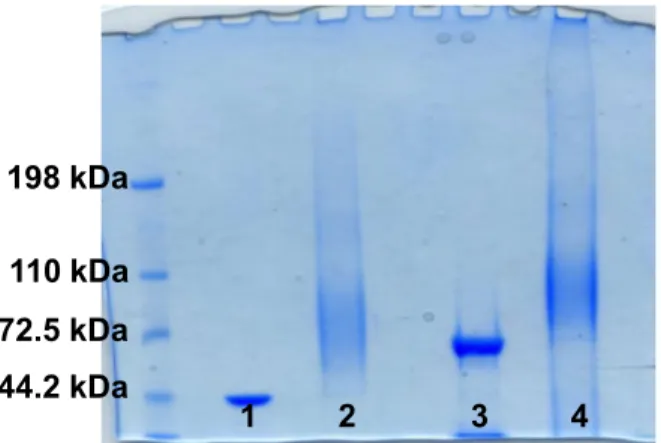
oxidized with sodium periodate to introduce an aldehyde group useful for the coupling to the lysine residues of the protein by reductive amination23 (Fig. 1). The occurrence of complete conjugation was assessed by SDS-PAGE, and confirmed by the formation of a broad smear and the concomitant disappearance of the narrow band of the proteins (Fig. 2). Subsequently, the glycoconjugates were purified by size exclusion chromatography to remove unbound saccharide and analyzed for their protein content and in terms of total and free saccharide by HPAEC-PAD HPLC as described in literature.23
Table 1 summarizes the physico-chemical characterization of the purified glycoconjugates in term of total and free saccharide and protein content, compared to the PSII-CRM197 conjugate of which the
preparation and the characterization were previously reported.23 Notably, the degrees of glycosylation, ranging from 0.2 to 0.3 (w/w), was comparable for both the products.
Figure 2. SDS-Page of PSII-Toxin Conjugates 1. TcdA_B2 fragment protein; 2. PSII-TcdA_B2 conjugate, 3. TcdB_GT fragment protein; PSII-TcdB_GT conjugate.
Table 1. Characteristics of PSII Conjugates.
Conjugates PSII avDP Free Saccharide
% Saccharide/Protein w/w PSII-TcdA_B2 15 7.7 0.28 PSII-TcdB_GT 15 22.7 0.33 PSII-CRM197* 21 11.2 0.24
*characterization previously described23
2.2. Immunological Evaluation of PSII-toxins conjugates
To assess the ability of conjugates to induce anti-PSII antibodies, groups of 8 female BALB/c mice were intraperitoneally immunized three times with 2.5 µg carbohydrate based doses of conjugates, at three week-interval between the first and the second dose and two week-interval from the second and the third dose. The conjugates were formulated with the adjuvant MF59, an oil in water emulsion frequently used for seasonal flu vaccination.32 Adjuvant alone in phosphate buffer (PBS) was used as a negative control, while the PSII-CRM197 conjugate previously shown capable of inducing a robust
anti-198 kDa 110 kDa 72.5 kDa 44.2 kDa
polysaccharide immune response,23 and the TcdA_B2 and TcdB_GT fragment proteins already shown to be highly immunogenic,19 were used as a positive control.
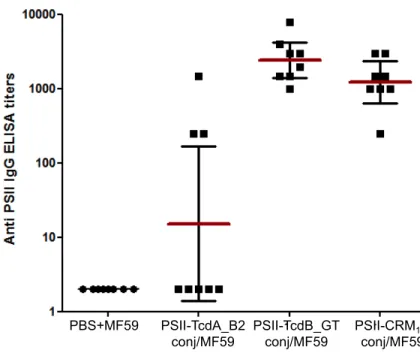
Sera obtained after two weeks from the third dose (post 3 sera) were analyzed by ELISA for their content of anti-PSII and anti-Toxin IgGs. Additionally, the functionality of the anti-Toxin antibodies was investigated in vitro to assess the capacity to neutralize the cytotoxicity of TcdA and TcdB.
The PSII-TcdB_GT conjugate was highly immunogenic, eliciting anti-PSII IgG titres comparable to those obtained with the PSII-CRM197 conjugate. Conversely, PSII-TcdA_B2 conjugate induced an
anti-polysaccharide response significantly lower than the CRM197 conjugate (p 0.006), where IgGs against
PSII were induced in three mice only (Fig. 3). These results evidenced a better capability of the TcdB_GT fragment protein to function as carrier for the PSII moiety in comparison to the TcdA_B2 peptide.
Figure 3. Anti-PSII IgG levels detected in individual post 3 sera of BALB/c mice; each dot represents single mouse sera; vertical bars indicate geometric mean titers of each group with 95% statistical confidence intervals as red bars.
PBS+MF59 PSII-TcdA_B2 conj/MF59 PSII-TcdB_GT conj/MF59 PSII-CRM197 conj/MF59