ContentslistsavailableatScienceDirect
Neuroscience
and
Biobehavioral
Reviews
j o u r n a l ho me p ag e :w w w . e l s e v i e r . c o m / l o c a t e / n e u b i o r e v
Review
The
contribution
of
TMS–EEG
coregistration
in
the
exploration
of
the
human
cortical
connectome
Marta
Bortoletto
a,∗,
Domenica
Veniero
b,
Gregor
Thut
b,
Carlo
Miniussi
a,c aCognitiveNeuroscienceSection,IRCCSCentroSanGiovannidiDioFatebenefratelli,Brescia,ItalybCentreforCognitiveNeuroimaging,InstituteofNeuroscienceandPsychology,UniversityofGlasgow,Glasgow,UK cNeuroscienceSection,DepartmentofClinicalandExperimentalSciences,UniversityofBrescia,Brescia,Italy
a
r
t
i
c
l
e
i
n
f
o
Articlehistory: Received11April2014
Receivedinrevisedform14October2014 Accepted11December2014
Availableonline22December2014 Keywords:
Effectiveconnectivity Functionalconnectivity TMS-evokedpotentialTEP Graphtheory
Transcranialmagneticstimulation(TMS) Electroencephalography(EEG) Coregistration
Connections
Non-invasivebrainstimulation(NIBS)
a
b
s
t
r
a
c
t
Recent developments in neuroscience have emphasised the importance of integrated distributed networksofbrainareasforsuccessfulcognitivefunctioning.Ourcurrentunderstandingisthatthebrain hasamodularorganisationinwhichsegregatednetworkssupportingspecialisedprocessingarelinked throughafewlong-rangeconnections,ensuringprocessingintegration.Althoughsucharchitectureis structurallystable,itappearstobeflexibleinitsfunctioning,enablinglong-rangeconnectionsto regu-latetheinformationflowandfacilitatecommunicationamongtherelevantmodules,dependingonthe contingentcognitivedemands.Hereweshowhowinsightsbroughtbythecoregistrationoftranscranial magneticstimulationandelectroencephalography(TMS–EEG)integrateandsupportrecentmodelsof functionalbrainarchitecture.Moreover,wewillhighlightthetypesofdatathatcanbeobtainedthrough TMS–EEG,suchasthetimingofsignalpropagation,theexcitatory/inhibitorynatureofconnectionsand causality.Last,wewilldiscussrecentemergingapplicationsofTMS–EEGinthestudyofbraindisorders. ©2014TheAuthors.PublishedbyElsevierLtd.ThisisanopenaccessarticleundertheCCBY-NC-ND license(http://creativecommons.org/licenses/by-nc-nd/4.0/).
Contents
1. Introduction... 115
2. EffectiveconnectivitythroughTMS–EEGcoregistration... 115
3. Distributednetworksfromneuroimagingandgraphtheory ... 116
4. Corticalnetworksinhealthycontrols:thecontributionofTMS–EEG ... 117
4.1. Cortico-corticalconnectivitywithinfunctionalnetworks... 117
4.2. Cortico-corticalconnectivityfromnodestobrainhubs... 117
4.3. Cortico-corticalconnectivityfrombrainhubstonodes... 118
5. CorticalnetworksandbrainoscillationsinTMS–EEGresearch... 119
6. Clinicalapplications... 119
7. Limitationsandopenquestions... 120
8. Conclusionsandfutureperspectives... 121
Acknowledgments... 121
References... 121
∗ Correspondingauthorat:IRCCSCentroSanGiovannidiDioFatebenefratelli,viaPilastroni4,25125Brescia,Italy.Tel.:+390303501597. E-mailaddress:[email protected](M.Bortoletto).
http://dx.doi.org/10.1016/j.neubiorev.2014.12.014
1. Introduction
In the past, the major focus of research defining the brain–behaviourrelationshipwastoidentifythesegregatedbrain regionsrecruitedbyagiventask.Morerecentdevelopmentshave emphasisedtheimportanceofdistributednetworksatalllevels, fromindividualneuronstoneuralpopulationsandbrainregions. Suchanapproachisthereforemovingneurosciencefroma topo-logicalperspectiveonthemappingof“importantbrainareas”to ahodologicalperspectiveon“systeminteractions”.Neuronsinthe brainareclusteredinregionswithdifferentcytoarchitectonicaland functionalproperties,whichareheavilyinterconnectedthrough bidirectionalconnectionsinlarge-scalenetworks.This architec-turerelatestofunctionalnetworks,inthatcognitiveefficiency– fromperceptiontomovementcontroland fromexecutive func-tionstoemotions –does notonly rely onthelocal processing ofinformationinspecialisedareasbutalsoontheintegrationof information(i.e.,connectivity)throughthecoordinatedactivityof multipleareas(Driveret al.,2009;Spornsetal.,2004). Accord-ingly,imbalancesinconnectivitypatternshavebeenproposedto bestronglyassociatedwithneurological(e.g.,Alzheimer’sdisease –AD:Tijmsetal.,2013;Vecchioetal.,2014andbraindamage: Catanietal.,2013)andpsychiatricdisorders(e.g.schizophrenia, BuckholtzandMeyer-Lindenberg,2012;Frantsevaetal.,2012).
Acrucialgoalofneurosciencestudiesistodefinethehuman “connectome”, a complete map of the neural connections in the human brain, both in terms of structural and functional connectivity, which will enable a better comprehension of the brain–behaviourrelationship(BehrensandSporns,2012;Sporns, 2013).Towardsthisgoal,neuroimagingtechniquessuchas func-tionalmagneticresonanceimaging(fMRI)andpositronemission tomography (PET) have beenemployed in thevast majority of studies.This field hasgreatly benefittedfrom multidisciplinary approachessuchastheintegrationofgraphtheoryin neuroimag-ing.Graphtheoryisamathematicaltooltoquantifytheproperties ofcomplexnetworksanddescribetheinterrelationshipsbetween networkelements(nodes)bymeansofconnections(edges),which inthiscaserepresentbrainregionsandtheirconnections, respec-tively(Baronchellietal.,2013;Minatietal.,2013).
Nevertheless,westillfaceseveralimportantchallengesinthis field,rangingfromthemethodologicallimitationsofour investiga-tiveequipment(Johansen-Berg,2013)tothelimitationsconcerning thecomplexityofanalysesandaprioriassumptionsthatareneeded withgraph theory(Buckneret al., 2013;De Reusand van den Heuvel,2013a;Fornitoetal.,2013).Forthisreason,crossvalidation throughindependentmethodologiesmaybecriticalfor overcom-ingthelimitationsofsinglemethodologiesin defininghowthe brainconnectomesupportscognitivefunctioning.
Here, we outline how a multimodal imaging approach combiningtranscranialmagneticstimulation(TMS)and electroen-cephalography(EEG)hascontributedandmightcontributetothe understandingof thefunctionalconnectome. TMS–EEG offersa uniqueinsightintoeffectiveconnectivity,thedescriptionofcausal interactionsbetweenregions,includinghowwelltheactivationof oneregionexplainstheactivationofanother(Fristonetal.,1993a). Notably,thisapproachprovidesacausalmodelontheoriginsof activationinneuralactivitypatternsandmightdefinethe func-tionalstrengthsbetweenregions.
We first review the TMS–EEG literature probing the motor systemduringtherestingstate.WethenreviewtheTMS–EEG lit-eratureonconnectivityindifferenttaskcontexts.Bycomparing TMS–EEGstudieswithfMRI-basedfunctional connectomics,we willshowhowitispossibletotestandvalidatethegeneral princi-plesoffunctionalbrainarchitectureinferredbygraphtheory,i.e., theorganisationandconfigurationofbrainnetworks,andprovide furtherinsightsintothepropertiesoftheconnectomeasafunction
ofabrainstateorspecificcognitivetask.Last,wewillunderline recentemergingapplicationsoftheTMS–EEGapproachinbrain disorders.
2. EffectiveconnectivitythroughTMS–EEGcoregistration
Neuroimagingtechniques,suchasEEG,fMRIandPET,canreveal thefunctionalconnectivitybetweenareasasresultof“temporal correlationsbetweenspatiallyremoteneurophysiologicalevents” (Fristonetal.,1993b).Obtainingmeasuresofeffective connectiv-itywiththesetechniquesrequirescomplexcausalmodels,such asdynamiccausalmodellingandGrangercausality(Fristonetal., 2013;StephanandRoebroeck,2012),basedonpre-existing neu-roanatomical, neuropsychological and functional neuroimaging data(Stephanetal.,2008).Effectiveconnectivityincludesa def-initionofcausality,whichthesetechniquescannotprovideperse. Therefore,theirinferentialpoweroneffectiveconnectivityrelies onaprioriassumptionsabouttheinvolvednetworkandaboutthe validityoftheimplementedmodel.
TheintegrationofTMSwiththeabovementioned neuroimag-ingtechniques,i.e.,PET,fMRIandEEG,maybeausefulempirical methodtotestfunctionalintegrationandcausality,i.e.,effective connectivity(Shafiet al.,2012,2013).Each combinationallows focusing on differentaspects of TMS-induced changes in brain activity. TMS–PET and TMS–fMRI coregistration can reveal the spatialprofilesoftranscranialbrainstimulationeffectswithhigh spatialresolution,includingsubcorticalstructures(Siebneretal., 2009).Nevertheless,thesetechniques havea reduced temporal resolutionandcanonlydetectmodulationsarisingafewseconds (fMRI) oreven minutes (PET)post-stimulus, becausesuch neu-roimaging techniquesare based on changes in blood flow and oxygenation.
TMS–EEG consistsof measuring electrical brain activity(via EEG)afterbriefnon-invasivebrainstimulation(viaTMS)and pro-videsanempiricalmeasureofeffectiveconnectivitybecausethe activation inducedby theTMS in thetargeted areapropagates toanatomicallyandfunctionallyconnectedregions(O’Sheaetal., 2008;Siebneretal.,2009).Themillisecondtemporalresolutionof theEEGprovidestwoimportantadvantagesinthestudyofbrain connectivity.First,informationaboutthetemporalpatternofthe responsesinducedbytheTMScontributestodefiningthecausal relationshipsintheconnectionsacrossbrainareas.Indeed,wecan assumethatifareaAisactivepriortoareaB,thenareaAmight causeincreased(orreduced)activityinareaBthroughexcitatory (orinhibitory)connectionsbetweenthetwoareas(Spornsetal., 2004).Second,itallowstheinvestigationofthetemporalevolution ofcommunicationbetweenregionsandtounfoldtheconnectivity patternsthroughouttaskexecution,highlightingshorttime win-dowsofinformationexchange.Inaddition,thedeliveryoftheTMS pulsesisunderexplicitexperimentalcontrol(Miniussietal.,2013; SackandLinden,2003),therebyallowingresearchersto success-fullydifferentiatetheconnectivitypatternofdifferentcognitive processes related tothe execution ofspecific tasks (Morishima etal.,2009)ortodifferentbrainstates(Massiminietal.,2005).Last, EEGprovidesadirectmeasureoftheelectricalsignalsgenerated byneuronalactivityandenablesresearcherstoderivethe excita-tory/inhibitorynatureofnetwork connections(Daskalakisetal., 2012).ThesefeaturesofTMS–EEGcanbeofgreathelptostudy thetemporalsequenceofneuralactivitythatdeterminethe inter-actionsbetweenbrainareas,thatistypicallymodelledbygraph theoryintheneuroimagingfiledtorepresentnetworksof commu-nication.
A simple wayto evaluatecortico-cortical connectivity is by studyingthespatio-temporaldistributionofTMS-evoked poten-tials (TEPs) after a single TMS pulse, also called the inductive
TMS–EEGapproach(MiniussiandThut,2010).AfteraTMSpulse overacorticalregion,thespreadoftheinducedactivitycanbe tracedviathewaveformandtopographyoftheTEPoverthescalp, providingadirectmeasureofbrainconnectivity(e.g.,Bonatoetal., 2006;Ilmoniemietal.,1997;Komssietal.,2002).
Single-pulseTMSandtrainsofpulsescanalsotrigger(Pausetal., 2001)orenhancebrainoscillations(ThutandMiniussi,2009;Thut etal.,2011),whenthesetrainsarefrequency-tunedtothe under-lyingbrainoscillationsofthetargetcorticalarea(entrainment), definedastherhythmicTMS–EEGapproach(MiniussiandThut, 2010).Brain oscillationsarethoughttorepresent a mechanism throughwhichinformationisprocessedwithinanetwork,forming dynamicassembliesofneuronsthroughsynchronisationinspecific frequencybands(Buzsaki,2006;Watrousetal.,2013).
Last,TMS-inducedcoherenceofcorticalareascanbeusedforthe identificationofeffectiveconnectionsduringatask,basedonthe notionthataboundiscreatedbythesynchronisationofoscillatory activity.Severalmethodshavebeendevelopedtoestimate inter-actionsbetweenbrainregionsbasedontheamplitudeandphase ofEEGoscillations,e.g.,directedcoherence,imaginarycoherence, phase-lockingvalue,phase-lagindex,etc.Adetaileddescriptionof thesemethods,theirbreakthroughsandtheirpitfalls,canbefound inrecentmethodologicalpapers(Greenblattetal.,2012;Sakkalis, 2011;SchoffelenandGross,2009).
Briefly,thespatial–temporalpatternofthebrainresponsesto TMScancontributetodefiningcausalrelationshipsinthe connec-tionsacrossbrainareasandcanrevealtheiractivationatthetimeof stimulation.Byexaminingtheresponseswithinthenetworkwhen oneofitsnodesisstimulated,TMS–EEGcoregistrationprovides
measuresofeffectiveconnectivitythatmaytestthepredictionsof graphtheorymodelsintermsofbraininteractionsmoredirectly andalongacausaldimension.
3. Distributednetworksfromneuroimagingandgraph
theory
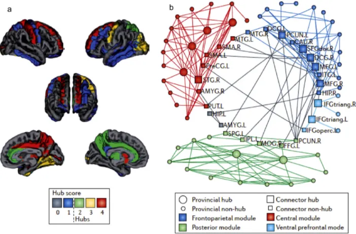
Graph theory analyses of structural and functional neu-roimaging have suggested a hierarchical modular organisation ofthehumanconnectome thatis organisedtorapidlyand effi-cientlytransferinformationthroughminimalwiring(Bassettand Bullmore,2006;BullmoreandSporns,2012;Ercsey-Ravaszetal., 2013). Brain connectivity architecture may resemble a “small world”networkthatischaracterisedbymodules(orsub-networks) thatperformsegregatedandhighlyspecialisedprocessingandthat arelinkedthroughafewlong-rangeconnectionsthatensure inte-gratedprocessing(Achardetal.,2006;BassettandBullmore,2006; Spornsetal.,2004)(Fig.1).
Modulesofnodescorrespondtofunctionalnetworksofareas, e.g.,themotornetwork,visualnetwork,dorsalattentionnetwork, defaultmodenetwork,mediallobememorynetworkand fronto-parietalcontrolnetwork(Poweretal.,2011;Smithetal.,2009). Areasofthesamenetworkhavestrongeranatomicalconnections (Wangetal.,2013)andinherentbiasesintheirinteractions(Chu etal.,2012;Decoetal.,2013;Smithetal.,2009),asindicatedby highlocalclustering,i.e.,thehighprobabilityoftwoneighbouring nodesbeingconnectedtoeachother.
Long-range connectionslink areas that participate in multi-plenetworks,called“hubs”,andensurefastinformationexchange
Fig.1.Hierarchicalmodularorganisationofthehumanconnectome.(a)Hubs:regionswithahighernumberofconnections,highervalueofbetweennesscentrality,a shorterpathlengthandhighlyclusteredamongthemselvesarecalled‘hubs’andareindicatedinthefigurebya‘hubscore’of2orhigher.Hubsincludefronto-parietal regionsandsubcorticalregions.(b)Modulesofnodes:functionallyrelatednodes(circles)arespatiallycloseanddenselyinterconnectedthroughshort-rangeconnections, formingmodulesorsub-networks.Thehubs(squares)ofeachmodulemediatemostofthelonger-distanceinter-modularconnections.Here,fourmajormodulesareshown, comprisingfrontal(darkblue),central(red)andposterior(green)brainregionsandinferiorfrontalregions(lightblue).(Forinterpretationofthereferencestocolourinthis figurelegend,thereaderisreferredtothewebversionofthisarticle.)
acrossnetworksby shorteningtheaveragepathlength,i.e.,the minimum path length between any two pairs of nodes in the network.Hubsarecentralandinfluentialonglobalnetwork func-tioningbecausetheyhaveahighernumberofconnections(high degree), are waypoints for the shortest path in the network (betweennesscentrality)andtendtobedenselyinterconnected withotherhubs,forminga“richclub”.Althoughthereisno com-pleteagreementonthelocalisationofareasincludedintherich club,hubshave beenfoundinassociationareaswithinbilateral fronto-parietalregionsandsubcorticalregions(Gongetal.,2009; VandenHeuvelandSporns,2011).
Although thestructural connectome is highly stable (atthe macroscale), the functional connectome appears to be flexible asdifferent cognitivestates areassociated withchanges inthe weightoffunctionalconnections.Thereisevidencethattheresting statemaybeassociatedwithastrongermodularstructure com-paredwithconditionsofhighcognitivedemand(Dietal.,2013; Kitzbichleretal.,2011).Duringtherestingstate,acost-efficient networkreconfigurationhasbeensuggested,inwhichtheactivity ofweaklong-rangeconnectionsisreducedandmoresegregated modulesofstronger connectionsincreasetheefficiencyoflocal communication(Ercsey-Ravaszetal.,2013).Inotherwords,areas thatworktogethertosubtendacognitivefunction(e.g.,movement, attentionandmemory)tendtobemorestronglyconnectedand alsomoresegregatedfromothermodulesintherestingstate.Graph theorymodelsfurthersuggestthatthecommunicationacrossthese locallyclusteredandspecialisednetworksisensuredbybrainhubs andthecentralinfrastructureoftherichclub(DeReusandvanden Heuvel,2013b;VandenHeuvelandSporns,2011).Theconnections ofbrainhubsareshapedbycontingentcognitivedemands(Chadick andGazzaley,2011;Cocchietal.,2013)tofacilitate communica-tionbetweenrelevantmodules,assuggestedbytheflexiblehub theory(Coleetal.,2013).Thissmall-worldarchitectureconstrains thefunctioningofbrainconnections,possiblybymaximisingthe complexityoradaptivityofafunctionwhilealsominimisingwiring costs(BassettandBullmore,2006;BullmoreandSporns,2012).
4. Corticalnetworksinhealthycontrols:thecontribution
ofTMS–EEG
Accordingto graph theory models, thenodes of specialised networksand hubsoftherich clubshouldshow different con-nectivityprofiles:theformer shouldmainlyconnect withother nodesofthesamefunctionalnetwork,especiallyduringtheresting state,whereasthelattershouldshowahighnumberof intermod-ularconnections.TMS–EEGcanbeemployedtoempiricallytest thesepredictionsaboutnetworkarchitecture.Wewillshowthat TMS–EEGstudiesonnetworkdynamicsatrestandduringcognitive processesareinlinewithspecialisednetworksegregation(which ismorepronouncedduringtherestingstate),withdense connec-tivitythroughbrainhubs,andsupportwhathasbeensuggestedby graphtheorymodels.
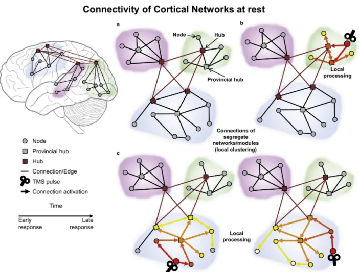
4.1. Cortico-corticalconnectivitywithinfunctionalnetworks Inlinewithresting-stateneuroimaging,TMS–EEGhasrevealed functionallyisolatednetworks whenparticipantsare atrest,as depictedinFig.2.Asanexample,letusconsiderthemotornetwork, identifiedbyfMRIasincludingtheleftandtherightmotorregions andthesupplementarymotorarea(Biswaletal.,1995;Patriatetal., 2013;Smithetal.,2009).
Sincethefirststudies,TMS–EEGhasrevealedthatinduced activ-ityspreadsfromthestimulatednodetoothernodesofthesame motornetwork.ActivationmapsfromTEPsusingminimum-norm estimates(Ilmoniemietal.,1997;Komssietal.,2002)andprecise
sourcelocalisation(Litvaketal.,2007)haveshownthatTMSofthe primarymotorcortexcausesthesucceedingactivationofipsilateral supplementary/premotorareasand contralateral motorregions, andthattheseactivationsdependontheamplitudeoftheresponse evokedinthetargetedmotorcortex(Giambattistellietal.,2014). Accordingly,transcallosalconnectivity(Ilmoniemietal.,1997)and fastdirectsignalconductionontheorderof9–20msbetweenthe homologousmotorregionshavebeenreportedusingTEPs(Komssi et al.,2002)and EEGoscillations(Manganottiet al.,2012).The shortconductiontimeoftheTMS-inducedactivitysuggestsdirect connections,inlinewithneurophysiologicalevidenceinhumans (Civardietal.,2001;Groppaetal.,2012)andmonkeys(Boussaoud etal.,2005;DumandStrick,1991;Rouilleretal.,1994).
Interestingly, TMS–EEG studies have revealed theinhibitory versus facilitatory nature of these connections depending on the level of cortical activation. Regarding transcallosal connec-tivity, a recent studycorrelated diffusion tensor imaging-based fractionalanisotropy incallosalmotor fibreswithTMS-induced interhemispheric signalpropagationin theprimary motor cor-tex(Voineskosetal.,2010).Crucially,theauthorsreportedthat sub- and supra-threshold TMS over the primary motor cortex induced,respectively,facilitatoryandinhibitoryeffectsover the contralateralhomologousarea.Suchdistincteffectsarelikelydue toadifferentthresholdofactivationofexcitatoryandinhibitory circuits, suggesting that the corpuscallosum mayregulate the communicationbetweenhemispheresdependingonthelevelof activation.Likewise, arecent TMS–EEG studyreportedthat the connectionsbetweenprimarymotorandpremotorareasmaybe mainlyinhibitoryduringtherestingstate(Venieroetal.,2012). Theauthorsreportedanegativecorrelationbetweenexcitabilityof theprimarymotorcortex,measuredastheamplitudeofthe mus-culartwitchinducedbytheTMS,andtheamplitudeofanearlyTEP componentlikelygeneratedintheipsilateralpremotorarea.Inline withfMRI-basedgraphtheorymodelsofcorticalconnectivity,that haveindividuatedsegregatednetworksofco-varyingactivity,TMS activationsatrestdonotreachareasclearlyembeddedinother specialisednetworks,e.g.,thevisualnetwork.
Insummary,thesestudiesonthemotorsystemhaveidentified singularnodesoftheprobednetworkatrestbythespatio-temporal decomposition ofTMS-induced activity.Theyillustratethat the cortico-corticalspreadingofTMS-inducedactivityremainslargely confinedtothespecialisedmotornetwork.Therefore,byshowing thatactivityisconfinedtoarestrictedspecialisednetwork,they supportamodularorganisationoffunctionalbrainarchitectureat rest.
4.2. Cortico-corticalconnectivityfromnodestobrainhubs
AlthoughearlylatencyTMSresponsesreflectdirectconnections withinfunctionalnetworks,latercomponentsofTEPsreveal fur-thernodesandmorecomplexinteractions,suggestingbottom-up signalpropagationfromlower-degreenodestobrainhubs.
For example, primary motor area stimulation generates the lateactivationofareasoutsidethestimulatedfunctionalnetwork, involvingthecingulategyrusandtemporo-parietaljunction(Litvak etal.,2007).Theseactivations,possiblyachievedthroughloopsor indirectconnectionswithothernodes,affectareasthathaveastrict functionalrelationshipwiththestimulatednetworkandmay cor-respondtobrainhubs.Compellingevidencehasalsobeenfoundin thevisualsystem.Garciaetal.(2011)appliedTMStodifferentareas ofthevisualsystem,includingtheleftandrightprimaryvisual area,middletemporalcortex,andaventraltemporalregion. Inter-estingly,bothsite-specificandsite-invariantEEGresponseswere obtained.Site-specificresponsesweremainlygeneratedatearlier latencies,whereassite-invariantresponsesincreasedwithlatency. Importantly, many of these site-invariant responses seemed to
Fig.2. Testingconnectivityduringtherestingstate.(a)Schematicfigureofthemodularorganisationofthebrainnetwork,includingnodes(greycircles),provincialhubs (greysquares)andhubsoftherichclub(redsquares),andtheirshort-range(blacklines)andlong-range(redlines)connections.(b)Thespatio-temporaldistributionofthe brainresponsetoTMSofalower-degreenodeisshown.Colouredarrowsrepresentthecausalinteractionsbetweennodesandthelatencyofsignalpropagationfromthe TMSpulse.AfterTMS,theactivationofthetargetareatravelstoothernodesofthesamemodulethroughshort-rangeconnections.(c)Whentwolower-degreenodesof thesamenetworkarestimulatedbyTMS,thesignalpropagateswithinthesamemodule.Atfirst,differentnodes(site-specificresponses)andeventuallythesamehubsare activated(site-invariantresponses).ThefiguresimulatesresultsfromGarciaetal.(2011).(Forinterpretationofthereferencestocolourinthisfigurelegend,thereaderis referredtothewebversionofthisarticle.)
convergeonafrontalandparietalEEGsignature,suggestingthat thespreadofactivationfromthestimulatedlower-degreevisual nodesconvergedtocommon,heavilyinterconnectedassociative areasthatcanbeidentifiedasbrainhubs.
Tosumup,theTMS–EEGliteratureonconnectivityshowsthat activityinducedindifferentareastendstoreachasubsetofareas thathavebeenidentifiedasbrainhubs.Theconvergenceofthe signalfromnodestohubsisinlinewiththeputativeroleofbrain hubsinthetransmissionofinformationacrossthebrainandwith graphtheoryoutcomesthatmostoftheconnectionroutespassby atleastahub(VandenHeuveletal.,2012).
4.3. Cortico-corticalconnectivityfrombrainhubstonodes
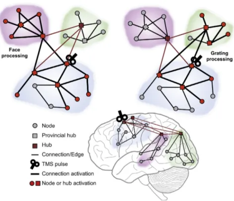
SomeTMS–EEGstudieshavetargetedmultimodalassociative andexecutiveareasresponsibleforhigher-ordercognitive func-tions (e.g., Morishima et al., 2009; Taylor et al., 2007), likely correspondingto brainhubs. Workby Morishima et al. (2009) usedTMSasaprobetoevaluateneuralimpulsetransmissionfrom theprefrontalcortextodownstream,specialisedposteriorregions whileparticipantswererequiredtoattendtospecificfeaturesof visualstimuli.Theauthorshypothesisedthatstimulatingthe pre-frontalareasoftheattentionalnetwork (CorbettaandShulman, 2002;DesimoneandDuncan,1995)wouldinduceaspreadof acti-vationtowardsanatomicallyconnectedposteriorregionsandthat
thedirectionandamountofthecurrentspreadcouldbedifferent accordingtothefunctionalnetworkengagedtoaccomplishthetask inaparticularcontext.Inlinewiththesepredictions,theyfound thatTMSoverthefrontaleyefieldactivatedtwodifferentnetworks, functionallyconnectingthetargetareatodistinctposteriorvisual areasdependingonthenatureoftheto-be-attendedvisualfeature (verticalgratingsversusfaces),asrepresentedinFig.3.Moreover, TMSeffectsoccurred20–40msfollowingthepulse,suggestingthat impulsepropagationwasnotduetoreroutingviaotherareasbut wasinsteadachievedbythedirecttransmissionofaneuralinput fromfrontaltoposteriorregions.
Othershaveconfirmedthatbytargetingassociatedareaswith TMSduringtaskperformanceandassessingtheTMSeffectson task-relatedEEGresponses,thetop-downsignalssenttodownstream networknodesspecialisinginstimulusprocessingcanbetracked. ThisuseofTMS–EEG,termedtheinteractiveTMS–EEGapproach (MiniussiandThut, 2010), hasbeensuccessfullyappliedin sev-eraldomains,includingperceptualdecision-making(Akaishietal., 2013), controlledbehaviour(Akaishiet al.,2010), goal-directed actions(Verhagenetal.,2013),visualsearch(Tayloretal.,2011), faceprocessing(Mattavellietal.,2013)andshort-termmemory (Johnsonetal.,2012).
Therefore,theTMS–EEGliteratureontask-relatedconnectivity frombrainhubsshowsdivergenceofTMS-inducedactivityfrom theseareasdependingonthetaskcontext.Theseobservationsare
inlinewithdenseconnectivitythroughbrainhubsandwiththe suggestedroleofbrainhubstomediatecommunicationbetween differentmodulesaccordingtothecognitivecontext(Coleetal., 2013).
Hence,TMS–EEGrevealsdynamicchangestofunctional con-nectivity asa result ofbrain stateand thehierarchicallevel of the targeted node. If the target area is highly interconnected acrossnetworks(constitutingahub oftherichclub),TMS–EEG hashighlightedtop-downmodulationsthatestablishthe informa-tionflowacrossdifferentnetworks.Incontrast,targetinganodeof aspecialisednetworkappearstomainlyillustratethebottom-up task-relatedmodulationsoftheconnectionswithinthatfunctional network. These state-dependent changes have been previously suggestedbyanalysingfMRIdatabymeansofgraphtheoryand byapplyingpaired-pulseTMSprotocolsshowingthatthe interac-tionsbetweenbrainareasareshapedbyongoingprocesses(e.g., Buch et al., 2010; Davare et al., 2008, 2009). Altogether,these resultsillustratetheuseofTMS–EEGintheselectiveinvestigation oftransientlyactivatedcorticalnetworksintheprocessof accom-plishingacognitiveact,reflectedinthespreadoflocalTMSeffects to connected areas in a state-dependent manner,as shown in Fig.3.
5. CorticalnetworksandbrainoscillationsinTMS–EEG
research
AnemerginglineofTMS–EEGresearchfocusesonTMS-induced brain oscillations, which extends the TEP approach tothe fre-quencydomain,focusingontheoscillatorycomponentsofthebrain responsegeneratedbyTMSpulsesortrains.TMSisexpectedto interactwithsuchoscillatorypatternsinthedirectlystimulated corticalarea(Thutetal.,2011)andindistantareasbelongingto thesameneuralnetwork(Thutetal.,2012).Therefore,ifagroup ofnetwork elementssynchroniseata locallevel afterTMS,we shouldexpectaconsequentlonger-rangesynchronisation (interre-gionalcoherence)representinginformationtransferamongbrain structures.Inshort,weshouldexpecttheinductionofthesame frequencyactivityafteradelay(phaseoffset)inall“synchronised” areasofthesamenetwork.Thisoutcomewouldfacilitatethe local-isationofinvolvedareas.Inductionofafrequencyinthestimulated areamayeveninduceadifferentfrequencyinaconnectedarea. Nevertheless,bothfrequencieswouldbetimelockedwiththeTMS pulse,therebypotentiallyenablingconnectedareastobelinked withthestimulatedarea.
Somepreliminaryindicationsthatdistinctcorticalnetworksare characterisedbydifferentoscillatoryactivityhavebeenreported byRosanovaetal.(2009)whotargetedtheleftfrontal,parietaland occipitalcortexwithsinglepulseTMSatrestandmeasuredthe evokedcorticaloscillatoryresponses.Eachcorticalarearesponded ata characteristicfrequency,itseigenfrequency (ornatural fre-quency).Mostimportantly,Rosanovaetal.(2009)alsoshowedthat thetopographyoftheevokedoscillatoryactivitywassubstantially dependentonthetargetedregion,withlittleoverlapacross stimu-lationsites,thussuggestingthatfunctionallysegregatednetworks canoscillateatdifferentfrequenciesatrest(seealsoBrignanietal., 2008;Venieroetal.,2011).
AfollowupstudyusingrhythmicTMS,frequency-tunedtothe naturaloscillatoryactivityofthetargetarea,revealedthe possi-bilityofa frequency-specificenhancementdue toaprogressive synchronisationofthenaturaloscillatortotheperiodicexternal stimulation (entrainment) (Thut et al., 2011). Interestingly,the stimulationofanode(intra-parietalsulcus)oftheattentional net-workwiththeTMSrhythmicapproach(targetingattention-related frequencies)alsoledtochangesinthebehaviouralperformance, biasingthesubjects’perception(Romeietal.,2010,2011,2012)and
suggestingthattheseoscillationsand presumablytheentrained networksarecausallyimplicatedintheprobedcognitiveprocess.
Finally,corticalconnectivityhasbeenshowntopotentiallybe promotedviapairedassociativeTMS,duringwhichtwopaired sin-gleTMSpulses (Ferreri etal.,2011;Venieroetal.,2013)ortwo pairedrhythmicTMStrainsatthesamefrequency(Plewniaetal., 2008)areapplied,withaslightdelay,over two interconnected corticalareas.Basedonthisconcept,Venieroetal.(2013) demon-stratedthattherepeatedco-activationoftwoareas(parietaland primarymotorcortex)selectivelyreinforcescommunication, mea-suredasinterregionalcoherence,betweenthetargetedregionsin twodifferentoscillatorycomponentsaccordingtotheinhibitoryor excitatorymotoroutcome.Likewise,Plewniaetal.(2008) demon-stratedthatbifocalrhythmicTMSinthealphafrequencyinduceda topographicallyselectiveenhancementofinterregionalcoherence, mainlyatthestimulatedfrequency,thatlastedupto10minafter stimulation.
FurtherstudieswillhavetofocusonhowTMSinteractswith oscillatorynetworkactivitywithinthecomplexmulti-frequency workspace of our brain, likely providing additional interesting informationonthehumanconnectomefromanoscillatory per-spective.
6. Clinicalapplications
Whenanodeisaltered,networkconnectionscanchangeintwo ways.First,theweightofedgesoftheaffectednodecanchange,i.e., theconnectionsbetweenthetargetareaandtheconnectedareas canbestrengthenedorweakened.Second,thelossoftheaffected nodeanditsconnectionscanmodulatedistantedgesandactivate alternative(compensatory)pathsofinformationflow.Graph the-oryindicatesthattheeffectsofneuraldamagemaydependonthe affectedarea,i.e.,whetheritconsistsofabrainhuboraspecialised lower-degreenode(Albertetal.,2000).Indeed,thelossofhubs mayleadtofragmentationofthebrainnetworkintodisconnected parts(Tijmsetal.,2013).Convergingresultsfrommodelling stud-iessupportthatinterventionsintobrainhubs,suchastheremoval orweakeningofahub’sconnections(Achardetal.,2006;Vanden HeuvelandSporns,2011)orlesionsinassociativecortices,have amuchstrongerimpactonbrainarchitecturethanlesionsin pri-maryareas(Alstottetal.,2009).Thesestudiesindicatethathubs aremoreimportantthanothernodesforglobalbrainfunctioning andthattheirlossmaybemoredifficulttocompensatefor.
Thedisruptionofneuralconnectivityhaslongbeenassociated withmanypathologicalconditions,e.g.,AD,autism,aphasic distur-bancesandagnosias(Frith,2001;Geschwind,1965;Vecchioetal., 2014).Therefore,theopportunitytoevaluateabnormal connectiv-itymightplayacentralroleindiagnosesandfuturetherapeutic interventions.TMS–EEGcanbeusedtoexaminenormaland mod-ified effectivebrainconnectivityunderspecificconditions,such asdifferentphysiological (Massimini etal.,2005)and patholog-icalstates(Ferreri etal.,2014;Ragazzonietal.,2013;Rosanova etal.,2012),andunderpharmacologicaltreatment(Ferrarellietal., 2010).Assuch,TMS–EEGcouldindicatethestrengtheningor weak-eningofexistingcortico-corticalconnectionsortherecruitmentof compensatorynetworks.
Afewstudieshaveemployedresting-stateTMS–EEGtoevaluate alteredconnectivityinspecificpathologiessuchasAD(Casarotto etal.,2011;Julkunenetal.,2008,2012)orasatoolfor diagnos-ticsandearlyidentificationofmildcognitiveimpairment(Julkunen et al.,2008).Julkunen etal. (2008) showedthat stimulationof themotorcortexinADpatientswasassociatedwithasignificant decreasein TMS-inducedactivityover severalbrain areas com-paredwithhealthycontrols.Theseprominentchangesinfunctional corticalconnectivitysuggestthatlarge-scalenetworksare abnor-mallyorganisedinADpatientsduetothealterationoflong-range
Fig.3.Testingrevealsconnectivityduringtaskexecution.Thefigureillustratesthesignaldistributioninthebrainschematicallyrepresentedbythreemodulesofnodes withlong-rangeconnectionsamongthem(asinFig.2).Taskexecutionincreasestheconnectivity(thicklines)betweenthefunctionallyrelevantmodules.TMS-induced responsestravelontheseconnections,i.e.,fromthetargetnodetothenodesthataremorestronglyconnectedduringthetask.Bystimulatingahub,“increasedconnectivity” withinthelocalstimulatedmoduleandalonglong-distanceconnectionstothefunctionallyrelevantdownstreammodulescanbeidentified.Thefiguresimulatesresults fromMorishimaetal.(2009).
connections.Similarly,theapplicationofgraphtheorytofunctional connectivitystudiesinADsupportsaglobaldecreaseinfunctional connectivityandahighervulnerabilityofhubconnections(Stam etal.,2007,2009;Tijmsetal.,2013;Vecchioetal.,2014).
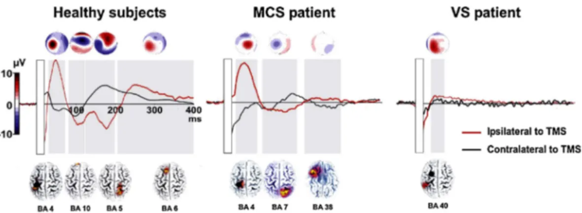
More prominently,TMS–EEG connectivity studies have pro-vided valuable information for the differential diagnosis of consciousness disorders (e.g., vegetative state and minimally conscious state) and hence have related consciousness to sig-naturesof cortico-corticalconnectivity.Consciousnesshasbeen describedasanemergentpropertyofthecollectiveactivityofa widespreadassociative network(Demertziet al.,2013; Laureys and Schiff, 2012; Tononi, 2004). Although to our knowledge, graphtheoryhasnotyetbeenappliedtopatientswithdisorders ofconsciousness,severalneuroimagingstudiesusingboth fMRI (Vanhaudenhuyseetal.,2010)andEEG(Varottoetal.,2014) sug-gestacloserelationshipbetweenresting-stateconnectivityin a fronto-temporo-parietal network (resembling thedefault mode network)andthelevelofconsciousness.Inlinewiththesedata,two independentTMS–EEGstudies(Ragazzonietal.,2013;Rosanova etal., 2012)havebeen abletodistinguish betweenpatientsin avegetativestateandaminimallyconsciousstatebasedon dif-ferencesin intra-and interhemisphericconnectivitypatternsas revealedbyTEPs(asillustratedinFig.4).Inminimallyconscious patients, both studies found TMS evoked local activity in the stimulatedareaandalsoinmoredistal,connectedcorticalsites. This distal activation was limited to the areas homologous to thestimulatedone (Ragazzoni etal.,2013).Incontrast,in veg-etative statepatients, TMS-evoked activity(when present)was locallyconfinedtothestimulatedhemisphere,indicatingstrongly compromisedeffectiveconnectivity.Interestingly,theabsenceof contralateralTEPssignificantlydiscriminatedbetweenthe vege-tativestateandminimalconsciousstategroups(Ragazzonietal., 2013).
Asreviewedabove,TMS–EEGconnectivitystudiesinpatients havehighlightedthevitalimportanceofstablebrainnetwork inter-actionstoensurenormalbrainfunction,characterisingdisordersby alteredconnectivityandadvancingknowledgeofthe pathophysi-ologicalstateofagivencondition.
7. Limitationsandopenquestions
SofarwehavehighlightedtheadvantagesoftheuseofTMS–EEG inthestudyoftheconnectome.Herewesummariselimitationsand confounderswiththepossiblestrategiesthathavebeendeveloped foraddressingthem.
Cautionshouldbetakenwhenstudyingconnectivitythrough EEG-basedcoherencemeasuresbecausetheactivityfromthesame sourcecan berecorded from differentEEG sensorsand induce spuriousconnectivitypatterns.Severalmethodshavebeen imple-mentedtoreduceorexcludespuriousconnectivity,mainlybased onthedetectionoflaggedinteractions,excludingzero-phase inter-actions(e.g.,imaginarycoherence,partialdirectedcoherenceand phase-lagindex)(Greenblattetal.,2012;PalvaandPalva,2012; Sakkalis,2011;SchoffelenandGross,2009).Moreover,volume con-ductionmaybefurtherreducedbyperformingtheconnectivity analysesatthesourcelevel.
Anotherlimitation is related to the useof TMS. Given that TMS induces spreading of activity along active functional con-nectionsatthetimeofstimulation,TMS–EEGmayhighlightthe excitatory/inhibitoryinterplaybetweennodeswithinandacross networks,butitmightnotrevealthecompletesetofconnections of a node. Secondly, most subcortical structures are silent in EEGrecordingsbecause onlycolumnar structurescontributeto surface recordings. Therefore some properties of the connec-tome and the distinction between some connectivity models mayhardlybeobtainedwiththistechnique,e.g.,thesparcityof
Fig.4. Alteredconnectivityindisordersofconsciousness.ThefigurepresentstheTEPsrecordedfromtheC3(ipsilateraltoTMS–redline)andC4electrodes(contralateral hemisphere–blackline)inagroupofhealthycontrols,aminimalconsciousstate(MCS)patientandavegetativestate(VS)patient.Inallsubjects,theleftmotorcortexwas stimulated,asindicatedwithablackdotonthesignaldistributionmapsreportedbelowwaveforms.Theresponsesobtainedduringtheshamconditionwerepoint-by-point subtractedfromthoseobtainedduringrealTMS.Thetimewindowsinwhichthesignalwassignificantareindicatedwithagreyrectangle(i.e.,EEGsignalexceedingthree timesthestandarddeviationofthepre-stimulusactivityforatleast20ms).Grand-averagedscalptopographiesandsLORETAlocalisationmapsarereportedonthetopand bottom,respectively,showingchangesovertimeoftheactivatedareas(BA=Brodmannarea).Thecolourscaleontheleftshowstherangeofvaluesfortopographymaps. (Forinterpretationofthereferencestocolourinthisfigurelegend,thereaderisreferredtothewebversionofthisarticle.)
ModifiedfromRagazzonietal.(2013).
intermodular connectivity (Bassett and Bullmore, 2006) or the declineofconnectionswithdistance(MarkovandErcsey-Ravasz, 2013).
AnopenquestionhereishowsensitiveTMS–EEGmaybefor trackingalternativeroutesofneuralinputtransmissionasa func-tion of task context and pathological conditions. Graph theory indicatesthat whenanodeisaltered, networkconnectionscan changeintwoways.First,theweightofedgesoftheaffectednode canchange,i.e.,theconnectionbetweenthetargetareaandthe con-nectedareascanbestrengthenedorweakened.Second,thelossof theaffectednodeanditsconnectionscanmodulatedistantedges andactivatealternativepathsofinformationflow.TMS–EEG stud-iesmighthighlightbothmechanisms,bylookingatthestrength oftheconnectionsbetweenthetargetareaandtheconnected net-work(Shafietal.,2013)andattheactivationofnewrouteswithin thenetwork. In theclinical context, it would beinteresting to useaTMS–EEGapproachtotrackcompensatoryactivityandthe reorganisationofconnectivity,i.e.,whetheralternativeroutesare adoptedtosolveataskinpathologicalconditionsandtounderstand theassociationbetweenthealternativeroutesandthepreserved abilities.Moreover,establishingwhetherahuboranodeisaltered wouldaidinthedesignofmorespecificneuromodulationand reha-bilitationprotocolstore-establishdegradedfunctions(Miniussi andRossini,2011).
Changesintheneuronalnetworkdynamicsareimportantalso forhealthybrainfunctioning,inthecontextoflearning.Indeed, changesincognitivefunctionsareintimatelytiedtothecapacity ofa systemtoacquireor improveskillsthrough plasticity pro-cesses.RepetitiveTMSprotocolshaveplasticityinducingproperties (Shafietal.,2013;Siebneretal.,2009),andthereforeTMScanbe usedtotemporarilymodifyactivityinatargetedcorticalregion. Theseplasticchangesinthetargetedareamayalsoinducecomplex widespreadalterationintheglobalfunctionalconnectivityand net-workefficiencythatmaydependonwhetherthetargetedareais abrainhuboraspecialisedareaoflowerdegree.Inthiscontext byrecordingEEGactivityduringplasticity-inducingTMS proto-colsitmaybepossibletoevaluatehowlongterm potentiation-orlong-termdepression-likeeffectsactatnetworklevel.
8. Conclusionsandfutureperspectives
TMS–EEGcoregistrationoffersauniqueopportunitytostudy effectiveconnectivityathightemporalresolutionthrough simul-taneous cortical stimulation and evaluation of induced cortical
activityatboth localandglobal(network)levels.Thereviewed findings,inlinewithprevioustheorizationsbymeansoffMRIstudy withgraphtheoryanalysis,suggestthatTMS-probed connectiv-itypatternsdependonthehierarchicallevelofthetargetedarea, revealedbydifferentphysiologicalandbehaviouralconsequences ofthestimulationofbrainhubsandlower-degreeareas.Induced activityinresponsetothestimulationoflower-degreeareas ini-tiallyspreadswithinthesegregatednetworkinwhichtheareais embeddedandeventuallyreachesmore interconnected, higher-degreenodes.Incontrast,inducedactivityfromthestimulationof abrainhubquicklyreachesspecialisedareasofalowerdegree, dependingontheactivationstateofitsconnections.
TheinterfacingofTMS–EEGstudieswithfindingsonbrain net-workarchitecturederivedfromotherneuroimagingtechniqueshas twoimportantadvantages.First,itprovidesempiricalvalidationto modelsofbrainnetworkarchitecture,suchasgraphtheory. Sec-ond,TMS–EEGcanprovidedynamicmeasuresoftheresponseof thebrainwhenonespecificnodeistargeted,i.e.,focusingon spe-cificsub-networksoronspecificconnections.Thisinformationis missingingraphtheorymodelsinwhichglobalindicesofnetwork functioningareemployed.However,morespecificindicesof net-workfunctioningarecrucialtodefinetherelationshipbetween theactivityinaspecialisednetworkandaspecificcognitive func-tionordysfunctionafterneuraldamage.TMS–EEGcoregistration canovercometheselimitationsandhighlightthedifferent contrib-utionsofbrainhubsandlower-degreespecialisednodesinhealthy anddysfunctionalbrainorganisation.
TheconvergenceofgraphtheorymodelsandTMS–EEG stud-ieswillprovideanexcellentmethodofmappingeffectivecortical connectivityinanetworkanditsrelationshipwithcognitive func-tioning,fosteringthedevelopmentofnewtoolsforthediagnosis ofneuraldiseases.
Acknowledgments
Theauthorsacknowledgedthefinancialsupportfortheresearch andpublicationofthisarticlefromRicercaCorrente2013Italian HealthMinistry.
References
Achard, S., Salvador, R., Whitcher, B., Suckling, J., Bullmore, E., 2006. A resilient, low-frequency, small-world human brain functional network with highly connected association cortical hubs. J. Neurosci. 26, 63–72, http://dx.doi.org/10.1523/JNEUROSCI.3874-05.2006.
Akaishi, R., Morishima, Y., Rajeswaren, V.P., Aoki, S., Sakai, K., 2010. Stimulation of the frontal eye field reveals persistent effective con-nectivity after controlled behavior. J. Neurosci. 30, 4295–4305, http://dx.doi.org/10.1523/JNEUROSCI.6198-09.2010.
Akaishi, R., Ueda, N., Sakai, K., 2013. Task-related modulation of effec-tiveconnectivityduringperceptualdecision making:dissociationbetween dorsal and ventral prefrontal cortex. Front. Hum. Neurosci. 7, 365, http://dx.doi.org/10.3389/fnhum.2013.00365.
Albert,R.,Jeong,H.,Barabasi,A., 2000.Errorandattacktoleranceofcomplex networks.Nature406,378–382,http://dx.doi.org/10.1038/35019019. Alstott,J.,Breakspear,M.,Hagmann,P.,Cammoun,L.,Sporns,O.,2009.Modeling
theimpactoflesionsinthehumanbrain.PLoSComput.Biol.5,e1000408, http://dx.doi.org/10.1371/journal.pcbi.1000408.
Baronchelli,A.,Ferrer-I-Cancho,R.,Pastor-Satorras,R.,Chater,N.,Christiansen, M.H.,2013. Networksincognitive science.Trends Cogn.Sci.17,348–360, http://dx.doi.org/10.1016/j.tics.2013.04.010.
Bassett,D.S.,Bullmore,E.,2006.Small-worldbrainnetworks.Neuroscience12, 512–523,http://dx.doi.org/10.1177/1073858406293182.
Behrens,T.,Sporns,O.,2012.Humanconnectomics.Curr.Opin.Neurobiol.22, 144–153,http://dx.doi.org/10.1016/j.conb.2011.08.005.
Biswal,B.,Yetkin,Z.F.,Haughton,V.M.,Hyde,J.S.,1995.Functionalconnectivityin themotorcortexofrestinghumanbrainusingecho-planarmri.Magn.Reson. Med.,537–541.
Bonato,C.,Miniussi,C.,Rossini,P.M.,2006.Transcranialmagneticstimulationand corticalevokedpotentials:aTMS/EEGco-registrationstudy.Clin.Neurophysiol. 117,1699–1707,http://dx.doi.org/10.1016/j.clinph.2006.05.006.
Boussaoud,D.,Tanné-Gariépy,J.,Wannier,T.,Rouiller,E.,2005.Callosalconnections ofdorsalversusventralpremotorareasinthemacaquemonkey:amultiple retrogradetracingstudy.BMCNeurosci.25,6–67.
Brignani,D.,Manganotti,P.,Rossini,P.M.,Miniussi,C.,2008.Modulationofcortical oscillatoryactivityduringtranscranialmagneticstimulation.Hum.BrainMapp. 29,603–612,http://dx.doi.org/10.1002/hbm.20423.
Buch,E.R.,Mars,R.B.,Boorman,E.D.,Rushworth,M.F.S.,2010.Anetworkcentered onventralpremotorcortexexertsbothfacilitatoryandinhibitorycontrolover primarymotorcortexduringactionreprogramming.J.Neurosci.30,1395–1401, http://dx.doi.org/10.1523/JNEUROSCI.4882-09.2010.
Buckholtz,J.W.,Meyer-Lindenberg,A.,2012.Psychopathologyandthehuman con-nectome:towardatransdiagnosticmodelofriskformentalillness.Neuron74, 990–1004,http://dx.doi.org/10.1016/j.neuron.2012.06.002.
Buckner, R.L., Krienen, F.M., Yeo, B.T.T., 2013. Opportunities and limita-tionsofintrinsicfunctionalconnectivityMRI.Nat.Neurosci.16,832–837, http://dx.doi.org/10.1038/nn.3423.
Bullmore,E.,Sporns,O.,2012.Theeconomyofbrainnetworkorganization.Nat.Rev. Neurosci.13,336–349,http://dx.doi.org/10.1038/nrn3214.
Buzsaki,G.,2006.RhythmsoftheBrain.OxfordUniversityPress,Oxford. Casarotto,S., Määttä,S., Herukka, S.K., Pigorini,A., Napolitani, M., Gosseries,
O., Niskanen, E., Könönen, M., Mervaala, E., Rosanova, M., Soininen, H., Massimini,M.,2011.Transcranialmagneticstimulation-evokedEEG/cortical potentialsinphysiologicalandpathologicalaging.Neuroreport22,592–597, http://dx.doi.org/10.1097/WNR.0b013e328349433a.
Catani, M., Thiebaut de Schotten, M., Slater, D., Dell’Acqua, F., 2013. Con-nectomic approaches before the connectome. Neuroimage 80, 2–13, http://dx.doi.org/10.1016/j.neuroimage.2013.05.109.
Chadick,J.Z.,Gazzaley,A.,2011.Differentialcouplingofvisualcortexwithdefault or frontal–parietal network based on goals. Nat. Neurosci. 14, 830–832, http://dx.doi.org/10.1038/nn.2823.
Chu, C.J., Kramer, M.A., Pathmanathan, J., Bianchi, M.T., Westover, M.B., Wizon, L., Cash, S.S., 2012. Emergence of stable functional networks in long-term human electroencephalography. J. Neurosci. 32, 2703–2713, http://dx.doi.org/10.1523/JNEUROSCI.5669-11.2012.
Civardi,C., Cantello, R., Asselman, P., Rothwell, J.C., 2001. Transcranial mag-neticstimulationcanbeusedtotestconnectionsto primarymotorareas from frontal and medial cortex in humans. Neuroimage 14, 1444–1453, http://dx.doi.org/10.1006/nimg.2001.0918.
Cocchi,L.,Zalesky,A.,Fornito,A.,Mattingley,J.B.,2013.Dynamiccooperationand competitionbetweenbrainsystemsduringcognitivecontrol.TrendsCogn.Sci. 17,493–501,http://dx.doi.org/10.1016/j.tics.2013.08.006.
Cole,M.W.,Reynolds,J.R.,Power,J.D.,Repovs,G.,Anticevic,A.,Braver,T.S.,2013. Multi-taskconnectivityrevealsflexiblehubsforadaptivetaskcontrol.Nat. Neu-rosci.16,http://dx.doi.org/10.1038/nn.3470.
Corbetta, M., Shulman, G.L., 2002. Control of goal-directed and stimulus-driven attention in the brain. Nat. Rev. Neurosci. 3, 201–215, http://dx.doi.org/10.1038/nrn755.
Daskalakis,Z.J.,Farzan,F.,Radhu,N.,Fitzgerald,P.B.,2012.Combinedtranscranial magneticstimulationandelectroencephalography:itspast,presentandfuture. BrainRes.1463,93–107,http://dx.doi.org/10.1016/j.brainres.2012.04.045. Davare, M., Lemon, R., Olivier, E., 2008. Selective modulation of
inter-actions between ventral premotor cortex and primary motor cortex during precision grasping in humans. J. Physiol. 586, 2735–2742, http://dx.doi.org/10.1113/jphysiol.2008.152603.
Davare,M.,Montague,K.,Olivier,E.,Rothwell,J.C.,Lemon,R.N.,2009.Ventral premo-tortoprimarymotorcorticalinteractionsduringobject-drivengraspinhumans. Cortex45,1050–1057,http://dx.doi.org/10.1016/j.cortex.2009.02.011. De Reus, M.A., van den Heuvel, M.P., 2013a. The parcellation-based
connectome: limitations and extensions. Neuroimage 80, 397–404, http://dx.doi.org/10.1016/j.neuroimage.2013.03.053.
DeReus,M.A.,vandenHeuvel,M.P.,2013b.Richcluborganizationand inter-modulecommunicationinthecatconnectome.J.Neurosci.33,12929–12939, http://dx.doi.org/10.1523/JNEUROSCI.1448-13.2013.
Deco,G.,Ponce-Alvarez,A.,Mantini,D.,Romani,G.L.,Hagmann,P.,Corbetta,M., 2013. Resting-state functionalconnectivityemergesfrom structurally and dynamicallyshapedslowlinear fluctuations.J.Neurosci.33,11239–11252, http://dx.doi.org/10.1523/JNEUROSCI.1091-13.2013.
Demertzi,A.,Soddu,A.,Laureys,S.,2013.Consciousnesssupportingnetworks.Curr. Opin.Neurobiol.23,239–244,http://dx.doi.org/10.1016/j.conb.2012.12.003. Desimone, R., Duncan, J., 1995. Neural mechanisms of
selec-tive visual attention. Annu. Rev. Neurosci. 18, 193–222, http://dx.doi.org/10.1146/annurev.ne.18.030195.001205.
Di,X.,Gohel,S.,Kim,E.H.,Biswal,B.B.,2013.Taskvs.rest—differentnetwork config-urationsbetweenthecoactivationandtheresting-statebrainnetworks.Front. Hum.Neurosci.7,1–9,http://dx.doi.org/10.3389/fnhum.2013.00493. Driver,J.,Blankenburg,F.,Bestmann,S.,Vanduffel,W.,Ruff,C.C.,2009.Concurrent
brain-stimulationandneuroimagingforstudiesofcognition.TrendsCogn.Sci. 13,319–327,http://dx.doi.org/10.1016/j.tics.2009.04.007.
Dum,R.,Strick,P.,1991.Theoriginofcorticospinalprojectionsfromthepremotor areasinthefrontallobe.J.Neurosci.7,667–689.
Ercsey-Ravasz,M.,Markov,N.,Lamy,C.,2013.Apredictive networkmodelof cerebralcorticalconnectivitybasedonadistancerule.Neuron80,184–197, http://dx.doi.org/10.1016/j.neuron.2013.07.036.
Ferrarelli,F.,Massimini,M.,Sarasso,S.,Casali,A.,Riedner,B.A.,Angelini,F.,Tononi, G.,Pearce R.A.,2010. Breakdown in cortical effectiveconnectivity during midazolam-inducedlossofconsciousness.Proc.Natl.Acad.Sci.U.S.A.107, 2681–2686,http://dx.doi.org/10.1073/pnas.0913008107.
Ferreri,F.,Pasqualetti,P.,Määttä,S.,Ponzo,D.,Ferrarelli,F.,Tononi,G.,Mervaala, E.,Miniussi,C.,Rossini,P.M.,2011.Humanbrainconnectivityduringsingle andpairedpulsetranscranialmagneticstimulation.Neuroimage54,90–102, http://dx.doi.org/10.1016/j.neuroimage.2010.07.056.
Ferreri,F.,Ponzo,D.,Vollero,L.,Guerra,A.,DiPino,G.,Petrichella,S.,Benvenuto, A.,Tombini,M.,Rossini,L.,Denaro,L.,Micera,S.,Iannello,G.,Guglielmelli,E., Denaro,V.,Rossini,P.M.,2014.Doesanintraneuralinterfaceshort-termimplant forrobotichandcontrolmodulatesensorimotorcorticalintegration?An EEG-TMSco-registrationstudyonahumanamputee.Restor.Neurol.Neurosci.32, 281–292,http://dx.doi.org/10.3233/RNN-130347.
Fornito, A., Zalesky, A., Breakspear, M., 2013. Graph analysis of the human connectome: promise, progress, and pitfalls. Neuroimage 80, 426–444, http://dx.doi.org/10.1016/j.neuroimage.2013.04.087.
Frantseva,M.,Cui,J.,Farzan,F.,Chinta,L.V.,Velazquez,J.,Daskalakis,Z.J.,2012. Dis-ruptedcorticalconductivityinschizophrenia:TMS–EEGstudy.Cereb.Cortex24, 211–221,http://dx.doi.org/10.1093/cercor/bhs304.
Friston,K.,Frith,C.,Frackowiak,R.,1993a.Time-dependentchangesineffective connectivitymeasuredwithPET.Hum.BrainMapp.1,69–80.
Friston,K.,Frith,C.,Liddle,P.,Frackowiak,R.,1993b.Functionalconnectivity:the principal-componentanalysisoflarge(PET)datasets.J.Cereb.BloodFlow Metab.13,5–14.
Friston, K., Moran, R., Seth, A.K., 2013. Analysing connectivity with Granger causalityanddynamiccausalmodelling.Curr.Opin.Neurobiol.23,172–178, http://dx.doi.org/10.1016/j.conb.2012.11.010.
Frith,U.,2001.Mindblindnessandthebraininautism.Neuron32,969–979. Garcia,J.O.,Grossman,E.D.,Srinivasan,R.,2011.Evokedpotentialsinlarge-scale
corticalnetworkselicitedbyTMSofthevisualcortex.J.Neurophysiol.106, 1734–1746,http://dx.doi.org/10.1152/jn.00739.2010.
Geschwind,N.,1965.Disconnexionsyndromesinanimalsandman.I.Brain88, 237–294.
Giambattistelli,F.,Tomasevic,L.,Pellegrino,G.,Porcaro,C.,Melgari,J.M.,Rossini, P.M.,Tecchio,F.,2014.Thespontaneousfluctuationoftheexcitabilityofa sin-glenodemodulatestheinternodesconnectivity:aTMS–EEGstudy.Hum.Brain Mapp.35,1740–1749,http://dx.doi.org/10.1002/hbm.22288.
Gong,G.,He,Y.,Concha,L.,Lebel,C.,Gross,D.W.,Evans,A.C.,Beaulieu,C.,2009. Mappinganatomicalconnectivitypatterns ofhumancerebralcortexusing invivo diffusiontensorimagingtractography. Cereb.Cortex19, 524–536, http://dx.doi.org/10.1093/cercor/bhn102.
Greenblatt,R.E.,Pflieger,M.E.,Ossadtchi,A.E.,2012.Connectivitymeasuresapplied tohuman brainelectrophysiologicaldata.J. Neurosci.Methods207,1–16, http://dx.doi.org/10.1016/j.jneumeth.2012.02.025.
Groppa,S.,Schlaak,B.H.,Münchau,A.,Werner-Petroll,N.,Dünnweber,J.,Bäumer, T., van Nuenen, B.F.L., Siebner, H.R., 2012. The human dorsal premo-tor cortex facilitates the excitability of ipsilateral primary motor cortex viaashortlatency cortico-corticalroute.Hum.Brain Mapp.33, 419–430, http://dx.doi.org/10.1002/hbm.21221.
Ilmoniemi,R.J.,Virtanen,J.,Ruohonen,J.,Karhu,J.,Aronen,H.J.,Näätänen,R.,Katila, T.,1997.Neuronalresponsestomagneticstimulationrevealcorticalreactivity andconnectivity.Neuroreport8,3537–3540.
Johansen-Berg,H.,2013.Humanconnectomics–whatwillthefuturedemand? Neuroimage80,541–544,http://dx.doi.org/10.1016/j.neuroimage.2013.05.082. Johnson,J.S.,Kundu,B.,Casali,A.G.,Postle,B.R.,2012.Task-dependentchangesin corticalexcitabilityandeffectiveconnectivity:acombinedTMS–EEGstudy.J. Neurophysiol.107,2383–2392,http://dx.doi.org/10.1152/jn.00707.2011. Julkunen, P., Jauhiainen, A.M., Westerén-Punnonen, S., Pirinen, E.,
Soini-nen, H., Könönen, M., Pääkkönen, A., Määttä, S., Karhu, J., 2008. Nav-igated TMS combined with EEG in mild cognitive impairment and Alzheimer’s disease: a pilot study. J. Neurosci. Methods 172, 270–276, http://dx.doi.org/10.1016/j.jneumeth.2008.04.021.
Julkunen,P., Säisänen,L.,Hukkanen, T.,Danner, N.,Könönen, M.,2012. Does second-scaleintertrialintervalaffect motor evokedpotentials inducedby single-pulsetranscranial magnetic stimulation? Brain Stimul. 5, 526–532, http://dx.doi.org/10.1016/j.brs.2011.07.006.
Kitzbichler, M.G., Henson, R.N.A., Smith, M.L., Nathan, P.J., Bullmore, E.T., 2011. Cognitive effort drives workspace configuration of human brain functional networks. J. Neurosci. 31, 8259–8270, http://dx.doi.org/10.1523/JNEUROSCI.0440-11.2011.
Komssi,S.,Aronen,H.J.,Huttunen,J.,Kesäniemi,M.,Soinne,L.,Nikouline,V.V., Ollikainen,M.,Roine,R.O.,Karhu,J.,Savolainen,S.,Ilmoniemi,R.J.,2002. Ipsi-andcontralateralEEGreactionstotranscranialmagneticstimulation.Clin. Neu-rophysiol.113,175–184.
Laureys, S., Schiff, N.D., 2012. Coma and consciousness: paradigms (re)framed by neuroimaging. Neuroimage 61, 478–491, http://dx.doi.org/10.1016/j.neuroimage.2011.12.041.
Litvak, V., Komssi, S., Scherg, M., Hoechstetter, K., Classen, J., Zaaroor, M., Pratt, H., Kahkonen, S., 2007. Artifact correction and source analysis of early electroencephalographic responses evoked by transcranial mag-netic stimulation over primary motor cortex. Neuroimage 37, 56–70, http://dx.doi.org/10.1016/j.neuroimage.2007.05.015.
Manganotti, P., Formaggio, E., Storti, S.F., De Massari, D., Zamboni, A., Bertoldo, A., Fiaschi, A., Toffolo, G.M., 2012. Time–frequency analysis of short-lasting modulation of EEG induced by intracortical and transcal-losal paired TMS over motor areas. J. Neurophysiol. 107, 2475–2484, http://dx.doi.org/10.1152/jn.00543.2011.
Markov,N.,Ercsey-Ravasz,M.,2013.Theroleoflong-rangeconnectionsonthe speci-ficityofthemacaqueinterarealcorticalnetwork.Proc.Natl.Acad.Sci.U.S.A. 110,17160,http://dx.doi.org/10.1073/pnas.1317701110.
Massimini,M.,Ferrarelli,F.,Huber,R.,Esser,S.K.,Singh,H.,Tononi,G.,2005. Break-downofcorticaleffectiveconnectivityduringsleep.Science309,2228–2232, http://dx.doi.org/10.1126/science.1117256.
Mattavelli, G., Rosanova, M., Casali, A.G., Papagno, C., Romero Lauro, L.J., 2013. Top-down interference and cortical responsiveness in face processing: a TMS–EEG study. Neuroimage 76, 24–32, http://dx.doi.org/10.1016/j.neuroimage.2013.03.020.
Minati,L.,Varotto,G.,D’Incerti,L.,Panzica,F.,Chan,D.,2013.Frombrain topogra-phytobraintopology:relevanceofgraphtheorytofunctionalneuroscience. Neuroreport24,536–543,http://dx.doi.org/10.1097/WNR.0b013e3283621234. Miniussi, C., Rossini, P.M., 2011. Transcranial magnetic stimulation in cognitive rehabilitation. Neuropsychol. Rehabil. 21, 579–601, http://dx.doi.org/10.1080/09602011.2011.562689.
Miniussi, C., Thut, G., 2010. Combining TMS and EEG offers new prospects in cognitive neuroscience. Brain Topogr. 22, 249–256, http://dx.doi.org/10.1007/s10548-009-0083-8.
Miniussi,C.,Harris,J.A.,Ruzzoli,M.,2013.Modelling non-invasivebrain stim-ulationincognitiveneuroscience.Neurosci.Biobehav.Rev.37,1702–1712, http://dx.doi.org/10.1016/j.neubiorev.2013.06.014.
Morishima,Y.,Akaishi,R.,Yamada,Y.,Okuda,J.,Toma,K.,Sakai,K.,2009. Task-specificsignaltransmissionfromprefrontalcortexinvisualselectiveattention. Nat.Neurosci.12,85–91,http://dx.doi.org/10.1038/nn.2237.
O’Shea, J., Taylor, P.C.J., Rushworth, M.F.S., 2008. Imaging causal inter-actions during sensorimotor processing. Cortex 44, 598–608, http://dx.doi.org/10.1016/j.cortex.2007.08.012.
Palva,S., Palva, J.M., 2012. Discovering oscillatory interaction networks with M/EEG: challenges and breakthroughs. Trends Cogn. Sci. 16, 219–230, http://dx.doi.org/10.1016/j.tics.2012.02.004.
Patriat, R., Molloy, E.K., Meier, T.B., Kirk, G.R., Nair, V.A., Meyerand, M.E., Prabhakaran, V., Birn, R.M., 2013. The effect of resting condition on resting-state fMRI reliability and consistency: a comparison between resting with eyes open, closed, and fixated. Neuroimage 78, 463–473, http://dx.doi.org/10.1016/j.neuroimage.2013.04.013.
Paus,T.,Sipila,P.K.,Strafella,A.P.,2001.Synchronizationofneuronalactivityin thehumanprimarymotorcortexbytranscranialmagneticstimulation:anEEG study.J.Neurophysiol.86,1983–1990.
Plewnia,C.,Rilk,A.J.,Soekadar,S.R.,Arfeller,C.,Huber,H.S.,Sauseng,P.,Hummel,F., Gerloff,C.,2008.Enhancementoflong-rangeEEGcoherencebysynchronous bifocaltranscranialmagneticstimulation. Eur.J. Neurosci.27,1577–1583, http://dx.doi.org/10.1111/j.1460-9568.2008.06124.x.
Power,J.D.,Cohen,A.L.,Nelson,S.M.,Wig,G.S.,Barnes,K.A.,Church,J.A.,Vogel, A.C.,Laumann,T.O.,Miezin,F.M.,Schlaggar,B.L.,Petersen,S.E.,2011. Func-tional network organization of the human brain. Neuron 72, 665–678, http://dx.doi.org/10.1016/j.neuron.2011.09.006.
Ragazzoni,A.,Pirulli,C.,Veniero,D.,Feurra,M.,Cincotta,M.,Giovannelli,F., Chiara-monti,R.,Lino,M.,Rossi,S.,Miniussi,C.,2013.Vegetativeversusminimally consciousstates:astudyusingTMS–EEG,sensoryandevent-relatedpotentials. PLOSONE8,e57069,http://dx.doi.org/10.1371/journal.pone.0057069. Romei,V.,Gross,J.,Thut,G.,2010.Ontheroleofprestimulusalpharhythmsover
occipito-parietalareasinvisualinputregulation:correlationorcausation?J. Neurosci.30,8692–8697,http://dx.doi.org/10.1523/JNEUROSCI.0160-10.2010. Romei,V.,Driver,J.,Schyns,P.G.,Thut,G.,2011.RhythmicTMSoverparietalcortex
linksdistinctbrainfrequenciestoglobalversuslocalvisualprocessing.Curr. Biol.21,334–337,http://dx.doi.org/10.1016/j.cub.2011.01.035.
Romei, V., Thut, G., Mok, R.M., Schyns, P.G., Driver, J., 2012. Causal impli-cation by rhythmic transcranial magnetic stimulation of alpha frequency in feature-based localvs. global attention.Eur. J. Neurosci. 35, 968–974, http://dx.doi.org/10.1111/j.1460-9568.2012.08020.x.
Rosanova,M.,Casali,A.,Bellina,V.,Resta,F.,Mariotti,M.,Massimini,M.,2009. Nat-uralfrequenciesofhumancorticothalamiccircuits.J.Neurosci.29,7679–7685, http://dx.doi.org/10.1523/JNEUROSCI.0445-09.2009.
Rosanova,M.,Gosseries,O.,Casarotto,S.,Boly,M.,Casali,A.G.,Bruno,M.-A.,Mariotti, M.,Boveroux,P.,Tononi,G.,Laureys,S.,Massimini,M.,2012.Recoveryof corti-caleffectiveconnectivityandrecoveryofconsciousnessinvegetativepatients. Brain135,1308–1320,http://dx.doi.org/10.1093/brain/awr340.
Rouiller,E.,Babalian,A.,Kazennikov,O.,Moret,V.,Yu,X.,Wiesendanger,M.,1994. Transcallosalconnectionsofthedistalforelimbrepresentationsoftheprimary andsupplementarymotorcorticalareasinmacaquemonkeys.Exp.BrainRes. 102,227–243.
Sack,A.T.,Linden,D.E.J.,2003.Combiningtranscranialmagneticstimulationand functionalimagingincognitivebrainresearch:possibilitiesandlimitations. BrainRes.Rev.43,41–56.
Sakkalis,V., 2011.Reviewofadvancedtechniquesfortheestimationofbrain connectivitymeasuredwithEEG/MEG. Comput.Biol.Med.41,1110–1117, http://dx.doi.org/10.1016/j.compbiomed.2011.06.020.
Schoffelen,J.-M.,Gross,J.,2009.SourceconnectivityanalysiswithMEGandEEG. Hum.BrainMapp.30,1857–1865,http://dx.doi.org/10.1002/hbm.20745. Shafi,M.M.,Westover,M.B.,Fox,M.D.,Pascual-Leone,A.,2012.Explorationand
modulation of brain network interactions with noninvasive brain stimu-lation in combination with neuroimaging. Eur. J. Neurosci. 35, 805–825, http://dx.doi.org/10.1111/j.1460-9568.2012.08035.x.
Shafi,M.M.,Westover,M.,Oberman,L.,2013.ModulationofEEGfunctional con-nectivitynetworks insubjectsundergoingrepetitivetranscranialmagnetic stimulation.BrainTopogr.,http://dx.doi.org/10.1007/s10548-013-0277-y. Siebner,H.R.,Bergmann, T.O.,Bestmann, S.,Massimini,M.,Johansen-Berg,H.,
Mochizuki,H.,Bohning,D.E.,Boorman,E.D.,Groppa,S.,Miniussi,C., Pascual-Leone,A.,Huber,R.,Taylor,P.C.J.,Ilmoniemi,R.J.,De Gennaro,L.,Strafella, A.P.,Kähkönen,S.,Klöppel,S.,Frisoni,G.B.,George,M.S.,Hallett,M.,Brandt, S.A., Rushworth, M.F., Ziemann, U., Rothwell,J.C., Ward, N., Cohen, L.G., Baudewig,J.,Paus,T.,Ugawa,Y.,Rossini,P.M.,2009.Consensuspaper: com-biningtranscranialstimulationwithneuroimaging. BrainStimul.2,58–80, http://dx.doi.org/10.1016/j.brs.2008.11.002.
Smith,S.M.,Fox,P.T.,Miller,K.L.,Glahn,D.C.,Fox,P.M.,Mackay,C.E.,Filippini,N., Watkins,K.E.,Toro,R.,Laird,A.R.,Beckmann,C.F.,2009.Correspondenceofthe brain’sfunctionalarchitectureduringactivationandrest.Proc.Natl.Acad.Sci. U.S.A.106,13040–13045,http://dx.doi.org/10.1073/pnas.0905267106. Sporns, O., 2013. Network attributes for segregation and
integra-tion in the human brain. Curr. Opin. Neurobiol. 23, 162–171, http://dx.doi.org/10.1016/j.conb.2012.11.015.
Sporns,O.,Chialvo,D.R.,Kaiser,M.,Hilgetag,C.C.,2004.Organization, develop-mentandfunctionofcomplexbrainnetworks.TrendsCogn.Sci.8,418–425, http://dx.doi.org/10.1016/j.tics.2004.07.008.
Stam,C.J.,Jones,B.F.,Nolte,G.,Breakspear,M.,Scheltens,P.,2007.Small-world networksandfunctionalconnectivityinAlzheimer’sdisease.Cereb.Cortex17, 92–99,http://dx.doi.org/10.1093/cercor/bhj127.
Stam,C.J.,deHaan,W.,Daffertshofer,A.,Jones,B.F.,Manshanden,I.,van Cap-pellenvanWalsum,A.M.,Montez,T.,Verbunt,J.P.A.,deMunck,J.C.,vanDijk, B.W.,Berendse,H.W.,Scheltens,P.,2009.Graphtheoreticalanalysisof magne-toencephalographicfunctionalconnectivityinAlzheimer’sdisease.Brain132, 213–224,http://dx.doi.org/10.1093/brain/awn262.
Stephan,K.E.,Roebroeck,A.,2012.AshorthistoryofcausalmodelingoffMRIdata. Neuroimage62,856–863,http://dx.doi.org/10.1016/j.neuroimage.2012.01.034. Stephan, K.E., Riera, J.J., Deco, G., Horwitz, B., 2008. The brain connectivity workshops:movingthefrontiersofcomputationalsystemsneuroscience. Neu-roimage42,1–9,http://dx.doi.org/10.1016/j.neuroimage.2008.04.167. Taylor,P.C.J.,Nobre,A.C.,Rushworth,M.F.S.,2007.FEFTMSaffectsvisualcortical
activity.Cereb.Cortex17,391–399,http://dx.doi.org/10.1093/cercor/bhj156. Taylor, P.C.J., Muggleton, N.G., Kalla, R., Walsh, V., Eimer, M., 2011. TMS
of the right angular gyrus modulates priming of pop-out in visual search: combined TMS–ERP evidence. J. Neurophysiol. 106, 3001–3009, http://dx.doi.org/10.1152/jn.00121.2011.
Thut, G., Miniussi, C., 2009. New insights into rhythmic brain activ-ity from TMS–EEG studies. Trends Cogn. Sci. 13, 182–189, http://dx.doi.org/10.1016/j.tics.2009.01.004.
Thut,G.,Veniero,D.,Romei,V.,Miniussi,C.,Schyns,P.,Gross,J.,2011.Rhythmic TMScauseslocalentrainmentofnaturaloscillatorysignatures.Curr.Biol.21, 1176–1185,http://dx.doi.org/10.1016/j.cub.2011.05.049.
Thut,G.,Miniussi,C.,Gross,J.,2012.Thefunctionalimportanceofrhythmicactivity inthebrain.Curr.Biol.22,658–663.
Tijms,B.M., Wink, A.M., de Haan,W., vander Flier, W.M.,Stam,C.J., Schel-tens, P., Barkhof, F., 2013. Alzheimer’s disease: connectingfindings from graphtheoreticalstudiesofbrainnetworks.Neurobiol.Aging34,2023–2036, http://dx.doi.org/10.1016/j.neurobiolaging.2013.02.020.
Tononi,G.,2004.Aninformationintegrationtheoryofconsciousness.BMCNeurosci. 5,42,http://dx.doi.org/10.1186/1471-2202-5-42.
Van den Heuvel, M.P., Sporns, O., 2011. Rich-club organiza-tion of the human connectome. J. Neurosci. 31, 15775–15786, http://dx.doi.org/10.1523/JNEUROSCI.3539-11.2011.
Van den Heuvel, M., Kahn, R., Goni, J., Sporns, O., 2012. High-cost, high-capacitybackboneforglobalbraincommunication.PNAS109,11372–11377, http://dx.doi.org/10.1073/pnas.1203593109.
Vanhaudenhuyse,A.,Noirhomme,Q.,Tshibanda,L.J.-F.,Bruno,M.-A.,Boveroux, P.,Schnakers,C.,Soddu,A.,Perlbarg,V.,Ledoux,D.,Brichant,J.-F.,Moonen, G.,Maquet,P., Greicius,M.D., Laureys,S.,Boly, M.,2010.Default network
connectivity reflects the level of consciousness in non-communicative brain-damagedpatients.Brain133,161–171,http://dx.doi.org/10.1093/brain/ awp313.
Varotto,G.,Fazio,P.,RossiSebastiano,D.,Duran,D.,D’Incerti,L.,Parati,E.,Sattin, D.,Leonardi,M.,Franceschetti,S.,Panzica,F.,2014.Alteredrestingstate effec-tiveconnectivityinlong-standingvegetativestatepatients:anEEGstudy.Clin. Neurophysiol.125,63–68,http://dx.doi.org/10.1016/j.clinph.2013.06.016. Vecchio,F.,Miraglia,F.,Marra,C.,Quaranta,D.,Vita,M.G.,Bramanti,P.,Rossini,
P.M.,2014.Humanbrainnetworksincognitivedecline:agraphtheoretical analysisofcorticalconnectivityfromEEGdata.J.AlzheimersDis.41,113–127, http://dx.doi.org/10.3233/JAD-132087.
Veniero,D.,Brignani,D.,Thut,G.,Miniussi,C.,2011.Alpha-generationasbasic response-signaturetotranscranialmagneticstimulation(TMS)targetingthe humanrestingmotorcortex:aTMS/EEGco-registrationstudy. Psychophysio-logy48,1381–1389,http://dx.doi.org/10.1111/j.1469-8986.2011.01218.x. Veniero, D., Bortoletto, M., Miniussi, C., 2012. Cortical modulation of
short-latency TMS-evoked potentials. Front. Hum. Neurosci. 6, 352, http://dx.doi.org/10.3389/fnhum.2012.00352.
Veniero,D.,Ponzo,V.,Koch,G.,2013.Pairedassociativestimulationenforcesthe communicationbetweeninterconnectedareas.J.Neurosci.33,13773–13783, http://dx.doi.org/10.1523/JNEUROSCI.1777-13.2013.
Verhagen,L.,Dijkerman,H.C.,Medendorp,W.P.,Toni,I.,2013.Hierarchical orga-nizationofparietofrontalcircuitsduringgoal-directedaction.J.Neurosci.33, 6492–6503,http://dx.doi.org/10.1523/JNEUROSCI.3928-12.2013.
Voineskos,A.N.,Farzan,F.,Barr,M.S.,Lobaugh,N.J.,Mulsant,B.H.,Chen,R.,Fitzgerald, P.B.,Daskalakis,Z.J.,2010.Theroleofthecorpuscallosumintranscranial mag-neticstimulationinducedinterhemisphericsignalpropagation.Biol.Psychiatry 68,825–831,http://dx.doi.org/10.1016/j.biopsych.2010.06.021.
Wang,Z.,Chen,L.M.,Négyessy,L.,Friedman,R.M.,Mishra,A.,Gore,J.C.,Roe,A.W., 2013.Therelationshipofanatomicalandfunctionalconnectivityto resting-stateconnectivityinprimatesomatosensorycortex.Neuron78,1116–1126, http://dx.doi.org/10.1016/j.neuron.2013.04.023.
Watrous,A.,Tandon,N.,Conner,C.,2013.Frequency-specificnetworkconnectivity increasesunderlieaccuratespatiotemporalmemoryretrieval.Nat.Neurosci.16, 349–356,http://dx.doi.org/10.1038/nn.3315.