Use of Dual-Point Fluorodeoxyglucose
Imaging to Enhance Sensitivity and Specificity
Orazio Schillaci, MD, PhD*
,†Positron emission tomography (PET) and positron emission tomography/computed tomog-raphy imaging with fluorodeoxyglucose (FDG) are widely used as a powerful evaluation modality in oncological nuclear medicine not only for detecting tumors but also for staging and for therapy monitoring. Nevertheless, there are numerous causes of FDG uptake in benign processes seen on PET images. In fact, the degree of FDG uptake is related to the cellular metabolic rate and the number of glucose transporters. FDG accumulation in tumors is due, in part, to an increased number of glucose transporters in malignant cells. However, FDG is not specific for neoplasms: a similar situation exists in inflammation; activated inflammatory cells demonstrate increased expression of glucose transporters. Therefore, there is growing interest in improving the specificity of FDG-PET in patients with cancer. Preliminary studies showed that in several neoplasms, the uptake of FDG contin-ues to increase for hours after radiopharmaceutical injection, and this difference in the time course of FDG uptake could be useful to improve the accuracy of PET to distinguish benign lesions from malignant ones. Also in experimental cultures, dual-point acquisition (early at 40-60 minutes postinjection and delayed at 90-270 minutes) demonstrated that it is able to differentiate inflammatory from neoplastic tissue. In general, inflammatory tissue is ex-pected to reduce FDG uptake as the time goes by, whereas the uptake in the neoplastic lesions is supposed to be increasing. There is evidence in the recent literature of the clinical usefulness of dual-time-point FDG-PET imaging in a wide variety of malignancies, including those of head and neck, lung, breast, gallbladder, cervix, liver, and in brain tumors. A lesion is likely to be malignant if the standard uptake value increases over time, whereas it is likely to be benign if the standard uptake value is stable or decreases. It is worth noting that in many of these studies, dual-time-point PET improved not only the specificity but also the sensitivity in assessing breast, pulmonary, liver, and other tumors because of increased lesion-to-background ratio, as a consequence of FDG washout from the surrounding normal tissues and increasing neoplastic uptake.
Semin Nucl Med 42:267-280 © 2012 Elsevier Inc. All rights reserved.
T
he introduction of positron emission tomography (PET) and positron emission tomography/computed tomogra-phy (PET/CT) in the clinical setting have substantially changed the diagnostic algorithm in oncology and the man-agement of patients with cancer.1,2 The usefulness of thiskind of images for the differential diagnosis of undefined lesions, initial tumor staging, detection of relapse, and ther-apeutic response monitoring has been extensively reported.3
Depending on the clinical needs, several radiopharmaceu-ticals are currently employed for functional imaging with PET; nevertheless, the most common radiotracer in use today is certainly18F-fluorodeoxyglucose (FDG), which is a
radio-labeled sugar able to detect sites of abnormal glucose me-tabolism and can localize many types of tumors.4FDG is
transported into cells by glucose transporters and is phos-phorylated by hexokinase enzyme to18F-2’-FDG-6
phos-phate but is not metabolized. The degree of FDG uptake is related to the cellular metabolic rate and the number of glu-cose transporters. Enhanced FDG uptake in tumors is due, in part, to an increased number of glucose transporters in ma-lignant cells.5
However, FDG is not specific for malignancies: a similar situation exists in inflammation; activated inflammatory cells demonstrate increased expression of glucose transporters.6
*Department of Biopathology and Diagnostic Imaging, University “Tor Ver-gata”, Rome, Italy.
†IRCCS Neuromed, Pozzilli, Italy.
Address reprint requests to Orazio Schillaci, MD, PhD, Department of Bio-pathology and Diagnostic Imaging, University “Tor Vergata”, Viale Mazzini 121, 00195 Rome, Italy. E-mail:[email protected]
267
0001-2998/12/$-see front matter © 2012 Elsevier Inc. All rights reserved. doi:10.1053/j.semnuclmed.2012.02.003
In addition, in inflammatory conditions, the affinity of glu-cose transporters for deoxygluglu-cose is apparently increased by various cytokines and growth factors. FDG uptake in infec-tions is related to the granulocytes and mononuclear cells using large quantity of glucose by way of the hexose mono-phosphate shunt.6,7As a matter of fact, because of the
grow-ing use of FDG-PET, there are also increasgrow-ing literature data reporting nonspecific FDG accumulation in a wide spectrum of anatomical variants and physiological processes as well as in several benign pathologies.8Therefore, FDG uptake in the
unknown benign lesions gives rise to false-positive results when a patient is managed for a potential malignant disorder, with a decreased positive predictive value (PPV) of PET and PET/CT in such settings.9
In addition to visual assessment, various reports suggest standard uptake value (SUV) measurement as a useful tool for differentiating between benign and malignant lesions.10
Traditionally, a threshold for a single-time-point SUV of 2.5 has been proposed as the optimal one for separating malig-nant from benign lung lesions11; subsequently, this threshold
has been used by many groups also to diagnose other malig-nancies. However, a considerable overlap exists between the uptake values of malignant and benign lesions: there is a wide range of FDG accumulation among malignant processes, and also in inflammatory and infectious conditions, high SUVs can be observed.10,12
About 10 years ago, dual-time-point (DP) FDG-PET imag-ing emerged as a possible strategy for distimag-inguishimag-ing malig-nant from benign FDG-avid processes.10This approach was
based on the results of previous studies demonstrating that the uptake of FDG continues to increase in malignant tumors for several hours after the injection of the radiotracer.13,14
From these findings it has been deduced that the difference in the time course of FDG uptake could be used to improve the accuracy of PET to differentiate benign lesions from malig-nant ones (Fig. 1).
In the course of developing a clinical protocol for FDG-PET imaging of nonsmall cell lung cancer (NSCLC), Fis-chman and Alpert13 performed dynamic PET images in a
series of patients to determine the optimal time for scanning. In most of the evaluated cases, even at 90 minutes after FDG administration, the plateau concentrations of radioactivity within the neoplasms were not achieved. These preliminary observations were confirmed in a following study aimed to evaluate the glucose metabolism in malignant tumors.14In 8
patients with stage III NSCLC, quantitative dynamic FDG-PET before and after treatment was performed. It is worth noting that the tumor concentrations of FDG did not reach a plateau within the 90-minute imaging in any of the pretreat-ment studies and only in 1 case posttreatpretreat-ment. Moreover, the average time to reach 95% of the plateau value was 298⫾ 42 minutes (range: 130-500) pretreatment and 154⫾ 31 min-utes (range: 65-24) posttreatment; the difference between the plateau and the 60-minute value was 46%⫾ 6% pretreat-ment and 17%⫾ 5% posttreatment, respectively.
In an article investing the accumulation of FDG in an ex-perimentally induced inflammatory tissue, rats were subcu-taneously inoculated with turpentine oil.15The time-course
study of FDG tissue distribution showed that the uptake of the radiopharmaceutical in inflammatory tissue gradually in-creased until 60 minutes and then dein-creased. These results suggest a different trend between malignant and benign/in-flammatory lesions with regard to FDG accumulation: the first ones show an increase with time, whereas the second ones a decrease. Based on this logic, Lodge et al16performed
a study of FDG uptake in soft tissue masses to investigate some of the methodological factors potentially affecting the ability of PET in assessing tumor malignancy. Twenty-nine patients with soft tissue masses were evaluated using a 6-hour scanning protocol, consisting of a 2-hour dynamic acquisition, which commenced immediately after tracer in-jection, followed by 2 further static scans, which started 4 and 6 hours after FDG administration. The histological eval-uation of each biopsy specimen resulted in 17 benign masses and 12 soft tissue sarcomas (10 were high-grade and 2 low-grade tumors). The findings in high-low-grade soft tissue sarco-Figure 1 A 23-year-old young man with Hodgkin lymphoma,
eval-uated at initial staging. PET/CT fusion images (transaxial views) show a focal area of pathologic FDG uptake in a lymph node of the hepatic hilum, with standard uptake values (SUV) of 6.1 at 45-minute scan (A) and 13.8 at 105-45-minute scan (B), respectively. (Color version of figure is available online.)
mas confirmed the data of Hamberg et al,14which had
sug-gested that FDG uptake in certain malignant tumors does not reach a maximum until approximately 5 hours after injec-tion. In fact, the time course of SUVs in benign lesions and high-grade sarcomas was significantly different, with the lat-ter ones tending to demonstrate grealat-ter uptake than benign masses, and their time to reach a maximum was also greater. In particular, high-grade sarcomas reached a peak activity concentration approximately 4 hours after FDG injection, whereas benign lesions reached a maximum within 30 min-utes. Therefore, measuring SUVs derived from images ac-quired at later times, an improved differentiation between these 2 kind of soft tissue masses, is achievable, with the SUV calculated 4 hours postinjection as an useful index of tumor malignancy. However, high-grade but not low-grade tumors could be distinguished from benign lesions.
The optimal scan time for FDG-PET was evaluated in a group of 29 patients with histologically proven primary breast cancer with a diameter ofⱖ2 cm.17Images were
ac-quired 0-40 minutes, 1.5 hours, and 3 hours after radiotracer injection; SUVs and tumor-to-nontumor and tumor-to-organ ratios were calculated. At visual analysis, the primary breast cancer was more clearly delineated on the later scans: 24 of 29 patients were diagnosed on the imaging at 1.5 hours and 27 on the scans acquired 3 hours after FDG injection. It is worth noting that 13 multifocal tumors appeared diffuse on the early scans but were clearly visualized on the 3-hour images, whereas lymph node metastases was correctly de-picted in 18 of 18 patients on the later scans but only in 12 of 18 on the 1.5-hour images. Tumor-to-nontumor and tumor-to-organ ratios resulted significantly higher for the 3-hour images than for the 1.5 hour images, whereas SUVs did not rise to the same extent. Therefore, tumor contrast in breast cancer is improved by acquiring the PET images at 3 hours, with a primary lesion detectability of 93% compared with 83% in the 1.5-hour scans.
Subsequently, Hustinx et al18studied the utility of DP
im-aging to differentiate malignancy from inflammation and nor-mal tissue in the head and neck cancers. Twenty-one patients were submitted to PET at 70 minutes (range 47-112) and then at 98 minutes (range 77-142) after FDG injection, with a mean interval between scans of 28 minutes (range 13-49). Regions of interest were drawn on every focus of increased uptake and on a control contralateral location, and also for the tongue, larynx, and cerebellum, to measure initial SUVs and changes over time for both pathologic and normal structures. Tumor SUVs in-creased over time (by 12%), whereas the uptakes of normal and inflamed tissue remained almost unchanged on average. More-over, the ratio tumor/contralateral SUV rose by 23% between early and delayed scans, whereas this ratio for inflamed sites increased by only 5%. These preliminary results indicated that the pattern of tumor uptake of FDG may be distinguishable from those of normal and inflamed tissues in the head and neck on DP PET, and so the acquisition of serial images could improve both the sensitivity and the specificity of FDG-PET in this oncological application.
The usefulness of delayed FDG-PET imaging was then evaluated in the pancreas in differentiating benign from
ma-lignant lesions in a group of 47 patients with suspected pan-creatic carcinoma.19All of them were imaged 1 and 2 hours
after radiotracer administration; also, a subset of 19 patients was scanned at 3 hours postinjection. The SUV was deter-mined, and the retention index (RI) was calculated by divid-ing the increase in the SUV between 1 and 2 hours by the SUV at 1 hours. The SUV increased 2 hours postinjection in 22 of 27 malignant lesions and decreased in 17 of 20 benign le-sions; the SUVs at 3 hours were higher than those at 2 hours in 9 of 14 malignant and in 2 of 5 benign lesions, respectively. The difference of RIs between malignant (12.36) and benign (⫺7.05) lesions were statistically significant. These data sug-gested that a delayed FDG-PET scan at 2 hours could be useful for distinguishing malignant from benign pancreatic lesions, especially in those patients in whom routine PET findings at 1 hour are not conclusive.
The advantage of delayed whole-body FDG-PET imaging for tumor detection was subsequently confirmed by Kubota et al20in a group of 22 subjects, with different kind of cancers
evaluated with images acquired both at 1 and at 2 hours. Most malignant tumors, especially those in the lung, medias-tinum, and upper abdomen, showed significant increase in FDG uptake at 2 hours than at 1 hour, whereas benign le-sions—with the exception of sarcoidosis—and normal tis-sues demonstrated lower uptake of FDG at 2 hours than at 1 hour. In particular, image interpretation was changed in 3 lesions (2 lung cancers and 1 malignant lymphoma) from equivocal on 1-hour scan to positive for cancer on delayed images. Therefore, patient-based sensitivity resulted im-proved from 78% to 94%, and lesion-based sensitivity from 92% to 98%, respectively.
After these previous reports, the first article on DP FDG-PET imaging for differentiating malignant from inflammatory processes, including in vitro, animal, and patients’ studies, was published in 2001 by Zhuang et al.10 In the in vitro
studies, the uptake by different tumor cell lines and periph-eral blood mononuclear cells (isolated from healthy human volunteers) was measured 20 and 60 minutes after adding FDG into growth medium. The tumor cell lines tested showed significantly increased FDG uptake over time (⫹100.3%), whereas it decreased in inflammatory cells (⫺13%). In the animal studies, mice mesothelioma cells were implanted into the left flank of rats and a focal inflammatory reaction (mechanical irritation) was generated in the right flank; PET images were acquired at 45 and 90 minutes. The SUVs of tumors from 90-minute imaging were higher than those from 45-minute imaging (18%), whereas the SUVs of inflammatory lesions decreased over time (⫺17%). The av-erage SUV of tumors was 3.49 for early and 4.12 for delayed images, whereas that of inflammatory processes was 2.33 for early and 1.93 for delayed, respectively. In the human stud-ies, 76 patients who had DP FDG-PET imaging were retro-spectively analyzed: 31 with malignant lesions, 12 with sur-gical pathology confirmed or clinical follow-up suggested, 12 with benign lung nodules showing increased FDG accumu-lation, 8 with inflammation caused by radiation therapy, and 25 with painful lower limb prostheses. PET acquisition began 48-63 minutes after FDG injection, and the average interval
between the first time point and the second time point scan was 52 minutes (range 41-65 minutes). The average SUV of the lesions in known cancer patients increased (19.2%) from 3.96 at the first time point to 4.72 at the second time point, whereas in benign lung nodules, it slightly decreased over time (⫺6.3%) from 2.37 to 2.22. The average SUVs of the inflammatory lesions due to radiotherapy were almost con-stant over time (1.2%), changing from 2.56 at the first time point to 2.59 at the second time point; similarly, the average SUVs of the lesions of painful lower limb prostheses resulted relatively stable over time (from 2.61 to 2.71). The results of this study, including an in vitro system, a small tumor-bear-ing animal model, and clinical patients, were consistent, and they clearly demonstrated that the analysis of change in SUV over time can improve the accuracy in separating malignant from benign abnormalities when compared with absolute SUV measurements at a single time. Moreover, the outcome of the human studies suggested that the changes of SUV over shorter intervals between the early and the delayed PET ac-quisition provide sufficient information for imaging interpre-tation.
The relationship between temporal changes in FDG up-take and expression of hexokinase or glucose transporter was then evaluated by Higashi et al21in 21 patients with
pancre-atic cancer before surgery. The percentages of cells strongly expressing hexokinase type-II and glucose transporter-1 were visually analyzed by immunohistochemical staining of paraffin sections from the tumor specimens. In this series, the RI obtained from DP (1 and 2 hours) FDG-PET predicted hexokinase II and demonstrated that the SUV at 1 hour had a positive correlation with glucose transporter-1 expression but not with hexokinase II expression. Therefore, these re-sults indicated that DP FDG-PET imaging can noninvasively evaluate 2 important factors in cellular glycolysis of cancers, as glucose transporters and glycolytic enzymes.
The findings of the previous mentioned articles have en-couraged the acquisition of DP imaging to enhance both sen-sitivity and specificity of FDG-PET, and there is considerable evidence in the recent literature of the possible usefulness of this technique, which has been evaluated in a wide variety of tumors.22The theoretical basis for this approach is that the
dephosphorylation of FDG-6-phosphate in neoplastic cells is likely to be slower than in normal cells owing to the low glucose-6-phosphatase content.23,24 Therefore, in general,
benign and inflammatory tissues are expected to reduce the FDG uptake as the time goes by, whereas the accumulation in the malignant tissue is supposed to be increasing. This article now deals with the clinical application of DP FDG-PET im-aging in various kind of tumors.
Lung Cancer
One of the most common diagnostic indications for FDG-PET in lung cancer is the differentiation between benign and malignant pulmonary nodules.25-27Matthies et al28compared
the diagnostic accuracy of standard single-time FDG-PET imaging with those of DP acquisition in 36 patients with 38 suspected malignant pulmonary nodules. Patients
under-went chest PET at 70 minutes (point 1, range 56-110 min-utes) and at 123 minutes (point 2, range 100-163 minmin-utes) after FDG injection, with a mean interval between the 2 scans of 56 minutes (range 49-64 minutes). Surgical pathology and follow-up revealed 19 patients with 20 malignant tumors. Single-time-point PET (at time point 1) with a threshold SUV of 2.5 reached a sensitivity of 80% and a specificity of 94%; DP imaging (with a threshold value of 10% increase between scan 1 and scan 2) had a sensitivity of 100% and a specificity of 89%, respectively. In particular, 4 of 20 of malignant nod-ules had SUVs⬍2.5 on scan 1, whereas 2 lesions with a high likelihood of benign etiology showing increase ⬎10% of their SUVs over time had very low uptake (0.43 and 0.63, respectively) on scan 1. These findings suggested that DP technique can improve the sensitivity of FDG-PET in the diagnosis of pulmonary nodules (Fig. 2); in contrast, this protocol might be useful to increase the specificity only for lung lesions with SUV⬎1 on scan 1.
Subsequently, a retrospective study including 265 patients with solitary pulmonary nodules (SPNs) on CT was con-ducted.29 All underwent total-body FDG-PET 60 minutes
after tracer injection, and 255 patients underwent a second FDG-PET for chest 100 minutes after injection. Partial vol-ume correction for first time SUV (corrSUV1) was applied using recovery coefficient method adopted and modified from the literature.30Malignancy of pulmonary nodules was
defined using the following criteria: (1) visual assessment, (2) SUV1 ⱖ2.5, (3) corrSUV1 ⱖ2.5, (4) SUV2 ⱖ2.5, (5) any increase in SUV between the first and second scans (SUV2⬎SUV1), (6) increase or no change between SUV1 and SUV2 (SUV2ⱖSUV1), and (7) combination of SUV1 ⱖ2.5 and/or SUV2ⱖSUV1. All nodules with no FDG uptake that had SUVs equal to lung background activity were considered as benign in all criteria. Of the 265 SPNs evaluated, 72 (27%) proved to be malignant and 193 (73%) benign. In assessing the diagnostic value of FDG-PET, sensitivity, specificity, and accuracy were the following for each criterion used: (1) 97%, 58%, and 68%; (2) 65%, 92%, and 85%; (3) 90%, 80%, and 83%; (4) 84, % 91%, and 89%; (5) 84%, 95%, and 92%; (6) 92%, 92%, and 92%; (7) 95%, 90%, and 91%. The results obtained adopting the last 3 criteria—that have comparable accuracies— clearly showed the benefit of DP imaging in im-proving the sensitivity and maintaining good specificity (Fig. 3). However, in this series, there were several benign lung nodules characterized by FDG uptake: the pathologic diag-nosis of these lesions, which had an increase or no change in SUV over time, included inflammation, granuloma, histo-plasmoma, mycobacterial infection, and sarcoidosis. The outcomes of this article led the authors to suggest the inclu-sion of DP FDG-PET imaging in the clinical workup of pa-tients with SPN, despite the increased length of each exami-nation. In particular, this technique proved to be useful for detecting small lung lesions with lower SUVs.
We have recently performed a prospective study to com-pare the diagnostic accuracy of early, delayed and DP FDG-PET, and of contrast-enhanced CT within a PET/CT exami-nation in the evaluation of SPN.31Thirty patients underwent
and at 90 minutes after injection (delayed). SUV was calcu-lated on both images, and lung nodules with SUVⱖ2.5 were considered malignant. SUV increasing ⱖ10% (⌬ SUV) was considered suggestive of malignancy. Absence of significant nodule enhancement (⬍15 ⌬ HU) at CT was considered strongly predictive of benignity. For the CT morphological assessment, the irregularity of the shape of each lesion was
rated. PET/CT results were related to histological assays and clinical records, and the diagnostic accuracy was assessed by area under the receiver operating characteristic curves (AUC) analysis. Eighteen of the 30 nodules (60%) were malignant, and 12 of 30 were benign (40%); both early and delayed SUV of malignant nodules were significantly higher than those of benign disease (early: 5.49 vs 2.15; delayed: 7.87 vs 2.27). Moreover, ⌬ SUVs of malignant lesions were significantly higher than those of benign ones (46.23% vs⫺2.17%). No statistical difference was found between nodule enhance-ment of malignant lesions versus enhanceenhance-ment of benign ones (36.22 vs 23.83), and no significant association was found between the CT morphologic categories and malig-nancy. The⌬ HU sensitivity, specificity, PPV, and negative predictive value (NPV) resulted: 94%, 34%, 67%, and 96%, respectively; the CT morphologic evaluation obtained these results: sensitivity was 61%, specificity 46%, PPV 60%, and NPV 47%. SUVdelayed corresponding values were 77%, 66%, 74%, and 66%; SUVearly values were 77%, 91%, 79%, and 67%; DP SUV values were 83%, 70%, 75%, and 74%, respectively. The AUC for SUVearly was 0.79, for SUVde-layed 0.8, for DP SUV 0.85, for ⌬ HU 0.63, and for CT morphologic assessment 0.58. Therefore, our findings in this small series of patients indicated that early and delayed SUV showed comparable accuracies, whereas morphologic and contrast-enhanced CT evaluations had the lowest accuracies.
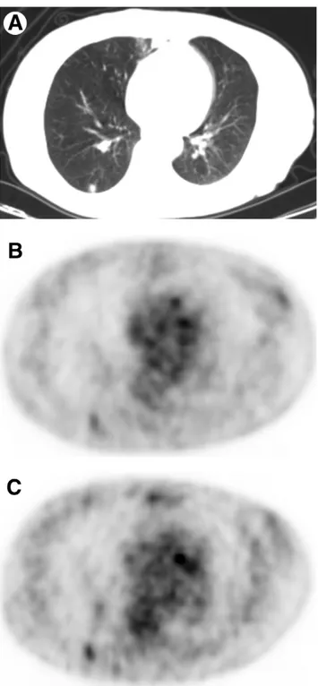
Figure 2 Dual-point fluorodeoxyglucose (FDG)-PET imaging of
67-year-old woman with 0.9 cm solitary pulmonary nodule in the right lower lobe on CT (A). Early PET (B) shows a focal area with faint FDG uptake (SUV 1.2); delayed PET (C) shows more prominent FDG accumulation (SUV 1.6) in the solitary pulmonary nodule. Pathologic diagnosis of this nodule was lung adenocarcinoma.
Figure 3 A 71-year-old man presented with 2.3-cm density in left
lower lobe. SUV was not significantly different between early PET at 55 minutes after FDG injection (A) and delayed imaging at 120 minutes (B): 6.1 and 5.8, respectively. Excised lesion was granu-loma.
It is worth noting that DP SUV showed the largest AUC. However, DP SUV was most sensitive, whereas single-time-point SUV was most specific.
The role of DP FDG-PET imaging in the evaluation of CT detected SPNs with an initial only mild metabolic activity (ie, SUV⬍2.5 at 60 minutes) was recently investigated in 36 patients.32Delayed scanning included 3-4 bed positions in
the thorax only, approximately 180 minutes after FDG injec-tion. Two methods of interpreting delayed PET imaging were applied: the first one analyzed the SUV of SPNs on 180-minute scan, in which a SUV of 2.5 or more was regarded as a criterion for malignancy; the second one was based on the calculation of RI, in which an increase of 10% or more in SUV between the initial and delayed images was considered an indication of malignancy. The sensitivity, specificity, and ac-curacy of using a SUV of 2.5 or more as a criterion for malig-nancy on the delayed acquisition resulted 36%, 96%, and 78%, respectively; when a RI of⬎10% between the initial and delayed images was used, the sensitivity, specificity, and accuracy was 73%, 80%, and 78%, respectively.
These findings are similar to those previously reported by Xiu et al,33whose second image acquisition occurred earlier,
on average 114 minutes after tracer injection, and despite a different prevalence of granulomatous disease between the 2 study populations. In fact, sensitivity, specificity, and accu-racy resulted 44%, 87%, and 72% when using the criterion of delayed SUVⱖ2.5, and 81%, 87%, and 85% when applying a RI of⬎10%, respectively.
Nevertheless, in geographic regions with epidemic granu-lomatous disease, such as tuberculosis or in patients at high risk of granulomatous inflammation, delayed FDG-PET did not prove useful for differentiating malignant from benign pulmonary nodules with an initial mean SUV ⬍2.5.34 DP
FDG-PET studies were conducted with imaging 1 and 2 hours after radiopharmaceutical injection in 27 patients with 31 lesions. The differences in 1-hour SUV, 2-hour SUV, RI, and size between the benign (n⫽ 15) and malignant (n ⫽ 16) lung pulmonary nodules were not statistically significant, and the AUC did not differ from 0.5. In particular, 60% of the benign lesions had a RI⬎10% and 62% of the malignant lesions showed a RI⬎10%. Therefore, according to these data, for pulmonary lesions with an initial mean SUV⬍2.5, further increases in FDG uptake are frequent in granuloma-tous inflammation.
A comprehensive meta-analysis evaluating the diagnostic performance of DP FDG-PET in the diagnosis of pulmonary nodules has been recently made, including studies published between January 2000 and January 2011, which used pathol-ogy or clinical follow-up as the reference standard, and ab-solute number of true-positive, true-negative, false-positive, and false-negative results or stated sufficient data to derive these values.35Ten publications28,29,31,33,34,36-40fulfilled all of
the inclusion criteria, with a total of 816 patients and 890 pul-monary nodules. Multiple different thresholds were used to de-fine a pulmonary nodule as malignant between the initial and later images. DP FDG-PET demonstrated a summary sensitivity of 85% and a summary specificity of 77%. Based on these out-comes, the authors’ conclusion is that the accuracy of DP
FDG-PET is not superior to single-time-point imaging in determining benign versus malignant etiology of pulmonary nodules because the obtained values for sensitivity and specificity are similar to those reported in a previous meta-analysis of single-time-point FDG-PET.27For the authors, the main reasons for evaluating as
questionable the additive value of the DP FDG-PET in the diag-nosis of lung nodules are the significant overlap of benign and malignant nodule FDG-PET characteristics and the lack of con-sensus criteria for quantitative thresholds to define nodules as malignant.
DP FDG-PET has been proposed also for staging lung can-cer and for differentiating metastatic from nonmetastatic lung cancer lesions.41In a group of 153 lung cancer patients with
known or suspected mediastinal and hilar lymph node in-volvement or distant metastases, total-body FDG-PET was acquired at 60 minutes (early imaging) and at 180 minutes (delayed imaging). A strong correlation between the RI SUV of the primary lesion and the metastatic lesions was found; FDG uptake in metastases was almost indistinguishable from that in nonmetastatic lesions on both early and delayed scans, and there was no relationship between the RI SUV results of primary lesions and those of nonmetastatic lesions. The ac-curacy in detecting lymph node and distant metastases was improved when RI SUV was used because of the significant increase in specificity relative to early and delayed SUV. In fact, patient-by-patient analysis for nodal metastasis staging gave these results: early imaging sensitivity 93%, specificity 64%, and accuracy 76%; delayed images sensitivity 97%, specificity 73%, and accuracy 82%; RI SUV sensitivity 100%, specificity 98%, and accuracy 99%. For distant me-tastasis staging, these values were: early imaging sensitivity 100%, specificity 59%, and accuracy 77%; delayed images sensitivity 100%, specificity 66%, and accuracy 81%; RI SUV sensitivity 100%, specificity 98%, and accuracy 99%. There-fore, these results suggested that RI SUV is able to raise the accuracy for diagnosis of metastases and was superior to early and delayed imaging for the differentiation of malignancy from nonmetastatic uptake.
In 34 patients with NSCLC, the diagnostic capacity of DP FDG-PET/CT for staging intrathoracic lymph node metasta-ses was determined.42 Images were acquired 60 minutes
(early) and 2 hours (delayed) after tracer administration. Sig-nificant differences were detected between metastatic and nonmetastatic lymph nodes and between metastatic and in-flammatory lymph nodes for early and delayed SUV and RI, whereas no difference was found between nonmetastatic and inflammatory lymph nodes in early and delayed SUV and RI. For early SUV, the cut-off value for highest accuracy with metastatic lymph nodes was 3.61, yielding a sensitivity of 87% and a specificity of 88%; for delayed SUV, the cut-off value was 4, yielding a sensitivity of 92% and specificity of 93%. For RI, the cut-off value resulted 20.91%, yielding a sensitivity of 74%, and specificity of 76%. These findings indicated that in patients with NSCLC, the 2-hour images have the potential to improve the accuracy of FDG-PET/CT for intrathoracic lymph node staging.
Nevertheless, the usefulness of DP FDG-PET for regional lymph nodes staging in NSCLC cancer is still controversial.
As a matter of fact, Kim et al43reported a limited predictive
value of DP (1 and 2 hours) FDG-PET/CT in the evaluation of pathologic N1 status in 70 patients with stage I and II NSCLC. Lymph node metastases (N1) were present in 15 of 70 patients (21.4%). The N1 group showed statistically sig-nificant higher value of both early and delayed SUV than N0 group; nevertheless, the⌬ SUV did not result statistically dif-ferent between pathologic N1 and N0 groups. Yen et al44
dem-onstrated that in tuberculosis endemic country, PET is more accurate than CT in staging chest lymph node in NSCLC, but semiquantitative indices of DP FDG-PET do not have better diagnostic accuracy than visual analysis of PET images. Further-more, Kasai et al45reported that single-time imaging of
FDG-PET/CT is sufficiently useful for diagnosing mediastinal and hi-lar lymph node metastases in NSCLC patients.
In a recent study aimed to evaluate the efficacy of an addi-tional CT attenuation and a DP scan in determining the status of regional lymph nodes in NSCLC, 39 nodal groups with positive FDG uptake in the initial scan underwent DP imag-ing, and the difference in the RI between benign and malig-nant groups showed no statistical significance.46However, in
patients with NSCLC with lung comorbidity, DP FDG-PET/CT was more effective for mediastinal nodal staging than single-time-point imaging.47In fact, on a per patient basis,
the sensitivity, specificity, accuracy, PPV, and NPV of single-time-point scan were 87%, 59%, 68%, 48%, and 92%, and those values of DP imaging resulted 94%, 68%, 75%, 56%, and 96%, respectively. On a per nodal station basis, the spec-ificity, accuracy, and PPV of DP scan were statistically better than those of single point.
DP FDG-PET can improve the diagnostic accuracy in dif-ferentiating benign from malignant pleural disease and
would help to avoid many unnecessary biopsies. In a retro-spective study, including 61 NSCLC patients with pleural ef-fusion,48all of them were submitted to total-body FDG-PET/CT
imaging at 60⫾ 10 minutes after tracer injection, and 31 pa-tients had second delayed imaging for the chest (at 90⫾ 10 minutes). In this series, the change in SUVs over time calculated with DP technique was helpful in differentiating malignant pleu-ral disease from benign inflammatory process (sensitivity 100%, specificity 94%, and accuracy 97%).
Conflicting results have been reported on the possible role of DP FDG-PET as a prognostic factor for lung cancer. In a retrospective analysis of 100 consecutive patients with newly diagnosed lung adenocarcinoma, pretherapy SUV change over time between the early (⬃60 minutes) and the delayed (⬃90 minutes) PET was a strong independent predictor of outcome.49A cutoff of 25% change for SUV over time showed
the best discriminative value: patients with⬎25% increase in SUV had a median survival of 15 months, compared with 39 months for those with⬍25% increase in SUV (Fig. 4). An-other retrospective review of 66 patients with surgically re-sected early (stage I and II) NSCLC investigated the prognos-tic value of DP (at 60 and 120 minutes) FDG-PET/CT at cancer diagnosis.50The overall survival and disease-free
sur-vival (DFS) resulted better in patients with early tumor SUV ⱖ5.75 than in the patients with early tumor SUV ⱕ5.75. Seventeen (51.5%) patients with a delayed tumor SUVⱖ6.8 and 4 (12.1%) patients with a delayed tumor SUV ⱕ6.8 recurred during follow-up period. The median DFS of the patients with delayed tumor SUVⱖ6.8 was 31.7 months and was significantly worse than the patients with delayed tumor SUV ⱕ6.8. However, the ⌬ SUV did not have prognostic values for overall survival and DFS. Both early and delayed
Figure 4 Coronal CT, PET, and PET/CT fusion images of a 58-years-old patient: early (A) and delayed (B) scans.
Histopathology was moderately differentiated adenocarcinoma of the right upper lobe. Percentage change of SUV, between early and delayed images, measures 44%, and survival was 11 months. (Color version of figure is available online.)
SUVs were predictors for overall survival; early SUV was also a predictor of DFS. Therefore, these data indicated that ⌬ SUV calculated by DP FDG-PET/CT might not have a prog-nostic value for overall survival and DFS in surgically re-sected early stage NSCLC.
Breast Cancer
After the aforementioned manuscript of Boerner et al,17 a
study was conducted to measure how SUV changes over time in breast cancer.51FDG-PET was performed as 60-minute
dynamic imaging with an additional image acquired at⬃75 minutes after injection in 20 newly diagnosed untreated lo-cally advanced breast cancer patients. In this group, SUV changed approximately linearly after 27 minutes at a rate ranging from⫺0.02 to 0.15 per minutes, and the rate of SUV change was linearly correlated with the instantaneous SUV measured at different times after injection.
Subsequently, the potential clinical usefulness of DP (at ⬃63 and ⬃101 minutes) FDG-PET for identifying malignant lesions in the breast was evaluated in 54 patients with 57 breast lesions.52Among the 39 invasive carcinoma lesions, 33
(85%) showed an increase and 6 (15%) showed either no change or a decrease in SUVs over time; the change in SUVs from time point 1 to time point 2 were 12.6%⫾ 11.4%. Of the 18 inflammatory lesions, 3 (17%) showed an increase and 15 (83%) showed either no change or a decrease in SUVs; the change in SUVs from time point 1 to time point 2 resulted ⫺10.2% ⫾ 16.5%. Of the 57 normal contralateral breasts, 2 (3.5%) showed an increase and 55 (96.5%) showed either no change or a decrease in SUVs, and the change in SUVs over time was ⫺15.8% ⫾ 17%. These data indicated that over time, there is an increase in the uptake of FDG in breast malignancies, whereas the uptake in inflammatory lesions and normal breast tissues decreases. A percentage change of 3.75 or more in SUV differentiated inflammatory lesions from malignant ones. The same group then used the DP technique in a prospective study aimed to assess the utility of this kind of imaging in detecting primary breast cancer and to determine whether there is a relationship FDG uptake and its change over time and the histopathologic subtypes.53 One
hundred fifty-two patients with newly diagnosed breast can-cer were divided into 2 groups according to histopathology as invasive and noninvasive. The mean early SUV, delayed SUV, and the⌬ SUV were 3.9 ⫾ 3.7, 4.3 ⫾ 4.0, and 8.3% ⫾ 11.5% for invasive; 2.0 ⫾ 0.6, 2.1 ⫾ 0.6, and 3.4% ⫾ 13% for noninvasive; and 1.2 ⫾ 0.3, 1.1% ⫾ 0.2%, and ⫺10% ⫾ 10.8% for the contralateral normal breasts, respectively. Moreover, DP was especially useful in detecting noninvasive (carcinoma in situ) breast cancer, invasive small lesions (⬍10 mm), and invasive lobular and mixed types of carcinomas. No data are actually available on the possible role of DP FDG-PET imaging in patients with suspicious recurrent breast cancer (Fig. 5).
The value of the reacquisition of PET/CT images 3 hours after FDG administration in the diagnostic evaluation of sus-picious breast lesions has been then confirmed in a series of 48 patients.54Malignant breast lesions showed an increase in
FDG uptake over time; in contrast, benign lesions demon-strated a reduction. In particular, the DP acquisition yielded an accuracy of 85% for lesions with a SUV ⱖ2.5 and/or positive⌬ SUV, with sensitivity and specificity values of 81% and 100%, compared with 69%, 63%, and 100%, respec-tively, for the single-point acquisition.
The accuracy of DP FDG-PET/CT in primary breast cancer with minimally increased FDG uptake (SUVⱕ2.5) has been evaluated in a group of 49 patients,55who underwent 2
se-quential PET/CT scans (55-60 and 100-120 minutes after tracer injection). Statistical analysis indicated that early SUV and delayed SUV singular measurements were unsatisfactory for discriminating between breast cancer with minimal FDG uptake and normal breast tissue, whereas the % change in SUV over time was adequate for the discrimination of cancer lesions. The highest accuracy for the characterization of ma-lignant breast lesions was obtained when a ⌬ SUV cut-off value 0% was used as criteria for malignant FDG uptake change over time (sensitivity 76%, false-positive rate 20%).
A recent article has reported that DP imaging is not able to improve the overall performance of FDG-PET/CT in the de-tection axillary lymph node metastasis in breast cancer pa-tients.56 The study included 171 patients who underwent
preoperative DP FDG-PET/CT (1 and 3 hours after radio-tracer injection). Where FDG uptake in the axilla was per-ceptibly increased, the SUVs for both time points and the RI were calculated. The results of early and delayed PET/CT images in detecting axillary lymph nodes were equal, with sensitivity 60% and specificity 85%. Sixty patients showed increased FDG uptake in the axillas on 1- or 3-hour images, but there was no significant difference in RI between the metastatic group and the lymph node-negative group. The AUC for early SUV was 0.90 and 0.87 for delayed SUV.
Digestive System Tumors
In the liver, it has been reported that the acquisition of de-layed images improves the hepatic detection of pathologic FDG uptake.57In a series of 95 consecutive patients
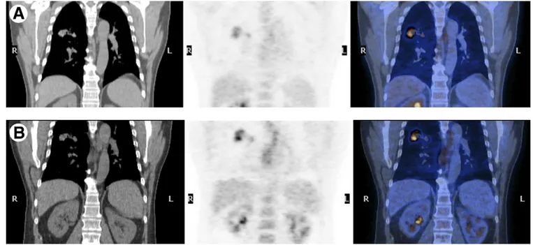
previ-ously treated for neoplastic disease and with suspected liver metastases, a DP PET/CT was acquired (1 and 2 hours after FDG injection). Thirty-seven of 95 patients (38.9%) pre-sented liver lesions at both PET/CT scans, whereas there were 2 (2.2%) only at the second PET/CT. Eighty-one liver lesions were identified at both PET studies, whereas there were 9 (11.1%) only at the second PET. Moreover, at the second PET, a statistically significant reduction of background SUV and an increase of lesions’ SUV were observed. These find-ings demonstrated that acquiring a delayed PET scan allows both a better visualization of liver pathologic uptake and a sensitivity increase—the second acquisition is particular use-ful in patients with clinically suspected liver metastases when the first PET scan is negative (Fig. 6).
The previous reported results have been subsequently confirmed in a recent article, including 39 with colorectal cancer and suspicion of hepatic metastases.58The first
whole-body PET/CT scan was performed 67 ⫾ 11 minutes after FDG injection; the delayed regional PET/CT scan was
ac-quired 113⫾ 20 minutes after tracer administration. A total of 91 hepatic lesions were detected in 39 patients, and 86 lesions in 34 patients were histopathologically proven to be hepatic metastases; visual analysis detected 77% of them on the first scan and 87% on the delayed images. The SUV and tumor-to-liver uptake ratio (TLR) of the metastatic lesions on the delayed images were higher than those on the early scan; moreover, the TLR and SUV of the delayed scan showed the highest detection rates (92% and 88%, respectively).
The importance of acquiring a delayed PET/CT scan for all hepatic lesions with indeterminate FDG uptake on routine 60-minute images has been highlighted in a case report de-scribing the pattern of FDG accumulation in a patient with hepatic epithelioid hemangioendothelioma, which demon-strated no pathologic uptake in the 60-minute scans, whereas the 3-hour PET/CT images showed intense FDG accumula-tion in the liver masses.59
In patients with suspected gallbladder carcinoma, delayed FDG-PET scans were more helpful than early imaging for evaluating malignant lesions because of increased lesion up-take and lesion-to-background contrast.60In fact, both SUV
and TLRs derived from delayed images were significantly higher than the ratios from early images. When a RI of⫺8 was chosen as arbitrary cut-off for differentiating between malignant and benign conditions, the accuracy was 84%. Nevertheless, it is interesting to highlight that the diagnostic performance of FDG-PET was highly dependent on C-reac-tive protein (CRP) levels (specificity was 80% in the group with a normal level of CRP and 0% in the group with an elevated CRP level, respectively).
In patients with esophageal squamous cell carcinoma, DP PET/CT demonstrated limited value in detection of primary tumor and locoregional lymph nodes metastasis; however, for the distant metastasis, the sensitivity improved if RI ⱖ10% was used as a supplemental criterion.61
In pancreatic cancer, after the study of Nakamoto et al,19it
has been recently reported that FDG-PET with DP (1 and 2-hours) evaluation is a useful modality for the detection of small invasive ductal carcinomas with a diameter of ⬍20 mm.62Moreover, the RI calculated with DP FDG-PET can be
used not only as a tool for initial diagnosis and staging of pancreatic cancer but also as a strong independent prognos-tic parameter for an accurate identification of those patients who will benefit from intensive therapy at different stages of the disease.63 In particular, when combined with tumor
stage, RI allowed even more precise prognostic evaluation: patients at stage I-III with RI⬎10% survived longer than did patients at the same stage with RI ⬍10% (15.3 vs 11.5 months), patients at stage IV with RI⬎10% had an interme-diate prognosis (median survival of 9.5 months), and patients at stage IV with RI⬍10% showed the worst prognosis (4.9 months).
The normal variants of the physiological FDG uptake in DP imaging have been described in a retrospective review of 206 consecutive asymptomatic subjects who underwent whole-body FDG-PET/CT for medical checkup.64The
find-ings of this study indicated that physiological FDG uptake in the colon increases from the early (50 minutes) to the delayed Figure 5 Dual-point FDG-PET imaging of 68-year-old woman,
pre-viously submitted to left mastectomy for breast cancer. CT scan (A) shows a suspicious local recurrence. Early PET (B) depicts an area of faint FDG uptake (arrow) that is more prominent in the delayed imaging (C). Pathologic diagnosis was relapsing invasive ductal breast carcinoma.
(100 minutes) images in a DP study, with half of the normal colon areas showing an increased uptake in the later scans, whereas the average SUV of the liver significantly decreased. Therefore, these results should be carefully taken into ac-count to avoid misdiagnosing malignant lesions in the colon.
Other Tumors
In cervical cancer, the clinical value of DP FDG-PET has been evaluated both in patients who had an initial diagnosis and in patients with recurrence.65-67 In a study, including 94
pa-tients, DP (40 minutes and 3 hour) PET imaging resulted superior to conventional PET or CT/magnetic resonance im-aging (MRI) in the evaluation of metastatic lesions in locally advanced or recurrent cervical cancer.65 These data have
been confirmed in restaging cervical carcinoma at the time of first recurrence in a group of 40 patients, in whom DP FDG-PET was significantly superior to CT/MRI in identifying met-astatic lesions, and so modifying the treatment in 55% of cases.66Moreover, the delayed images proved to be
particu-larly useful for the detection of lower para-aortic lymph node metastases.67
The efficacy of DP FDG-PET/CT in grading brain tumors was prospectively evaluated in 21 lesions of 18 consecutive patients with primary or metastatic brain tumors.68
Patho-logic diagnosis was obtained by stereotactic biopsy or open
surgery; grading of the tumor was performed according to the World Health Organization (WHO) classification. The early scans were started 40-45 minutes after the tracer injection, and the delayed PET/CT scans were performed at 72.8 ⫾ 29.7 minutes after the early ones. SUVs of the delayed images were more efficient than those of early scans to classify lesions by the grade of tumor, and RI had no statistical significance to this aim. In particular, the SUVs on the later imaging were 6.3⫾ 2.2, 5 ⫾ 2.5, 8.1 ⫾ 2.3, and 10 ⫾ 3.4 of grade I, II, III, and IV tumors, respectively. Moreover, in 25 patients with suspected high-grade brain tumors and inconclusive MRI find-ings, quantitative DP FDG-PET improved the sensitivity for the identification and volume delineation of such neoplasms, when compared with standard single-time studies.69
DP FDG-PET provided superior assessment of recurrence versus posttreatment necrosis in a group of 32 patients with treated brain metastases, lesion size⬎0.5 cm3, and suspected
recurrence on MRI.70Early (45-60 minutes postinjection) or
delayed (⬃225 minutes later) SUVs of the lesion alone did not accurately differentiate between tumor and necrosis, whereas a change⬎19% of lesion to gray matter SUV ratios as a function of time was 95% sensitive, 100% specific, and 96% accurate (AUC 0.97) in making this differential diagno-sis, regardless of histological type.
Normal FDG gray matter uptake usually decreases with time;71therefore, delayed-phase PET scan (3 hours) was
use-Figure 6 A 52-year-old male patient with rectal cancer. Early (40 minutes) PET (A) and PET/CT (B) transaxial views are
ful to enhance the detectability of tiny residual skull base osteosarcoma initially concealed by adjacent high physiolog-ical brain activity.72
In evaluating the locoregional status of nasopharyngeal carcinoma, the results of a prospective study, including 84 patients, indicated that FDG-PET is superior to MRI in identifying lower neck nodal metastasis, but 3-hour scans did not give further information in detecting both primary tumors or locoregional metastatic nodes with respect to 40-minute images.73Nevertheless, it is worth noting that
in this study, diazepam 5 mg per os, was routinely given before FDG injection, and this fact might decrease the hexokinase/glucose-6-phosphatase ratio and so render the DP technique ineffective under these circumstances.74 In
fact, diazepam can reduce phosphorylation of glucose by inhibiting hexokinase activity, and its effect after orally administration commences approximately 30 minutes af-ter ingestion and lasts many hours; therefore, it will likely minimally affect the early images (within the first hour), whereas it will significantly influence the FDG uptake on delayed images.74
The accuracy of DP FDG-PET for the diagnosis of the var-ious subtypes of thymic epithelial tumors based on the WHO classification was assessed in 23 patients.75High SUV values
suggested the presence of high-risk tumors, and a very high SUV value (early⬎7.1) was useful for the differentiation of thymic carcinomas from other types of tumors. The delayed SUV values were higher than the early SUV values in all types of tumors.
In patients with incidentally detected thyroid nodules dur-ing FDG-PET/CT, the DP protocol (60 and 120 minutes) was of limited value for differentiating malignant form benign nodules.76
Conclusions
Now FDG-PET and PET/CT imaging plays a fundamental role in oncology; however, with its wide application through-out the world, a growing number of limitations have been found. One of the main causes is certainly the nonspecificity of FDG uptake in tumor; it is well known that false-positive results could be because of inflammation, infection, or other unknown benign tumors,77,78and so, it is really difficult to
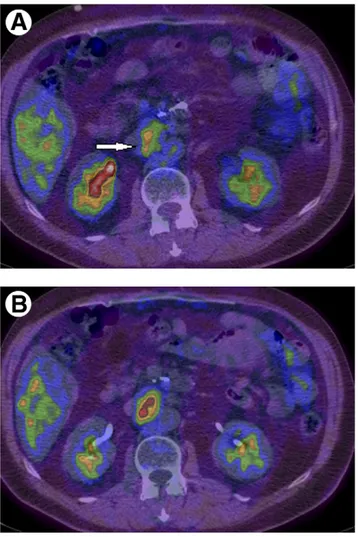
Figure 7 A 52-year-old male patient with previously resected
pan-creatic cancer. Early (60 minutes) PET/CT (A) shows a moderate FDG uptake in an enlarged abdominal lymph node (arrow, SUV 2.8), which increases in the delayed imaging (110 minutes, SUV 3.3) (B). The final diagnosis was lymph node metastasis.
Figure 8 A 24-year-old young female patient with Hodgkin
lym-phoma, evaluated after chemotherapy. Early (60 minutes) PET/CT (A) demonstrated a focal area of FDG uptake in an abdominal lymph node (arrow, SUV 3.5), which decreases (SUV 2.9) in the later imaging at 120 minutes (B). This pattern is strongly suggestive for the presence of inflammatory tissue rather than for residual disease.
distinguish malignant from benign lesions in such clinical situations.
Several experimental approaches both in vitro and through PET images in vivo suggest that the time-activity of FDG uptake between malignant and benign lesions is differ-ent—tumor uptake increases for hours, whereas in inflam-matory lesions it decreases gradually from 60 minutes after injection. Therefore, this difference in the time course could be useful to improve the specificity of FDG studies for differ-entiating benign from malignant lesions by means of the ac-quisitions of delayed scans. Moreover, it is important to high-light that in many of these studies, the DP technique was able to improve the sensitivity of PET in assessing various tumors. This can be explained by 2 factors: first, there may be clear-ance of background activity between the 2 time points, im-proving target-to-background ratio, that leads to higher sen-sitivity; second, a significant increase in SUV within malignant lesions over time might also improves lesion de-tectability.
Nevertheless, some points should be clarified before sug-gesting the inclusion of DP imaging in routine FDG-PET/CT protocols for cancer patients.79In particular, further
investi-gations are needed to identify patient populations that would most benefit from this technique. As a matter of fact, in a recent article assessing the utility of DP FDG-PET/CT in the Australian population, this approach differentiated malig-nant from benign intra-abdominal lesions (Figs. 7and8) but did not improve the overall evaluation of pulmonary le-sions.80
If imaging should be performed as late as reasonably pos-sible after FDG injection to increase tumor to background contrast and thereby improve the sensitivity in demonstrat-ing additional sites of disease, it is clear that the various tumor types may be affected differently by delayed scans. Tumor-specific studies would be useful to assess the optimal timing of imaging for tumors, which could have differing levels of hexokinase/glucose-6-phosphatase ratio (eg, hema-tological malignancy, sarcoma, breast and other carcinoma subtypes). Therefore, the most appropriate time for a delayed scan should be defined for each kind of neoplasm81;
more-over, the method of SUV calculation should be standardized, as well as the criteria for quantitative thresholds to define lesions as malignant, for example, for the lung nodules. Fi-nally, larger prospective studies may be helpful to better eval-uate the clinical role of DP FDG-PET/CT imaging in oncology patients, in particular taking into account any possible result-ing change in management.
References
1. Langer A: A systematic review of PET and PET/CT in oncology: A way to personalize cancer treatment in a cost-effective manner? BMC Health Serv Res 2010;10:283
2. Saif MW, Tzannou I, Makrilia N, et al: Role and cost effectiveness of PET/CT in management of patients with cancer. Yale J Biol Med 2010; 83:53-65
3. Hillner BE, Siegel BA, Shields AF, et al: Relationship between cancer type and impact of PET and PET/CT on intended management: Find-ings of the national oncologic PET registry. J Nucl Med 2008;49:1928-1935
4. Rice SL, Roney CA, Daumar P, et al: The next generation of positron emission tomography radiopharmaceuticals in oncology. Semin Nucl Med 2011;41:265-282
5. Pauwels EK, Ribeiro MJ, Stoot JH, et al: FDG accumulation and tumor biology. Nucl Med Biol 1998;25:317-322
6. Love C, Tomas MB, Tronco GG, et al: FDG-PET of infection and in-flammation. Radiographics 2005;25:1357-1368
7. Schillaci O: Hybrid imaging systems in the diagnosis of osteomyelitis and prosthetic joint infection. Q J Nucl Med Mol Imaging 2009;53:95-104
8. Metser U, Even-Sapir E: Increased (18)F-fluorodeoxyglucose uptake in benign, nonphysiologic lesions found on whole-body positron emis-sion tomography/computed tomography (PET/CT): Accumulated data from four years of experience with PET/CT. Semin Nucl Med 2007;37: 206-222
9. Strauss LG: Fluorine-18 deoxyglucose and false-positive results: A ma-jor problem in the diagnostics of oncological patients. Eur J Nucl Med 1996;23:1409-1415
10. Zhuang H, Pourdehnad M, Lambright ES, et al: Dual time point 18F-FDG-PET imaging for differentiating malignant from inflammatory processes. J Nucl Med 2001;42:1412-1417
11. Lowe VJ, Duhaylongsod FG, Patz EF, et al: Pulmonary abnormalities and PET data analysis: A retrospective study. Radiology 1997;202:435-439
12. Nguyen NC, Kaushik A, Wolverson MK, et al: Is there a common SUV threshold in oncological FDG-PET/CT, at least for some common indi-cations? A retrospective study. Acta Oncol 2011;50:670-677 13. Fischman AJ, Alpert NM: FDG-PET in oncology: There’s more to it than
looking at pictures. J Nucl Med 1993;34:6-11 [editorial]
14. Hamberg LM, Hunter GJ, Alpert NM, et al: The dose uptake ratio as an index of glucose metabolism: Useful parameter or oversimplification? J Nucl Med 1994;35:1308-1312
15. Yamada S, Kubota K, Kubota R, et al: High accumulation of fluorine-18-fluorodeoxyglucose in turpentine-induced inflammatory tissue. J Nucl Med 1995;36:1301-1306
16. Lodge MA, Lucas JD, Marsden PK, et al: A PET study of 18FDG uptake in soft tissue masses. Eur J Nucl Med 1999;26:22-30
17. Boerner AR, Weckesser M, Herzog H, et al: Optimal scan time for fluorine-18 fluorodeoxyglucose positron emission tomography in breast cancer. Eur J Nucl Med 1999;26:226-230
18. Hustinx R, Smith RJ, Benard F, et al: Dual time point fluorine-18 fluorodeoxyglucose positron emission tomography: A potential method to differentiate malignancy from inflammation and normal tissue in the head and neck. Eur J Nucl Med 1999;26:1345-1348 19. Nakamoto Y, Higashi T, Sakahara H, et al: Delayed
(18)F-fluoro-2-deoxy-D-glucose positron emission tomography scan for differentia-tion between malignant and benign lesions in the pancreas. Cancer 2000;89:2547-2554
20. Kubota K, Itoh M, Ozaki K, et al: Advantage of delayed whole-body FDG-PET imaging for tumour detection. Eur J Nucl Med 2001;28:696-703
21. Higashi T, Saga T, Nakamoto Y, et al: Relationship between retention index in dual-phase (18)F-FDG-PET, and hexokinase-II and glucose transporter-1 expression in pancreatic cancer. J Nucl Med 2002;43: 173-180
22. Basu S, Alavi A: Partial volume correction of standardized uptake values and the dual time point in FDG-PET imaging: Should these be routinely employed in assessing patients with cancer? Eur J Nucl Med Mol Im-aging 2007;34:1527-1529
23. Gallagher BM, Fowler JS, Gutterson NI, et al: Metabolic trapping as a principle of radiopharmaceutical design: Some factors responsible for the biodistribution of [18F] 2-deoxy-2-fluoro-D-glucose. J Nucl Med 1978;19:1154-1161
24. Nelson CA, Wang JQ, Leav I, et al: The interaction among glucose transport, hexokinase, and glucose-6-phosphatase with respect to 3H-2-deoxyglucose retention in murine tumor models. Nucl Med Biol 1996;23:533-541
25. Vansteenkiste JF, Stroobants SS: PET scan in lung cancer: Current recommendations and innovation. J Thorac Oncol 2006;1:71-73
26. Ung YC, Maziak DE, Vanderveen JA, et al: 18Fluorodeoxyglucose pos-itron emission tomography in the diagnosis and staging of lung cancer: A systematic review. J Natl Cancer Inst 2007;99:1753-1767 27. Gould MK, Maclean CC, Kuschner WG, et al: Accuracy of positron
emission tomography for diagnosis of pulmonary nodules and mass lesions: A meta-analysis. JAMA 2001;21:914-924
28. Matthies A, Hickeson M, Cuchiara A, et al: Dual time point 18F-FDG-PET for the evaluation of pulmonary nodules. J Nucl Med 2002;43: 871-875
29. Alkhawaldeh K, Bural G, Kumar R, et al: Impact of dual-time-point (18)F-FDG-PET imaging and partial volume correction in the assess-ment of solitary pulmonary nodules. Eur J Nucl Med Mol Imaging 2008;35:246-252
30. Lubberink M, Tolmachev V, Widström C, et al: 110mIn-DTPA-D
-Phe1-octreotide for imaging of neuroendocrine tumours with PET. J Nucl Med 2002;43:1391-1397
31. Schillaci O, Travascio L, Bolacchi F, et al: Accuracy of early and delayed FDG-PET-CT and of contrast-enhanced CT in the evaluation of lung nodules: A preliminary study on 30 patients. Radiol Med 2009;114: 890-906
32. MacDonald K, Searle J, Lyburn I: The role of dual time point FDG-PET imaging in the evaluation of solitary pulmonary nodules with an initial standard uptake value less than 2.5. Clin Radiol 2011;66:244-250 33. Xiu Y, Bhutani C, Dhurairaj T, et al: Dual-time point FDG-PET imaging
in the evaluation of pulmonary nodules with minimally increased met-abolic activity. Clin Nucl Med 2007;32:101-105
34. Chen CJ, Lee BF, Yao WJ, et al: Dual-phase 18F-FDG-PET in the diag-nosis of pulmonary nodules with an initial standard uptake value less than 2.5. Am J Roentgenol 2008;191:475-479
35. Barger RL Jr, Nandalur KR: Diagnostic performance of dual-time 18F-FDG-PET in the diagnosis of pulmonary nodules: A meta-analysis. Acad Radiol 2012;19:153-158
36. Laffon E, de Clermont H, Begueret H, et al: Assessment of dual-time-point 18F-FDG-PET imaging for pulmonary lesions. Nucl Med Com-mun 2009;30:455-461
37. Cloran FJ, Banks KP, Song WS, et al: Limitations of dual time point PET in the assessment of lung nodules with low FDG avidity. Lung Cancer 2010;68:66-71
38. Lan XL, Zhang YX, Wu ZJ, et al: The value of dual time point (18)F-FDG-PET imaging for the differentiation between malignant and be-nign lesions. Clin Radiol 2008;63:756-764
39. Núñez R, Kalapparambath A, Varela J: Improvement in sensitivity with delayed imaging of pulmonary lesions with FDG-PET. Rev Esp Med Nucl 2007;26:196-207
40. Suga K, Kawakami Y, Hiyama A, et al: Dual-time point 18F-FDG-PET/CT scan for differentiation between 18F-FDG avid non-small cell lung cancer and benign lesions. Ann Nucl Med 2009;23:427-435 41. Uesaka D, Demura Y, Ishizaki T, et al: Evaluation of dual-time-point
18F-FDG-PET for staging in patients with lung cancer. J Nucl Med 2008;49:1606-1161
42. Shinya T, Rai K, Okumura Y, et al: Dual-time-point F-18 FDG-PET/CT for evaluation of intrathoracic lymph nodes in patients with non-small cell lung cancer. Clin Nucl Med 2009;34:216-221
43. Kim SJ, Kim YK, Kim IJ, et al: Limited predictive value of dual-time-point F-18 FDG-PET/CT for evaluation of pathologic N1 status in NSCLC patients. Clin Nucl Med 2011;36:434-439
44. Yen RF, Chen KC, Lee JM, et al: 18F-FDG-PET for the lymph node staging of non-small cell lung cancer in a tuberculosis-endemic coun-try: Is dual time point imaging worth the effort? Eur J Nucl Med Mol Imaging 2008;35:1305-1315
45. Kasai T, Motoori K, Horikoshi T, et al: Dual-time point scanning of integrated FDG-PET/CT for the evaluation of mediastinal and hilar lymph nodes in non-small cell lung cancer diagnosed as operable by contrast-enhanced CT. Eur J Radiol 2010;75:143-146
46. Li M, Wu N, Liu Y, et al: Regional nodal staging with (18)F-FDG-PET-CT in non-small cell lung cancer: Additional diagnostic value of CT attenuation and dual-time-point imaging. Eur J Radiol 2011 Apr 19 [Epub ahead of print]. Available at: http://dx.doi.org/10.1016/j. ejrad.2011.03.074
47. Hu M, Han A, Xing L, et al: Value of dual-time-point FDG-PET/CT for mediastinal nodal staging in non-small-cell lung cancer patients with lung comorbidity. Clin Nucl Med 2011;36:429-433
48. Alkhawaldeh K, Biersack HJ, Henke A, et al: Impact of dual-time-point F-18 FDG-PET/CT in the assessment of pleural effusion in patients with non-small-cell lung cancer. Clin Nucl Med 2011;36:423-428 49. Houseni M, Chamroonrat W, Zhuang J, et al: Prognostic implication of
dual-phase PET in adenocarcinoma of the lung. J Nucl Med 2010;51: 535-542
50. Kim SJ, Kim YK, Kim IJ, et al: Limited prognostic value of dual time point F-18 FDG-PET/CT in patients with early stage (stage I & II) non-small cell lung cancer (NSCLC). Radiother Oncol 2011;98:105-108
51. Beaulieu S, Kinahan P, Tseng J, et al: SUV varies with time after injec-tion in (18)F-FDG-PET of breast cancer: Characterizainjec-tion and method to adjust for time differences. J Nucl Med 2003;44:1044-1050 52. Kumar R, Loving VA, Chauhan A, et al: Potential of dual-time-point
imaging to improve breast cancer diagnosis with (18)F-FDG-PET. J Nucl Med 2005;46:1819-1824
53. Mavi A, Urhan M, Yu JQ, et al: Dual time point 18F-FDG-PET imaging detects breast cancer with high sensitivity and correlates well with histologic subtypes. J Nucl Med 2006;47:1440-1446
54. Caprio MG, Cangiano A, Imbriaco M, et al: Dual-time-point [18F]-FDG-PET/CT in the diagnostic evaluation of suspicious breast lesions. Radiol Med 2010;115:215-224
55. Zytoon AA, Murakami K, El-Kholy MR, et al: Breast cancer with low FDG uptake: Characterization by means of dual-time point FDG-PET/ CT. Eur J Radiol 2009;70:530-538
56. Choi WH, Yoo IR, O JH, et al: The value of dual-time-point 18F-FDG-PET/CT for identifying axillary lymph node metastasis in breast cancer patients. Br J Radiol 2011;84:593-599
57. Arena V, Skanjeti A, Casoni R, et al: Dual-phase FDG-PET: Delayed acquisition improves hepatic detectability of pathological uptake. Ra-diol Med 2008;113:875-886
58. Lee JW, Kim SK, Lee SM, et al: Detection of hepatic metastases using dual-time-point FDG-PET/CT scans in patients with colorectal cancer. Mol Imaging Biol 2011;13:565-572
59. Kitapci MT, Akkas¸ BE, Gullu I, et al: FDG-PET/CT in the evaluation of epithelioid hemangioendothelioma of the liver: The role of dual-time-point imaging. A case presentation and review of the literature. Ann Nucl Med 2010;24:549-553
60. Nishiyama Y, Yamamoto Y, Fukunaga K, et al: Dual-time-point 18F-FDG-PET for the evaluation of gallbladder carcinoma. J Nucl Med 2006;47:633-638
61. Shum WY, Hsieh TC, Yeh JJ, et al: Clinical usefulness of dual-time FDG-PET–CT in assessment of esophageal squamous cell carcinoma. Eur J Ra-diol 2011 Mar 31 [Epub ahead of print]. Available at:http://dx.doi.org/ 10.1016/j.ejrad.2011.03.018
62. Okano K, Kakinoki K, Akamoto S, et al: 18F-fluorodeoxyglucose pos-itron emission tomography in the diagnosis of small pancreatic cancer. World J Gastroenterol 2011;17:231-235
63. Lyshchik A, Higashi T, Nakamoto Y, et al: Dual-phase 18F-fluoro-2-deoxy-D-glucose positron emission tomography as a prognostic param-eter in patients with pancreatic cancer. Eur J Nucl Med Mol Imaging 2005;32:389-397
64. Toriihara A, Yoshida K, Umehara I, et al: Normal variants of bowel FDG uptake in dual-time-point PET/CT imaging. Ann Nucl Med 2011;25: 173-178
65. Yen TC, Ng KK, Ma SY, et al: Value of dual-phase 2-fluoro-2-deoxy-d-glucose positron emission tomography in cervical cancer. J Clin Oncol 2003;21:3651-3658
66. Lai CH, Huang KG, See LC, et al: Restaging of recurrent cervical carci-noma with dual-phase [18F]fluoro-2-deoxy-D-glucose positron emis-sion tomography. Cancer 2004;100:544-552
67. Ma SY, See LC, Lai CH, et al: Delayed 18F-FDG-PET for detection of paraaortic lymph node metastases in cervical cancer patients. J Nucl Med 2003;44:1775-1783
FDG-PET imaging for grading of brain tumors. Clin Nucl Med 2010;35:400-403
69. Prieto E, Martí-Climent JM, Domínguez-Prado I, et al: Voxel-based analysis of dual-time-point 18F-FDG-PET images for brain tumor iden-tification and delineation. J Nucl Med 2011;52:865-872
70. Horky LL, Hsiao EM, Weiss SE, et al: Dual phase FDG-PET imaging of brain metastases provides superior assessment of recurrence versus post-treatment necrosis. J Neuro Oncol 2011;103:137-146
71. Spence AM, Muzi M, Mankoff DA, et al: 18F-FDG-PET of gliomas at delayed intervals: Improved distinction between tumor and normal gray matter. J Nucl Med 2004;45:1653-1659
72. Chen YK, Kao CH, Sun SS, et al: Exposing the evil in the dark: The usefulness of delayed-phase FDG-PET scan to enhance the detectability of tiny residual skull base osteosarcoma initially concealed by adjacent high physiological brain activity. Clin Nucl Med 2010;35:630-632 73. Yen TC, Chang YC, Chan SC, et al: Are dual-phase F-18-FDG-PET
scans necessary in nasopharyngeal carcinoma to assess the primary tumour and locoregional nodes? Eur J Nucl Med Mol Imaging 2005; 32:541-548
74. Zhuang H, Hustinx R, Alavi A: Effect of diazepam on the efficacy of dual-phase FDG-PET imaging. Eur J Nucl Med Mol Imaging 2006;33: 228-229
75. Inoue A, Tomiyama N, Tatsumi M, et al: (18)F-FDG-PET for the eval-uation of thymic epithelial tumors: Correlation with the world Health Organization classification in addition to dual-time-point imaging. Eur J Nucl Med Mol Imaging 2009;36:1219-1225
76. Kim SJ, Kim BH, Jeon YK, et al: Limited diagnostic and predictive values of dual-time-point 18F FDG-PET/CT for differentiation of inci-dentally detected thyroid nodules. Ann Nucl Med 2011;25:347-353 77. Basu S, Chryssikos T, Moghadam-Kia S, et al: Positron emission
tomog-raphy as a diagnostic tool in infection: Present role and future possibil-ities. Semin Nucl Med 2009;39:36-51
78. Cook GJ, Fogelman I, Maisey MN: Normal physiological and benign pathological variants of 18-fluoro-2-deoxyglucose positron-emission tomography scanning: Potential for error in interpretation. Semin Nucl Med 1999;26:308-314
79. Lee ST, Scott AM: Are we ready for dual-time point FDG-PET imaging? J Med Imaging Radiat Oncol 2011;55:351-352 (editorial)
80. Chan WL, Ramsay SC, Szeto ER, et al: Dual-time-point 18F-FDG-PET/CT imaging in the assessment of suspected malignancy. J Med Imaging Radiat Oncol 2011;55:379-390
81. Chen YM, Huang G, Sun XG, et al: Optimizing delayed scan time for FDG-PET: Comparison of the early and late delayed scan. Nucl Med Commun 2008;29:425-430