A
A
l
l
m
m
a
a
M
M
a
a
t
t
e
e
r
r
S
S
t
t
u
u
d
d
i
i
o
o
r
r
u
u
m
m
–
–
U
U
n
n
i
i
v
v
e
e
r
r
s
s
i
i
t
t
à
à
d
d
i
i
B
B
o
o
l
l
o
o
g
g
n
n
a
a
DOTTORATO DI RICERCA
BIODIVERSITA’ ED EVOLUZIONE
Ciclo XXI
Settore/i scientifico disciplinari di afferenza: BIO-05
TITOLO TESI
Methodological approaches in order to investigate the
mechanisms of sexual determination and sexual
differentiation in the common toad Bufo bufo L.
(Amphibia, Anura).
Presentata da: Jean-David Durussel
Coordinatore Dottorato
Relatore
Prof.G.Cristofolini
Prof.F.Zaccanti
[1]
INDEX
1.INTRODUCTION Pag.4 1.1) Sexual determination and sexual differentiation: generalities. Pag.5
1.2) Sexual determination in the animal kingdom: comparative study in different
models organisms. Pag.5
1.3) Invertebrate. Pag.6 a) Hymenoptera.
b) Dipterans (Drosophila melanogaster). c) Nematodes (Caenorabditis elegans).
1.4) Vertebrates. Pag.13 a) Fishes. b) Reptiles. c) Birds. d) Mammals. e) Amphibians.
1.5) Reproduction strategies in the animal kingdom: costs and benefits. Pag.22 1.6) Sexual differentiation in vertebrates: interpretative models. Pag.23
a) Symmetric model. b) Asymmetric model.
1.7) Development of the germ line in vertebrate. Pag.25
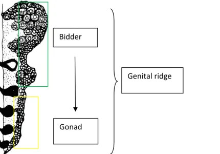
1.8) Peculiarities of the germinal cells in anuran: nuage and germinal plasm. Pag.27 1.9) Sex differentiation and gonadal development in amphibians. Pag.28 1.10) Architecture of the genital body in bufonids. Pag.29 a) Bidder’s organ.
b) Gonad.
[2]
3. MATERIALS AND METHODS Pag.35 3.1) Samples. Pag.35
3.2) Staging of tadpoles. Pag.35 3.3) Sexual determination: Bio-molecular approaches. Pag.36 a) Introduction Pag.36
b) RAPD (Random Amplified Polymorphic DNA). Pag.36 c) Genomic DNA extraction.
d) Primers.
e) Amplification conditions. f) Band patterns.
g) AAT (ADP/ATP translocase). Pag.39
h) Sexing by PCR-RFLP in Rana rugosa.
i) Sequence alignment and primers. j) Amplification conditions.
k) Purification of the PCR products. l) Results.
m) Bio-molecular approaches: discussion. Pag.45 3.4) Sex reversal in amphibians. Pag.46
a) Introduction. Pag.46 b) Hormonal sex reversal Pag.46 c) Hormonal treatments in B.bufo tadpoles
at early developmental stages( III1...III9). d) Results and discussion.
e) Sex-reversal by surgical castration. Pag.49 f) Surgical castration of B.bufo adult males.
g) Hormonal treatments (DiEtilStilbestrol dipropionate). h) Results and discussion.
i) Sex reversal in amphibians: discussion. Pag.54 3.5) Sexual differentiation: methodological approaches. Pag.55
a) Introduction. Pag.55 b) Immuno-detection and localization of α- tubulin
in B.bufo specimen (stage IV17) by
confocal laser scanning microscopy. Pag.56 c) Cytoskeleton: generalities.
d) Cytoskeleton organization in Xenopus oocytes. e) Confocal microscopy: protocol.
[3]
g) Distribution of α-tubulin in Bidder’s organ
and in the gonads: discussion. Pag.71 h) Immuno-detection and localization of Vasa protein
in B.bufo samples (stage IV17) by confocal
laser scanning microscopy. Pag.73 i) Function and regulation of vasa gene in different organisms.
j) Confocal microscopy: protocol. k) Results.
l) Spatial distribution of Vasa protein in the Bidder’s
organ and in the gonads: discussion. Pag.81 m) Spatiotemporal distribution of Sox9 in the gonad and
in the Bidder organ of B.bufo. Pag.83 n) Sox9: a marker of male sexual development in vertebrate?
o) Whole mount is situ hybridization for the detection of mRNA. p) Whole mount in situ hybridization: protocol.
q) Results and discussion.
r) Spatiotemporal distribution of Sox9 in the gonad and
in the Bidder’s organ of B.bufo: discussion. Pag.93
4. DISCUSSION Pag.94 5. REFERENCES Pag.99
[4]
1.INTRODUCTION
1.1) Sexual determination and sexual differentiation: generalities.
In gonochoristic animals, sexual determination and sexual differentiation are two different but closely related mechanisms involved in the development of the alternative sexual phenotypes. Although often used interchangeably, there is a fundamental difference between sexual determination and sexual differentiation. Sexual determination is referred to mechanisms that direct sexual differentiation, whereas sexual differentiation is referred to the development of the alternative gonadal architectures (testes or ovaries) starting from the bi-potential and undifferentiated gonad.
The different sexes develop because individuals are directed through alternative developmental pathways as a result of the activation of the sex determining genes or as a consequence of an enviromental induction that not only include the sexual differentiation of reproductive organs, but also affects almost every aspect of an organism such as behavior, physiology, and morphology. All individuals may have the capacity to develop into either sex. This capacity is evident from the ability to experimental sex reverse as in amphibians: depending on the species, genetic males can be experimentally induced to develop ovaries and vice versa with steroid hormonal treatments. From an evolutionary point of view, sexual determination is considered as a convergent adaptive phenomenon that takes place in accordance with a wide range of alternatives. Starting from an hermaphrodite information the alternative pathways are imposed by the conversion of the analogical signal into the binary signal initiating male or female development.
On the contrary, sexual differentiation is considered as a conservative phenomenon, quite similar in each classes of vertebrate, that leads to the development of testicular or ovarian architectures. For these reasons, is very important to understand the different competences of the somatic and germinal districts and their interactions during the development of the alternative typologies of gonadal architectures.
From the first years of the 1900 the issues about sexual differentiation were analyzed and described resulting in manifold interpretative models that diverge on the attribution of the primary inductive role exerted by the germinal cells or by the somatic cells during male or female gonadal development.
These difference are resumed in few interpretative models. The “symmetric” model argue that the bi-potential condition of the germinal cells is a previous condition before of somatic sexualizing actions: the germinal cells, as passive targets, would be able to differentiate only in presence of a differential somatic inductor derived by different somatic districts .
Another model termed “asymmetric”, recognize at the germinal cells the innate tendency to develop, in absence of somatic inputs, trough a female development. The bi-potent condition of the germinal cells would be the result of a somatic inhibitory signal on a primary sexual development program where female differentiation would be the “default” pathway.
[5]
1.2) Sexual determination in the animal kingdom: comparative study in different models organisms.
Sex determination is an integral part of reproduction and an essential process for the evolvement and enrichment of the genome. It has thus been the subject of many studies in reference to species across the entire animal kingdom. From insects to mammals the animals display different strategies to determine the sexual fate: for this reason we can consider the sexual determination as an adaptive mechanism during the evolutionary pathway. Interestingly, data so far accumulated by a variety of model organisms has shown a relative economy in the molecular regulation of sex determination. More specifically, sex determination has so far proven to be the result of one of the following three mechanisms:
1) enviromental action on the embryo at a crucial stage of development. In this kind of mechanism the temperature or better the range of temperatures, determinate the sexual choice. Other enviromental variables such as pH or social variables (the size of an organism relative to other members of its population) can condition the sexual fate.
Concerning enviromental action, the temperature is consider the principal factor of the enviromental sex determination (ESD); in fact this mechanism is also described as temperature-dependent sex determination and the developmental stage of sex determination is referred to as the thermosensitive period (TSP). This kind of mechanism is observed in lower vertebrate classes like fishes and reptiles (in which the body temperature depends by the enviromental temperature);
2) genetic action, when at least one specific gene is considered to be the central regulator, the switch, in a cascade of events leading to the determination of sexual phenotype. We can find this mechanism starting from the analysis of invertebrates and reaching to high vertebrates, but at today there is not sufficient knowledge about these switch genes;
3) the presence of distinct sex chromosomes or gonsome. The identical pair may be present in both males (birds) and females (mammals) and their major sex determining gene may be either known (the mammalian SRY) or still suspected.
Although sex determination has been suggested to promote specific function at a universal level, such as selective cell proliferation (Mittwoch) or steroid hormone accumulation (Howard), this issue remains debatable. Interesting, the animal kingdom displays of relative limited regulatory patterns to reach the determination of the sex; this fact could be the result of a single general regulatory scheme, at least in vertebrate, potentially involving or incorporating both hormonal elements and dosage compensation epigenetic regulatory phenomena.
The description and the study of different models organisms is a primary necessity to value the general regulatory scheme and to reach high levels of knowledge among the entire animal kingdom. We must also consider that all the information about the ancestral classes of vertebrate like fishes, amphibians and reptiles could give us the keys to understand the complexity of this mechanism in mammals and maybe to understand some dysfunctions in the human being connected with an abnormal development of the gonads.
[6]
1.3) Invertebrate
A variety of sex determining mechanisms are known for insect including male and female heterogamety, haplodiploidy, parental genome loss and systems with X chromosome elimination (Beukeboom,1995; Beukeboom et al.2000)[1,2].
The comparison between invertebrate and vertebrate model organisms share some interesting analogy to define the sexual choice and in the sex determining pathways.
a) Hymenoptera
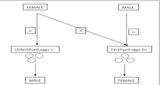
One of the most interesting model to consider is certainly the class of hymenoptera. This class count about 200’000 spices spread in all the world and all of these are showing the mechanism of haplodiploidy.
The dominant and ancestral mode of sex determination in the hymenoptera is arrehenotokous parthenogenesis: a subset of haplodiploidy in which diploid females develop from fertilized eggs and haploid males develop from unfertilized eggs (George et al.2008) [12].
Figure 1.1: haplodiploidy reproduction. In Hymenoptera, unfertilized eggs develop into uniparental haploid males whereas fertilized eggs into biparental diploid females.
This mode of reproduction holds a specific position in sex determination because uniparental males inherit a random half of the maternal genome, while females inherit both the maternal and paternal genes rendering any chromosomal-based sex-determining system impossible (Bull, 1983) [3]. The best-studied genetic example of sex determination in haplodiploids can be attributed in some hymenopteran insects (bees, wasps, ants) to a single, highly polymorphic sex locus in which a single locus with several alleles directs sexual development (Cook, 1993)[4]: the so-called single locus complementary sex-determining mechanism (sl-CSD).
For the honeybee, Apis mellifera, the estimated numbers of sex-determining alleles segregating in populations range from 11 to 19.
[7]
Under single-locus complementary sex determination (sl-CSD), eggs that are heterozygous at the sex locus develop as females while hemizygous and homozygous eggs develop as haploid and diploid males, respectively. Diploid males may be unviable, sterile, or functionally reproductive (Stouthamer et al, 1992; Cowan and Stahlhut, 2004) [5,6], and they are produced under inbreeding, or in populations with low sex allele diversity. While sterile or unviable diploid males constitute a genetic load associated with sl-CSD (Werren, 1993; Cook and Crozier, 1995) [7,8], the recent finding of functionally reproductive diploid males in the vespid Euodynerus foraminatus suggests that the production of diploid males need not always represent a reproductive dead-end (Cowan and Stahlhut, 2004) [6].
The population dynamics of sex-determining alleles have some notable parallels to the self-incompatibility loci in plants and fungi. When an allele is shared between pollen and pistil or between fungi with the same mating type then an incompatibility response will follow. Typically, these systems are controlled by a single genetic locus having multiple allelic versions or specificities. Investigations into the molecular bases have revealed diversity in the mode of recognition and rejection in these systems.
The 1453 base long csd sequence consists of nine exons and contains an open reading frame (ORF) of 385 amino acid residues. Sequence comparisons indicated that CSD is a member of an arginine-serine-rich (RS) protein. CSD protein is a member of RS protein; previous studies have suggested that proteins with an arginine-serine (RS) rich domain are involved in protein-protein interactions and have a dominant role in constitutive and regulated pre-mRNA splicing and metabolism.
Proteins with RS domains fall into two distinct groups: those with an RNA recognition motif (RRM) at its N terminus that binds directly to RNA, the SR proteins (Hastings and Krainer, 2001) [9], and those that are structurally distinct from the SR proteins, the so-called SR-related proteins. The csd gene constitutes a divergent member of the SR-related protein family that does not contain any RNA binding domain.
CSD protein shows some sequence identity to various RS domains that include some conserved SR protein members involved in pre-mRNA splicing, mRNA processing and RNA binding; highest similarity was found for its C terminus to Tra (transformer) protein which achieves female-specific splicing of dsx transcripts in the sex-determining cascade of Drosophila melanogaster. No homology, however, is found for the N terminus that is supposed to be conserved among RS protein orthologs, supporting its function in the specialized process of complementary sex determination.
SR-related proteins can generate multimeric proteins that govern development by specialized splicing. A challenging problem is to determine how the polymorphic signal of various allelic combinations of csd is transformed into the binary switch initiating male and female development. Results with RNAi show that a heterozygous allelic composition encodes a functional product that initiates female development; repression of the csd transcript in female honey bees led to male development, providing conclusive evidence that csd controls sex determination.
The hemi or homozygous allelic composition produces a nonfunctional product that regulates male development by default.
The model proposed is that only the combination of polypetides from different alleles yields an active heteromeric protein complex. Most putative amino acid differences that characterize the
[8]
various alleles are found in the very variable C terminus, in the RS domain, in the proline-rich region, and in a hypervariable region between them. RS domains and proline-rich regions in general have protein binding abilities suggesting that differences in the amino acid sequence may result in modification of protein-protein interactions that are critical in the regulation of splicing. CSD does not constitute a universal explanation in the hymenoptera, however, as it is absent from a number of species.
Homozygotes at csd have zero or near zero fitness in honey bees and most other hymenopterans with CSD, and the sex locus is therefore excepted to be under both balancing selection (balancing selection refers to forms of natural selection which work to maintain genetic polymorphisms ,or multiple alleles, within a population) and diversifying selection (disruptive selection, also called diversifying selection, is a term used to describe changes in population genetics that simultaneously favor individuals at both extremes of the distribution. When disruptive selection operates, individuals at the extremes contribute more offspring than those in the center, producing two peaks in the distribution of a particular trait). This means that selection should maintain diverse allelic variants within a population, with rare alleles enjoying a selective advantage over common alleles. This balancing selection is reflected in honey bees by a surfeit of nonsynonimous nucleotide changes across the different csd alleles, particularly in young alleles. Nonsynonymous substitution are by definition necessary in the formation of novel functional sex alleles, and the finding of an excess of these classes of substitution in the RS domain is consistent with both the specification of functional allele differences in this area and a history of selection for novel alleles. The csd locus supports low levels of recombination, a feature that can lead to the long-term preservation of difference among alleles. In contrast, genomic areas flanking the sex-determining locus have abnormally high recombination rates, which may reduce potential conflicts between csd and neighboring genes (Hasselmann et al.2006) [10]. The discovery of the csd gene will likely lead to elucidation of the molecular mechanism by which csd controls sex determination. csd shows moderate homology to transformer, one of the major sex-determing gene in Drosophila melanogaster that also contains an RS domain. In Drosophila, transformer is involved in female-specific splicing of the doublesex transcript, which initiates the female development pathway. The conservation of transformer function between Drosophila and Apis, if confirmed, would support the hypothesis that most evolution in sex-determining pathways has occurred at or near the beginning of the pathway, with downstream components more conserved in function (George et al.2008) [11].
b) Dipterans (Drosophila melanogaster)
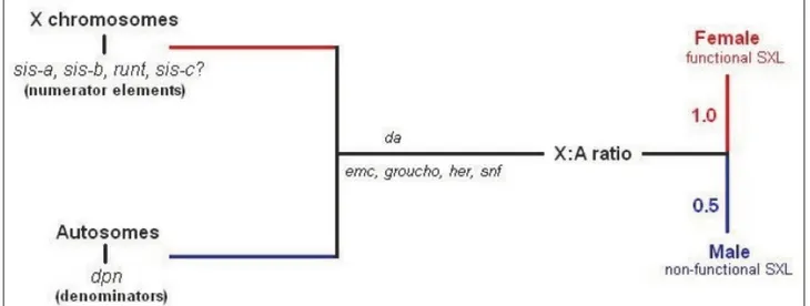
Certainly, the most investigated model organism in genetics and genomics is the fruit-fly Drosophila melanogaster. As reviewed by Gilbert,2000 [17] Drosophila sex determination is due by the presence of a single feminizing switch gene called sex-lethal (sxl) in response to the balance of female determinants on the X chromosomes versus male determinants on the autosomes (X:A ratio). Normally, fruit-flies have either one or two X chromosomes and two sets of autosomes: the presence of a single X chromosome in a diploid cell (1X:2A) develop a male phenotype while two X
[9]
chromosomes in a diploid cell (2X:2A) lead to a female phenotype (Bridges 1921, 1925) [14,15]. Thus, XO Drosophila are sterile males.
In XY cells, sxl remains inactive during the early stages of development (Cline 1983; Salz et al. 1987) [16,19]. In XX Drosophila, sxl is activated during the first 2 hours after fertilization, and this gene transcribes a particular embryonic type of sxl mRNA that is found for only about 2 hours more (Salz et al. 1989) [20]. Once activated, the sxl gene remains active because its protein product is able to bind to and activate its own promoter (Sánchez and Nöthiger 1983) [21]: sxl regulates the splicing of its own pre-mRNA with a typical positive feedback loop.
This female-specific activation of sxl is thought to be stimulated by “numerator proteins” encoded by the X chromosome. These constitute the X part of the X:A ratio. Cline (1988) [18] has demonstrated that these numerator proteins include Sisterless-a,b,c. These proteins bind to the “early” promoter of the sxl gene to promote its transcription shortly after fertilization.
The “denominator proteins” are autosomally encoded proteins such as Deadpan and Extramacrochaetae. These proteins block the binding or activity of the numerator proteins (Younger-Shepherd et al. 1992)[22]. The denominator proteins may actually be able to form inactive heterodimers with the numerator proteins. It appears, then, that the X:A ratio is measured by the balance between X-encoded activators genes at the numerator and autosomally encoded repressors genes of the promoter of the sxl gene in conjunction with maternally derived products. All this take place early in development, leading to the activation of the sxl gene through an early promoter only in females.
Figure 1.2: the X:A ratio determines sex in D.melanogaster. In Drosophila melonogaster sex is the result of the X:A ratio as result of the balance between the X numerator elements and the autosomal denominators in the presence of several maternally derived proteins. An X:A ratio of 0,5 leads to nonfunctional SXL and male development, whereas an X:A ratio of 1 maintains SXL in its active state (early promoter) to female development (from Manolakou, 2006)[47].
This early form of the SXL protein absent in males, orchestrate a different splicing activating a “late” promoter on the Sex-lethal gene that is now transcribed in both males and females. Only in females, trough an autoregulatory feedback loop, that sxl manages to keep itself in an active and functional state through this alternative sex-specific splicing. The autoregulatory loop exemplifies
[10]
how an early developmental decision can be “remembered” for the rest of development, even after the initial signals that established the decision have long disappeared.
Analysis of the cDNA from sxl mRNA shows sex specific difference: the sxl mRNA of males differs from sxl mRNA of females (Bopp et al. 1991) [23]. While the male specific sxl mRNA transcript is nonfunctional, the female-specific sxl message encodes a protein of 354 amino acids. The major difference between the alternative transcript is that male-specific sxl transcript contains an early translation termination codon (UGA) as consequence of the alternative splicing of sxl mRNA: splicing control by sxl allows the production of a functional protein product only in presence of early promoter. Protein–protein interactions, such as competition between normal and inhibitory subunits for dimer formation, can be triggers for controlling developmental switches. The protein made by the female-specific sxl transcript contains two regions that are important for binding to RNA. These regions are similar to regions found in nuclear RNA-binding proteins. Research have shown that there are two targets for the female-specific SXL protein (Kelley et al.1997) [24]. One of these targets is the pre-mRNA of sxl itself. The second is the pre-mRNA of the downstream target gene on the pathway, transformer.
The sxl gene regulates somatic sex determination by controlling the processing of the transformer (tra) gene transcript. The tra message is alternatively spliced to create a female-specific mRNA as well as a nonspecific mRNA that is found in both females and males. Like the male sxl message, the nonspecific tra mRNA contains a termination codon early in the message, making the protein nonfunctional (Belote et al. 1989) [25]. The female-specific tra product acts in concert with the product of the transformer-2 (tra2) gene to help generate the female phenotype. Tra forms a heterodimer with the Transformer-2 (Tra-2) protein that modulates the splicing of other two genes: double sex (dsx) and fruitless (fru). Once that SXL active state has been established, it then goes on to regulate a series of other proteins that control female development as tra/tra2, leading finally to the two alternative products of the doublesex gene (dsx), DSXf and DSXm.
The doublesex (dsx) gene is active in both males and females, but its primary transcript is processed in a sex-specific manner (Baker et al. 1987) [13]. This alternative RNA processing appears to be the result of the action of the transformer gene products on the dsx gene. If the Tra-2 and specific Tra proteins are both present, the dsx transcript is processed in a female-specific manner. The female splicing pattern produces a female-female-specific protein that activates female-specific genes (such as those of the yolk proteins) and inhibits male development while if functional Tra is not produced, a male-specific transcript of dsx is made. This transcript encodes an active protein that inhibits female traits and promotes male traits. If the dsx gene is absent, both the male and the female primordia develop, and intersexual genitalia are produced.
This chain of alternative genes splicing drives the development of the appropriate sex. It is interesting to highlight how the Y chromosome, present in males, is not involved in determining sex, it only helps the correct completion in differentiation of the male germline during the spermatogenesis. According to this model (Baker 1989) [11], the result of the sex determination cascade comes down to what type of mRNA is going to be processed from the dsx transcript. If the X:A ratio is 1, then Sxl makes a female-specific splicing factor that causes the tra gene transcript to be spliced in a female-specific manner. This female-specific protein interacts with the Tra2 splicing factor to cause the doublesex pre-mRNA to be spliced in a female-specific manner. If the doublesex
[11]
transcript is not acted on in this way, it will be processed in a “default” manner to make the male-specific message.
c) Nematodes (Caenorabditis elegans)
The current model of sex determination in C.elegans begin with a start signal that is based on the number of X chromosomes in a diploid animal. In 1949, Nigon demonstrated that C. elegans used an XX/XO sex chromosome system, and the importance of the rapport between X chromosomes and autosome was clarified by Madl & Herman (1979) [26].
The wild-type sexes in diploid C.elegans are the self-fertile hermaphrodite, which has two X chromosomes and the male which has one X chromosome (X0).Therefore males have an X:A ratio of 0.5 and hermaphrodites a ratio of 1.0.
In order to equalize the amount of gene products between the sexes, the hermaphrodite transcript level of the X-linked genes are reduced by half (Meyer and Casson, 1986; Donahue et al.,1987) [27,28]. This differ from how dosage compensation is achieved in Drosophila where the single X chromosome in the male is twice as active as each of the two X chromosomes in female. C.elegans sex determining system provides another opportunity for examining the regulatory interaction between a small set of genes that control the choice of the sexuality.
The development depends by the worm’s ability to interpret the balancing between determinants on the autosomic chromosomes and determinants on the sexual chromosomes: the X:A ratio regulates the activity of xol-1 via the combined action of a set of “numerator” or X-signal elements (fox-1, sex-1) on the X chromosome and “denominator” (sea-1,sea-2) autosomic signal elements elsewhere in the genome.
A potential numerator element is called fox-1 (feminizing locus on X) and another X-linked numerator element is sex-1(signal element on X). sex-1 and fox-1 have additive effects in establishing the X:A ratio. SEX-1 (Signal Element on X) and the post-transcriptional protein FOX-1 (Feminizing locus On X), in association with others elements that are still not identified, inhibit the expression of X0L-1 (X0 lethal) the protein that is present in male pathway.
Concerning the denominators elements, sea genes denominators have yet been identified. Thus, somatic sex, germ-line sex and X-chromosome gene expression are controlled by the dosage of several genes on the X chromosome which seem to act additively by a mechanism that is still not clear. All numerator elements act to control the expression of a single master switch gene, xol-1 (X0 lethal) (Nicoll et al,1997) [29].
According to the model, xol-1 expression promote male development and allow for male X-transcript levels in X0 males while it is inactive in XX animals to allow for the hermaphrodite fate as well as a lowering of X-linked transcript levels. In male animals with genetic asset X0, xol-1 appears to act by repressing the activity of the three sdc (sex and dosage compensation) genes. xol-1 seem to be regulated in a number of ways: one of these is the regulation by SEX-1. SEX-1 acts as a transcriptional repressor of xol-1 by directly binding to its promoter (Carmi et al.,1998) [30]. All these genetic elements play a role in both sex determination and dosage compensation. In contrast, all factors acting epistatically downstream of the sdc genes are involved in either dosage compensation or sex determination, but not both.
[12]
In somatic sex determination the sdc genes promote the hermaphrodite fate in XX organisms, at least in part, by lowering the transcript level of the male specific gene, her-1 (hermaphrodization) (Schauer and Wood,1990) [31]. her-1 is the first gene in the pathway that responds most directly to the primary sex determining signal (xol-1) that depends by the ratio of X chromosomes. The other genes respond indirectly, so that we can define one set of genes in XX animals and another one established in X0 animals. The current model of protein interactions involved in somatic sex determination differ in X0 and XX organisms by the specific bounding of the protein HER-1 at the cell membrane protein TRA-2 only in males.
The binging inactivates TRA-2 allowing the FEM proteins to exert their inhibitory influence on TRA-1. With TRA-1 repressed, the cell is able to take on the male fate. In organism with XX chromosomes asset, HER-1 is not present and TRA-1 is able to bind DNA promoting the female development. tra-1 is thought to be the terminal regulator of somatic sex determination by functioning as a transcription factor that regulates sex specific genes. tra-1 locus makes two mRNA that encode two different proteins (Zarkower and Hodgin, 1992) [32]. The smaller protein contains two zinc finger motifs while the larger contains five, and only the larger is capable of binding DNA in vitro (Zarkower and Hodgin, 1993) [33] supporting the idea that TRA-1 functions as a transcriptional regulator that controls the expression of sex specific genes. Mab-3 (male abnormal) function downstream of tra-1.
Another mechanism is responsible for the specification of the sex in the germinal cells.
Since the C.elegans XX animal makes sperm and oocyte in each of its ovotestes, it is reasonable to assume that additional levels of genetic control are needed in germline compared to the soma. One of the major obstacles that hermaphrodite must addressed is how a differentiated male tissue can be made in a female soma while the environment remains competent for subsequent oocyte production. Spermatogenesis occurs for a short period of time, before switching to oogenesis, implying that the regulation of genetic factors involved in promoting sperm and oocyte production must change at this point. Gamete production in the XX germ line is divided into two temporal phases: spermatogenesis and oogenesis. The principal issue is about how the male fate is reached in organisms with a balancing of chromosomal determinants culminating in a female X/A ratio. First of all, the terminal regulator in the germ line differs from that in somatic districts. tra-1 is the terminal regulator only in somatic cells and function by promoting the female fate when active and allowing for a male soma development when absent. In the germ line, epistatic analysis suggest that tra-1 is not the terminal regulator although is necessary for continued spermatogenesia.
Two others genes also work with the fems to promote spermatogenesis namely fog-1 and fog-3. fem and fog genes termed tgfs must be active to promote spermatogenesis and their activity is dependent upon the inactivity of tra-2. Active tra-2 serves to repress at least one of the tgfs. A possible mechanism for tra-2 inactivation in the germ line involve in hermaphrodite another fog gene: fog-2.
Another mechanism regulating tra-2 in the hermaphrodite may involve a signal emanating from the somatic cells. These cells may secrete an inhibitory ligand for TRA-2 similar to the action of HER-1 in the male soma. The transient down regulation of tra-2 thereby allows the tgfs to promote a brief period of spermatogenesis sufficient for approximately 150 sperm to be made per
[13]
gonad arm, after which a switch is made to oocyte production. This required that one of more tgfs genes be turned off and oocyte promoting factors turned on. At least, in part this may be accomplished by repressing translation of fem-3 through its 3’ UTR. Six other genes (mog1..6) are also involved in the switch from sperm to oocyte production (Graham et al.,1993) [34]. Spermatogenesis in the hermaphrodite could be the result from a transient repression of tra-2 activity in the XX larval germline, which permits sufficient fem gene activity to promote a phase of spermatogenesis (Hansen et al.1999) [35].
1.4) Vertebrates
According to experimental evidence, vertebrates display different strategies leading to the development of alternative sexual phenotype. Among vertebrates, we can find classes in which coexist different mechanisms of GSD (such as XX/XY,ZZ/ZW) and other in which coexist GSD, TSD and also hermaphrodites. From an evolutionary point of view we can argue that at lover evolutionistic levels hermaphrodites and enviromental sex determination are represented and then, at higher levels, they are replaced by genetic sex determination.
a) Fishes.
There are numerous species of fishes in the animal kingdom, with estimation as to their current number reaching a mean price of 25000. Among such a variety of living organisms, research has been focused on relatively few specific model organisms each of which has been considered representative of the reproductive physiology of several other closely related species. Among the mechanisms observed one may refer to:
1- the presence of true hermaphrodites: a strategy usually associated with lower evolutionary levels,
2-temperature dependent sex determination,
3-genetic sex determination and sex chromosomal sex determination (XX/XY or ZZ/ZW patterns). It will be possible that this different kinds of mechanism have evolved separately through the evolution.
While hermaphrodites is not uncommon in worms and insects, it is rarely seen in vertebrates. In birds and mammals, hermaphroditism is usually a pathological condition causing infertility. The most common vertebrate hermaphrodites are fishes, which display several typology of hermaphroditism (Yamamoto, 1969) [36].
Some fishes, however, are gonochoristic; that is, they have a chromosomally determined sex that is either male or female or an enviromental sex determination.
Hermaphroditic fishes species can be divided into three groups. The first are the synchronous hermaphrodites, in which ovaries and testicular tissues exist at the same time and in which both sperm and eggs are produced. One such species is Servanus scriba. In nature and in aquaria, these fish form spawning pairs. As soon as one of the fish spawns its eggs, the other fish fertilizes them. Then the fish reverse their roles, and the fish that was formerly male spawns its eggs so that they can be fertilized by the sperm of its partner. In other hermaphroditic species, an individual
[14]
undergoes a genetically programmed sex change during its development. In these cases, the gonads are dimorphic, having both male and female areas. One or the other is predominant during a certain phase of life.
In protogynous "female-first" hermaphrodites, an animal begins its life as a female, but later becomes male. The reverse is the case in protandrous "male-first" species. In protogynous fishes all the organisms born as females and then, in adult age, invert the sex into males. One of the most explicit examples is the case of Labroides dimidiatus. In this specie exist an α-male: it dominates a little number of adult females and youth organisms that live in a restricted area. Also the adult females present a precise hierarchy: at the mating the male fruitful the dominant female, then the second one, then the third one following the hierarchy supremacy. If the dominant males die, the dominant female turn into male with a mechanism of complete sexual inversion that take place during only one week. After this period the ex dominant female begin its new life as dominant male.
An evolutionistic hypothesis to explain the protogynous model is that it would be favored by the natural selection when the females have a reproductive advantage connected with the short size and when the male has big size as in labridi. The explanation of this statement is referred to the model of selective advantage in size. Males with big size are advantaged in mating and so at the little ones do not invert the sex remaining females. Only after a long period of development and a great increment in size they will be able to compete against the dominant male. So, we can say that in this case of hermaphroditism, sexual dimorphism is represented by females with short size and male with big size.
We can find also other species that display a proterandric hermaphroditism (when the females are bigger than males). This fact could find an evolutionistic answer considering the correlation between number of eggs and size. The size of the eggs is steady in each species; if a female display a bigger size it is able to product more eggs and so is able to increase its fitness.
Concerning species which display temperature sex determination TSD, there are no consistent genetic differences between sexes. The earliest ontogenetic difference between sexes is an environmental one because the ambient temperature during sensitive period of early development irreversibly determines phenotypic sex and, therefore, the sex ratio.
In fishes, the first evidence of TSD was obtained investigating the Atlantic silverside, Menidia menidia. Fishes with TSD have readily been grouped according to three patterns of sex ratio response to environmental temperature during the thermosensitive period: more males at high temperature; more males at low temperature; and more males at extreme (high and low) temperatures. In any case, the presence of TSD in a given species is not incompatible with the existence of a different genotype. However, too often assignment of TSD in many fish species has proceeded regardless of evidence such as the presence of sex chromosomes, which is strongly indicative of GSD. Thus, evidence to support the presence of TSD has been obtained in many cases using temperatures in the laboratory that the species will rarely experience in nature (Conover, 2004) [37].
It has been pointed out that observed sex ratio shifts under these circumstances might be the consequence of thermal effects on GSD (GSD+TE) rather than proof of the presence of TSD. Thus, there is concern regarding the actual prevalence of TSD in fish. In particular, to discern true cases
[15]
of TSD from GSD+TE (Conover, 2004) [37]. Nevertheless, the existence of TSD in fishes is now widely accepted, assumed to be widespread and expected to be found in more species as new studies become available.
Finally, GSD in teleost fishes display an amazing diversity of sex-determination systems. Male heterogamety and female heterogamety are sometimes observed within the same genus and even the same species. More complicated systems can involve multiple sex chromosomes and multiple gene loci (influence from autosomal loci on sex determination and polyfactorial sex determination).
For example, in platyfish (Xiphophorus maculatus), the genome display three different kinds of sexual chromosomes: X, Y e W. This permits a great number of sexual chromosomes pairs in contrast to the classical vision that is based on the segregation of a single sex chromosome pair that give an homogametic and an heterogametic sex. The WX, WY, XX pairs give a female phenotype whereas XY and YY pairs develop a male phenotype (Schartl et al.2004) [38].
No sex determining genes are detected; research is focused on genes that are located on the W chromosome because all the organisms that carry a W are female.
Another model organism, which display a chromosomal sex determination is the teleost medaka fish, Oryzias latipes.
Sex determination in this organisms is founded on male heterogamety as in the mammals but, in contrast to the situation observed in humans, the medaka Y chromosome is very similar to the X; there is no cytogenetic difference between X and Y and X-Y pairing occurs along almost the complete chromosome length.
This suggests that the male-determining region on the Y chromosome should be relatively small. A putative sex determining gene in medaka is DMY: it is expressed exclusively in XY males because it is linked at the Y chromosome. The DMY gene contain a DNA-binding domain called DM domain which is also present in some proteins involved in sex determination in Drosophila melanogaster (DSX) and C. elegans (MAB3).
DMY is homolog to DMRT1, a transcription factor involved in male development in other vertebrates (humans): it should represent the result of a duplication of DMRT1. The sequence of DMY highlight a higher substitution rate if compared to the autosomical maternal gene DMRT1. This evidence support the Y erosion theory and it is reasonable to think that DMY and DMRT1 act as necessary determinants in the male developmental pathway. DMY is shown to be essential for male medaka development, as demonstrated by spontaneous XY female (sex reversal) that produced a truncated DMY protein as a result of a single insertion in exon 3. A second typology of XY female presented a very low level of expression of DMY. DMY expression was detected only in the somatic cells surrounding the germ cells in XY embryos. Taken together, the results of Matsuda et al. 2002 [39] indicate that DMY is a Y-specific gene required for male development in the medaka fish. In female specimen, the role of the aromatase is central although its induction is not under temperature control but under genetic control. The specific expression of gene like FIGa (factor in germ line a) only in females could be related with the genetic induction of the aromatase.
[16]
b) Reptiles.
Two prevailing paradigms explain the diversity of sex determining models in reptiles. Many researcher, consider genetic and enviromental sex determining mechanism to be fundamentally different and that one can be demonstrated experimentally to the exclusion of the other. The conventional view is that these mechanisms are mutually exclusive and they can therefore be viewed as discrete variables. Other researcher argue that no clear boundaries exist between them and that is probable that all sex determining mechanisms have some genetic component.
Recent research on the genes involved in sex differentiation in alligators and turtles with TSD which demonstrates remarkable homology in structure, function and expression of the sex differentiation genes of mammals and reptiles, lends considerable support to that view. Genetic and enviromental sex determination in reptiles should be seen as a continuum of states represented by species whose sex is determined primary by genotype, species where genetic and enviromental mechanisms coexist and interact in lesser or greater measure to bring about sex phenotypes, and species where sex is determinate primarily by environment. In particular exist an intra-generic distribution within some families of enviromental and genetics modes of sex determination and the apparent interaction of both models within some species. In contrast with both mammals and birds, reptiles shows an impressive array of sex determining modes comparable to the variety observed in fishes and frogs. Male heterogamety (XY or XXY) is known in turtles, female heterogamety (ZW, ZZW or ZWW) is known in snakes and both are known in lizards (Graves,2001) [40]. Many species display GSD in the absence of any heteromorphy in the sex chromosomes. Many others have temperature dependent sex determination TSD a form of enviromental sex determination.
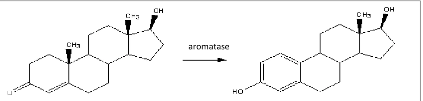
In TSD sex determination, the temperature induce the sexual choice during a particular period known as thermosensitive period (TSP). It is during this period that a very specific enzyme enters into the equation. Aromatase, a cytocrome P450 enzyme, is responsible for the conversion of androgens into estrogens and it is common among many organisms: it acts as an important factor in sexual development.
Figure 1.3: aromatase. Aromatase is an enzyme of the cytochrome P450 superfamily that allows the conversion of androgens into estrogens.
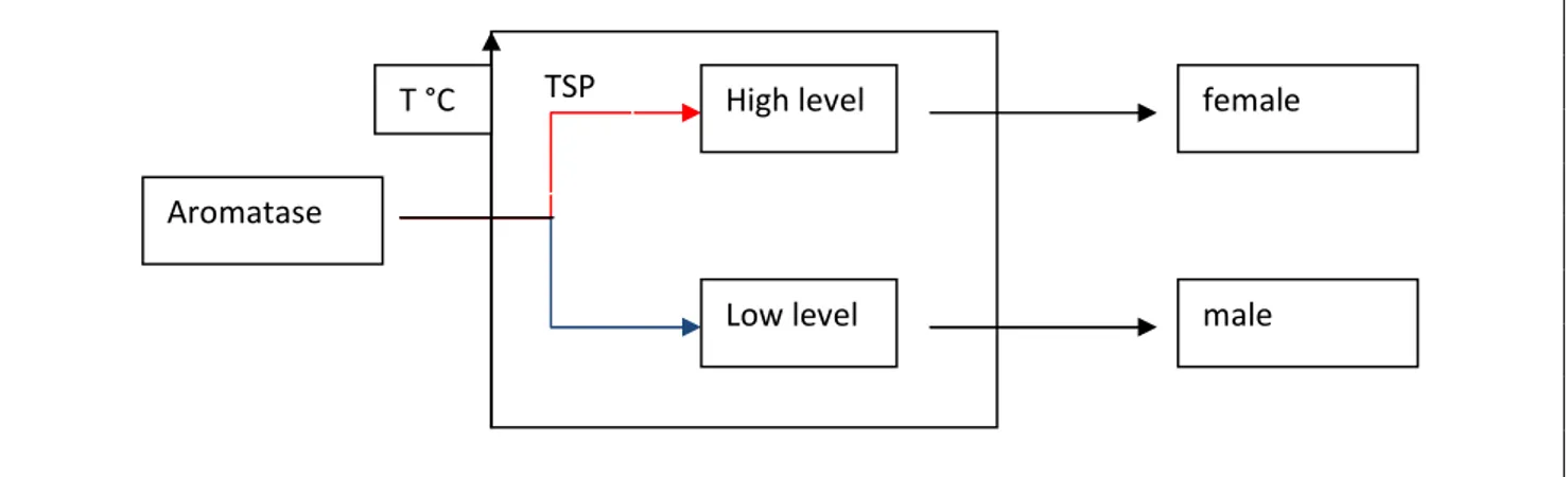
In reptiles while steroidogenesis begins very early, prior even to the thermosensitive period, aromatase activity remains universally low. With the onset of the thermosensitive period aromatase activity seems to increase in certain temperatures, which vary for each species. In
[17]
marine turtles higher temperatures cause an increment of aromatase activity, whereas in lower temperatures aromatase remains low. The two different levels of aromatase drive the development of the undifferentiated gonad through the female or the male pathways. Once the thermosensitive period is over and the fate of the gonad has been established, further changes in temperature seem to have no effects.
Figure 1.3bis: aromatase and sexual determination. During TSP aromatase levels are regulated by the temperature. Higher temperatures increase the aromatase activity developing ovarian whereas lower temperatures decrease the aromatase activity developing testes.
So, in many species we can consider GSD and TSD as transitional forms. An example provide from the study of the interaction between GSD and TSD in Pogona vitticeps. This lizard shows a ZZ/ZW sex determining mechanism but high incubation temperatures reverse genotypic males (ZZ) to phenotypic females. The W chromosome is thus unnecessary for female differentiation, which suggests that molecular mechanism directing sex determination is the result of the dosage balancing of a gene on the Z chromosome rather than the presence of a female determining gene on the W. That is, male differentiation requires two copies of a Z borne gene: the expression or gene activity is sufficient for male development only at optimal temperatures (Quinn et al.2007) [41].
c) Birds.
Reptiles represent the common ancestor for birds and mammals. From reptiles the evolution take two different pathways: one give rise to the branch of birds and the other one that culminate with the evolution of mammals.
Birds display a sex determining mechanism based on female heterogamety (ZZ/ZW). The sexual chromosomes Z and W have not relations with the mammalian X and Y; they could be evolved separately by different ancestral autosomic pairs and this could be a reason to explain the lack of knowledge about the localization of the sex determining gene in this class.
To this day there are two different theories under investigation. The first one considers the sex determination as the result of dosage of Z chromosomes in a quite similar scheme as described for Drosophila or C.elegans. One candidate gene could be DMRT1 located on the Z chromosomes, escapes dosage compensation and is expressed specifically at gonadal level, and is thus capable of linking the numbers of Z chromosomes with gonadal differentiation. On the contrary, sex
Aromatase High level Low level TSP female male T °C
[18]
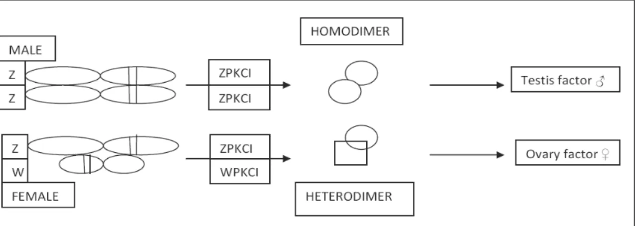
determination could be the consequence of a female induction exerted by the presence of the W chromosome following the example of Y chromosome in eutherian mammals. Two different mechanisms are proposed to support this theory. The first one include the FET1 gene which is localized exclusively in the W chromosome and it is expressed only in the female urogenital system. The second one refers to the homologues genes WPKCI and ZPKCI that are located respectively on the W and on the Z chromosome and to the different functional role resulting by dimerisation of their products (Smith and Sinclair,2004) [42].
The male genome produce an homodimer ZPKCI/ZPKCI acting as testis factor while the WPKCI/ZPKCI heterodimer prevent this effect.
Figure 1.4: hypothetical role of ZPKCI in sexual determination. ZPKCI/ZPKCI homodimer stimulate a factor required for sexual determination in ZZ genetic males, whereas ZPKCI/WPKCI prevent the activation of the factor or stimulate directly ovarian differentiation in ZW genetic females.
To support this theory different combination of Z and W chromosomes caused by aneuploidy are investigated. So, organism with ZZZ present a testis but are not fertile, ZWW die early, but ZZW present a condition of intersexuality: these organism appear as female on hatching but slowly turn into males reaching the sexual maturity. These evidences could support the second theory. d) Mammals.
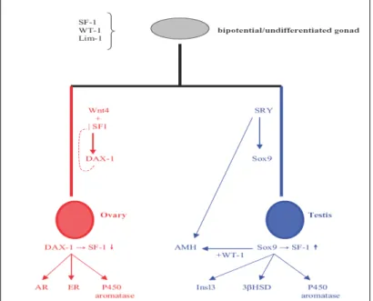
Sex determination in mammals has been more extensively studied than in any other species, most probably due to its direct relevance to human physiology and pathophysiology. A large number of genes have already been described and many more are expected to be added in the process, since relevant research constantly reveals new players in the complex network of reactions related to sex determination. Even in the common, in bi-potential gonad, the expression of several genes is considered crucial for subsequent development and normal sexual dimorphism. These include, among others, WT1, FtzF1/SF1 and Lim1 (Angelopoulou,2005) [43]. Absence of any on these products at this stage, especially WT1, is inconsistent with further gonadal development and may also cause malformations. Sex determination in eutherian mammals is initiated by the Sry locus on the Y chromosome. Sry expression in the bi-potential, undifferentiated gonad directs the support cells precursors to differentiate as Sertoli cells, thus initiating the testis differentiation pathway. In
[19]
the absence of Sry, or if Sry is expressed at insufficient levels, the support cells precursors differentiate as granulosae cells, thus initiating the ovarian pathway.
Sry is a member of the large Sox HMG gene family postulated to have evolved from an ancestor of its paralogue on the Y chromosome, Sox3 (Thompson 2004)[44]. This makes sense, as Sox3 lies on the X chromosome and the Y chromosome is thought to have evolved from the X, and is consistent with the fact that birds and reptiles have neither a Sry gene nor a Y chromosome. Marsupials have the Sry gene, but monotremes appear not to, which suggest, but certainly doesn’t prove, that Sry evolved after the divergence of monotremes from the mammalian lineage.
The SRY protein is a transcription factor a SOX (Sry-like bOX) HMG (High Mobility Group) box type involved in the determination of cell fate and in the regulation of embryonic development (Koopman et al,1990) [45]. All SOX protein are transcription factors and interact with DNA to affect transcription (that is the generation of message RNA from the DNA template), either negatively or positively. SRY isn’t a typical transcription factor, though, and all the players in mammalian sex determination pathway are not understood. Various elements such as WT1+KTS, GATA4, FOG2 are implicated in the regulation of Sry; Sox9 is a downstream target of Sry and Amh, Fgf9 and Dax1 may be subsequent downstream targets involved in the cascade toward testis development. In the female embryo, the Y chromosome is not present and, therefore, Sry is not expressed. The genetic cascade regulating female reproductive system differentiation is not as extensively studied as in men, but DAX1 (and its regulatory system, including genes such as Wnt4 and SF1) is generally considered as a significant player in this process, which is how it came to acquire the rather simplistic description of the “antitestis gene” (Koopman et al.2001) [46].
Figure 1.5: genetic model of sex determination in humans. The formation of the undifferentiated/bipotential gonad is controlled by several genes acting simultaneously, such as WT1, SF1 and Lim 1. Primary sex differentiation is based on the presence of the Y chromosome and its main sex-determining gene SRY. In this case SOX9, FtzF1/SF1 and AMH expression divert the gonad and the reproductive tracts towards the male phenotype. This differentiation process is regulated by several other genes, including DAX1, GATA4,FOXL2and, possibly, DMRT1 and 2. In females, SRY absence allows gonadal development towards female phenotype, mediated by genes such as DAX1, Wnt4 and SF1, resulting in aromatase upregulation. The exact role of Stra8 in this process remains unclear (from Manolakou, 2006) [47].
[20]
Two relatively recently described genes with a potential role in sex determination and differentiation are DMRT1 and Stra8 (stimulated by retinoic acid gene 8). The first has been already discussed previously as a conserved sex-related gene bearing a DM domain originally studied in nematodes. In humans, XY sex reversal in cases of 9p chromosome deletions have been attributed to impaired action of DMRT1 or its homologue DMRT2.
Stra8 on the other hand, is exclusively expressed in female germ cells and its presence signals their sexual gradual differentiation in an anterior to posterior direction. However, it has not yet been established whether the gene’s product directly induces sex determinations towards the female pathway, or rather acts as a simple marker of this phenomenon, without active participation in the process.
e) Amphibians.
Amphibians employ a genetic mechanism of sex determination according to all available information on sex chromosomes or breeding tests. Several study have suggested that some amphibian species display ESD (Dournon et al,1990)[48]. In frogs, one species of Bufo and four species of Rana (Piquet,1930) [49] were examined. These studies all showed that the temperature of the rearing water can alter the sex ratios of tadpoles. In all of these studies, however, effects were obtained by exposure to temperatures that are not normally experienced by the species under study. In frogs 100% males were produce at high temperature (about 32°C), whereas 100% males or 100% females were produce in salamanders. A single study (Uchida,1937b) [50] showed that low temperatures (10°C) can produce 100% female also. When reared at temperatures within the ranges experienced naturally by the species under study, a 50:50 sex ratio was obtained in all of the studies cited above. The natural role of enviromental temperature in sex determination in reptiles or fishes is much more convincing, because effects have been observed at temperatures within ranges experienced by the species in the wild and effects have been shown in natural nests in turtles. Thus, given the lack of effects on sex ratio at appropriate temperatures for the species in the studies on amphibians, it is not likely that temperature is important in normal sex determination in amphibians. The reported enviromental influences are probably due to artifacts of the abnormally high temperatures at which the animals are reared. In addition, reptiles and fish displaying ESD typically lack sex chromosomes which is not the case in amphibians. Thus the available evidence suggest that sex determination in amphibian is under genetic control in natural condition (Wallace et al.1999) [51].
Certainly the presence of sex chromosomes is the clearest evidence that a species possesses a GSD. Sex chromosome can differ in morphology as result of loss of genetic material during the evolution or can be present no difference in shape and morphology if compared with the autosomic pairs. In the past, different techniques were adopted to examine preparations of metaphase spreads, in order to detect difference between the sex chromosomes. About 1500 species were investigated and only less than 4% of the amphibians examined cytologically posses morphologically distinguishable sex chromosomes. The inability to distinguish sex chromosomes suggest that a much smaller region may be involved in sex determination in amphibian sex
[21]
chromosomes relative to other vertebrates with morphologically distinct sex chromosomes (Hillis and Green,1990) [52].
Other experimental techniques that not require morphological identification of sex chromosomes have been used to identify the sex determining systems in amphibians. In most cases, experimental manipulation were used to obtain information regarding heterogamety. The natural condition of sex reversal in this class allows breeding tests between “sex-reversed” organisms to normal individuals, to establish which sex is heterogametic providing an indication of the mechanism of sex determination.
To induce the sex reversion, sex differentiation can be altered by treatment with exogenous steroid hormones or surgically . The effect of these treatments vary between species; the role of sex steroids in gonadal differentiation is not well known and also the sex steroids receptors have not been examined in amphibians.
Concerning the sex determining switch in this class no sex determining genes have been identified. The methodological approach used to investigate some putative genes is the comparative analysis of the conservative genes connected to sexual determination from invertebrate to high vertebrate throughout the evolution.
In addition to Sry, other genes such as WT1, Fgf9, Dax1, Dmrt1 and Sox9 are widely accepted to be involved in the sex determination in vertebrates. However, the roles of these genes during sex determination in amphibians is still unclear (Eggert 2004) [53].
Comparisons of WT1 and Fgf9 sequences between R.rugosa and other vertebrate species showed that both proteins have been highly conserved. The expression of these genes in the undifferentiated gonads began prior to sex determination but neither WT1 nor Fgf9 displayed a sexual dimorphism expression pattern at early developmental stages (Yamamura et al.2005) [54]. Concerning Dmrt1, the DM domain has a highly intertwined structure that chelates two zinc atoms, and makes specific DNA contacts predominantly in the minor groove (Zhu et al., 2000) [55]. The DM domain gene family has some members with a conserved DNA-binding DM domain encoding putative transcription factors related to the sexual regulators Doublesex (DSX) from Drosophila melanogaster and Male abnormal 3 (MAB-3) from Caenorhabditis elegans (Erdman and Burtis, 1993; Raymond et al., 1998) [56,57]. Doublesex and MAB-3 related transcription factor (Dmrt) gene is expressed in the frog Rana rugosa in the differentiating testes but is not detectable during ovarian differentiation (Shibata et al.2002) [58]. As a prelude to understanding the involvement of the Dmrt genes in sexual development in toads, the DM domain gene family of B. gargarizans was cloned and compared with other vertebrates (Chen and al.2007) [59]. Dmrt genes that are duplicated in B. gargarizans are present in mammals as single copies. For example, the human genome contains single copy of DMRT3 (DMRTA3), whereas this gene is present in at least three copies in B. gargarizans. Gene duplication is a mechanism by which new gene functions may be acquired. The very recent duplication of the Dmrt1 has apparently led to the formation of the master male-determining DMY gene in the medaka fish, and similar scenarios might have generated new paralogues of other Dmrt genes in different taxa (Mastuda et al., 2002; Volff et al., 2003) [60,61]. The Dmrt gene duplication in B. gargarizans may have been the result of such a process, unlike the situation in teleost fish, which may have been the result of the ancient
[22]
duplication of the whole genome. However, how these Dmrt genes function in the sexual development B. gargarizans still needs further experimental exploration.
Finally, the role of Sox9 in the gonad will be discuss and examined later in section 3.5. 1.5) Reproduction strategies in the animal kingdom: costs and benefits.
In the animal kingdom are known two different strategies to perpetuate the species: asexual and sexual reproduction. Each one present costs and benefits associated with the typology of cellular division, number of progeny and in reposing at enviromental modifications.
Considering that each kind of reproduction is still now present we can argue that all of them are winning strategies from an evolutionistic point of view.
Asexual reproduction is a form of reproduction which does not involve meiosis, ploidy reduction and fertilization. This kinds of reproduction is typical of ancestral organisms that utilize only mitotic (equational) divisions in their reproduction strategy. Because asexual reproduction does not require the formation of gametes (as in gonochoristic animals) and bringing them together for fertilization, it occurs much faster than sexual reproduction and requires less energy. Asexual lineages can increase their numbers rapidly because all members can reproduce viable offspring. Other advantages connected to this kind of reproductive strategy include the ability to generate offspring without a partner in situations where the population density is low, reducing the chance of finding a mate, or during colonization of isolated habitats such as oceanic islands, where a single member of the species is enough to start a population.
A limitation related to asexual reproduction is that offspring are typically genetically similar to their parents; this genetic similarity may be beneficial if the genotype is well-suited to a stable environment, but disadvantageous if the environment is changing.
The second strategy is the sexual reproduction. The evolution of sex and sexual reproduction contains two related themes: its origin and its maintenance. However, since the hypotheses for the origins of sex are difficult to test experimentally, most current work has been focused on the maintenance of sexual reproduction.
It seems that a sexual cycle is maintained because it improves the quality of progeny (fitness), despite reducing the overall number of offspring. In order for sex to be evolutionarily advantageous, it must be associated with a significant increase in the fitness of offspring. One of the most widely accepted explanations for the advantage of sex lies in the creation of genetic variation. Another explanation is based on two molecular advantages. First is the advantage of recombinational DNA repair (promoted during meiosis because homologous chromosomes pair at that time), and second is the advantage of complementation (also known as hybrid vigor, heterosis or masking of mutations).
For the advantage due to creation of genetic variation, there are three possible reasons this might happen. First, sexual reproduction can bring together mutations that are beneficial into the same individual (sex aids in the spread of advantageous traits). Second, sex acts to bring together currently deleterious mutations to create severely unfit individuals that are then eliminated from the population (sex aids in the removal of deleterious genes). Last, sex creates new gene combinations that may be more fit than previously existing ones, or may simply lead to reduced competition among relatives.
[23]
For the advantage due to DNA repair, there is an immediate large benefit to removal of DNA damage by recombinational DNA repair during meiosis, since this removal allows greater survival of progeny with undamaged DNA. The advantage of complementation to each sexual partner is avoidance of the bad effects of their deleterious recessive genes in progeny by the masking effect of normal dominant genes contributed by the other partner.
In these organisms that display the sexual reproduction the genetic information is passed from parents to offspring via the germline, which segregates from the soma early in development and undergoes a complex developmental program to give rise to the adult gametes. Many aspects of germline development and germline proprieties such as cell fate, maintenance of cell identity, the migration of germ cells to the somatic gonadal primordium and proliferation of germ cells during development have been conserved throughout the animal kingdom from invertebrates to vertebrates.
1.6) Sexual differentiation in vertebrates: interpretative models.
Sexual differentiation is considered a conservative mechanism quite similar among vertebrates. Starting from an hermaphrodite information the undifferentiated and bi-potential gonad is induced to follow one of the two alternative pathways reaching the development of a male or female sexuality.
The nature of the inductive signal is a controversy argument in developmental biology. Researchers supported different hypothesis that diverge in the attribution of the cause-effect relationships between somatic and germinal cells. These are the principals lineages:
- the classical and ancient conception identify the somatic cells as primary and causal inductors in sexual differentiation: in this model, germinal cells would represent undifferentiated and bi-potential passive targets that only under somatic inductions, exerted by different gonadal district, would be able to develop the two alternative phenotypes (symmetric model);
- in asymmetric model, germinal cells have an innate tendency to develop, without a somatic inductor, a “default” sexual phenotype. The alternative phenotype would be the result of a somatic inhibitory induction on a default program;
a) Symmetric model.
In the first year of the 1900 Witchi conduced a great number of observations on the gonadal organogenesis in amphibians by which he postulated a general developmental model of gonadal differentiation for the vertebrate classes.
In this hypothesis sex differentiation is seen as the result of sexualizing actions exercised by the somatic lineage on the germinal linage and the subsequently sexual development is the result of this cause-effect relationship.
In this first description Witchi, 1957 [62] illustrate the early embryonic gonadal architecture as an undifferentiated bisexual primordium with the capacity to differentiate into either sex; male or