UNIVERSITY OF CALABRIA
Department of Biology, Ecology and Earth Sciences
PhD Program in Operative Research
CYCLE XXVII
“La presente tesi è cofinanziata con il sostegno della Commissione Europea, Fondo Sociale Europeo e della Regione Calabria. L’autore è il solo responsabile di questa tesi e la Commissione Europea e la Regione Calabria
declinano ogni responsabilità sull’uso che potrà essere fatto delle informazioni in essa contenute.”
Study of two epigenetic age-associated modifications:
DNA methylation of the rRNA gene promoter in
humans and histone tail acetylation at the
sub-telomeric region in S. cerevisiae
Co-ordinator:
Prof. Lucio Grandinetti
Candidate:
Teresa Scafone
Supervisor:
Prof. Dina Bellizzi
ii
“
The two grand tyrants of
the
Earth,
Time
and
Chance”
iii
iv Index ... iii Abbreviations ... v Index of figures ... ix Sommario ... xi Summary ... xiv
CHAPTER 1: GENERAL INTRODUCTION ... 1
1.1AGING AND AGING PHENOTYPES... 2
1.2EPIGENETICS ... 4
1.3EPIGENETICS AND GENE REGULATION ... 7
1.4EPIGENETICS AND AGING ... 8
CHAPTER 2: METHYLATION OF THE RIBOSOMAL RNA GENE PROMOTER IS ASSOCIATED WITH HUMAN AGING AND AGING PHENOTYPES ... 11
2.1 INTRODUCTION ... 12
2.1.1 The Ribosome: Structure and Function ... 12
2.1.2 Ribosomal DNA (rDNA) arrangement ... 14
2.1.3 Structure and function of Ribosomal RNA ... 15
2.1.4 Ribosomal RNA transcription ... 17
2.1.5 DNA methylation ... 19
2.1.6 DNA methylation and aging ... 22
2.1.7 Ribosomal DNA methylation ... 25
2.1.8 Aging and ribosomal RNA ... 28
2.1.9 Aim of the study ... 31
2.2 MATERIALS AND METHODS ... 32
2.3 RESULTS ... 39
2.3.1 Methylation levels of the rRNA gene promoter correlate with aging . ... 40
2.3.2 Methylation levels of the rRNA gene promoter are associated of with frailty ... 42
2.3.3 Methylation levels of the rRNA gene promoter in association with frailty shows sex-related differences ... 43
v
CHAPTER 3: SIR2 DEACETILATION ACTIVITY AT SUB-TELOMERIC REGION
AFFECTS CELLULAR STRESS RESISTANCE BUT NOT CLS EXTENSION ... 48
3.1 INTRODUCTION ... 49
3.1.1 Saccharomyces cerevisiae, a model organism in aging research ... 49
3.1.2 Sir2, transcriptional silencing mediator ... 51
3.1.3 Regulation at telomeric and sub-telomeric regions ... 54
3.1.4 Sir2, a longevity regulator in yeast ... 55
3.1.5 Aim of the study ... 59
3.2 MATERIALS AND METHODS ... 60
3.3 RESULTS ... 63
3.3.1 The lack of sas2, the antagonist of sir2, shows divergent effects on CLS and oxidative damage ... 63
3.3.2 Tolerance to oxidative stress is reduced in rpd3Δ mutant in a Sir2 dependent manner ... 66
3.3.3 qPCR analysis reveals no difference in sub-telomeric gene expression, but an increasing trend of this expression through aging ... 68
3.3.4 CR is still effective in CLS extension of each mutant, although no one of these mutants seems to be involved in Sir2 pathway ... 69
3.4DISCUSSION... 73
CONCLUSIVE REMARKS ... 76
REFERENCES ... 78
Appendix A ... 87
v
v
5-hmC 5-Hydroxymethylcytosine
5Mc 5-methylcytosine
Å Angstrom
Adh2 Alcohol dehydrogenase
ADP Adenosine diphosphate
ATP Adenosine triphosphate
aza-dC 5-aza-2_-deoxycytidine
CGI CpG island
CLS Chronological lifespan
CPE Core promoter element
CpG Cytosine phosphate Guanine
CR Calorie restriction
DNA Deoxyribonucleic Acid
DNMT DNA methyltransferase
dNTPs Nucleoside triphosphate
DZ Di-Zygotic
ER Endoplasmic reticulum
ERCs Extrachromosomal rDNA circles
ETS External transcribed spacer
HDACs Histone deacetylases
HERV-K Human endogenous retrovirus K
HP1 Heterochromatin protein 1
IRSs Interspersed ripetitive sequences
ITS Internal transcribed spacer
Kb Kilo base
lncRNA Long non-coding RNA
LSU Large sub unit
miRNA MicroRNA
MBPs Methyl-Binding Proteins
mRNA Messenger RNA
mtDNA Mitochondrial DNA
vi
NAD Nicotinamide adenine dinucleotide
NOR Nucleolar organizer region
NoRC Nucleolar remodeling complex
NTS Non-transcribed spacers
OD Optical Density
ORF Open reading frame
p53 Protein 53
Pck1 Phosphoenolpyruvate carboxykinase
qPCR quantitative PCR
RB Retinoblastoma protein
RLS Replicative lifespan
RNA Ribonucleic acid
rDNA Ribosomal DNA
rRNA Ribosomal RNA
S Svedberg
SAM S-adenosyl-lmethionine
SAP Shrimp Alkaline Phosphatase
SC Synthetic complete medium
Sir Silent information regulator
SL1 Selectivity factor
Sod Superoxide dismutase
SSU Small sub unit
TBP TATA- binding protein
TAFs Transcription activating factors
TIF Transcription initiation factor
TPE Telomere position effect
UBF Upstream binding factor
UPE Upstream promoter element
WS Werner syndrome
ix
ix
Figure 1.1 Representation the Homeodynamic. ... 3
Figure 1.2 Waddington’s Epigenetic Landscape. ... 5
Figure 2.1 Ribosome structure ... 13
Figure 2.2 Organization of the human rDNA repeats in eukaryotes. ... 14
Figure 2.3 Secondary rRNA structures. ... 16
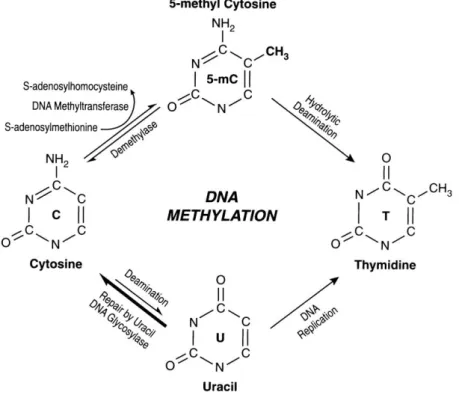
Figure 2.4 Schematic representation of the biochemical pathways for cytosine methylation, demethylation and mutagenesis of cytosine and 5mC. ... 20
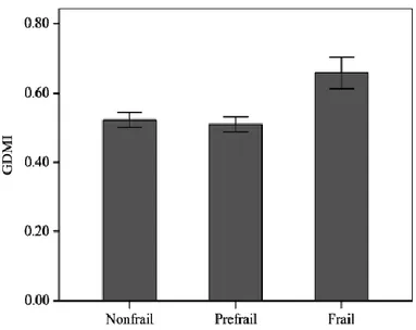
Figure 2.5 Distribution of methylation pattern in a sample of old subjects arranged according to frailty. ... 24
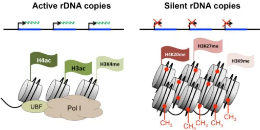
Figure 2.6 The two chromatin states of rDNA repeats. ... 25
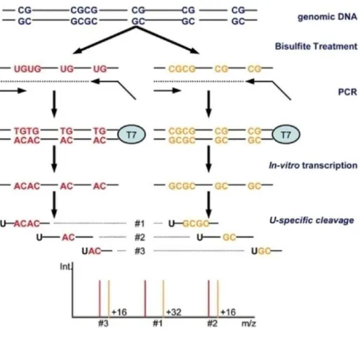
Figure 2.7 Schematic outline of the EpiTYPER process ... 35
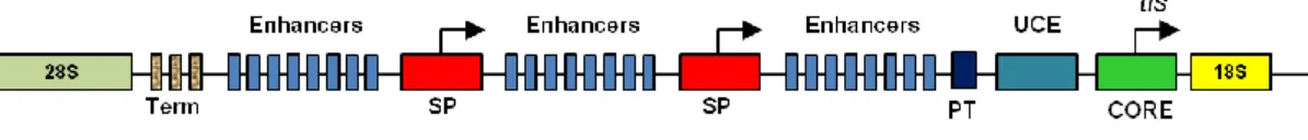
Figure 2.8 Basic structure of the regulator region of the eukaryotic rRNA genes. ... 39
Figure 2.9 Sequence of the rDNA promoter region... 40
Figure 2.10 Median percentage of DNA methylation at each of the unit CpGs located within the rDNA promoter. ... 41
Figure 2.11 Correlation of the methylation levels of the rDNA promoter with frailty in two age-related groups, <90 (A) and 90+ (B) years old. ... 42
Figure 2.12 Correlation of methylation levels of the rDNA promoter with frailty in the group of <90 years old subdivided in genders ... 43
Figure 2.13 Correlation of methylation levels of the rDNA promoter with frailty in the group of 90+ years old subdivided in genders ... 44
Figure 3.1 The aging models in yeast. ... 50
Figure 3.2 Yeast culture growth curve. ... 51
Figure 3.3 Sir2 model of silencing at the mating type loci, telomeres, and rDNA loci. ... 53
Figure 3.4 Chronological lifespan under calorie restriction and oxidative stress resistance at day 3 of Sir2Δ mutant ... 56
Figure 3.5 List of primers utilized for qPCR analysis ... 62
Figure 3.6 Oxidative stress resistance of Sir2Δ and Sas2Δ at day 3 ... 64
Figure 3.7 8 Chronological Lifespan measurements under standard and CR conditions for Sas2Δ compared with Sch9Δ e Sch9ΔSir2Δ previous described (153). ... 65
Figure 3.9 Oxidative stress resistance at day 3 of sir2Δ, sas2Δ, rpd3Δ and the double mutant sir2Δrpd3Δ. ... 66
x
Figure 3.10 Chronological Lifespan assays under normal and CR conditions for Rpd3Δ. ... 67 Figure 3.11 Exemplifying graph of the expression levels of candidate genes analyzed by qPCR. ... 68 Figure 3.12 Synthetic list of analyzed genes with a short description ... 69 Figure 3.13 Oxidative stress resistance of Sir2Δ and Sas2Δ Rpd3Δ and six sub-telomeric genes deleted mutants at day 3 ... 70 Figure 3.14 Chronological Lifespan assays under standard and CR conditions of six sub-telomeric genes deleted mutants. ... 71
xi
xii
Il presente lavoro di tesi è stato realizzato in parte presso il laboratorio di genetica dell'Università della Calabria, sotto la supervisione della Professoressa Dina Bellizzi e in parte presso la Davis School of Gerontology, in collaborazione con il Professor Valter Longo direttore del USC Longevity Institute.
L'invecchiamento è un processo complesso. I fenotipi d’invecchiamento variano da organismo a organismo, ma un generale declino fisiologico si realizza con l’avanzare dell'età.
Una correlazione tra modificazioni epigenetiche e invecchiamento è stato proposto molti anni fa, e nel corso degli anni molteplici studi hanno fornito le prove dell’esistenza di tale connessione, suggerendo che si tratta di un fenomeno conservato lungo il processo evolutivo.
In questo lavoro sono stati investigati due dei più importanti meccanismi epigenetici associati all’invecchiamento: la metilazione del DNA e l’acetilazione degli istoni. È noto che a livello globale la metilazione del DNA tende a diminuire nel corso dell’invecchiamento, con un concomitante aumento, invece, ai promotori di specifici geni. Presso il laboratorio di genetica dell’Università della Calabria è stata investigata la presenza di citosine metilate in siti CpG all’interno della regione del promotore dei geni che codificano per l’RNA ribosomiale umano nonché l’associazione tra tali livelli, l’invecchiamento e la frailty.
L’attenzione è stata focalizzata su questo sito a causa di alcune peculiari caratteristiche che esso presenta: il ruolo cruciale dell’rRNA ribosomiale nelle funzioni cellulari, l’alto livello di conservazione di questa sequenza nucleotidica lungo il processo evolutivo, nonchè l’organizzazione in cluster dei geni che codificano per l’rRNA.
I livelli di metilazione sono stati valutati in campioni di sangue estratti da individui di età differente e con differenti fenotipi d’invecchiamento mediante la piattaforma Sequenom MassARRAY EpiTYPER. Dall’analisi è emersa l’esistenza di una correlazione tra i livelli di metilazione di specifici dinucleotidi CpG, presenti nel promotore genico da noi investigato, l’invecchiamento e i diversi fenotipi ad esso correlati. I risultati di questo studio sono mostrati nel Capitolo 2.
Studiare l’invecchiamento negli esseri umani, tuttavia, comporta numerose difficoltà, per questa ragione sono spesso utilizzati sistemi modello.
xiii
Uno dei più importanti organismi modello è il lievito Saccharomyces cerevisiae attraverso cui è stato possibile individuare numerosi geni rilevanti nel processo di invecchiamento, tra cui il gene Sir2 che codifica per un’istone deacetilasi.
Sir2 è un gene associato con resistenza cellulare allo stress e con la regolazione della durata della vita sia replicativa che cronologica nel lievito. Presso il laboratorio del Prof. Longo è stato in precedenza dimostrato che Sir2 blocca l’estensione della durata della vita cronologica causata dalla restrizione calorica o da mutazioni nei pathway Tor/Sch9, Ras/cAMP/PKA e promuove, inoltre, la protezione cellulare sia contro lo stress termico che ossidativo.
Evidenze crescenti dimostrano che Sir2 e la sua controparte, Sas2, regolano la durata della vita di S. cerevisiae mediante acetilazione e deacetilazione degli istoni H4K16 nella regione sub-telomerica.
In questo studio, abbiamo identificato un nuovo meccanismo attraverso cui Sir2, agendo sulla regione sub-telomerica, regola la protezione contro il danno ossidativo, senza influenzare l'invecchiamento cronologico, segno che questi eventi sono regolati da pathway differenti. I dati riportati supportano i risultati ottenuti in precedenza e allo stesso tempo forniscono una spiegazione parziale del meccanismo sottostante alla maggiore resistenza mostrata dalle cellule mancanti del gene Sir2, come riportato nel Capitolo 3.
Durante il periodo trascorso presso i laboratori del Longevity Institute sono stata inoltre coinvolta in un secondo progetto, dimostrando che una dieta ipocalorica e non solo il digiuno sono efficaci nel ridurre la progressione tumorale e gli effetti collaterali associati alla chemioterapia, identificando inoltre nel sistema immunitario un attore fondamentale di questo processo. Data la rilevanza di questi dati, la USC si riserva la possibilità di bloccarne la divulgazione prima della pubblicazione. Tuttavia, una breve descrizione di questo lavoro è fornita in appendice A.
Nella prima parte del mio programma di dottorato di ricerca, condotta presso l'Università della Calabria, ho inoltre collaborato ad uno studio volto a risolvere il dibattito circa la possibile presenza di citosine metilate all'interno del DNA mitocondriale (mtDNA). I risultati di questo studio sono illustrati nell’articolo scientifico “The Control Region of Mitochondrial DNA Shows an Unusual CpG and Non-CpG Methylation Pattern”, DNA Research, riportato in appendice B.
xiv
xv
The work presented in this thesis has been realized partially at the Calabria University in the Genetics laboratory, under the supervision of Prof. Dina Bellizzi and partially at the Davis School of Gerontology, in collaboration with Prof. Valter Longo, director of the USC Longevity Institute.
Aging is a complex process. The phenotypes of aging vary from organism to organism, but a general physiological decline is recorded with age.
The relationship between epigenetics and aging was proposed many years ago, and today the evidence about this relationship come from several studies, suggesting this as an evolutionarily conserved process.
In this work I investigated two of the most important epigenetic aging-associated features: DNA methylation, and histone tail acetylation.
As is well known, the global DNA methylation tends to decrease during aging, with a coinciding increase localized within the promoters of specific genes. At the Genetics laboratory of the University of Calabria, I investigated the presence of methylated cytosine in CpG sites within the promoter region of the genes coding for ribosomal RNA in association with human aging and frailty.
The attention has been focused on this region because of some unusual characteristics that it presents: ribosomal rRNA’s critical role in cellular function, high conservation of this nucleotide sequence along the evolutionary process, as well as organization in cluster of rDNA genes.
The methylation levels were measured in blood samples of people of different ages and with different frailty phenotypes by means of the MassARRAY EpiTYPER Sequenom platform. The analysis demonstrated a correlation between the methylation levels of specific CpG dinucleotides, aging and the different aging phenotypes. The results are shown in Chapter 2.
Studying aging in humans, however, involves several problems, for this reason model systems are often used.
One of the most important model organisms is the budding yeast Saccharomyces
cerevisiae through which several important genes in the aging process have been
identified, including the gene Sir2, which encodes for a histone deacetylases
Sir2 is associated with cellular stress resistance and lifespan regulation both replicative and chronological in yeast. The Longo lab has previously demonstrated
xvi
that Sir2 blocks the extreme chronological lifespan extension due to calorie restriction or mutations in the Tor/Sch9, and Ras/cAMP/PKA pathways and promotes cellular protection against thermo and oxidative stresses.
Growing evidence demonstrate that Sir2 and its counterparty, Sas2, regulate lifespan in S. cerevisiae by means of histone H4K16 acetylation-deacetylation at sub-telomeric region.
In this study, we identified a novel mechanism through which Sir2 regulates the protection against oxidative damage, without affecting the chronological aging, sign that different pathways regulate these events. Data reported here support previous results and at the same time provide a partial explanation of the underlying mechanism to the higher cell protection in the lack of Sir2, as reported in Chapter 3. During the period I spent at the Longevity Institute laboratories, I was also involved in a second project, showing that a diet and not only fasting is effective in reducing tumor progression and chemotherapy-associated side effects thus identifying in the immune system a crucial player. Given the relevance of these data, the USC reserves the right to prevent the release of them if they are still in press. However a brief description of this work is provided in the appendix A.
Lastly in the first part of my PhD program, performed at the University of Calabria, I collaborated in a study to solve the debate about the possible presence of methylated cytosine within the mitochondrial DNA (mtDNA).
The results of this study are illustrated in the published paper “The Control Region of Mitochondrial DNA Shows an Unusual CpG and Non-CpG Methylation Pattern”, DNA Research, presented in the appendix B.
1
2
1.1
A
GING AND AGING PHENOTYPESAging is a highly complex phenomenon, in fact it is not only ‘the passage of time’, but it is the ‘biological process of growing older’. Aging can be considered as the amount of all changes in structure and function which are deleterious for a cell or an organism, and it leads to an unavoidable consequence, the failure of survival and therefore the death. However the lifespan of each organism is determined both by the innate rate of physiological decline and by the importance of the environmental phenomena destabilizing its homeostatic balance (1).
Despite the large amount of data available about aging and age-related changes in a wide range of organisms, and the abundance of knowledge produced, a final definition of aging and its reasons have been the object of countless debates and many theories of aging from evolutionary, molecular, cellular or systemic point of view have been proposed during the years, sometimes with contradictory results, highlighting the need of more investigation in the aging field.
Nonetheless, the majority of bio-gerontologists today seems to agree at least on the fundamental mechanisms of the aging process.
Life is characterized by a continuous series of environmental changes. The aging process depends on the individual´s ability to respond, control, and adapt to them, maintaining a state of dynamic equilibrium, known as homeodynamics, which in turn depends on a maintenance and repair system (2).
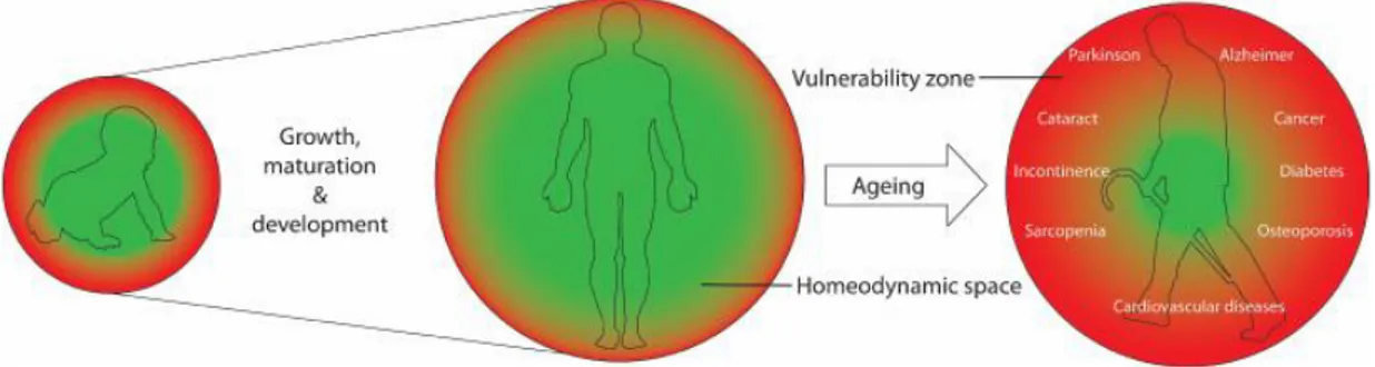
The individual’s ability to respond and to survive the environmental damages can be described as having a homeodynamic space. However, because of the imperfections of the maintenance and repair systems, a vulnerability zone is always present. When the failure starts to be severe, the system can break down or even collapse bringing to aging first, and consequently to death (Figure 1.1) (3).
Recent evidence points to DNA damage accumulation, due to environmental damage and failure of DNA repair systems, as one of the major drivers of the aging process
(4). Aging therefore involves a main feature: fitness decline, with the increase of
mortality rate and a parallel reduction in fertility due to a progressive decline in the ability to respond to environmental challenge (5).
3
Moreover, on a microscopic level, the consequences of age-related changes to the cellular macromolecular components lead to gradual loss of normal structure and function called ‘‘chronological aging’’, due simply by the passage of time. For continuously dividing cells, instead, there is another challenge of ‘‘replicative aging’’. We could say that normal cells are designed for a limited number of successive cell growth-and-division cycles. This limit is known as the Hayflick Limit
(6). Cells go on growing until 40-60 divisions, and then they slow down and finally
stop. This state is known as senescence, and it is an irreversible state because of the accumulation of cellular damage, such as telomere shortening and replication-associated DNA mutations, that occurs during the process of cell division (7).
Figure 1.1 Representation the Homeodynamic.
Homeodynamic space is the ability of the living systems to respond and counteract stress, to repair and remove the damage, and to undergo constant remodeling and adaptation. Genetic polymorphism and epigenetic factors including prenatal exposures and lifestyle establish a personalized functional homeodynamic space during growth, development and maturation, within the evolutionary constraints of essential lifespan of the species. Due to the imperfections of the maintenance and repair systems, there is always a small vulnerability zone. Aging is the progressive shrinkage of the homeodynamic space, resulting in an increase in the vulnerability zone and in the probability of emergence of age-related diseases and the eventually death.
Aging in humans is broadly a linear process, characterized by changes in appearance, due to loss of muscle and bone mass, a lower metabolic rate, longer reaction times, declines in certain memory functions, as well as in all body functions. Although no
4
specific mutation is known to increase human lifespan, several genetic diseases appear to accelerate many features of the normal aging (8).
Aging studies are increasing in biomedical research, especially those attempting to slow this process down. In fact aging itself is the leading risk factor for a variety of widespread diseases, such as selected cancers, cardiovascular disease, and Alzheimer’s disease, but also conditions such as frailty which, in principle, was distinct from diagnosed diseases or disability (9, 10). The concept of frailty is suggested to help in understanding the heterogeneity of functional decline observed with chronological aging. The presence of some components identifies a person as being frail, pre-frail or not-frail (11).
In the last years many definitions of frailty have arisen. The most valued has been proposed by Schuurmans and colleagues “frailty is a loss of resources in several domains of functioning, which leads to a declining reserve capacity for dealing with stressors” (12). The main consequence of such vulnerability is an increased risk of multiple adverse health-related outcomes.
Nonetheless, to directly study aging in humans can be very difficult, especially because of the long lifespan and the ethical concerns. However well characterized systems for aging research are available: mice, rats, flies, worms and yeast, are the most popular. In the last century model organisms have acted as engines of genes and mechanisms involved in extension of lifespan discovery (13).
1.2
E
PIGENETICSIn the last sixty years, it became evident that the genomic code of DNA is not the only carrier of information within a cell or an organism. With “epigenome” we refer to the whole epigenetic modifications occurring on a genome-wide scale (14). Epigenetic represents heritable information that is not directly encoded by the DNA sequence itself but involves chemical alterations of chromatin. The concept of epigenetics was first proposed by Conrad Waddington in 1942. According to him, environmental inputs could be converted into an internal genetic factor by “canalization of development”, explaining how complex phenotypes could form from interaction between genes and environment both internal and external (15).
5
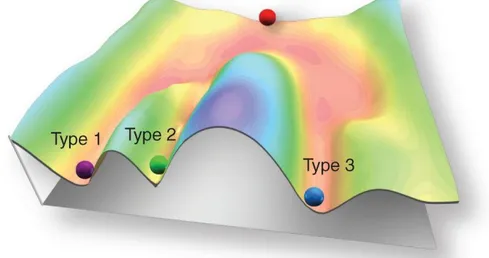
To explain this concept Waddington uses a rolling ball to symbolize a genotype and a downward sloping hill, upon which the ball rests, to symbolize epigenetics (Figure
1.2).
Figure 1.2 Waddington’s EpigeneticLandscape.
The development of a cellular state is represented by a ball rolling down a landscape of bifurcating valleys. At various points in this dynamic visual metaphor, the ball that represents a stem cell can take specific permitted trajectories, each representing different cell types. Source (16).
As the ball travels down the hill it encounters a number of paths, each one bringing the ball in a variety of different directions and leading to a variety of different outcomes. The idea is that a same genotype encounters a certain number of different signals as it progresses through development. The path each gene takes will confer ultimate phenotypic expression (17).
More recently, Berger and colleagues established a new definition of epigenetics. From this point of view, “An epigenetic trait is a stably heritable phenotype resulting from changes in a chromosome without alterations in the DNA sequence” (18). Epigenetic changes are stables, however at the same time they can be regulated by internal factors as physiologic and pathologic conditions, as well as by the external environment (19, 20), or by stochastic factors (21).
6
Gene expression control at the chromatin level is essential for all eukaryotic organisms, and it is important especially in multi-cellular organisms to orchestrate the biological processes, such as differentiation, imprinting, X-chromosome inactivation and aging. Cellular differentiation is the best example to understand how epigenetic changes work. In fact, although all cells in an organism contain essentially the same DNA, cell types and functions, they differ because of qualitative and quantitative differences in their gene expression. Control of gene expression is therefore at the heart of differentiation and development. The patterns of gene expression that characterize differentiated cells are established during fetal development, when each cell gains a specific set of epigenetic marks, that will be maintained as the cells divide by mitosis. However some of these marks will change all lifelong responding to environmental stimuli while the cellular genome will be still intact (22). Therefore, if we imagine the ball that rolls down the hill as a cell, the epigenetic landscape would represent all possible paths the cell can take leading to differentiation and lineage commitment (Figure 1.2).
The key epigenetic marks are DNA methylation, histone tails modifications and non-coding RNAs silencing (23).
DNA methylation is a process whereby a methyl (CH3) group is added, most
commonly to a cytosine of a DNA sequence. In vertebrates, DNA methylation occurs almost exclusively at CpG islands and correlates with transcriptional repression (24). Lower eukaryotes show either no or very little traces of DNA methylation. Nonetheless, three kinds of modifications (5-methylcytosine, N4-methylcytosine and N6-methyladenine) are found in various bacteria, where they are associated with restriction enzyme-based defense.
Histone modifications. The organization of the eukaryotic genome into chromatin
allows DNA to fit inside the nucleus while also regulating the access of proteins to the DNA to facilitate genomic functions such as transcription, replication and repair. The basic unit of chromatin is the nucleosome, comprised of five histone molecules. Interestingly, the N-terminal tails of histones can be subjected to at least 8 types of known post-translational
7
modifications. The modifications include acetylation, methylation, phosphorylation, sumoylation, ubiquitination, ADP-ribosylation, deimination and prolineisomerization (25). Covalent post-translational modifications can impact the conformation of the nucleosome-nucleosome architecture within chromatin and influence its function such that some modifications are associated with an active chromatin state and others with a repressive state
(26).
Noncoding RNA. Recent evidence has emerged showing that noncoding RNAs
regulate multiple epigenetic phenomena. Non coding RNAs can be divided in two classes: microRNAs (miRNAs) and long non-coding RNAs (lncRNAs). Roles for miRNAs and lncRNAs have been demonstrated in the regulation of a broad range of biological activities and diseases (27)
1.3
E
PIGENETICS AND GENE REGULATIONEpigenetic modifications are broadly involved in three key activities:
control of chromosome architecture ensuring stability and proper chromosome segregation during cell division; regulation of the silencing of repetitive elements; and finally, they can initiate and maintain the activity and silencing of genes or their clusters (26).
Gene expression is a complex process involving numerous steps. Transcription, translation and protein modification represent the fundamental steps to transfer genetic information from the store copy of DNA to mRNA, broadly with the following protein synthesis (28).
Epigenetic processes influence gene expression mostly at the transcription level. In general, DNA methylation is associated with gene repression (29) while the histone modification can be related to both silencing and activation of gene expression (30). DNA methylation leads to transcriptional silencing by means of a mechanism involving a group of protein known as DNA methyl-binding proteins (MBPs) that are transcriptional repressor proteins (31). These patterns can also be maintained after DNA replication and mitosis.
8
In contrast to DNA methylation the effects of histone modifications are more complex and the scientific literature is constantly expanding. Early studies indicated that histone acetylation positively correlates with transcription (as opposed to low levels of acetylation associated with transcriptionally silent chromatin).
Acetylation of lysine residues neutralizes the positively charged histone side chains, reducing the strength of the binding of histone tails to negatively charged DNA, ‘opening’ the chromatin structure and facilitating transcription, similarly to lysine phosphorylation (30).
Of the various methylated residues that have been identified, several have been highly characterized.
The consequences of methylation as well as ubiquitylation can be either positive or negative with respect to transcriptional activity, depending on the position of the modified residue, whereas lysine sumoylation is always connected to gene repression. The stability of these modifications is variable. Acetylation and ubiquitylation are dynamic and transient, whereas methylation is stable and longer lasting (32).
In summary, the covalent modification status of histone proteins, together with nucleosome composition and arrangement comprises an epigenetic layer of information that facilitates or inhibits gene expression.
Discovered less than a decade ago, miRNAs, the best characterized of all non-coding RNAs, have emerged as important regulators of gene expression. miRNAs repress gene expression degrading target mRNAs and/or inhibiting their translation. Nonetheless RNA-based mechanisms of epigenetic regulation are still less well understood than mechanisms based on DNA methylation and histone tails modifications (33).
1.4
E
PIGENETICS AND AGINGAging is associated with the accumulation of DNA mutations both at nuclear and mitochondrial level (34). Although the accumulation of these mutations has been correlated with aging, no experiment has demonstrated that a reduction in DNA mutations leads to an extension of lifespan. As such, there is currently much interest in the role of epigenetics as a mediator of the aging process (35).
9
The relationship between epigenetics and aging was proposed many years ago. Pioneering studies showed that genomic global DNA methylation decreases with age in spawning humpbacked salmon and in rat brain and heart (36, 37). More recently, this trend has been confirmed in various mouse tissues and human cells. The definitive validation was provided in 2008 through a longitudinal study of DNA methylation in more than 100 individuals (38).
Moreover the evidence about the relationship between epigenetics and aging come from studies on multiple model organisms, suggesting this as an evolutionarily conserved process (39).
It was observed that the aging clock of C. elegans can be halted by environmental influences; this suggested that the causes of aging may be largely epigenetic. The hypothesis is further supported by the finding of transgenerational epigenetic inheritance of extended lifespan in C. elegans (40). Aged cells show several distinctive features on their chromatin and not only a global reduction in DNA methylation. Depletion of DNMT1, the enzyme responsible for maintenance of cytosine methylation, can be related to the observed age-associated increase in stochastic gene expression (transcriptional noise) in some aging tissues (41). The sirtuin family of NAD+-dependent lysine deacetylases has also long been associated with the control of longevity, although the precise mechanisms remain controversial
(42).
Furthermore studies on mono- (MZ) and di-zygotic (DZ) twins provided significant evidence that epigenetic variants accumulate during aging independently of the genetic sequence (43). It was observed that lower epigenetic differences occur between MZ than DZ twins. Despite MZ twins are epigenetically indistinguishable during the early stage of life, older individuals exhibited significant differences in their epigenome, elucidating the effect of environmental characteristics on gene function and phenotype (44).
Recent researches on C. elegans indicate that the miRNA might also become altered with aging. However, very little is known about miRNA function (45).
Epigenetic changes are, therefore both responsive to, and effectors of the aging process (46). With DNA damage and environmental stresses leading to changes in chromatin, the epigenome adapts to age-related changes as a sensor of cellular
10
dysfunction, sensing changes that accompany aging. However, the epigenome is also an effector of the aging process, enforcing different patterns of gene expression resulting in cellular phenotypes associated with aging such as senescence and metaplasia (47).
11
C
HAPTER
2:
M
ETHYLATION OF THE
RIBOSOMAL
RNA
GENE PROMOTER IS
ASSOCIATED WITH HUMAN AGING AND AGING
PHENOTYPES
12
2.1 I
NTRODUCTION2.1.1 The Ribosome: Structure and Function
Life, as we know it, is possible thanks to interactions between nucleic acids, proteins and lipids. Protein biosynthesis is an essential process borne by ribosomes, a molecular machine in charge for translating genetic information into proteins (48). Ribosomes are ribonucleoprotein complexes lie in ribosomal RNA (rRNA) and over 50 ribosomal proteins. Albert Claude first discovered rRNA during his experiments on sarcoma virus infected cells in the late 1930s (49). Later George Palade characterized the ribosome, a new cytoplasmic component with a small, round body of about 100 to 150Åin diameter (50), and after a few years, protein synthesis was shown to take place on the ribosome (51).
Ribosomes are composed of two unequal subunits called small and large subunit respectively (Figure 2.1). The structure has been conserved throughout the kingdoms
(52), in fact the ribosomal subunits of prokaryotes and eukaryotes are quite similar.
However, the size is different, and the difference is due to the protein and rRNA composition. Prokaryotes have smaller ribosomes with a sedimentation coefficient of 70 Svedberg units (70S) consisting of a small (30S) and a large (50S) subunit, compared to eukaryotic ribosomes of 80S composed of a small (40S) and a large (60S) subunit (53).
Eukaryotic large subunit contains a 5, 25, 28, and a 5.8S rRNA (53), and about 49 proteins, while the small one contains an 18S rRNA and about 33 proteins (54). Ribosomal proteins work in a cooperative way; this means that no one protein alone is able to catalyze an enzymatic reaction. Moreover in the later years a more important role has arisen for rRNAs, in fact, evidence has shown their plausible catalytic activity inside the ribosome, displayed as a super-enzyme complex (55). Ribosomes are abundant cellular elements, placed in several cellular compartments:
in cytoplasm, where they synthesize cytoplasmatic, nuclear, mitochondrial and
13
on the endoplasmic reticulum (ER), originating the rough ER. Here membrane-bound
proteins, proteins residing in the endoplasmic reticulum (ER), Golgi and endosomes are synthesized, following the co-translational targeting;
inside mitochondria and chloroplasts, where proteins codified by their own genes are
synthesized.
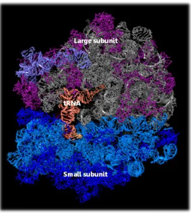
Figure 2.1 Ribosome structure
Ribosomes are complex molecular machines that synthesize proteins in all living cells. This view of a ribosome shows its components in different colors: the small subunit on the left contains an RNA molecule (cyan) and proteins (dark blue); the large subunit on the right contains RNA molecules (grey and slate) and proteins (magenta). The image also shows a transfer RNA (orange) bound to the active site of the ribosome. Source (56).
In both prokaryotic and eukaryotic cells from 20000 to 50000 ribosomes are present, depending on protein synthesis activity. In fact cells must govern both the amounts of specific proteins synthesized and the total protein synthesized in response to environmental signals and internal programming (57).
14
2.1.2 Ribosomal DNA (rDNA) arrangement
The rRNA genes are the most conserved DNA sequences in cells, also in distantly related organisms. For this reason, rDNA has been a useful tool in taxonomy and phylogeny studies (58).
Most prokaryotes have three rRNAs, called the 5S, 16S and 23S rRNA organized in operons. Genes for ribosomal RNAs in eukaryotic genomes have been found in separate clusters, consisting of several hundred tandemly repeated copies of the transcription unit and non-transcribed spacer (Figure 2.2) (59). The number of copies of this transcription unit may be variable within different species.
Figure 2.2 Organization of the human rDNA repeats in eukaryotes.
The coding regions for the 18S, 5.8S and 28S rRNA are shown as yellow boxes. The green boxes represent external (ETS) and internal (ITS) transcribed spacer regions. The start site and direction of transcription are indicated by the arrow. Tandemly repeated rRNA coding units are separated by nontranscribed intergenic spacers (NTS/IGS) (60).
rRNA genes are arranged as clusters of tandem repeats. One cluster consists of hundreds of copies of the 5S gene. Human 5S RNA genes are arranged as tandem repeats in a large cluster on chromosome 1. The number of repeats is variable from 35 to 175 copies. The other genes are found in similar structures to the bacterial operons on the short arms of five chromosomes, which consist of repeated sequences, including rDNA and several kinds of satellites (61). In humans, there are five clusters of rDNA located at the p region of 13, 14, 15, 21, and 22 chromosomes. These regions are called nucleolar organizer regions (NORs), where nucleolus originates. The nucleolus is a dense region of the nucleus where massive transcription of ribosomal RNA genes takes place (62).
There is considerable variation in the size of a transcription unit from one species to the next. This fluctuation is due to the length of the external and internal transcribed
15
sequences (ETS) and (ITS) (Figure 2.2). RNA precursor of about 13,000 bp, known as 47 RNA, originates the remaining 3 RNA molecules, 28S, 18S, 5.8S (63).
2.1.3 Structure and function of Ribosomal RNA
Analyzing the primary rRNA structure, it appears as a hodgepodge of evolutionarily conserved interspersed within non-conserved sequences (64). Efforts to predict the secondary and tertiary structure of rRNAs has been made since the late 1970s.
The rRNA secondary structure is mostly characterized by double strand segments, however many hairpin loops have been found. The secondary structure is highly conserved (Figure 2.3), for instance, the E. coli 23S molecular structure was found to be conserved in the yeast 25S rRNA with extra blocks of sequence and it was also reported in the 28S molecule of multicellular eukaryotes with other extra blocks inserted into the structure. These extra blocks, which occupy the same relative positions in all organisms, have been called expansion segments or variable regions. The increase in size from the E. coli 16S and 23S rRNA to the mammalian 18S and 28S rRNA has been attributed to the differences in the number and size of the expansion segments. The small rRNAs of the ribosomal large sub unit LSU (5.8S and 5S) do not contain significant expansion segments as is the case for the large rRNA molecules. However, as mentioned earlier, the 5.8S rRNA is not free but is associated with the 25-28S rRNA (64). Studies on the primary and secondary structures of 5S RNAs also demonstrated that the nucleotide sequence is highly conserved in the course of evolution.
Modeling of eukaryotic ribosomal RNA (rRNA) has been limited to available crystal structures from eubacterial and archaeal sources. Although recent progresses have been made, it is still missing approximately 50% of the rRNA sequence because of expansion segments (64). The tertiary structure of the rRNAs available from X-ray diffraction analysis has shown that the substitution rates, during evolution, are generally low near the centre of the ribosome, where the essential nucleotides for its function are situated, and that they increase towards the surface (65).
The tertiary structure of the eukaryotic ribosomal 5S rRNA, examined using chemical modifications and enzymatic cleavage reactions, revealed a structure
16
resembling a lollipop. Although the tertiary structure of 5S rRNA has been obtained further studies are still required (66).
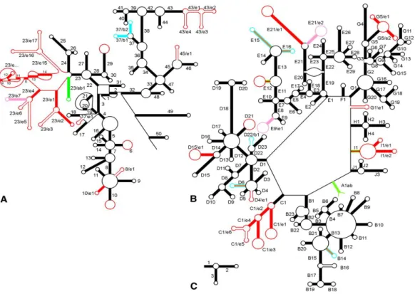
Figure 2.3 Secondary rRNA structures.
Structure and helix numbering of (A) SSU rRNA, (B) LSU rRNA (C) 5S rRNA. The presence of helices in the three domains is indicated as follows: Bacteria only, blue; Eucarya only, red or pink; Bacteria + Archaea, green; Eucarya + Archaea, orange; all three domains, black. Source (65).
Efforts to identify the responsible components for the different ribosomal functions have been made since the discovery of the role of the ribosome in the translation process.
In the early studies the researchers focused their attention on the ribosomal proteins as carriers of the enzymatic functions. The biological role of ribosomal RNA was commonly accepted to be only structural (67). However, after the discovery of catalytic RNAs and RNA catalytic function of RNase P the role of rRNA in translation has been investigated (68).
During the last forty years many experiments have been made to better understand the rRNAs activity, identifying many important features. Shine and Dalgarno, in
17
1974, discovered that 3' region of the 16S rRNA was involved in mRNA binding and this sequence is still known as Shine and Dalgarno sequence (69).
Later it has been demonstrated that mutations in 16S or 23S RNA affected the resistance to several antibiotics (70). Moreover the 16S rRNA molecule is involved at the A site and it takes also contact with the tRNA molecule at the P site, the crystal structures directly showed the participation of 16S rRNA in tRNA binding to the 30S P site (71, 72).
In 1992, Noller and colleagues showed that the 50S subunit of T. thermiphilus retained a peptidyl transferase activity after proteins degradation, referring the catalytic functionality of rRNA (73).
A functional role of 5.8S rRNAs, instead, has emerged more recently, suggesting that the 5.8S rRNA plays a direct role in ribosome translocation (74).
The 5S rRNA molecule is the most highly conserved rRNA, with an important role in ribosome function, coordinating the multiple events catalyzed by the ribosome. Evidence demonstrated that if 5S molecule is not incorporated into the LSU, the LSU becomes unstable and quickly degrades (75).
All these proofs show the functional roles of rRNAs, which are constantly increasing. Thus, rRNA is involved in all the translation process: mRNA and tRNA binding, peptidyl transferase activity, association of ribosomal subunits, translocation, and termination as well as in drugs interaction.
2.1.4 Ribosomal RNA transcription
The nucleolus is a nuclear compartment formed around the ribosomal DNA repeats, called nucleolar organizing regions (NORs). Nucleolus can be considered as a factory where these ribosomal RNAs are transcribed, processed and assembled into ribosomal subunits (76).
In higher eukaryotes, a single transcription unit encodes a precursor transcript (45S pre-rRNA) containing the sequence for three rRNA molecules (5.8S, 18S and 28S). rRNA synthesis, by means of transcriptional activity changes, meets the demand for ribosome production and protein synthesis. rRNA genes activity during development and differentiation of each cell type requires a strict regulation through a wide range
18
of pathways among which JNK2-MAPK-mTOR, MYC, PKC, p53 and RAS/ERK
(62, 77–80).
The transcription of rRNA genes can be defined as a check point for the ribosome biogenesis. The number of active rRNA genes varies between different cell types, and during different stages of cell development and differentiation. It is highly regulated and also linked not only to cellular growth, proliferation and stress response, but also to energetic requirements of cells (77, 81). Nutrients availability, growth factors, and ATP levels alter rDNA transcription, mainly modulating the activity of TIF-IA (77). Transcription initiation factor TIF-1A was, in fact, suggested to be a key player in regulation of rDNA transcription (82).
The rate of cellular growth is directly dependent on the rate of protein synthesis, which, itself, is intricately linked to ribosome synthesis and rRNA transcription. Several reports suggested that yeast rRNA synthesis could be modulated by varying the transcription rate of rDNA genes or by varying the number of active genes, although an a electron microscopy study revealed that the overall initiation rate determines the rate of rDNA transcription (83, 84).
Almost all signaling pathways that influence growth converge at the RNA polymerase I (Pol I) transcription machinery (62). However not only alterations in the amounts or activity of trans-acting factors, but also the gene promoter chromatin accessibility by Pol I regulate rDNA transcription in eukaryotes (76).
The rDNA promoter contains two domains, the core promoter element (CPE) and the upstream promoter element (UPE). The other sequences involved in promotion of rDNA transcription include the promoter proximal terminator elements and the enhancers. The mammalian rRNA gene core promoter is located from -45 to +18 relative to the start site (+1), that is essential for accurate transcription initiation. The Upstream Promoter Element (UPE), from -156 to -107, shows a regulatory role. The crucial role of both of them has been demonstrated for a well-functioning of the transcriptional machinery (85, 86).
In mammalian cells, efficient transcription of rDNA requires the interaction of the core promoter element with three major proteins:
pol I complex, a large complex enzyme with an approximate molecular weight of
19
upstream binding factor (UBF), that binds as a dimer to the CPE, UPE, spacer
promoters and the enhancer repeats in the intergenic spacer,
selectivity factor termed SL1 in humans or TIF-IB in mouse cells. Selectivity factor
in turn is a protein complex consisting of TATA- binding protein (TBP), three transcription activating factors (TAFs), transcription initiation factors TIF-IA and TIF-IC and several others (81, 87).
2.1.5 DNA methylation
DNA methylation together with histone tail modification is the most important DNA epigenetic change. In eukaryotes it involves the covalent addition of a methyl group to the carbon 5 position of the cytosine ring. This modification is generally found on cytosines followed by a guanine, in a sequence called CpG dinucleotides (CpGs)
(88). CpG dinucleotides are not evenly distributed throughout the mammalian
genome and are also greatly underrepresented (89). For instance, human genome presents only 5-10% of the CpG dinucleotides compared to what would be statistically predicted (88). One possible explanation for this distribution is that over a long evolutionary period, the number of CpG dinucleotides has declined as a result of the conversion of CpG to TpG because of the trend of methylated cytosines to deaminate to timine (89).
Despite their underrepresentation into the genome, CpG dinucleotides can be collected in small DNA sections. GC-rich sequences are frequent in satellite repeat, rDNA, centromeric repeat sequences and CpG islands (CGIs) (90).
DNA methylation prevalently involves repetitive sequences, broadly derived from transposable elements, as well as intergenic and intronic CpG-poor regions. Unmethylated CpG dinucleotides are instead gathered in the CpG islands (CGIs); sequences of about 0.5-1 kb in length, with a CG content, higher than 55%, often associated to the gene promoter regions and to the first exons of many genes among which most housekeeping genes and half of all tissue-specific genes (91).
In humans a very low percentage of all cytosine bases are methylated. However considering only CpGs dinucleotides, this number grows to approximately 70-80%
non-20
CpG methylation in promoter regions of different genes. This kind of methylation has been also reported in plants and in the D-loop of human mitochondrial DNA, although its biological significance is unclear, it seems to be a part of a specific pluripotent cell mechanism. After differentiation of the ESC line, cytosine methylation in non-CpGs disappeared, while it is present lifelong in the mitochondrial control region (93, 94).
Basically two enzyme families are involved in DNA methylation establishment: DNA methyltranferases (DNMTs) and DNA demethylase. DNA methylation is mediated by a family of DNA methyltransferases (DNMTs) that includes DNMT1, DNMT3A, and DNMT3B. These enzymes transfer a methyl group from adenosylmethionine (SAM) to deoxycytosine, producing 5-methylcytosine and S-adenosylhomocysteine (Figure 2.4).
Figure 2.4 Schematic representation of the biochemical pathways for cytosine methylation, demethylation and mutagenesis of cytosine and 5mC.
Cytosine can be methylated to form 5-methylcytosine, which deamination rises to thymine. Deamination of cytosine rises to uracil that however it is recognized by the uracil-DNA glycosylase. Source (88).
21
DNMT1 is responsible for maintaining CpG methylation during DNA replication. It acts on the hemimethylated sites of DNA produced during the replication process, maintaining the methylation pattern after DNA syntesis. DNMT3A and DNMT3B, homologous enzymes are in charge for de novo DNA methylation during development. While DNMT3B is prevalent in the early stages of embryonic development, DNMT3A acts later and it is also involved in the gametes methylation patterns establishment. DNA methylation patterns are also determined by DNA demethylases that operate in several ways (95). More recently another epigenetic marker, 5-hmC, is emerging and it seems to have important roles in epigenetic reprogramming and regulation of tissue-specific gene expression (96).
In a methylated DNA sequence, the methyl group is located in the major groove and does not interfere with the base pairing.DNA methylation, in the promoter regions, can repress gene transcription interfering with transcriptional activators or favoring the formation of repressive chromatin by methyl DNA-binding proteins. Almost all housekeeping genes and the majority of genes with tissue-specific expression contain one or more CpG islands in their promoter region. The methylation of the CpGs in these promoter regions correlates inversely with promoter activity and gene expression (97).
In higher eukaryotes, DNA methylation is crucial for a wide range of cellular activities such as genome stability and protection, imprinting, X-chromosome inactivation, tissue specific gene regulation, carcinogenesis and aging (98).
In mammals, DNA methylation is of vital importance for genome stability, as confirmed by homozygous mice with a disruptive mutations in the Dnmt1 gene, they die at the embryonic stage (99).
Changes in DNA methylation are involved in the pathogenesis of many human diseases. Differences in the global methylation pattern between cancers and their healthy tissue have been reported. DNA methylation has also been correlated with several non-malignant diseases, and physiological conditions such as the aging process (100).
22
2.1.6 DNA methylation and aging
The aging process, as described in the first chapter, is characterized by a gradual deterioration of the functional capabilities, with an increased susceptibility to environmental challenges and illnesses, leading to an unavoidable consequence, the failure of survival and finally, the death.
Acquired changes in epigenetic marks have been suggested to be a part of the aging process as well as age-dependent onset of many human diseases (101).
Several experiments have been made over the years, demonstrating the strong association between epigenetic changes and aging. One of the hallmarks of epigenetic drift is a progressive change in DNA methylation. In general two specific modifications of DNA methylation occur during aging: a solid and progressive increase in DNA methylation levels through lifespan in many specific loci and a hypomethylation, across all genome, of repetitive elements. In humans, studies on mono- (MZ) and di-zygotic (DZ) twins provided significant evidence that epigenetic variants accumulate during aging independently of the genetic sequence (43). It was also observed that lower epigenetic differences occur between MZ than DZ twins. In fact DNA methylation changes in aging exhibit familiar clustering, suggesting that the DNA methylation stability is partially genetically determined (38).
Despite MZ twins are epigenetically indistinguishable during the early stage of life, older individuals exhibited significant differences in their epigenome. Remarkably, those twin pairs who reported having spent less of their life together demonstrated the maximum differences in these global epigenetic marks. This suggests that different amount of shared environment might explain the observed differences in the pattern of epigenetic marks, elucidating the effect of environmental characteristics on gene function and phenotype (44). One DNA methylation feature is the age-associated increase of methylation of the regulatory regions of specific genes, partially due to over expression of DNMT3B (102–104).
An example is the increased methylation of the estrogen receptor (ER) gene promoter in colon. It was the first showing an association between aging and promoter DNA methylation. Since then, a growing number of specific loci have been described to become hypermethylated with aging. Hypermethylation was found in genes encoding for ribosomal DNA clusters and for DNA binding and transcription
23
regulatory proteins. In turn both rRNA and regulatory proteins can affect protein synthesis and biogenesis in general, as well as a huge range of pathways involved in a wide variety of cellular functions (105, 106).
Genes involved in tumor suppression, development and growth, cell-cell adhesion, metabolism, DNA repair, control of signal transmission exhibit altered DNA methylation patterns in aging, in some cases displaying also tissue and cellular specificity (107). Remarkably, methylation changes of certain genes such as the
EDARADD, TOM1L1, NPTX2, ELOVL2 , FHL2, and PENK can be age predictive (108). Moreover global genome methylation gradually decreases with age in the
majority of tissues. Loss of 5-methylcytosine content occurs mainly into non CpG-islands within repeated sequences such as interspersed ripetitive sequences (IRSs), Alu and human endogenous retrovirus K (HERV-K). This might be associated with a reduced expression of DNMT1, the enzyme responsible for maintenance of cytosine methylation (103, 104). As demonstrated by Dnmt1+/- mice, they show insufficient DNA methylation, and it has been associated to immunosenescence (41).
It should be considered that significant inter-individual differences in DNA methylation have been discovered in longitudinal studies with both increase and decrease of the global genome methylation over the period of more than 10 years. It is evident how the effect of age on epigenetic marks can be difficult to interpret, as several possibly confounding genetic and environmental variables remain unaccounted for. Furthermore, as described above, changes in epigenetic marks are not all uni-directional. All these studies demonstrated a loss of the epigenetic control in aging, suggesting a correlation with pathological and physiological processes, among which the frailty phenotype. In particular, Bellizzi et al. reported that global DNA methylation levels were correlated to the frailty status in middle/advanced-aged subjects (Figure 2.5) (38, 109).
24
Figure 2.5 Distribution of methylation pattern in a sample of old subjects arranged according to frailty.
The human life expectancy has known a remarkable increase during the last century. Advances in biomedical technology with improved healthcare and disease treatment has to be considered in explaining these changes. However other factors have to be evaluated because genetic and environmental factors work together to determine many phenotypes. As described so far, several evidence gave weight to the epigenetic influence in the aging process as a bridge between aging itself and the environment. One of the most important environmental factors involved in the epigenetic control of aging is nutrition; dietary constituents can affect many phenotypes acting on the DNA methylation status, affecting also aging. These are enzyme co-factors such as folate and vitamins B12 and B6, as well as methyl group donors such as methionine, choline, betaine and serine that increase methylation, and selenium, green tea polyphenols and bioflavonoids that reduce methylation. Maternal behavior or diet can affect epigenetic patterns in her offspring. A study carried out on Agouti pregnant mice showed that a diet rich in methyl donors influences coat color, body weight, and health of their progeny, as well as a diet supplemented with antioxidants like folate, choline and vitamins B6 and B12 have been demonstrated to be effective in aging prevention. Calorie restriction (CR) is the most effective environmental manipulation that can extend maximum lifespan in different species. Recent data suggest that DNA methylation modifications, involving specific genes, play an important role in CR-dependent aging and longevity (110).
25
2.1.7 Ribosomal DNA methylation
In mouse, about half of the rRNA genes are always transcriptionally silent. Although all the rDNA transcription units of a cluster are almost identical, an important part is generally silenced by means of epigenetic marks and thus, not all rRNA genes are available for transcription. Two classes of rRNA genes exist: active rRNA genes are characterized by an ‘open’ chromatin structure defined by DNA hypomethylation, acetylation of histone H4 and dimethylation of histone H3 and associated with emerging pre-rRNA, on the other hand silent rRNA genes are inaccessible, they present CpG hypermethylation, histone H4 hypoacetylation, methylation of H3K9 and H4K20 and they are not bound to transcription factors or Pol I (Figure 2.6) (62).
Figure 2.6 The two chromatin states of rDNA repeats.
The two classes of active and inactive NORs are epigenetically distinct. Potentially active rRNA genes exhibit an open chromatin structure, they are associated with Pol I, UBF and nascent pre-rRNA (green lines) and are characterized by DNA hypomethylation, acetylation of histone H4 (Ac) and methylation of H3K4me2. Epigenetically silenced rRNA genes are demarcated by CpG hypermethylation (CH3), histone H4 hypoacetylation, and methylation of H3K9 and H4K20 (Me). Modified from (111).
DNA methylation is commonly associated with gene silencing. Vertebrate rDNA regulatory elements and transcribed sequences show an unusual pattern. They are both rich in CpG dinucleotides and densely methylated. In the early studies, the methylation status of CpGs within the sequence CCGG of mouse or rat rDNA were
26
determined using the isoschizomers HpaII and MspI. These studies showed an interesting link between the activity state and the methylation levels of the rRNA genes. The methylated sequences matched the silent repeats present in the promoter and enhancer of inactive genes (Figure 2.6) (112).
The correlation between promoter methylation and transcriptional silencing was further investigated; finding that 5-aza-2-deoxycytidine (aza-dC) was able to incentivate rDNA transcription, suggesting that transcriptional silencing of rDNA was due to DNA methylation. However methylation did not prevent transcription directly, but only when DNA was assembled into chromatin, indicating a connection between DNA methylation and chromatin modifications to silence the rDNA units
(112).
It has been demonstrated that rRNA synthesis is determined by the RNA polymerase I activity rather than the number of active genes. However the coexistence of active and silent rDNA units in each cell implied that only through some phases of early development all gene copies might be activated; as during the oogenesis, for example, where large quantities of ribosomes and proteins are required. This thesis is supported by the lower level of global CpG methylation in germ line cells compared with somatic cells, indicating a cell- or tissue-specific epigenetic difference. The same mechanisms may be related to the augment ribosome production in cancer cells, where rDNA methylation is decreased and the number of active rRNA genes is increased (111).
In mice, CpG dinucleotide at position –133 within the UCE is sufficient to compromise the UBF transcription factor and the Pol I to nucleosomal rDNA binding, preventing initiation complex formation. Impairment of UBF binding to methylated rDNA promoter was specific to rRNA genes assembled in chromatin. This implied that cytosine at position -133 is exposed on the surface of the positioned nucleosome and that the methyl group represents a steric barrier for the binding of UBF transcription factor. In human cells, there are at least 25 CpGs residing within the bounds of the Pol I promoter, although only the CpG methylation at positions -60 and -68 seems to act similarly to the mouse -133 CpG dinucleotide (113).
Mouse and human promoters of active rDNA are associated with Pol I transcription complex and with active histone marks such as acetylated histone H4 (H4Ac) and