POLITECNICO DI MILANO
Scuola di Ingegneria Industriale e dell’Informazione
Corso di Laurea Magistrale in Ingegneria Biomedica
Cardiorespiratory Interactions in Newborns during Sleep:
Linear and Nonlinear Multiparametric Analysis
Relatore: Prof. Maria Gabriella SIGNORINI Correlatori: Maristella LUCCHINI
William FIFER, PhD
Tesi di Laurea di: Nicolò PINI Matr. 841423
I
Contents
List of figures ... III List of tables ... VII Ringraziamenti ... IX Acknowledgements ... X Sommario ... XI Summary ... XXII Chapter 1 ... 1 1 Introduction ... 2
1.1 Sudden Infant Death Syndrome ... 2
1.2 Intrinsic risk: Prematurity ... 4
1.3 Extrinsic risk: Sleep Position ... 6
1.4 Fetal and newborn cardiovascular systems ... 8
1.4.1 Cardiovascular system generation ... 8
1.4.2 Circulation before birth ... 8
1.4.3 Circulation after birth ... 10
1.5 Autonomic Nervous System ... 12
1.6 Cardiorespiratory coupling ... 14
1.7 Sleep states ... 15
1.8 SIDS and cardiorespiratory control ... 16
Chapter 2 ... 21
2 Materials and Methods ... 22
2.1 Study population and acquisition system ... 22
2.2 Time domain analysis ... 25
2.3 Entropy analysis: univariate and bivariate estimators ... 26
2.3.1 Approximate Entropy ... 27
2.3.2 Sample Entropy ... 28
II
2.3.4 Transfer Entropy ... 30
2.4 Phase locking analysis ... 35
2.5 Directionality Index analysis ... 41
2.6 Statistical analysis ... 44
Chapter 3 ... 45
3 Results ... 46
3.1 Time domain ... 46
3.2 Sample Entropy and Quadratic Sample Entropy ... 50
3.3 Transfer Entropy ... 55
3.4 Phase synchronization ... 59
3.5 Directionality index ... 67
Chapter 4 ... 79
4 Discussions ... 80
4.1 Time and Frequency domain ... 80
4.2 Sample Entropy and Quadratic Sample Entropy ... 82
4.3 Transfer Entropy ... 83
4.4 Phase synchronization ... 85
4.5 Directionality Index ... 86
4.6 Conclusions ... 88
4.7 Further developments and future work ... 90
III
List of figures
Figure 1.1.1 Triple-risk model for SIDS by Filiano et al. ... 3 Figure 1.2.1 Preterm birth rates with respect to the total life birth in each state in 2015. .... 4 Figure 1.3.1 Trend of SIDS rate, the diminishing trend can be observed as a consequence of
the Back to Sleep Campaign. The green line indicates the percentage of babies sleeping supine with respect to the total percentage of premature infants in the USA ... 6
Figure 1.4.1 The heart and peripheral circulations in fetus ... 9 Figure 1.4.2 Heart and peripheral circulation before and after birth ... 10 Figure 1.4.3 A schematic of the fetal circulation before birth and the changes in flow in a
newborn subject. Before birth the major supply of preload for the left ventricle is derived from the placental circulation, which passes from several structures and enters the left side of the heart; thereby bypassing the right side of the heart and the lungs (panel A, pathway is shown by red arrow). As most blood exiting the right ventricle passes through the ductus arteriosus (A, red arrow) and enters the descending aorta, very little blood flows into the fetal lungs before birth. ... 11
Figure 1.8.1 Five Steps in the terminal respiratory pathway associated with the SIDS results
from one or more failures in protective mechanisms against a life-threatening event during sleep in the vulnerable infant during a critical period. ... 18
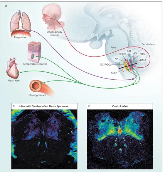
Figure 1.8.2 The serotonergic system is considered to be critical for the modulation and
integration of diverse homeostatic functions. The medullary level of the brain stem (black line in Panel A) includes regions involved in the regulation of upper-airway control, respiration, temperature, autonomic function, and the sympathetic nervous system. In the medulla of an infant with the sudden infant death syndrome (SIDS), tissue autoradiography shows a generalized reduction in binding to the 5- hydroxytryptamine type 1A receptor (Panel B), as compared with that in a control infant at the same postconceptional age (Panel C). ARC denotes arcuate nucleus, DMX dorsal motor nucleus of the vagus nerve, GC ganglion cells, HG hypoglossal nucleus, NA noradrenaline, NTS
IV
nucleus tractus solitarius, PGCL paragigantocellularis lateralis, PreBot pre-Bötzinger complexes, and ROb raphe obscurus ... 20
Figure 2.3.1 Scheme of the main steps involved in TE calculation: ... 34 Figure 2.4.1 From top to bottom: examples of a 60-second segment of RR series, respiration,
synchrogram and relative λ index of 3:1 synchronization order ... 36
Figure 3.1.1 Boxplot of time domain parameters computed from RR series (RR mean, RR
IQR, SDNN, RMSDD, IBI mean, IBI IQR) and from respiratory signal (IBI mean, IBI IQR) ... 47
Figure 3.1.2 Boxplot of time domain parameters computed from RR series (RR mean, RR
IQR, SDNN, RMSDD, IBI mean, IBI IQR) and from respiratory signal (IBI mean, IBI IQR) ... 49
Figure 3.2.1 Boxplots of Sample Entropy computed for different embedding dimensions
when state-related analysis is performed, active (red) versus quiet (green) in newborns and one month infants ... 51
Figure 3.2.2 Boxplots of Sample Entropy computed for different embedding dimensions
when state-related analysis is performed, AS (red) versus QS (green) in newborns and one month infants ... 52
Figure 3.2.3 Boxplots of Quadratic Sample Entropy computed for different embedding
dimensions when state-related analysis is performed, AS (red) versus QS (green) in newborns and one month infants ... 53
Figure 3.3.1 Boxplots of Transfer Entropy computed for newborn cohort. Comparisons are
made considering same direction and different sleep state or same sleep state and different direction, N represents the number of segments in the considered population ... 56
Figure 3.3.2 Boxplots of Transfer Entropy computed for one month cohort. Comparisons
are made considering same direction and different sleep state or same sleep state and different direction, N represents the number of segments in the considered population ... 57
Figure 3.3.3 Boxplots of Transfer Entropy computed for both newborns and one month
V
different time points, N represents the number of segments in the considered population ... 58
Figure 3.4.1 Boxplots of total percentage of synchronization, sum of ratios with respect a
single breathing cycle and two consecutive breathing cycles ... 60
Figure 3.4.2 Boxplots of total duration of synchronization, sum of ratios with respect a
single breathing cycle and two consecutive breathing cycles ... 61
Figure 3.4.3 Boxplots of total percentage of synchronization, sum of ratios with respect a
single breathing cycle and two consecutive breathing cycles ... 62
Figure 3.4.4 Boxplots of total duration of synchronization, sum of ratios with respect a
single breathing cycle and two consecutive breathing cycles ... 63
Figure 3.4.5 Entity of increase in synchronization between AS and QS for subjects with
both state during the recorded baseline. Each colored line represents a subject ... 65
Figure 3.4.6 Bar graph of specific ratio of synchronization comparing same sleep state for
different time points ... 66
Figure 3.5.1 Scatter plot of breathing frequency and directionality index for newborns ... 67 Figure 3.5.2 Boxplots of directionality index and breathing frequency in AS versus QS
considering newborns ... 68
Figure 3.5.3 Scatter plot of breathing frequency and directionality index for one months 69 Figure 3.5.4 Boxplots of directionality index and breathing frequency in AS versus QS
considering one months ... 70
Figure 3.5.5 Scatter plot of breathing frequency and directionality index in AS for both
newborns and one months ... 72
Figure 3.5.6 Boxplots of directionality index and breathing frequency comparing newborns
versus one months in AS ... 72
Figure 3.5.7 Scatter plot of breathing frequency and directionality index in QS for both
newborns and one months ... 73
Figure 3.5.8 Boxplots of directionality index and breathing frequency comparing newborns
versus one months in QS ... 73
Figure 3.5.9 Histogram of newborn cohort grouped based on breathing frequency, darker
VI
Figure 3.5.10 Boxplots of directionality index and breathing comparing newborns grouped
based on breathing frequency ... 75
Figure 3.5.11 Histogram of one month cohort grouped based on breathing frequency, darker
green indicates bins where the distributions overlap ... 76
Figure 3.5.12 Boxplots of directionality index and breathing frequency comparing one
months grouped based on breathing frequency ... 77
Figure 3.5.13 Boxplots of breathing and directionality index comparing newborns and one
VII
List of tables
Table 2.1.1 Subject information, percentages are computed with respect to the total number
of subjects for each cohort; 151 newborns and 33 one month infants ... 23
Table 3.1.1 Time domain parameters extracted from RR series and respiratory signal. IBI
measures the mean distance between adjacent respiratory onsets and IBI IQR the interquartile range of this distribution. P-values are relative to statistics comparing AS and QS parameters within the same age ... 46
Table 3.1.2 Time domain parameters extracted from RR series and respiratory signal.
P-values are relative to statistics comparing same sleep state (AS and QS) parameters for the two different time points ... 48
Table 3.2.1 Sample Entropy and Quadratic Sample Entropy considering 300 beats of RR
series only. P-values are relative to statistics comparing AS and QS parameters within the same age ... 51
Table 3.2.2 Sample Entropy and Quadratic Sample Entropy considering 300 beats of RR
series only. P-values are relative to statistics comparing same sleep state (AS and QS) parameters for the two different time points ... 54
Table 3.3.1 Transfer Entropy considering 300 beats of RR series and respiratory sampled at
RR instants. P-values are relative to statistics comparing AS and QS parameters within the same age and directionality or within the same age and different directionality ... 55
Table 3.3.2 Transfer Entropy considering 300 beats of RR series and respiratory sampled at
RR instants. -values are relative to statistics comparing same sleep state (AS and QS) parameters for the two different time points ... 58
Table 3.4.1 Total synchronization parameters extracted from the analysis of phase
relationship between RR series and respiratory signal. P-values are relative to statistics comparing AS and QS parameters within the same age ... 59
Table 3.4.2 Total synchronization parameters extracted from the analysis of phase
VIII
statistics comparing same sleep state (AS or QS) parameters for the two different time points ... 62
Table 3.4.3 Paired comparison of AS versus QS in newborns and one months ... 64 Table 3.5.1 Computed directionality index and extracted breathing frequency. P-values are
relative to statistics comparing AS and QS parameters within the same age 67
Table 3.5.2 Extracted directionality index and breathing frequency computed. P-values are
relative to statistics comparing same sleep state (AS or QS) parameters for the two different time points ... 71
Table 3.5.3 Percentage indicating the portion of subjects associated with a negative
directionality index and positive directionality index ... 74
Table 3.5.4 Extracted breathing frequency and computed directionality index. P-values are
relative to statistics comparing AS and QS parameters within the same age 74
Table 3.5.5 Computed directionality index and extracted breathing frequency. P-values are
relative to statistics comparing newborns and one months grouped based on breathing frequency threshold ... 77
IX
Ringraziamenti
Questa tesi è la realizzazione di un progetto di collaborazione tra il Politecnico di Milano e Columbia University Medical Center, ideato e sviluppatosi durante l’anno accademico 2015-2016.
In particolare, hanno reso possibile questa esperienza il costante supporto e l’entusiasmo di William Fifer PhD, Direttore del Clinical Developmental Neuroscience Division at the Sackler Institute for Developmental Psychobiology, mio relatore durante i mesi di permanenza a New York.
Un ringraziamento al gruppo di lavoro del laboratorio di ricerca per la loro disponibilità.
Esprimo la mia profonda gratitudine alla mia correlatrice Maristella Lucchini, dottoranda presso il Politecnico di Milano, per il suo incessante aiuto, la pazienza ed il supporto durante questi mesi.
Un ringraziamento speciale va alla mia relatrice Maria Gabriella Signorini, Professore Associato presso il Dipartimento di Bioingegneria del Politecnico di Milano, per aver creduto in me ed avermi permesso di vivere questa esperienza unica, nonché per il supporto costante fin dall’inizio di questa tesi.
Infine un ringraziamento al sostegno economico fornito dal Politecnico di Milano per l’erogazione della borsa di studio “Tesi all’estero”.
X
Acknowledgements
This thesis is the realization of a collaboration project between the Politecnico di Milano and Columbia University Medical Center, conceived and developed during the academic year 2015-2016.
Particularly, this has been possible thanks to the consistent support and enthusiasm of William Fifer PhD, Director of the Clinical Developmental Neuroscience Division at the Sackler Institute for Developmental Psychobiology, my Advisor during my stay in New York.
I would like to thank the entire staff of the research lab for their availability.
I would like to express my deep gratitude to my advisor Maristella Lucchini, PhD student at Politecnico di Milano, for her endless help, her patience and her support during these months.
A special thank goes to my advisor Maria Gabriella Signorini, Professore Associato at the Dipartimento di Bioingegneria del Politecnico di Milano, for believing in me and giving me this unique opportunity along with her constant presence since the beginning of this thesis.
Lastly, thanks to the Politecnico di Milano for the economic support given through the scholarship “Tesi all’estero”.
XI
Sommario
Introduzione e scopo del lavoro
La sindrome della morte improvvisa infantile (Sudden Infants Death Syndrome, SIDS) è descritta come la morte improvvisa di un neonato sano durante il sonno e rappresenta una delle maggiori cause di mortalità infantile nei paesi sviluppati. Nonostante il marcato declino dell’incidenza della SIDS a partire dal 1990, grazie a campagne globali di educazione sul tema, SIDS rimane una delle maggiori cause di morte per i neonati di età 1-12 mesi di età. A causa del suo drammatico impatto, questa sindrome è stata studiata per lungo tempo, ma i meccanismi fisiologici di base rimangono ancora da essere chiariti.
Attualmente, la spiegazione più supportata in letteratura riguardo SIDS, è il modello del triplo rischio (Triple-risk model), proposto da Filiano e Kinney.
L’ipotesi di Filiano e Kinney riguardo SIDS è basata sulla concomitanza di tre fattori: 1) un neonato fragile, 2) un periodo critico dello sviluppo, 3) un fattore di stress esogeno.
I neonati hanno una maggiore probabilità di morire di SIDS se posseggono tutti questi tre fattori: la vulnerabilità congenita dei neonati rimane latente finché questi ultimi entrano nel periodo critico della SIDS e sono soggetti ad uno stress esterno.
Lo sviluppo del controllo cardiorespiratorio può essere classificato come un fattore di rischio, in accordo con il modello del triplo rischio. In particolare può essere pensato come una sottoclasse del cosiddetto controllo omeostatico.
A seguito di queste considerazioni, il presente studio si propone di analizzare una popolazione di neonati a termine sani ed una popolazione di infanti sani di un mese di età. Il lavoro di tesi si concentra sull’analisi dell’interazione cardiorespiratoria, la sua relazione con gli stati del sonno e la sua evoluzione dalla nascita ad un mese di vita. Lo scopo della ricerca è la descrizione dell’accoppiamento cardiorespiratorio di tipo fisiologico, al fine di metterne in luce le differenze rispetto all’accoppiamento cardiorespiratorio delle vittime di SIDS.
SOMMARIO
XII
Lo studio di questa interazione è eseguita su soggetti durante il sonno. Il sonno ha un ruolo fondamentale nello sviluppo del sistema nervoso e nella regolazione omeostatica.
Il sonno nei neonati e negli infanti può essere classificato in tre tipi: Sonno Quieto (Quiet Sleep, QS) (equivalente a NREM), Sonno Attivo (Active Sleep, AS) (equivalente a REM) e Sonno Indeterminato (Indeterminate Sleep, IS).
È stato ipotizzato che gli infanti morti di SIDS avrebbero mostrato anormalità nell’organizzazione degli stati del sonno precedentemente allo loro morte.
Il meccanismo alla base di SIDS appare avere origini nell’ambiente fetale con il risultato di danni neuronali e di sviluppo del sistema nervoso autonomo (Autonomic Nervous System, ANS). Questo meccanismo ancora sconosciuto in seguito, comprometterebbe l’adeguata risposta alle sfide respiratorie e pressorie durante il sonno.
Questo deficit coinvolge alterazioni dei recettori delle regioni coinvolte nel controllo chemocettivo, cardiovascolare e cardiorespiratorio.
Risulta fondamentale evidenziare che l’indagine dell’accoppiamento cardiorespiratorio è in grado di fornire una conoscenza non invasiva dei meccanismi di interazione fra il sistema cardiaco e respiratorio ed è in grado di aiutare a comprendere i fattori che contribuiscono all’occorrenza di SIDS.
Lo studio descritto in questa tesi è stato possibile grazie alla collaborazione fra il centro clinico di eccellenza Columbia University Medical Center (CUMC) e il Politecnico di Milano, Dipartimento DEIB.
L’indagine di tesi è stata condotta al Politecnico di Milano e al CUMC durante i miei 6 mesi di permanenza nella città di New York. L’incontro fra il contributo dell’ingegneria biomedica e le competenze mediche hanno permesso di proporre nuove soluzioni per la quantificazione dell’interazione cardiorespiratoria e la validazione dei risultati ottenuti.
Materiali e Metodi
Il dataset dei neonati include 151 infanti nati al Morgan Stanley Children’s Hospital di New York at CUMC fra 38 e 40 settimane di età gestazionale (Gestational Age (GA)), mentre il gruppo degli infanti di un mese di età include 33 soggetti che si sono sottoposti ad un follow-up ad un mese, la selezione di questi ultimi soggetti è basata sul medesimo criterio
SOMMARIO
XIII
relativo all’età gestazionale. Nessuno degli infanti è stato ricoverato nell’unità di terapia intensiva neonatale (Neonatal Intensive Care Unit, NICU) o è stato diagnosticato di gravi patologie o disordini genetici conosciuti.
Fra i vari segnali registrati, in questo contesto sono stati analizzati ECG e segnale respiratorio. Gli stati del sonno sono stati codificati, sulla base del respiro, da medici esperti nel settore.
Durante i 10 minuti di acquisizione della baseline, i neonati dormivano in posizione supina, entro ~ 30 minuti in seguito all’allattamento. Sono stati analizzati segmenti di 3 minuti di durata duranti i quale non vi sono cambiamenti nello stato del sonno: 514 epoche di durata tre minuti per la popolazione dei neonati (239 sonno quieto, 275 sonno attivo), mentre 247 epoche per gli infanti di un mese di età (144 sonno quieto, 103 sonno attivo).
I picchi R sono stati individuati sul tracciato ECG per mezzo dell’algoritmo di Pan-Tompkins. Un filtro adattativo è stato successivamente applicato al fine di rimuovere i battiti ectopici o artefatti.
Il segnale respiratorio è stato filtrato con un filtro passa-banda (0.05 – 3.5 Hz). I picchi di inspirazione sono stati individuati per mezzo di un software automatico di riconoscimento ed ogni segmento è stato controllato manualmente al fine di eliminare i picchi incorretti.
L’analisi dell’interazione cardiorespiratoria durante il sonno è svolta per mezzo di un approcciato univariato ed uno bivariato, per mezzo di metodi lineari e non lineari. Lo scopo dell’analisi è la caratterizzazione dell’interazione in relazione agli stati del sonno e la corrispondente evoluzione dovuta all’età.
La prima analisi riguarda l’estrazione dei parametri univariati nel dominio del tempo, calcolati dalla serie RR e dal respiro. Vi è una mancanza di linee guida per l’applicabilità di questi metodi per i neonati. È evidente che modalità di analisi applicate agli adulti non possano essere applicate a questo contesto, considerato che la frequenza cardiaca media (Heart Rate, HR) dei neonati risulta circa doppia rispetto a quella degli adulti e presenta caratteristiche peculiari. Date queste considerazioni, l’analisi nel dominio del tempo per i neonati utilizza parametri adattati dal contesto dell’analisi per gli adulti.
SOMMARIO
XIV
Come indicato nella HRV Task Force, i parametri relativi ad HR sono RR medio, RR IQR, SDNN and RMSSD, calcolati per ogni segmento di durata 3 minuti. Riguardo il segnale respiratorio, i parametri calcolati sono Inter Breath Interval (IBI) medio and IBI IQR.
L’analisi nel dominio delle frequenze è effettuata considerando tre differenti bande specificatamente scelte for la popolazione di infanti di questo studio: Very Low Frequency (VLF), 0.01-0.04 Hz, Low Frequency (LF), 0.04-0.2 Hz, and High Frequency (HF), 0.35-1.5 Hz.
Riguardo l’analisi non lineare dell’interazione cardiorespiratoria, stimatori di entropia univariati e bivariati sono stati calcolati.
È ampiamente riportata in letteratura la capacità degli stimatori di entropia di discriminare i segnali fisiologici per mezzo di misure di complessità. In questa tesi sono stati calcolati parametri di entropia classici ed in aggiunta ad essi, nuovi indici capaci di descrivere la direzionalità dell’interazione fra sottosistemi.
Sample Entropy (SampEn) e Quadratic Sample Entropy (QSE) sono stimatori di entropia univariati basati sull’analisi della serie RR. Possono essere intesi come l’evoluzione dell’Approximate Entropy (ApEn) di Pincus. Entrambi mostrano un leggero bias nella stima in dipendenza alla lunghezza del segnale analizzato, per questa ragione in questa analisi, è stato considerato un numero fisso di battiti per ogni segmento.
L’approccio innovativo proposto in questo lavoro è l’utilizzo della Transfer Entropy (TE) per lo studio della coordinazione cardiorespiratoria. TE stima la direzionalità del trasferimento di informazione fra il segnale HR e il segnale respiratorio senza nessuna assunzione a priori riguardo la natura dell’interazione fra i sottosistemi, in questo modo è in grado di coglierne i contributi lineari e non lineari. La stima di TE riguarda il calcolo della funzione di densità di probabilità di entrambi i segnali e la funzione di densità di probabilità capace di descrivere la relazione reciproca fra serie RR e respiro.
TE è una misura di predicibilità e complessità. In questa contesto, TE calcolata per la direzionalità 1→2, quantifica il miglioramento nella predizione del futuro del segnale 2, nel caso in cui si tenga in considerazione non solo il passato del segnale stesso ma anche l’informazione del passato del segnale 1.
SOMMARIO
XV
Al fine di fornire una differente prospettiva alla quantificazione dell’interazione cardiorespiratoria l’analisi conclusiva di questo lavoro si focalizza sull’interazione delle fasi della serie RR e del segnale respiratorio.
Gli stimatori presentati sono la quantificazione del locking di fase (phase locking) e l’indice di direzionalità (Directionality Index, DI).
Entrambi sono metodi non lineari bivariati che quantificano, per mezzo dell’analisi della fase di due sistemi, sincronizzazione e direzionalità rispettivamente. In accordo con questa ipotesi è possibile indagare la sincronizzazione cardiorespiratoria per mezzo dell’analisi di fase della serie RR e del segnale respiratorio rispetto ad una classica analisi sulle ampiezze.
Questo presupposto supporta l’assunzione che l’ampiezza di due oscillatori possa rimanere scorrelata nonostante le loro fasi interagiscano mutualmente.
La sincronizzazione è stata in prima luogo analizzata per mezzo del sincrogramma, uno strumento visuale che rappresenta le distanze relative fra i picchi R e i picchi di inspirazione respiratoria. Al fine di quantificare la presenza o l’assenza di interazione e valutare la forza dell’accoppiamento fra i sistemi in analisi, l’indice di sincronizzazione λ è stato calcolato.
L’analisi della sincronizzazione non è in grado di spiegare la modalità con cui i sistemi interagiscono e mutualmente perturbano loro stessi. L’indice di direzionalità è capace di stimare l’interazione causale fra HR e segnale respiratorio, osservando l’evoluzione delle fasi dei sottosistemi.
In questo studio, l’algoritmo Evolution Map Approach (EMA) è stato utilizzato. Questo metodo si basa sulla mutua predicibilità in modo simile alla causalità di Granger.
Risultati
In questa tesi, sono state eseguite due tipologie di analisi: una riguardante gli stati del sonno (state-related) ed una riguardante le età (age-related). Il primo caso tratta il confronto di un parametro in AS rispetto allo stesso parametro in QS considerando una specifica età (neonati oppure infanti di un mese d’età), il secondo caso riguarda invece il confronto di un parametro in uno specifico state del sonno valutato per le due differenti età.
SOMMARIO
XVI
L’analisi statistica è stata eseguita utilizzando il criterio IQR per l’outlier rejection. Le differenze fra gruppi sono state valutate per mezzo di un unpaired T-test se l’ipotesi di distribuzione Gaussiana per la distribuzione della popolazione risulta verificata; nel caso l’ipotesi di Gaussianità non sussiste, è stato utilizzando il test non parametrico Wilcoxon signed-rank.
I risultati ottenuti in questa tesi mostrano come i nuovi parametri descritti nella sezione precedente siano in grado di aumentare la conoscenza della sincronizzazione cardiorespiratoria e della regolazione del sistema nervoso autonomo durante il sonno.
Risulta importante sottolineare come questi indici siano da utilizzarsi in combinazione con gli stimatori nel dominio del tempo, nel dominio delle frequenze, con gli stimatori di entropia classici al fine di ottenere una descrizione completa dell’interazione fra sistema cardiaco e sistema respiratorio.
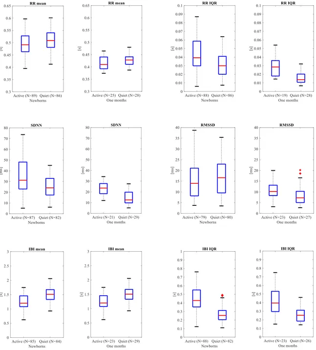
Dominio del tempo e dominio delle frequenze: i risultati confermano un aumento di HR dalla nascita ad un mese di età come riportato da vari autori. I parametri estratti dalla serie RR e dal segnale respiratorio mostrano differenze significative per l’analisi state-related, ad eccezione del parametro RMSSD. Le differenze nella variabilità battito-battito possono essere estratte per mezzo dell’analisi in frequenza, il contributo delle alte frequenze aumenta in modo significativo comparando AS contro QS. Questa considerazioni sono valide per entrambe le popolazioni presentate in questa tesi. Riguardo l’analisi age-related, solo i parametri nel dominio del tempo relativi ad HR mostrano una evoluzione relativamente all’età.
Sample Entropy e Quadratic Sample Entropy: è stato possibile osservare in studi precedenti che l’entropia è maggiore in QS rispetto che in AS. I risultati ottenuti in questo lavoro confermano un aumento di entropia in QS per entrambi gli stimatori. Queste considerazioni sono consistenti indipendentemente la dimensione di embedding (m), in questo contesto m=1, 2, 3. L’analisi age-related non mostra differenze nel confronto fra le due età.
Transfer Entropy: in questa tesi, TE è stata utilizzata per la prima volta per caratterizzare il cambiamento in termini di flusso di informazione, per entrambi i tipi
SOMMARIO
XVII
di analisi: state-related e age-related (paper sottomesso, Entropy Journal 2017). In AS non è evidenziabile nessuna differenza di flusso di informazione comparando le direzionalità RR→RESP e RESP→RR. Al contrario in QS, un aumento netto di informazione è rintracciabile in direzione RESP→RR rispetto alla direzionalità opposta. Le precedenti considerazioni sono valide per entrambe le età.
Riguardo l’analisi age-related, l’evoluzione in termini di flusso di informazione è rintracciabile in QS per entrambe le direzioni (RR→RESP e RESP→RR), al contrario AS non mostra nessuna evoluzione in termini di trasferimento di informazione fra i sottosistemi, confrontando neonati e infanti di un mese d’età. L’analisi state-related dell’interazione cardiorespiratoria per mezzo dell’analisi delle fasi del sistema cardiaco e respiratorio ha evidenziato interessanti peculiarità. È evidente un significativo aumento della sincronizzazione confrontando QS ed AS (poster session Dynamics Days, January 2017; conference paper EMBEC, June 2017).
Sono state individuate differenze in termini di direzionalità relativamente agli stati del sonno, i risultati supportano le analisi di TE sopra riportate. Le presenti conclusioni sono in contrasto con la precedente letteratura in cui è riportata una assenza di cambio di direzionalità nel confronto AS contro QS.
Locking di fase: l’analisi state-related mostra un aumento di sincronizzazione significativo confrontando AS contro QS. L’aumento è calcolato in termini di percentuale di sincronizzazione (la durata di sincronizzazione percentuale rispetto alla durata del segmento analizzato) e di durata media di sincronizzazione (la durata media degli episodi di sincronizzazione durante il segmento analizzato). L’indagine age-related mostra un aumento di sincronizzazione in termini di percentuale, mentre la durata media della sincronizzazione resta invariata, questi risultati sono relativi solamente a QS.
Indice di direzionalità: l’analisi state-related ha evidenziato un’assenza di direzionalità chiara in AS ed una predominanza di direzionalità RESP→RR in QS. I presenti risultati supportano le considerazioni ottenute per mezzo di TE, è da evidenziare però la differente modalità di indagine, basata sulla fase del segnale
SOMMARIO
XVIII
cardiaco e respiratorio. L’analisi age-related non mostra alcuna evoluzione nel confronto fra neonati ed infanti di un mese di età. Un’ulteriore analisi indaga la direzionalità dell’interazione quando la suddivisione delle popolazioni è basata sulla frequenza respiratoria indipendentemente dallo stato del sonno. È evidente una direzionalità predominante (RESP→RR) per frequenze respiratorie < 0.6 Hz, mentre non è individuabile una direzionalità chiara per frequenze respiratorie ≥ 0.6 Hz.
Discussioni
I risultati presentati nelle sezioni precedenti hanno contribuito alla descrizione dell’interazione cardiorespiratoria attraverso metodi non invasivi di analisi del segnale.
Il tentativo di caratterizzare la relazione reciproca fra il sistema cardiaco e il sistema respiratorio ha confermato gli studi precedenti e ha fornito interessanti nuovi risultati.
I parametri nel dominio del tempo hanno evidenziato differenze fra gli stati del sonno, con un aumento di RR medio in QS rispetto ad AS esclusivamente nei neonati e una decrescita della variabilità cardiaca (Heart Rate Variability, HRV) sia nel caso dei neonati che negli infanti.
Considerando il confronto tra gli stati del sonno, i parametri nel dominio del tempo indicano una maggiore variabilità in AS (SDNN), mentre non vi sono differenze nella variabilità battito-battito (RMSSD) alla nascita, ad un mese di età è evidenziabile una lieve e significativa decrescita in QS rispetto ad AS.
In conclusione, la maggioranza dei parametri nel dominio del tempo mostra una adeguata capacità nell’individuare differenze in relazione agli stati del sonno.
Riguardo il confronto fra neonati ed infanti di un mese di età, solo i parametri nel dominio del tempo relativi all’analisi delle serie RR mostrano differenze significative, mentre i parametri estratti dall’analisi del segnale respiratorio mostrano una decrescita non significativa con l’età.
La complessità del segnale HR è stata analizzata per mezzo dell’entropia univariata: Sample Entropy (SampEn) e Quadratic Sample Entropy (QSE).
Un aumento di entropia in QS rispetto ad AS è consistente indipendentemente dalla dimensione di embedding, questo risultato conferma la predominanza del controllo
SOMMARIO
XIX
simpatico in QS. Un’ulteriore conferma a quest’ultima considerazione è stata proposta in studi precedenti, i quali interpretano una semplificazione della variabilità cardiaca e quindi una diminuzione della stima di entropia, in conseguenza di una diminuzione dell’attività parasimpatica ed attivazione del sistema simpatico.
TE è uno stimatore di entropia bivariato, il quale può essere impiegato per stimare il flusso di informazione fra due serie temporali.
I risultati riportati in questa tesi mostrano valori di TE maggiori in QS rispetto ad AS: QS può essere interpretato come una condizione in cui l’accoppiamento cardiorespiratorio è maggiormente evidente e la mutua influenza dei segnali risulta evidente. Inoltre, la principale direzione di interazione è RESP→RR in QS, mentre in AS non è rintracciabile una direzione preferenziale. Data le più lente e regolari frequenze respiratorie associate a QS, è possibile ipotizzare una più stabile relazione fra respiro e segnale cardiaco. L’analisi della popolazione degli infanti di un mese d’età mostra risultati analoghi rispetto a quanto emerso dall’analisi dei neonati.
L’analisi age-related mostra una evoluzione in termine di flusso di informazione in QS per entrambe le direzionalità (RESP→RR, RR → RESP). AS risulta invece uno stato di minore accoppiamento cardiorespiratorio e la stima di TE non varia in modo significativo con l’età
I risultati ottenuti per mezzo dell’analisi di TE fornisco una descrizione innovativa ed una quantificazione del controllo cardiorespiratorio e della regolazione autonomica negli infanti.
L’analisi della sincronizzazione di fase permette la quantificazione dell’interazione cardiorespiratoria per mezzo dell’analisi della fase degli oscillatori cardiaco e respiratorio.
Il confronto fra AS e QS nei neonati mostra un incremento di sincronizzazione significativo in QS in accordo con studi precedenti i quali riportano gli stati del sonno come un aspetto fondamentale per la regolazione della sincronizzazione cardiorespiratoria, la quale è più frequente in QS.
La medesima analisi considerando gli infanti di un mese d’età evidenzia trend di aumento di sincronizzazione analoghi a quanto riportato nei neonati.
SOMMARIO
XX
Nell’analisi age-related, un aumento di sincronizzazione è rintracciabile solo in QS. Il seguente risultato è coerente con l’analisi dei parametri del dominio del tempo e di TE, i quali riportano una evoluzione relativa all’età solo in QS.
L’analisi dell’indice di direzionalità (DI) permette una quantificazione della relazione di causalità fra HR e respiro, in modo simile alla stima di TE.
QS è caratterizzato da una prevalenza di direzionalità RESP→RR, mentre AS non mostra nessuna direzionalità preferenziale nell’interazione fra i due sistemi. Questi risultati sono simili all’analisi di TE: il respiro appare il principale driver dell’interazione in QS. Frequenze respiratorie più elevate ed associate ad AS non risultano in grado di modulare HR e la direzionalità conseguentemente si sposta verso la direzionalità opposta (RR→RESP). Risultati analoghi sono ottenuti considerando le due popolazioni: neonati e infanti di un mese d’età.
Gli indici tradizionali ed innovativi calcolati in questa tesi, hanno dimostrato la loro capacità di discriminare fra i differenti stati del sonno e descrivere l’interazione cardiorespiratoria attraverso una nuova prospettiva.
Alcune fra le più recenti teorie riguardo SIDS attribuiscono alla mancanza di coordinazione cardiorespiratoria un ruolo fondamentale nell’aumento del rischio associato alla patologia. È stato ipotizzato che gli stati del sonno (AS e QS) siano due condizioni che differiscono in termini di sincronizzazione cardiorespiratoria e la continua alternanza fra stati di maggiore e minore accoppiamento sia in grado di stressare il sistema nervoso autonomo al fine di renderlo capace di fronteggiare le numerose sfide cui è sottoposto.
I risultati ottenuti in questa tesi possono essere interpretati in accordo con l’ipotesi del sistema nervoso autonomo come capace di stressare se stesso. I soggetti analizzati in questa tesi erano distesi supini, e hanno presentato epoche di AS e di QS con una prevalenza di AS nei neonati. AS e QS sono stati descritti in questa indagine come due stati del sonno che differiscono in termini di sincronizzazione e direzionalità.
L’alternanza di questa due stati del sonno costituisce un fattore di stress positivo per il sistema nervoso autonomo, capace di stimolarne lo sviluppo.
SOMMARIO
XXI
Nonostante gli interessanti risultati, molte domande richiedono una futura approfondita indagine.
Al fine di studiare in profondità lo sviluppo del sistema nervoso autonomo in termini di sincronizzazione e direzionalità, sarebbe opportuno seguirne la crescita in funzione del tempo. La situazione ideale è rappresentata da una indagine dalla nascita fino all’anno di vita al fine di caratterizzare l’evoluzione fisiologica della coordinazione cardiorespiratoria durante il periodo critico per l’occorrenza di SIDS.
In seguito alla caratterizzazione della risposta fisiologica, un’indagine su soggetti prematuri potrebbe evidenziarne differenze nello sviluppo autonomico rispetto ai soggetti a termine.
Infine, l’analisi delle differenze nella coordinazione cardiorespiratoria in soggetti deceduti a causa di SIDS potrebbe aprire nuovi orizzonti nel monitoraggio e nella cura neonatale per mezzo di un’indagine non invasiva ed affidabile del sistema nervoso autonomo.
XXII
Summary
Introduction and aim of the work
Sudden Infants Death Syndrome (SIDS) is characterized by the sudden death of a healthy infant during a sleep period. It is one of the leading causes of infant mortality in developed countries. Despite the dramatic decline in the incidence of SIDS since 1990 following worldwide education programs, SIDS remains the major cause of death in infants of 1–12 months of age. Because of its dramatic impact, this syndrome has been studied for long, but the underlying physiologic mechanisms have not been cleared yet.
At the moment, the most supported explanation in literature regarding SIDS, is the Triple-risk model, proposed by of Filiano and Kinney.
Filiano and Kinney’s SIDS hypothesis is based on the concurrence of three factors: 1) a vulnerable infant, 2) a critical developmental period, 3) an exogenous stressor.
Infants are likely to die of SIDS if they possess all three factors: the already congenital infants’ vulnerability lies latent until they enter the crucial period and are subject to an exogenous stressor.
Cardiorespiratory control development can be classified as risk factor according to Triple-risk model. In particular, it can be thought as a subclass of the so-called homeostatic control.
In light of this issue, the present study proposes to analyze a population of healthy full-term newborns and one month infants with a deep focus on cardiorespiratory interaction, its relationship with sleep state and its evolution from birth to one month of age. The aim of the investigation is the description of physiological cardiorespiratory coupling, within the future prospective of highlighting differences with respect to cardiorespiratory coupling of SIDS victims.
SUMMARY
XXIII
The investigation of this interaction is performed on subjects during sleep. Sleep plays a fundamental role in neurodevelopment and homeostatic regulation.
Sleep in newborns and infants can be classified in three type: Quiet Sleep (QS) (NREM equivalent), Active Sleep (AS) (REM equivalent) and Indeterminate Sleep (IS).
It has been hypothesized that infants who were to die of SIDS would show abnormalities of sleep state distribution prior to their deaths.
The mechanism underlying SIDS appears to have origins in the fetal environment resulting in neural damage and ANS development which later compromises responses to breathing or blood pressure challenges during sleep. The deficits appear to be related to alterations in neurotransmitter receptors within regions involved in chemoreception and cardiovascular and cardiorespiratory control.
It is crucial to stress the fact that cardiorespiratory coupling can provide a noninvasive insight of the mechanism underlying the interaction between the cardiac and respiratory system and help understanding the factors contributing to SIDS occurrence.
The study reported in this thesis was possible thanks to collaboration of the outstanding Columbia University Medical Center (CUMC) and Politecnico di Milano, DEIB Department.
The thesis investigation has been conducted at Politecnico di Milano and at CUMC during my 6-months staying in New York. The encounter between the biomedical engineering contribution and the clinical expertise has allowed proposing new solutions for a quantification of cardiorespiratory interaction and validation of obtained results.
Materials and Methods
The newborn dataset includes 151 infants born at the Morgan Stanley Children’s Hospital of New York at CUMC between 38 and 40 weeks of gestation and the one month infants cohort includes 33 subjects who came back for a one month follow up, based on the same criterion about Gestational Age (GA). None of the infants were admitted to the Neonatal Intensive Care Unit (NICU) nor had any major illness or known genetic disorder. Among the various recorded signal, in this work ECG and respiratory signal were analyzed. Sleep states were coded based on respiration by expert clinicians.
SUMMARY
XXIV
During the 10-minutes baseline acquisition, babies were sleeping in supine position within ~ 30 minutes following feeding. Segments of 3-minute length in a continuous sleep state were analyzed: 514 three-minute epochs were considered for newborns (239 quiet, 275 active), while 247 epochs were considered for one month infants (144 quiet, 103 active).
The R peaks were detected on the ECG with the Pan-Tompkins algorithm. An adaptive filter was then applied to remove ectopic beats or artifacts.
The respiration signal was filtered with a bandpass filter (0.05 - 3.5 Hz). Peaks of inspiration were detected with automated marking software and each record was corrected for incorrect marks manually.
The analysis of cardiorespiratory interaction during sleep is performed with a univariate and bivariate approach by means of linear and nonlinear methods. The aim of this investigation is to characterize this interaction depending on sleep state and track its age-dependent evolution.
The first performed investigation was the extraction of time domain univariate parameters, computed from RR series and respiration. There is a lack in guidelines for the application of these methods on neonates. As a matter of fact, indications given for adults are not applicable, since the average heart rate (HR) of neonate is almost double adult’s one and it has peculiar characteristics. For this reason, time domain analysis takes adapted parameters from the adult approach.
As indicated in HRV Task Force, HR parameters employed in the analysis were RR mean, RR IQR, SDNN and RMSSD, computed on 3-minute length segments. On the other hand, computed respiratory parameters were Inter Breath Interval (IBI) mean and IBI IQR. Frequency domain analysis were performed considering three different bands specifically chosen for this population of infants: Very Low Frequency (VLF), 0.01-0.04 Hz, Low Frequency (LF), 0.04-0.2 Hz, and High Frequency (HF), 0.35-1.5 Hz.
Regarding nonlinear investigation of cardiorespiratory coordination, univariate and bivariate entropy estimates were computed.
Entropy estimators have already shown their capability in discriminating physiological signals based on complexity measurements. In this work, classical entropy
SUMMARY
XXV
parameters have been computed along with new indexes capable of describing directionality of interaction between subsystems within the information theory framework.
Sample Entropy (SampEn) and Quadratic Sample Entropy (QSE) are univariate entropy estimators based on RR series analysis. They can be seen as the evolution of Approximate Entropy (ApEn) by Pincus. Both SampEn and QSE have been reported to show a slight bias dependent on the length of the analyzed signal, due to this reason a fixed number of beats for each segment have been considered in this analysis.
The novel approach proposed in this work is the application of Transfer Entropy (TE) to investigate cardiorespiratory coordination. TE estimates the directionality of the information transfer between HR signal and respiratory signal without any assumption about the underlying nature of the interaction, in this way it is capable of catching both linear and nonlinear contributions. The estimation of TE deals with the computation of probability density function of both signals and the joint probability density function capable of describing the interrelationship between RR series and respiration.
TE is a measure of predictability and complexity. In this analysis when considering the direction 1→2, TE quantifies the improvement in predicting the future of signal 2 when the prediction takes into consideration not only the past of signal 2 but also information from the past of signal 1.
In order to provide a different prospective of cardiorespiratory quantification the last part of this work focuses on phase analysis of RR series and respiratory signal.
The last estimators presented in this work are phase locking quantification and directionality index (DI).
They both are nonlinear bivariate method to quantify synchronization and directionality respectively, by means of the analysis of systems’ phase rather than a traditional amplitude analysis.
This assumption supports the idea that amplitude of (two or more) oscillators may remain uncorrelated whereas their phases do mutually perturb.
Synchronization has been primarily investigated by means of synchrogram, a visual tool displaying the relative distances between R peaks and respiratory onsets. In order to
SUMMARY
XXVI
quantify the presence or absence of the interaction and the strength of the coupling between the systems under analysis, the synchronization index 𝜆 has been computed.
Even when considering phase-locking analysis, synchronization does not explain how systems interact and mutually perturb themselves. Directionality index is capable of estimating a casual interrelationship between the HR and respiration, looking at the evolution of phase of the subsystems.
In this work the Evolution Map Approach (EMA) algorithm has been used. This method deals with mutual predictability similarly to Granger causality. Considering system 1 interacting with system 2, if system 1 affects system 2, the future of system 2 can be better predicted taking into account the past samples of both system with respect to information of system 1 only.
Results
In this work, state-related and age-related analyses have been performed. The former case regards the comparison of a parameter in AS versus QS when a specific age is considered (newborns or one month infants), in the latter case a parameter in a specific state (AS or QS) is compared at two different time points.
The statistical analysis has been performed employing IQR outlier rejection criterion. Differences between groups have been tested by means of an unpaired T-test in case of Gaussian distributed populations; on the contrary the non-parametric Wilcoxon signed-rank test has been performed if the hypothesis of Gaussian-like distribution was not verified.
Results obtained in this thesis show that novel parameters are capable of improving the knowledge about cardiorespiratory synchronization and ANS regulation during sleep.
It is important to stress that they need to be employed in combination with traditional time domain, frequency domain and classical entropy estimators to achieve a complete description of the interaction between the cardiac and respiratory systems.
Time domain and frequency domain: results confirmed the HR increase from birth to one month of age as reported by many authors. Time domain parameters extracted from RR series and respiratory signal showed significant differences when
SUMMARY
XXVII
performing state-related analysis, with the exception of RMSSD. Beat-to-beat variability differences can be instead extracted by means of frequency analysis, high frequencies contribution is statistically increasing when comparing AS versus QS. These considerations are valid for both newborns and one month infants populations. Regarding age-related analysis, only time domain parameters extracted from HR showed an evolution with age when the two cohorts are compared.
Sample entropy and Quadratic Sample Entropy: it has been observed by previous studies that entropy is higher in QS than in AS. Results obtained in this work, confirmed an entropy increase in QS with respect to AS for both SampEn and QSE. This finding is consistent regardless the employed embedding dimension (m), in this work m=1, 2, 3. Age-related analysis showed no differences when comparing newborns and one month infants.
Transfer Entropy: in this work, TE analysis has been applied for the first time to characterize the information flow changes when performing state-related and age-related analyses (submitted paper, Entropy Journal 2017).
In AS no clear differences in information flow has been found when comparing RR→RESP and RESP→RR directionalities. In QS instead, a net TE increase is found for RESP→RR with respect to the opposite directionality. This result is valid both for newborns and one month infants.
Regarding the age-related analysis, the evolution of information flow is only seen in QS for both RR→RESP and RESP→RR directionalities, on the other hand AS did not show any evolution in term of information transfer between subsystems when comparing newborns versus one month infants.
The state-related investigation of cardiorespiratory interaction by means of phase analysis highlighted interesting peculiarities. A net increase in synchronization in QS with respect to AS is seen (poster session Dynamics Days, January 2017; conference paper EMBEC, June 2017).
SUMMARY
XXVIII
Differences in directionality related to sleep states have been found, supporting this work results about TE. The obtained finding is in contrast with previous works reporting the absence of a clear change in directionality when comparing AS versus QS.
Phase locking: state-related analysis showed a statistically significant increase in synchronization comparing AS versus QS. The increase is computed in term of percentage of synchronization (the percentage duration of synchronization with respect to length of the analyzed record) and mean duration in seconds (the mean duration of various synchronization episodes during the analyzed segment). Age-related investigation showed an increase in synchronization in term of percentage while the average duration stayed the same, in QS only.
Directionality Index: state-related analysis highlighted the absence of a clear directionality in AS and a predominance of RESP→RR directionality in QS. This result support findings of TE analysis, within a different investigation procedure based on oscillators’ phase. Age-related analysis shows no trend of evolution when comparing newborns and one month infants. A further analysis investigates the directionality of interaction when population grouping is performed based on breathing frequency rate regardless the sleep state. A predominant directionality (RESP→RR) is clearly seen for breathing frequency < 0.6 Hz, while no clear directionality is present when considering breathing frequency ≥ 0.6 Hz.
Discussions
The thesis results presented in the previous section have helped describing in details the cardiorespiratory interaction by means of noninvasive methods of signal processing.
The effort in characterizing the interrelationship between the cardiac and respiratory system confirmed previous studies and provided interesting new findings.
Time domain parameters from this data analysis enhance differences between sleep states, with an increase of RR mean in QS with respect to AS in newborns only, and a decrease of Heart Rate Variability (HRV) in both newborns and one month infants cohorts.
SUMMARY
XXIX
Considering the comparison between sleep states, time domain parameters indicate an increase in variability in AS (SDNN), while no difference was found in beat-to-beat variability (RMSSD) at birth and a slightly significant decrease in QS with respect to AS at one month of age.
Concluding, almost all the time domain parameters when performing state-related analysis have proved their ability to find significant differences between sleep states.
Regarding the comparison between newborns and one month infants within the same sleep state, only time domain parameters computed on RR series show significant differences while parameters extracted from respiratory signal show a non-significant decreasing trend with age.
The complexity of HR signal has been investigated by means of Univariate Entropy: Sample Entropy (SampEn) and Quadratic Sample Entropy (QSE).
An entropy increase in QS with respect to AS has been found for both SampEn and QSE despite the embedding dimension, confirming the predominance of parasympathetic control in QS. As a matter of fact, even previous studies have proposed that a simplification of HR dynamics and thus a lowering in entropy values, might follow a parasympathetic withdrawal and sympathetic activation.
Transfer entropy is a bivariate entropy estimator, which can be employed to assess the information flow between two time series.
Results reported in this work showed that TE is higher in QS than AS: QS can be seen as condition in which the cardiorespiratory coupling is more evident and the influence of RR over respiration and vice versa is noticeable. Moreover, the main direction of the information flow is RESP→RR in QS, while in AS no clear directionality is recognizable. Given that breathing in QS is slower and more regular, it potentially allows a more stable effect/relationship with HR.
The analysis performed on one month cohort shows analogous results of the ones obtained considering newborns.
An age dependent evolution in terms of information flow happens only in QS for both RR→RESP and RESP→RR directions. AS is per se a state of lower coupling between HR and respiration, changes in TE are not dramatically affected by age.
SUMMARY
XXX
These findings provide an innovative description and quantification of cardiorespiratory control and ANS regulation in infants, based on information theory.
Phase synchronization analysis allows quantifying the cardiorespiratory interaction between the cardiac and respiratory systems by means of oscillators’ phase analysis.
The comparison can be performed considering AS versus QS within the newborn cohort. A significant increase in synchronization in QS is noticeable and in agreement with previous studies, which established that sleep state is a relevant aspect for cardio-respiratory synchronization, which occurs more frequently in QS.
When one month population is analyzed, comparable trend of increase in synchronization are found.
When age-related analysis is performed, an increase in synchronization is found in QS only. This result is coherent with what previously found in time domain parameters and TE reporting an evolution with age in QS only.
Directionality index (DI) analysis performs a quantification of the causal directionality between HR and respiration similarly to Transfer Entropy.
QS is characterized by a prevalence of RESP→RR directionality while AS exhibit no clear directionality of interaction. These results are similar with what found in TE analysis: respiration appears to be the main driver in QS. Higher breathing frequencies associated with AS are no more capable of modulating the HR and the directionality shifts on the opposite direction, RR→RESP. The analogous analysis has been performed on one month infants: results are similar with what found for newborns.
The traditional and novel indexes have proven capability to discriminate between sleep states and to describe the cardiovascular interaction with a new insight.
Some of the most recent theories about SIDS started attributing the lack of cardiorespiratory control as the main driver of risk for SIDS. It has been hypothesized that AS and QS are two conditions that differs in term of cardiorespiratory synchronization and the continuous alternation between lower and higher coupling condition is capable of stressing the ANS, making it more ready to face several challenges.
SUMMARY
XXXI
It has been also reported that the state change from AS to QS increases the occurrence of sighs and gaps, two mechanisms those are crucial to overcome cardiorespiratory challenges and capable of driving the autoresuscitation mechanism.
The results obtained in this work could be interpreted with this hypothesis of ANS being the stressor of itself. The healthy and full-term subjects analyzed were lying supine and their sleep was characterized by both AS and QS with a prevalence of AS for newborns. AS and QS have been reported by this work as two different sleep states with different characteristics in term of synchronization and also directionality.
The alternation between these two sleep states constitutes a constant positive stressing condition that is probably capable of stimulating the ANS development.
Despite the interesting results many questions are still to be addressed.
In order to deeper investigate the ANS evolution in term of both synchronization and directionality, more time points are needed. The ideal situation would be tracking the ANS development from birth to one year of age to fully characterize the physiological behavior during the critical risk period for SIDS.
Once the quantification of interaction is established, an investigation on early and late preterm could help discovery the difference in term of ANS development on subjects born preterm.
The analysis on SIDS patients could highlight differences in coupling and synchronization and its quantification could open novel view about newborn state monitoring and care path by means of noninvasive and reliable investigations of ANS.
1
Chapter 1
In this chapter, Sudden Infant Death Syndrome (SIDS) will be defined and its epidemiology will be presented.
SIDS is characterized by the sudden death of a healthy infant during a sleep period. It is considered a multifactorial condition that associates inadequate cardiac, breathing, autonomic and/or arousal controls. The already immature control mechanisms can be aggravated by infections as well as by prenatal and postnatal life environmental factors.
These risk factors for SIDS were identified by epidemiological studies.
Afterwards, the fetal and newborn cardiovascular system evolution will be described, focusing on the Autonomic Nervous System (ANS) control.
Lastly, an overview of Cardiorespiratory Coupling (CRC) and sleep in infants is offered. Some of the most recent hypothesis about causes leading to SIDS are explained in term of lack of cardiorespiratory synchronization.
This work focuses on noninvasive signal processing techniques capable of assessing the physiological ANS coordination in a population of healthy newborns and one month infants.
CHAPTER 1. INTRODUCTION
2
1
Introduction
1.1 Sudden Infant Death Syndrome
Sudden Infant Death Syndrome (SIDS) is one of the leading causes of infant mortality in the developed countries and accounts for nearly 30% of deaths during the post neonatal period. Nonetheless, the physiologic mechanisms that underlie this syndrome are still unclear [1].
The first definition of SIDS was given by Bergman in 1970, who argued that SIDS did not depend on a “single characteristic that ordains an infant for death”, but on an interaction of risk factors with variable probabilities [2].
Later, Wedgwoog, Raring, Rognum and Saugstad developed the first “triple risk hypothesis” [2] which was succeeded by the most famous Triple Risk model by Filiano and Kinney. This model is based on invasive pathological studies of brainstem from SIDS victims.
Kinney at al. state that “many cases result from defects in brainstem-mediated protective responses to homeostatic stressors occurring during sleep in a critical developmental period” [2], [3], [4].
Given these hypothesis Kinney and Thach emphasize that SIDS occurs “only in infants with underlying abnormality”. The National Institute of Child Health and Development SIDS strategic plan 2000 quoting Kinney’s work, stated unequivocally that “SIDS is a developmental disorder. Its origins are during fetal development.” and later in 2001 “Knowledge acquired during the past decade supports the general hypothesis that infants who die from SIDS have abnormalities at birth that render them vulnerable to potentially life-threatening challenges during infancy” [4].
CHAPTER 1. INTRODUCTION
3
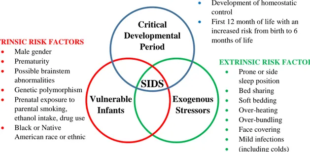
Filiano and Kinney’s SIDS hypothesis was based on the concurrence of three factors: 1) a vulnerable infant,
2) a critical developmental period for the homeostatic control with a peak at 2 - 4 postnatal months
3) an exogenous stressor, as summarized in Figure 1.1.1 [5].
Infants are likely to die of SIDS if they possess all three factors: the infants’ vulnerability lies latent until they enter the crucial period and the infants are subject to an exogenous stressor [2].
During the first year of life, rapid changes in the maturation of cardiorespiratory control and in cycling between sleeping and waking occur, first as the fetus transitions to extrauterine life and subsequently as the infants adjust to postnatal life.
Regarding the investigation of SIDS risk evolution with age, it appears crucial to investigate changes from birth to one year of age [6].
In the following sections two main intrinsic risk stressors will be investigated: Prematurity and Sleep Position.
Critical Developmental Period
SIDS
Vulnerable Infants Exogenous StressorsINTRINSIC RISK FACTORS
Male gender Prematurity Possible brainstem abnormalities Genetic polymorphism Prenatal exposure to parental smoking, ethanol intake, drug use
Black or Native American race or ethnic group
EXTRINSIC RISK FACTORS
Prone or side sleep position Bed sharing Soft bedding Over-heating Over-bundling Face covering Mild infections (including colds) Development of homeostatic control
First 12 month of life with an increased risk from birth to 6 months of life
CHAPTER 1. INTRODUCTION
4
1.2 Intrinsic risk: Prematurity
Any infant born before the 37th weeks of Gestational Age (GA) is defined as premature. Premature infant can be further divided into extremely (GA ≤ 26), early (26 < GA ≤ 34) and late preterm (34 < GA ≤ 36).
Reducing preterm birth is a national public health priority. Preterm birth rates decreased from 2007 to 2014 after about three decades of continuous increasing, from early 1980s through 2006. Despite this success, the preterm birth rate rose slightly in 2015 (Figure 1.2.1) and about 1 out of 10 babies (10%) was born premature in the United States. Additionally, racial and ethnic differences in preterm birth rates remain [6], [7], [8].
Prematurity is reported as intrinsic risk factor for SIDS in the Triple-risk model [9]. It should be reminded that preterm delivery is not a disease per se. Rather, preterm delivery raises the risk of adverse outcomes that would be present in normal delivery as well.
Prematurity sets the newborn to be exposed to the outer environment before his autonomic nervous system is fully developed and able to effectively face life challenging
Figure 1.2.1 Preterm birth rates with respect to the total life birth in each state in 2015.
The USA score (considering the whole country) is C, with a 9.6 rate
Source: Grades determined by March of Dimes based on preterm birth rates from National Center for Health Statistics, 2015 final natality data
CHAPTER 1. INTRODUCTION
5
situations like breathing and thermoregulation. A preterm baby is understandably more vulnerable than a full-term baby.
The immaturity of ANS has been hypothesized to be mainly reflected in cardiorespiratory coordination and consequently be responsible for SIDS episodes [10].
The lack of cardiorespiratory synchronization that represents per se a risk factor for SIDS, is mainly reflected in the frequent occurrence of apneic events. Apnea of prematurity is the most common disorder affecting infants born prematurely and the incidence and severity of apnea are also inversely related to GA.
Prolonged apneas in adults are generally resolved by arousal; however, in preterm infants this is not commonly found. Apnea of prematurity may not be inherently life threatening, but a deficient arousal response to the consequent asphyxia or hypoxia, could have fatal consequences. Abnormal arousal responsiveness to hypoxia and hypercarbia has been observed in infants with apnea of infancy, along with diminished ventilatory responsiveness [11], [12].
Preterm infants can experience a variety of cardiovascular disorders, ranging from major morphological defects to dysfunctional auto-regulation of blood vessels. Hypotension is a frequent concern in preterm infants, but there is no consensus as to what the blood pressure readings should be in preterm infants with gestational ages of less than 26 or 27 weeks.
Apnea and bradycardia are common in premature infants and are manifestations of immature cardiorespiratory control.
The non-physiological operation of these mechanisms exposes preterm infants to an increased risk of life-threating events, which cannot be successfully resolved because the absence of mature cardiorespiratory coordination. The lack of responsiveness between cardiac and respiratory systems is at base of many models trying to depict the SIDS manifestation.
CHAPTER 1. INTRODUCTION
6
Figure 1.3.1 Trend of SIDS rate, the diminishing trend can be observed as a consequence of the Back to Sleep
Campaign. The green line indicates the percentage of babies sleeping supine with respect to the total percentage of premature infants in the USA
SIDS Rate Source: CDC, National Center for Health Statistics Sleep Position Data: NICHD, National Infant Sleep Position Study
1.3 Extrinsic risk: Sleep Position
Prone position is thought to be one of the major risks for developing SIDS for a newborn. An association between prone sleep and an increased risk of SIDS was reported since the 1950s [1], [5].
The incidence of SIDS is highly reduced since the “Back to Sleep” campaign in 1994, initiated by the National Institute of Child Health and Development in the United States. In June 1992, the American Academy of Pediatrics (AAP) Task Force on Infant Positioning and SIDS published a recommendation that healthy full-term infants be placed laterally or supine to sleep. The SIDS rate in the United states declined by > 50 % in the 10 years after the initiation of the campaign (Figure 1.3.1) [13].