T
his article describes a biomechanical approach for treating an impacted, dilacerated maxillary central incisor in the mixed dentition. A new fixed appliance promotes proper sagittal and vertical traction of the incisor into the dental arch, while maintaining the patient’s periodontal health and preserving root length.The appliance is an .040" stainless steel fixed lingual arch, soldered to the lingual surfaces of the maxillary first molar bands (Fig. 1). A vertical arm of the same wire type, ending in a helix, is soldered to the arch at the center of the incisor space. Four metal occlusal rests are soldered to the lingual arch: two on either side of the impacted incisor and two at the first deciduous molars. These rests are bonded to the teeth with light-cured composite. Case Report
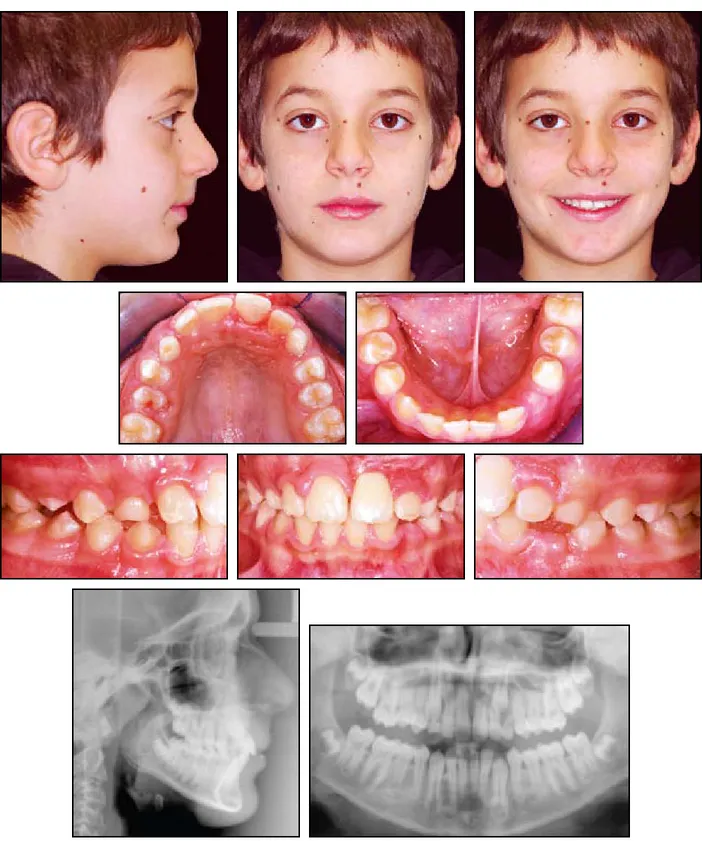
Use of the appliance is demonstrated in a 9-year-old male in the early mixed dentition who presented with the complaint of an unerupted maxillary left central incisor (Fig. 2). The boy’s parents indicated that the patient had lost the deciduous central incisor in a traumatic incident
Dilacerated Maxillary Central Incisors
ALDO GIANCOTTI, DDS PAOLA MOZZICATO, DDS FRANCESCO GERMANO, DDS
Fig. 1 Fixed lingual arch for traction of unerupted, dilacerated maxillary central incisor.
Dr. Germano Dr. Mozzicato
Dr. Giancotti is an Assistant Professor and Drs. Mozzicato and Germano are post-graduate residents, Department of Ortho-dontics, University of Rome “Tor Vergata”. Contact Dr. Giancotti at Viale Gorizia 24/c, Rome 00198, Italy; [email protected].
Fig. 2 9-year-old male patient showing dilacerated maxillary left central incisor, horizontally positioned with tip of crown near apex of right central incisor.
between age 2 and 3.
Clinical examination showed a Class I molar relationship, normal overjet and overbite, a midline deviation, and an unerupted maxillary left central incisor, which was not palpable either palatally or labially. Radiographs indicated a skeletal Class I malocclusion and a normal vertical growth pattern. A dilacerated maxillary left central incisor was positioned horizontally, with the tip of the crown near the apex of the right central incisor. The angle of dilaceration was about 60°; the incisor was rotated counterclockwise in relation to its long axis, with the crown in the usual apical area and the apex in the crown position.
The treatment plan involved extrusion of the dilacerated tooth with all its supporting tissue (alveolar bone and attached gingiva). A multidis-ciplinary approach involving both surgical and orthodontic treatment was implemented.
Procedure
Brackets were bonded to the three maxillary incisors, and space was opened for the misplaced left central incisor. After adequate space had been gained, a flap was raised to expose the surface of the dilacerated crown. A metal mesh attached to a specially fabricated metal chain (Fig. 3) was bonded directly to the exposed tooth surface. This thin metallic mesh can be easily adapted to the shape of the tooth, which minimizes irritation of the gingival tissues and promotes healing.
To simulate natural eruption, the orthodontic traction was first directed anteriorly, parallel to the occlusal plane, using an elastomeric ligature from the metal chain to the vertical arm of the lingual arch. A light force of about 50g was applied. This
anterior movement is designed to preserve the root length of the impacted incisor, avoiding undesir-able and potentially dangerous clockwise rotation of an already damaged root.
Tooth movement and soft-tissue status were monitored every three weeks (Fig. 4). Once the dilacerated tooth was visible clinically, a button was bonded to the facial surface of the crown, and orthodontic traction was redirected vertically by bending the vertical arm of the appliance down-ward (Fig. 5). At this point the anterior occlusal rests were removed from the traction appliance; space for the erupting central incisor was main-tained with an open-coil spring. Generally, the occlusal rests can be carefully removed from the lingual wire using a conventional handpiece, with-out removing the appliance.
The final alignment was completed with a standard twin bracket bonded to the dilacerated Fig. 4 Position of dilacerated incisor after six weeks of anterior traction.
Fig. 3 Thin metal mesh and specially fabricated chain for bonding to unerupted incisor.
Fig. 5 Button bonded to incisor, replacing metal mesh and chain; traction changed to vertical direction by bending vertical arm downward.
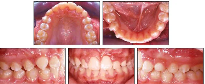
tooth and an .014" nickel titanium overlay wire, while the anterior teeth were stabilized with an .016" × .022" stainless steel archwire (Fig. 6). After 12 months of treatment, fixed appliances were removed and replaced with an upper Essix* retainer (Fig. 7).
Treatment Results
This treatment was successful in achieving a natural position for the maxillary central incisor, despite the root dilaceration. No adverse effects on the maxillary central incisor, including pulp pathology, color change, and mobility, were ob -served at post-treatment examinations. Radiograph-ically, the incisor showed proper root alignment and no apparent soft-tissue disease. The root of the dilacerated incisor continued to mature during the post-treatment phase. Although the hooked apex prevented a thorough evaluation of the apical third of the root, its development appeared normal.
Ten months after debonding, a second phase of orthodontic treatment was started using Invisalign Teen** appliances (Fig. 8). The patient was considered a good candidate for this technique because the permanent dentition had almost com-pletely erupted, and the space for the eruption of the upper left canine was adequate. The damaged front tooth suggested a less invasive approach with
Although the final evaluation of the impact-ed incisor’s gingival tissue was delayimpact-ed until the end of comprehensive treatment, positive changes can already be observed five months after starting the Invisalign Teen therapy. No periodontal treat-ment was carried out during the whole period, except for oral hygiene recommendations.
Discussion
The maxillary central incisors are the teeth most likely to be impacted, along with the third molars and maxillary canines.1,2 According to Andreasen, defective eruption of the upper central incisors can result from trauma to the primary incisor, presence of a supernumerary tooth, or obstruction of the eruption pathway.3 Other authors also believe dilaceration4 results from early trauma to the deciduous tooth5,6; Stewart attributes it in some cases to ectopic development of the tooth germ.6 Impaction is commonly associated with a severely dilacerated root, especially when the affected tooth is the maxillary incisor.
In adult patients, the most common therapeu-tic approach for a dilaceration is surgical extraction and replacement with a prosthetic implant. In the mixed dentition, the best solution is a multidisci-plinary treatment,6-8 including extraction when required by the severity of the tooth inversion. Some studies show that a dilacerated root with an obtuse angle, a lower position, and incomplete root formation has a better prognosis for treatment with orthodontic traction.5,9-13 Moreover, if a traumatic event occurred before root formation, and the dam-age did not involve the cementum and periodontal membrane, the root can be expected to mature normally.14
The ideal treatment for an impacted tooth directs eruption through the attached gingiva, rather than the alveolar mucosa.7,15,16 This general rule cannot be followed, however, when treating a deeply impacted tooth, because of the distance to Fig. 6 Bracket bonded to incisor for final
align-ment with overlay wire.
*Trademark of Raintree Essix, 6448 Parkland Drive, Sarasota, FL 34243; www.essix.com.
**Trademark of Align Technology, Inc., 881 Martin Ave., Santa Clara, CA 95050; www.aligntech.com.
the crest of the alveolus. In such a case, two surgi-cal steps are needed: the first to expose the impact-ed tooth and the second, at the end of the comprehensive phase of orthodontic treatment, to provide an adequate gingival attachment. An addi-tional concern in cases involving a dilacerated incisor is unfavorable tooth rotation, which can inhibit vertical traction. In this case, the tooth movement should occur in two stages, and precau-tions should be taken to avoid any traction that could damage the already compromised root.
The patient shown here was treated in the mixed dentition, when his root development was incomplete, using a light orthodontic force to allow normal root maturation.14 The results dem-onstrate that open eruption and orthodontic trac-tion using a two-step anchorage device can be an effective treatment for an impacted, dilacerated maxillary incisor.
REFERENCES
1. Chaushu, S.; Zilberman, Y.; and Becker, A.: Maxillary incisor impaction and its relationship to canine displacement, Am. J. Orthod. 124:144-150, 2003.
2. Chaushu, S.; Sharabi, S.; and Becker, A.: Dental morphologic characteristic of normal versus delayed developing dentitions with palatally displaced canines, Am. J. Orthod. 121:339-346, 2002.
3. Andreasen, J.O.; Petersen, J.K.; and Laskin, D.M.: Textbook
and Color Atlas of Tooth Impactions, Munksgaard International Publishers, Copenhagen, 1997, pp. 114-123.
4. Crawford, L.B.: Impacted maxillary central incisor in mixed dentition treatment, Am. J. Orthod. 112:1-7, 1997.
5. Smith, D.M. and Winter, G.B.: Root dilacerations of maxillary incisors, Br. Dent. J. 150:125-127, 1981.
6. Stewart, D.J.: Dilacerate unerupted maxillary central incisors, Br. Dent. J. 145:229-233, 1978.
7. Vermette, M.E.; Kokich, V.G.; and Kennedy, D.B.: Uncovering labially impacted teeth: Apically positioned flap and closed-eruption techniques, Angle Orthod. 65:23-32, 1995.
8. Maspero, C.; Farronato, D.; Alicino, C.; Santoro, G.; and Farronato, G.: Orthodontic surgical treatment on an upper cen-tral dilacerated incisor in an adult patient, Prog. Orthod. 8:314-321, 2007.
9. Kuvvetli, S.S.; Seymen, F.; and Gecay, K.: Management of an unerupted dilacerated maxillary central incisor: A case report, Dent. Traumatol. 23:257-261, 2007.
10. Bishara, S.E.: Treatment of unerupted incisors, Am. J. Orthod. 59:443-447, 1971.
11. De Echave-Krutwig, M. and Sánchez-Fernandez, L.: Impacted incisors with dilacerated roots, J. Clin. Orthod. 36:641-645, 2002.
12. Saldarriaga, J.R. and Patino, M.C.: Ectopic eruption and severe root resorption, Am. J. Orthod. 123:259-265, 2003.
13. Singh, G.P. and Sharma, V.P.: Eruption of an impacted maxil-lary central incisor with an unusual dilaceration, J. Clin. Orthod. 40:353-356, 2006.
14. Korbendau, J.M. and Patti, A.: Clinical Success in Surgical and
Orthodontic Treatment of Impacted Teeth, Quintessence, Chicago, 2006.
15. Vanarsdall, R.L. and Corn, H.: Soft-tissue management of labi-ally positioned unerupted teeth, Am. J. Orthod. 72:53-64, 1977.
16. Becker, A.; Brin, I.; Ben-Bassat, Y.; Zilberman, Y.; and Chaushu, S.: Periodontal status following surgical-orthodontic alignment of impacted maxillary incisors by closed-eruption technique, Am. J. Orthod. 122:9-14, 2002.