Review Article
High-Intensity Focused Ultrasound Circular Cyclocoagulation in
Glaucoma: A Step Forward for Cyclodestruction?
Rodolfo Mastropasqua,
1Vincenzo Fasanella,
2Alessandra Mastropasqua,
2Marco Ciancaglini,
3and Luca Agnifili
21Moorfields Eye Hospital, London EC1V 2PD, UK
2Ophthalmology Clinic, Department of Medicine and Ageing Science, University G. d’Annunzio of Chieti-Pescara, 66100 Chieti, Italy
3Ophthalmology Unit, Department of Life, Health and Environmental Sciences, University of L’Aquila, 67100 L’Aquila, Italy
Correspondence should be addressed to Luca Agnifili; l.agnifi[email protected]
Received 31 October 2016; Revised 28 January 2017; Accepted 20 February 2017; Published 23 April 2017 Academic Editor: Paul Harasymowycz
Copyright © 2017 Rodolfo Mastropasqua et al. This is an open access article distributed under the Creative Commons Attribution License, which permits unrestricted use, distribution, and reproduction in any medium, provided the original work is properly cited. The ciliary body ablation is still considered as a last resort treatment to reduce the intraocular pressure (IOP) in uncontrolled glaucoma. Several ablation techniques have been proposed over the years, all presenting a high rate of complications, nonselectivity for the target organ, and unpredictable dose-effect relationship. These drawbacks limited the application of cyclodestructive procedures almost exclusively to refractory glaucoma. High-intensity focused ultrasound (HIFU), proposed in
the early 1980s and later abandoned because of the complexity and side effects of the procedure, was recently reconsidered in
a new approach to destroy the ciliary body. Ultrasound circular cyclocoagulation (UC3), by using miniaturized transducers
embedded in a dedicated circular-shaped device, permits to selectively treat the ciliary body in a one-step, computer-assisted,
and non-operator-dependent procedure. UC3shows a high level of safety along with a predictable and sustained IOP reduction in
patients with refractory glaucoma. Because of this, the indication of UC3was recently extended also to naïve-to-surgery patients,
thus reconsidering the role and timing of ciliary body ablation in the surgical management of glaucoma. This article provides a
review of the most used cycloablative techniques with particular attention to UC3, summarizing the current knowledge about this
procedure and future possible developments.
1. Introduction
Lowering intraocular pressure (IOP) is the only proven approach to reduce the rate of retinal ganglion cell loss and the rate of progression in patients with glaucoma [1]. Never-theless, in many cases, medical and surgical approaches do not reach the required target IOP [2].
Refractory glaucoma comprises all forms of glaucoma in which the IOP remained uncontrolled despite maximum-tolerated medical therapy and previous laser or surgical pro-cedures. In this case, the IOP remains uncontrolled also after
repeated standard filtration surgeries. Surgical approaches
for refractory glaucoma include techniques increasing the aqueous humor (AH) outflow (filtrating procedures, drain-age devices) and techniques reducing the AH inflow, by destroying portions of the ciliary body [3].
Several cycloablative methods have been proposed over the years, such as cryotherapy, microwave heating, endoscopic laser coagulation, and transscleral diode laser photocoagula-tion, which remain the most used ablative procedure [4–7]. Given the occurrence of potential vision-threatening compli-cations, unpredictable dose-effect relationship, significant
variability in thefinal IOP lowering, and poor reproducibility,
the ciliary body ablation is still considered as a last resort treatment, recommended only in patients with refractory glaucoma [8].
The high-intensity focused ultrasound (HIFU) technology was proposed as a safer alternative of ciliary body destruction in the 1980s and 1990s (Therapeutic Ultrasound System;
Sonocare Inc., Ridgewood, NJ) [9–15]. This technique permits
a selective thermic effect on the target organ, limits dam-age to neighbouring tissues, and allows treating nonoptically
Volume 2017, Article ID 7136275, 14 pages https://doi.org/10.1155/2017/7136275
transparent structures [9]. Nevertheless, because of the exces-sive complexity and duration of the treatment, the technique was progressively abandoned despite evidence of good effi-cacy and safety [14, 15]. In the last years, HIFU has been
reconsidered after critical technical modifications and
signif-icant improvements in all steps of the procedure.
The ultrasonic circular cyclocoagulation (UC3) is an
automated, computer-assisted, non-operator-dependent cycloablative procedure that utilizes a circular-shaped probe matching the three-dimensional anatomy of the ciliary body. The particular geometry of the probe permits correctly focusing the target organ. Recent studies in patients with refractory glaucoma report encouraging
results after UC3in terms of both IOP reduction and safety
of the procedure [16–21].
This article provides a review of all cycloablative tech-niques proposed over the years, giving particular attention
to mechanisms of action, efficacy, safety, and possible future
developments of UC3.
2. Methods
PubMed searches were performed on June 20, 2016, using the following phrases: cyclo-ablation and refractory glaucoma or open angle glaucoma; cyclo-destruction and refractory glaucoma or open angle glaucoma; high-intensity focused ultrasound cyclo-ablation and refractory glaucoma or open angle glaucoma; ultrasonic circular cyclo-coagulation and refractory glaucoma or open angle glaucoma. The searches identified 86 unique publications in English, which were considered for the present review. Publications that were not in English were included only if they provided enough information in the English abstract. All studies considered in the present review met the following inclusion criteria: patients get and signed an informed consent after expla-nation of the nature and possible consequences of the study and were approved by an Ethics Committee and/or Institutional Review Board.
3. Cyclodestructive Techniques
Several cyclodestructive procedures have been proposed during the past 70 years.
The first report was the surgical excision of the ciliary
body, named as cyclectomy, which required a full thickness
scleral flap to expose the ciliary body, with a following up
to one-quarter removal of the organ [22]. Despite a substan-tial good efficacy, the procedure was rapidly abandoned because of serious complications such as phthisis bulbi and vitreous and expulsive hemorrhages [23].
The following procedures proposed to ablate the ciliary body avoided the excision and aimed at ablate-selected portions of the ciliary processes, by using different physical approaches. Generally, the epithelial cell destruction is considered as the major mechanism for the reduction of the AH secretion and, thus, the IOP.
In cyclodiathermy, heat was transsclerally delivered by using a round-tipped probe attached to a cautery unit in order to destroy selective portions of the ciliary body
epithelium [24]. Vogt subsequently modified the technique proposing the penetrating cyclodiathermy; in this procedure, the probe penetrates the sclera and directly treats the ciliary body [25]. Initial reports were encouraging, but studies with longer follow-up produced very poor results since only the 5% of the treated eyes presented a well-controlled IOP [26]. Moreover, serious postoperative complications similar to those described in cyclectomy were frequently described
[27]. Therefore, with the diffusion of newer and safer
cryodestructive techniques, the use of cyclodiathermy was progressively abandoned.
Cyclocryotherapy allowed treating the ciliary body in a less destructive and more predictable way than cyclo-diathermy, exploiting the effects of freezing. This approach was found to reduce the mean IOP from 7.9 to 24.3 mmHg in refractory open- or closed-angle glaucoma [28]. The IOP was better controlled in angle closure or primary open-angle glaucoma (POAG) (66.7% of cases) compared to that in secondary open-angle glaucoma (0%), with the success rate ranging from 57% to 76% [4, 29].
Despite good efficacy, cyclocryotherapy presents several postoperative complications, both mild (pain, anterior cham-ber inflammation, or a transient hyphema in neovascular glaucoma) and severe or vision threatening (persistent hypot-ony, choroidal detachment, visual acuity loss, and phthisis bulbi) (Table 1). The significant risk for vision-threatening adverse events limits the spectrum of application of this procedure, except for neovascular glaucoma where it is still
considered as a valid therapeutic option [3, 28, 30–42].
In other cases, the use of cyclocryotherapy is indicated in end-stage glaucoma and in patients with a poor visual acuity, because of the high risk of visual loss [29].
To date, cyclophotocoagulation still represents the most widely used cycloablative procedure. The transpupillary cyclocoagulation, which utilizes argon laser, has the advan-tage to directly treat the ciliary body without the need to pass through the sclera. However, the procedure presents
a poor efficacy in terms of IOP reduction [42–44]. The
trans-scleral cyclophotocoagulation uses lasers with shorter wave-lengths, with the neodymium-yttrium aluminum garnet
(Nd:YAG) being the most diffuse. This kind of laser allows
penetrating the sclera more effectively and with less back
scatter than other kinds of short-wavelength lasers [45]. Histopathology studies showed atrophy of the ciliary pro-cesses 1-2 months after Nd:YAG cyclophotocoagulation, with ablations of the secretive epithelium and vasculature necrosis, leading to significant IOP lowering [46–48]. Several studies
documented a good efficacy of this technique in reducing the
IOP in patients with refractive glaucoma [49–51].
Nd:YAG cyclophotocoagulation can be performed in a noncontact or a contact way. However, though noncontact Nd:YAG cyclophotocoagulation showed encouraging results, the high rate of complications related to the procedure (ante-rior chamber inflammation, choroidal detachment, transient hypotony, sympathetic ophthalmia, and scleral thinning) led the transscleral contact approach to become the most commonly used cyclophotocoagulative technique [52, 53].
The contact treatment induces damage to the pigmented and nonpigmented epithelia and the stroma of ciliary
processes, without a secondary effect to the overlying sclera [54]. The advantage of the contact procedure is to reduce the IOP using the same amount of energy than the that of the noncontact Nd:YAG procedure, but with an ability to deliver the energy sixty times lower, this leads to less tissue destruction and fewer postoperative complications. One of the most important studies on the efficacy of contact Nd:YAG cyclo-photocoagulation was conducted by Schuman et al. (mean follow-up, 3.2 months) [55]. In this short-term follow-up study, 62% of patients reduced IOP under 22 mmHg and 49% under 19 mmHg. The preoperative IOP was 36.7 mmHg and decreased to 21.2 mmHg, with a mean IOP reduction of
15.5 mmHg; notably, the final IOP reduction was achieved
soon after surgery, or within one week of treatment.
Afterwards, Brancato et al. used lower energy levels and fewer applications achieving similar results, even though IOP dropped under 20 mmHg in a limited number of cases [56]. In long-term follow-up studies conducted on contact cyclophotocoagulation in refractory glaucoma (2 to 10 years), the success rate of the procedure was reported to
range from 37% to 92% [55, 57–64]. In these studies, mean
preoperative IOP ranged from 29.9 to 40 mmHg and reduced from 15 to 21.8 mmHg. The most common postoperative
complications described after contact Nd:YAG
cyclophoto-coagulation are reported in Table 2 [38, 65–69].
In 1992, Uram [70] reported the initial results of a novel ciliary body photocoagulation delivered under direct visual-ization through endoscopy, in patients with neovascular glaucoma. With respect to transscleral cyclophotocoagulation, which is reserved to intractable and advanced glaucoma, the endoscopic cyclophotocoagulation (ECP) is used also in non-refractory cases, without absolute contraindications [71–73]. ECP has numerous advantages over transscleral cyclophoto-coagulation, since the target tissue is directly visualized and, therefore, overtreatment is usually avoided.
Because of this, ECP was used in both mild POAG and advanced secondary glaucoma, also in combination with cataract surgery. In POAG, the IOP reduction was found to range from 18% to 47% (3.9 to 10.9 mmHg), with a mean IOP decrease of 31% (7 mmHg). In advanced secondary glaucoma, the IOP reduction ranged from 26% to 68% (7 to 28 mmHg) or yielded a mean IOP decrease of
50% (18 mmHg) [70, 71, 74–76]. In the largest retrospective
study on ECP (7.4 years of follow-up), Lima et al. reported a postoperative IOP ranging between 6 and 21 mmHg in 79% of patients, with a mean number of medications of 1.9 [75]. Table 2: Complications after transscleral contact cyclophotocoagulation.
Complications Incidence Reference
Mild ACflare/uveitis 9%–28% [57], [58], [64], [65] Hyphema 0%–2% [55], [58], [59] Pain 9%–21% [55], [58], [64] Pupillary changes 0.8%–50% [57], [61] Severe/vision threatening VA loss 8.8%–47% [38], [54], [56], [64], [68] VA decrease∗ 38.5%–62.5% [56], [57], [59] Hypotony 0%–26% [3], [43], [56], [58], [61], [66] Phthisis bulbi 0%–10.7% [39], [54], [55], [61], [67] Retinal detachment 1% [55] Sympathetic ophthalmia ? [3], [41]
VA: visual acuity; ?: unsolved question.
∗≥2 Snellen lines.
Table 1: Complications after cyclocryotherapy.
Complications Incidence Reference
Mild
ACflare/uveitis 17.6%–100% [3], [28], [32], [40]
Hyphema 4%–17.6% [4], [29], [30], [40]
Sterile hypopyon 1.5% [28]
Lens dislocation 1 case reported [31]
Severe/vision threatening VA loss 5.3%–58% [3], [28], [29], [32], [36] VA decrease∗ 32.3–45.1% [28], [32] Hypotony 3.33%–32% [33–35] Phthisis bulbi 3.3%–40% [3], [29], [32], [37], [40] Choroidal detachment 2% [3], [4] Retinal detachment 1.6% [3], [29] Sympathetic ophthalmia ? [41]
VA: visual acuity; ?: unsolved question.
Usually, ECP presents transient complications such as anterior chamber inflammation (22%), hyphema (11%), or cystoid macular edema (10%). The serious and potentially vision-threatening complications are less frequent in external cyclophotocoagulation and are represented by persistent
hypotony (1–9%), phthisis (15 case reports), retinal
detach-ment (1–6%), and vision loss or reduction (3–24%), especially
in more advanced stages [71].
In closing, ECP is an effective and relatively safe proce-dure in recalcitrant glaucoma, which can be considered as a surgical option also in very selected cases of nonrefractive glaucoma.
4. High-Intensity Focused Ultrasounds (HIFU)
The HIFU technology, which is based on the favourable effects
of high-frequency ultrasounds, is used in manyfields of
med-icine. HIFU was initially proposed to treat different central nervous system diseases [77, 78]. Afterwards, in 1970s, its application was extended also in oncology, to induce a prolonged hyperthermia (elevation of tissue temperature to
43°C for one hour) in the entire tumor volume [79].
In ophthalmology, the HIFU technology was tested to treat retinal diseases, crystalline lens diseases, and choroid plexus diseases and to partially destroy the ciliary body. Baum and Greenwood showed that an ultrasound beam could disperse the ocular blood [80]; Purnell et al. published early results on cataract development and treatment of chorioretinal lesions [81]; Coleman et al. produced cataracts in rabbit lenses, observing the thermal mechanism underlying
thefinal effect of high-intensity ultrasounds [11]. They also
obtained thefirst in vivo threshold curves to induce
chorior-etinal lesions in albino rabbits, for the treatment of rchorior-etinal detachment.
In the 1980s, the device was investigated for treatment
of glaucoma. Coleman et al. conducted the first studies to
evaluate the efficacy and safety of high-intensity focused
ultrasounds (HIFU) in patients with uncontrolled IOP and advanced glaucoma [11, 12]. The strategy produced a commercially available device called as Sonocare Therapeutic Ultrasound System Model (Sonocare Inc., Ridgewood, New Jersey, USA) [82]. In the Sonocare system, the transducer was a single-spherical piezoceramic with 80 mm of diameter, working with a 4.6 MHz frequency. The system, which was
attached to an articulated arm, required a 37°bath of saline
solution to couple the eye with the transducer. The procedure was repeated to produce six pinpoint lesions of the ciliary body. In the study of Coleman et al. at the third month of fol-low-up, IOP was less than 25 mmHg or 18 mmHg in 83% and 62% of patients, respectively [12]. In a larger case series, Burgess et al. reported similar results, reporting IOP values less than 25 mmHg in 90% of patients three months after the procedure [13]. At one year of follow-up, IOP was ≤25 mmHg in 65% of patients. The authors also documented
the same efficacy of the procedure in retreating failing or
unresponsive cases. Sterk et al. reported a 42.2% of IOP reduction after three months of follow-up in the 44 eyes with uncontrolled refractory glaucoma [15].
Several mechanisms of action were proposed to explain
thefinal IOP lowering after HIFU, such as localized
destruc-tion of the ciliary-pigmented and nonpigmented epithelium, atrophy of the ciliary muscle, cyclodialysis cleft, and scleral
thinning [11–13, 83]. Despite encouraging initial evidence,
the ultrasound cyclodestruction was used only in advanced
and refractory glaucoma, because of the significant risk of
complications (scleral staphyloma, corneal thinning, persis-tent hypotony, phthisis bulbi, and loss of the visual acuity). Moreover, the particular complexity and duration of inso-nification with the Sonocare system led to progressively abandon the procedure in the middle of 1990s.
By refining the transducer design, the modes of energy delivery and the real-time imaging of the HIFU technology was rediscovered in oncology in the 1990s as an additional
effective strategy to treat cancer. Currently, this technology
is particularly used for primary solid tumors and metastatic diseases and to enhance the drug delivery through tissues.
Uterine fibroids, prostate cancer, pancreatic cancer, liver
tumors, and thyroid tumors are the main solid tumors acces-sible to the ultrasound energy benefit [84–87].
The availability of advanced imaging technologies such as the magnetic resonance thermometry and particular ultra-sound imaging techniques permits the real-time monitoring of treatment effects induced by HIFU.
5. Miniaturized High-Intensity Focused
Ultrasounds for Cyclodestruction in
Glaucoma
In the last years, a miniaturized HIFU device assembled to precisely match with the ocular globe geometry was devel-oped to insonify the ciliary body in uncontrolled refractory glaucoma. The device consists of a disposable therapeutic cir-cular probe, a coupling cone, and a touch screen console; the coupling cone and the probe are connected to the console by means of a tube and an electric cable, respectively, and a foot pedal allows the activation of the treatment. The procedure
was named as ultrasonic circular cyclocoagulation (UC3).
The device (Figure 1) allows a sequential, computer-assisted treatment of the cylinder-shaped regions of the ciliary body, in a quick one-step circular procedure, thus eliminating the need to move the probe during the treatment. The circular shape of the probe, reproducing the macro-scopic anatomy of the ciliary body, allows a high-precision coupling with the target organ (thus sparing the neighbour-ing structures) and permits a nonoperator-dependent treat-ment with highly reproducible lesions of the target organ [19]. To selectively impact with the ciliary body, the ultra-sound beam is focused at a depth of 2 mm below the sclera, corresponding to the spatial position of the ciliary body.
In order to be safe and efficient, the system respects four anatomical constraints: (i) avoiding insonification of the
cor-nea and the lens (obtained by a transducer aperture of 36°),
(ii) avoiding the nasal and temporal zones during treatment,
in order to preserve a sufficient aqueous humor production
(the angle between the two transducers in the nasal and
distance through tissues, with the aim to reduce the attenua-tion of the energy, and (iv) avoiding a retinal overexposure (obtained by choosing a cylindrically shaped transducer).
The probe, which is 30 mm in diameter and a 15 mm high ring, is divided in six cylindrical piezoceramic transducers generating six ultrasound beams that allow treating up to 30% of the ciliary body. Transducers were operated at 21 MHz of frequency with an acoustic power of 2 watts; ultra-sounds rapidly increase the local temperature of the ciliary
body (up to 90°to avoid tissue boiling). The transducers are
elliptic cylinder-shaped segments of a 10.2 mm radius, with a 4.5 mm width and a 7 mm length, generating an active
sur-face area of 35 mm2. The result is a highly precise focusing of
the target zone, not exceeding 0.1 mm× 1 mm in size.
Trans-ducers are equidistant between them, distributed three in the superior and three in the inferior regions. The focal volume of transducers presents an elliptic cylinder shape, which finally coagulates the same volume of the ciliary body.
The probe is inserted into a truncated polymer-made coupling cone and placed in direct contact with the eye; this allows the optimal positioning of the probe in terms of centering and distance and a stable alignment to the optical axis. The coupling cone is connected to a suction ring, which allows the application of a low-level vacuum to maintain the cone in contact with the ocular surface during the procedure, without movement and misalignment. A one dual-function
foot pedal allows activating the suction and thefiring phases
directly by the surgeon or the second operator. Probes are
commercialized in three different ring diameters (11, 12,
and 13 mm), which allow them tofit most ocular sizes, except
in cases of nanophthalmos or primary or secondary mega-lophthalmos. The probe size is determined before surgery by using ultrasound biomicroscopy (UBM), which permits to simulate the locations of the focal zones; the model that best targets the ciliary body is then chosen [16]. For UBM
assessment, radial and transverse scans are obtained at 0°,
45°, 90°, 135°, 180°, 235°, 270°, and 315°meridians.
The main module of the HIFU device is constituted by the following components: (i) a signal generator producing
an electrical voltage, (ii) an amplifier that enhances the electric voltage and allows transducers to be excited and produce ultrasounds, (iii) a watt meter that measures the incident and reflected electric power during the insonifica-tion, (iv) an electronic switch controller, which enables the electric voltage to be sent to transducers, and (v) a com-puter that controls the electronic switch and the signal generator and permits to set up the treatment parameters (frequency, power, duration, and number of sectors to treat). The computer sequentially activated sectors during treatment.
According to patient and physician preferences the procedure can be performed under topical, peribulbar, or general anaesthesia; nevertheless, anaesthesia is locally admin-istered in most parts of cases.
5.1. UC3Procedure. After registration of the surgeon name
and the patient demographic data, the operator connects the probe to the console and selects the eye. The device auto-matically recognizes the probe and the suction test starts after clumping of the suction tube. In the next step, the surgeon puts in contact the coupling cone with the ocular surface and gently moves the cone to obtain a correct positioning and centering (a homogeneous white scleral ring surround-ing the cornea should appear). The surgeon activates a 70 mmHg suction from the foot pedal, and when the optimal suction has been obtained (green bar on the screen), the probe is inserted into the cone and the position is maintained throughout the treatment. To facilitate the ultrasounds
trans-mission, the cone is finally filled with balanced saline
solu-tion. At this stage, the device is ready to use and the
treatment can start by selecting thefiring button of the foot
pedal. Transducers are sequentially activated clockwise, start-ing from the superior sectors both in the right and in the left eyes. Each transducer is activated for 4 or 6 seconds, with 20 seconds of interval between each sector, and the passage between sectors is completely automatic without the need to release the foot pedal. The particular interval between the activation of adjacent sectors allows the heat to be
(a) (b) (c) (d) (e)
Figure 1: UCCC procedure. HIFU device (new-generation probe) comprises two elements: the probe with the six piezoelectric transducers generating the ultrasound beam and the coupling cone (a). The correctly positioned cone must show a homogeneous ring of visible sclera; when this ring is regular, the cone is then maintained by a mild vacuum system (b). After verification of the
effective suction, the probe is inserted and stabilized into the cone (c). During the procedure, the cone is continuously filled with
saline solution (d), in order to allow the ultrasound transmission. The treatment starts in the superior sectors with a progressive activation of each transducer (e).
completely evacuated. The entire treatment, according to the selected regimen of insonification, lasts 2 minutes and 4 seconds (in the 4-second regimen) or 2 minutes and 16 seconds (in the 6-second regimen).
In the 4-second regimen, the volume of the destroyed
cil-iary process corresponds to 4.8 mm3, while in the 6-second
regimen, 7.8 mm3; the regimen dose selection generally
depends on the preoperative clinical status of the patient, in order to produce a lower or higher AH inflow reduction.
The console screen allows controlling the successful sequential activation of transducers during the whole procedure.
In the last year, a new-generation probe (Figures 1(a)–
1(d)) with a modified coupling cone was commercialized,
and it replaced thefirst-generation probe. The objectives of
the technical modifications were to make the UC3procedure
even more intuitive and surgeon-friendly and to further
boost clinical efficacy without compromising the favourable
safety profile. The device was successfully redesigned to make the intraoperative handling simpler and smoother. The treatment probe was modified to treat up to an average 45% of the entire circumference of the ciliary body, increas-ing the active surface of the transducers from 2.5 to 4 mm in width. In this way, the active surface now covers almost the
entire area of the transducer. Different from the original
pro-cedure, where the surgeon can select the desired time dose
regimen (4 or 6 seconds), the novel probe offers a unique
8-second dose exposure for each of the six transducers, maintaining the same interval between sectors. Therefore, the procedure currently lasts 2 minutes and 32 seconds.
At the end of the treatment, patients receive topical antibiotics and steroids three times a day for 1 week, according to surgeon preferences and the postoperative course, and cyclopentolate twice daily for 4 days. In
the first weeks after surgery, the IOP-lowering
medica-tions are generally maintained.
5.2. Efficacy and Safety of the Procedure. Clinical studies,
conducted with the first-generation probe in patients with
refractory glaucoma, reported encouraging results, especially in the early postoperative period. Mastropasqua et al. reported an overall success rate of 63.6% at month 1, with a higher success rate in the 6-second dose regimen (80%) compared to that in the 4-second dose regimen (41.6%)
(UC3 was considered successful when at least a 30%
reduction from preoperative IOP was obtained at one-month follow-up.) [88]. These results were in line with the 66.7% reported by Denis et al. in both groups of treat-ment at month 1 [17].
Considering the percentage IOP reduction, Mastropasqua et al. reported values of 30.1 and 38.7% in the 4-second and 6-second dose regimens, respectively, which were almost in line with literature that reported percentage reduction rang-ing from 22.8 to 26.4% in the 4-second regimen and from 28.2 to 38.2% in the 6-second regimen at month 1 [17].
The mild differences could probably depend on the different
stages of disease and the typology of refractory glaucoma enrolled in the studies. The same studies produced partially conflicting results when considering longer follow-up. At
12 months, Aptel et al. reported an overall success rate of 83.3%; successful procedures were complete in 50% of
cases and qualified in 68% of cases [16, 18]. Denis et al.
reported a success rate ranging from 48% to 57% (Groups 2 and 1, respectively), and Melamed et al. reported a success rate of 65% [17, 21].
In these studies, preoperative IOP ranged from 27.5 to 39.1 mmHg, whereas postoperative IOP ranged from 17.1
to 23 mmHg at the last follow-up [16–18, 21, 88, 89].
Overall, based on these results, it seems that the
proce-dure tends to maintain the IOP-lowering efficacy in the first
year, with a success rate ranging from 48% to 83.3%, without reduction of the topical IOP-lowering medications.
The mean number of the UC3procedure in thefirst 12
months ranged from 1.05 to 1.13 in the study of Aptel et al., while Denis et al. reported percentages of retreatment from 17.6 to 29.4% in the 6- and 4-second dose regimens, respectively [17, 89]. Finally, all these studies did not report
a significant reduction of the mean number of medications
after the procedure, especially in the long-term studies. On
this basis, the UC3 usually produces a qualified surgical
success. Though the new probes have been introduced to increase the efficacy of the procedure, to date, no direct comparative study has been published.
In all studies, the procedure was reported to be safe without serious intra- or postoperative complications. The most fre-quent complications were described in the early postoperative period (1 week) and were represented by conjunctival hyper-aemia, punctate keratitis, subconjunctival hemorrhage,
ante-rior chamber inflammation, and a transient IOP increase
(more than 10 mmHg from baseline) (Table 3).
The introduction of the new probe allowed maintaining a high level of safety, even though in our initial case series, we
Table 3: Complications after UC3.
Complications Incidence Mild Intraoperative pain 4.1%–10.7% Conjunctival hyperaemia 37.5%–75% Subconjunctival hemorrhage 4%–16.6% Superficial punctate keratitis 10.7%–40% Anterior chamber reaction 16.6%–40% IOP spikes∗ 6.6%–20.8%
Focal scleral thinning 3.3%
Severe/vision threatening Transient VA decrease∗∗ 10.7% Transient hypotony 1.6%–5% Corneal edema 7.1%–8.3% Corneal ulceration§ 8.3%–16.6% Transient macular edema 3.3%–3.6%
UC3: ultrasonic circular cyclocoagulation; VA: visual acuity.
∗IOP increase higher than 5 versus baseline, in thefirst week. ∗∗≥2 Snellen lines, transient.
§Patients with preexisting corneal disorders.
noted a slight higher incidence of anterior chamber in flam-mation (cellularity and proteins determined by a slit lamp examination and graded according to the Likert scale).
A transient IOP increase occurs also during the proce-dure, because of the suction needed to couple the device with the ocular surface; this should be carefully considered
in relation to the visualfield of the treated eye. Though the
occurrence of optic nerve and retinal vascular changes after
UC3 has been not documented, studies on subjects
under-going LASIK (that similarly requires suction) reported
cases of optic neuropathy and visual field loss related to
the suction process [90, 91]. Considering these potential Figure 2: Anterior segment-optical coherence tomography of the sclera before insonification. Preoperative normal sclera presenting a
relatively homogeneous stroma, with some scattered linear- (asterisk) or oval- (arrowhead) shaped hyporeflective spaces interspersed
between the collagenfibres.
(a) (b)
(c) (d)
(e) (f)
Figure 3: Anterior segment optical coherence tomography of scleral modifications after successful UCCC. (a–c) Second-generation probes
(8-second treatment; 4 mm wide active area); (d–f) first-generation probes (6-second treatment; 2.5 mm wide active area). Intrascleral
hyporeflective spaces (arrows and asterisks), with a different degree of internal reflectivity, are clearly recognizable within the stroma. These spaces are prominent after seven days from the treatment (a), and persist, even though reduced, after one (b) and three (c) months.
No significant macroscopic differences are detectable between the two generation probes, even though the current probes seem to induce a
complications, UC3 was not recommended in advanced/ end-stage glaucoma.
Serious complications such as persistent hypotony or phthisis, which were relatively common in other cycloablative techniques (occurring 6 to 30 months after surgery), were
never described after UC3. Given the high rate of safety
demonstrated in refractory glaucoma [16–21, 88], Aptel
et al. recently conducted a study to evaluate the efficacy
and safety of UC3in patients with early glaucoma, naïve of
any previous filtering surgery [89]. The authors reported a
complete and qualified success of 46.7% and 63%, respec-tively, with a mean IOP reduction of 37% at 12 months, using the 6-second regimen.
Based on these encouraging results, the indication for the procedure has been extended also to patients with
primary or secondary open-angle glaucoma naïve tofiltration
surgery.
5.3. Mechanisms of Action of UC3. The AH inflow reduction
following the thermic necrosis of the ciliary epithelium seems
to play the main role in thefinal IOP lowering after UC3. In
the pilot histopathology studies conducted on rabbits, Aptel et al. found that the distal and intermediate parts of the cili-ary processes presented necrotic changes of the stroma and epithelium, ranging from oedema to vascular congestion; conversely, the basal parts of the ciliary processes and the rest of the ciliary body appeared normal [19, 92]. Focal interrup-tions and disrupinterrup-tions of the ciliary processes and pars plana microvasculature were also observed with light and scanning electron microscopy.
On the other hand, the evenly delivered ultrasound dose did not induce significant inflammatory reactions in the treated portions and permitted a good preservation of the blood aqueous barrier. The adjacent untreated areas pre-sented normal ciliary epithelium and stroma, no signs of
inflammation, and a complete preservation of the 3D
vascu-lature. These anatomical aspects confirm that high-frequency
ultrasounds are precisely focused on the target volume, pro-ducing histological lesions strictly limited to the site of
soni-cation. Thesefindings also represent the basis for the higher
clinical safety of UC3compared to those of standard
cyclo-destructive procedures, which do not spare the neighbouring tissues during the energy delivery.
Besides the effects on the ciliary body, an increase of the suprachoroidal and transscleral AH outflow has been also
documented [16–19]. A hypoechogenic suprachoroidal space
was observed in 67% patients one month after the ciliary
body insonification; this indicated an increased uveoscleral
outflow through the supraciliary and suprachoroidal space,
in line with histologicalfindings.
In a recent study, our group observed significant modifi-cations of the scleral and conjunctival anatomy one month
after UC3, in patients insonified with either 4- or 6-second
regimen [88]. Anterior segment optical coherence tomogra-phy documented the formation of new (or the enlargement
of preexisting) intrascleral hyporeflective spaces (HSs) 1
month after the procedure. HSs were defined as intrastromal
cavities presenting a lower degree of reflectivity compared to
those of the surrounding sclera. Intrascleral HSs markedly increased from two to three times with respect to baseline Figure 4: The image shows an ocular surface thermogram obtained with a digital infrared camera, of a representative patient during 6-second dose insonification, immediately after removal of the 6 o’clock hour transducer. A single evident circular red spot is well distinguishable (arrowheads), which corresponds to an area of increased temperature at the site of transducer. Asterisk indicates the nose of the patient.
(a)
(b)
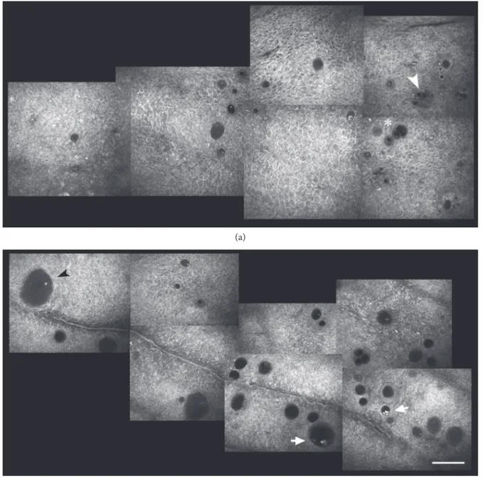
Figure 5: In vivo confocal microscopy of the superior temporal conjunctiva in the same patient scheduled to undergo a 4-second dose UCCC
(Group 1). (a) The baseline planar reconstruction shows small roundish microcysts, located at different levels within the epithelium, scattered,
and sometimes clustered (arrowhead). (b) Microcysts increased density and area (arrow) thirty days after insonification. Bar represents
(Figures 2 and 3) only at the site of transducer contact, with-out involvement of the surrounding sclera. We hypothesized that the HS increase was a consequence of a thermic-induced
scleral fibre delamination; in fact, a heating of the
supra-choroid, sclera, and conjunctiva during the procedure may occur, given that the transducer produces a thermic halo
(1.89 mm3) with a temperature gradient from the ciliary body
to the ocular surface. The preliminary results of an ongoing thermal infrared imaging study seem to support this
hypoth-esis, since we observed a significant increase in the ocular
sur-face temperature at the site of insonification, immediately
after the UC3(Figure 4). This thermic effect may also account
for the higher HS increase in patients treated with the 6-second dose regimen, which received a prolonged duration of the insonification. The increase of such HSs leads to an enhancement of the scleral hydraulic conductivity and, there-fore, of the AH transscleral outflow.
In vivo confocal microscopy confirmed the transscle-ral outflow enhancement one month after the procedure
by documenting a significant increase of conjunctival
microcysts at site of insonification (Figures 5 and 6).
These microcysts were proposed as an in vivo hallmark
of the AH passage through the sclera and finally the
conjunctiva [93–100]. (a)
(b)
Figure 6: In vivo confocal microscopy of the superior temporal conjunctiva in the same patient scheduled to undergo a 6-second dose UCCC. (a) The baseline planar reconstruction shows features similar to those observed in Group 1. Somewhere, microcysts appear encapsulated
(arrowhead) andfilled with amorphous material or punctate reflective elements (asterisk). (b) Epithelial microcysts increased density and,
especially, area (arrow) thirty days after UCCC. Microcysts may appear filled with amorphous material (black arrowhead) or reflective
The scleral architecture remodelling observed after UC3
may pose concerns in patients candidate to furtherfiltration
surgery, since the (intra- and postoperative) resistance of the
sclera and the AH permeability of collagen fibres could be
significantly altered, especially after repeated sonications. At
this moment, there are no studies that addressed this point; therefore, these aspects must be considered and carefully
pondered before proposing filtration surgery after HIFU,
either in refractory or (even more) in nonrefractory cases. In addition, there are no long-term studies that evaluated the risk of hypotony after repeated insonifications or the risk
of hypotony whether patients receive further filtration
surgery. Despite no comparative randomized clinical trials have been performed, an overview of literature leads to a
hypothesis that UC3 might have a little lower efficacy (also
in terms of reduction of number of medications) and provide
a shorter duration of the IOP-lowering effect compared to
that of the other cyclodestructive approaches, though with a greater safety profile [16–19, 42, 43, 54–69, 88]. On the other hand, the new probes seem to increase the IOP-lowering effi-cacy of the technique, maintaining the same level of safety.
In closing, even though promising and safe, the ultrasonic cyclocoagulation still requires correct positioning in terms of indication and timing, in the management of glaucoma.
6. Summary and Conclusions
Currently, cyclodestructive procedures are exclusively lim-ited to refractory/end-stage glaucoma, because of the high incidence of vision-threatening complications. All proposed procedures are noncompletely selective for the target organ, have an unpredictable dose-effect relationship, are operator
dependent, and are poorly reproducible. UC3is an emerging
and encouraging technique, which utilizes the HIFU technol-ogy to induce a one-step, automated, computer-assisted, non-operator-dependent, and highly reproducible thermal coagulation of the ciliary epithelium. This procedure allows
a selective destruction of the limited and predefined portions
of the ciliary body, thus reducing the AH inflow in a
con-trolled way. UC3presents several advantages over traditional
cyclodestructive techniques since it minimizes the intra- and postoperative complications, preserves neighbouring organs from undesired treatment, allows a faster postoperative recovery, and permits retreatments (by rotating transducers) because there is no dose limit.
Besides the reduction of the AH inflow, which is the main
mechanism that reduces IOP, UC3increases also the AH
out-flow, by favourably remodelling the anatomical architecture of suprachoroid, sclera, and conjunctiva. This indicates that
UC3may influence the entire hydrodynamic system,
exploit-ing different mechanisms to finally reduce the IOP. The promising results, along with the high level of safety reported in refractory glaucoma, allowed extending the indication for
UC3 also in glaucomatous patients naïve to surgery, thus
reconsidering the role and the timing of cyclodestruction in the management of glaucoma.
However, to date, no comparative study between UC3
and other cyclodestructive procedures has been published.
Therefore, whether UC3 represents a better solution for
refractory glaucoma with respect to standardized cycloablative techniques needs to be addressed.
The described effects of high-frequency ultrasounds on the sclera and conjunctiva might open future strategies to
lower IOP in glaucoma. In fact, the development of modified
HIFU probes that will focus the ultrasonic beam just within the sclera avoiding the ciliary body could stimulate the
uveoscleral outflow pathway by increasing the transscleral
AH resorption. This may have the great advantages to reduce the IOP by stimulating the physiological AH outflow routes and reduce the postoperative complications by pre-serving the ciliary body, which plays a critical role in the global health of the eye.
Conflicts of Interest
There are no competing interests.
References
[1] K. E. Kim, J. W. Jeoung, D. M. Kim, S. J. Ahn, K. H. Park, and
S. H. Kim,“Long-term follow-up in preperimetric open-angle
glaucoma: progression rates and associated factors,” American
Journal of Ophthalmology, vol. 159, no. 1, pp. 160–168, 2015.
[2] M. C. Leske, A. Heijl, L. Hyman et al., “Predictors of
long-term progression in the early manifest glaucoma trial,”
Ophthalmology, vol. 114, no. 11, pp. 1965–1972, 2007.
[3] J. M. Mastrobattista and M. Luntz, “Ciliary body ablation:
where are we and how did we get here?” Survey of
Ophthal-mology, vol. 41, no. 3, pp. 193–213, 1996.
[4] A. De Roetth Jr, “Cryosurgery for the treatment of
glaucoma,” Transactions of the American Ophthalmological
Society, vol. 63, no. 3, pp. 189–204, 1965.
[5] O. Kosoko, D. E. Gaasterland, I. P. Pollack, and C. L. Enger, “Long-term outcome of initial ciliary ablation with contact diode laser transscleral cyclophotocoagulation for severe
glaucoma. The Diode Laser Ciliary Ablation Study Group,”
Ophthalmology, vol. 103, no. 8, pp. 1294–1302, 1993.
[6] S. A. Vernon, J. M. Koppens, G. J. Menon, and A. K. Negi, “Diode laser cycloablation in adult glaucoma: long-term results of a standard protocol and review of current
litera-ture,” Clinical and Experimental Ophthalmology, vol. 34,
no. 5, pp. 411–420, 2006.
[7] P. T. Finger, P. D. Smith, R. W. Paglione, and H. D. Perry, “Transscleral microwave cyclodestruction,” Investigative Ophthalmology & Visual Science, vol. 31, no. 10, pp. 2151– 2155, 1990.
[8] M. Maus and L. J. Katz,“Choroidal detachment, flat anterior
chamber, and hypotony as complications of neodymium:
YAG laser cyclophotocoagulation,” Ophthalmology, vol. 97,
no. 1, pp. 69–72, 1990.
[9] D. J. Coleman, F. L. Lizzi, R. H. Silverman et al.,“Therapeutic
ultrasound,” Ultrasound in Medicine & Biology, vol. 12, no. 8,
pp. 633–638, 1986.
[10] F. Valtot, J. Kopel, and J. Haut,“Treatment of glaucoma with
high intensity focused ultrasound,” International
Ophthal-mology, vol. 13, no. 1-2, pp. 167–170, 1989.
[11] D. J. Coleman, F. L. Lizzi, J. Driller et al.,“Therapeutic
ultra-sound in the treatment of glaucoma: I. experimental model,”
[12] D. J. Coleman, F. L. Lizzi, J. Driller et al.,“Therapeutic ultra-sound in the treatment of glaucoma: II. Clinical applications,”
Ophthalmology, vol. 92, no. 3, pp. 347–353, 1985.
[13] S. E. Burgess, R. H. Silverman, D. J. Coleman et al., “Treatment of glaucoma with high-intensity focused ultra-sound,” Ophthalmology, vol. 93, no. 6, pp. 831–838, 1986. [14] S. L. Maskin, A. I. Mandell, J. A. Smith, R. C. Wood, and
S. A. Terry, “Therapeutic ultrasound for refractory
glau-coma: a three-center study,” Ophthalmic Surgery, vol. 20,
no. 3, pp. 86–192, 1989.
[15] C. C. Sterk, P. H. van der Valk, C. L. van Hees, J. L. van Delft,
J. A. van Best, and J. A. Oosterhuis,“The effect of therapeutic
ultrasound on the average of multiple intraocular pressures
throughout the day in therapy-resistant glaucoma,” Graefe's
Archive for Clinical and Experimental Ophthalmology,
vol. 227, no. 1, pp. 36–38, 1989.
[16] F. Aptel, T. Charrel, C. Lafon et al., “Miniaturized
high-intensity focused ultrasound device in patients with
glaucoma: a clinical pilot study,” Investigative
Ophthalmol-ogy & Visual Science, vol. 52, no. 12, pp. 8747–8753, 2011.
[17] P. Denis, F. Aptel, J. F. Rouland et al.,“Cyclocoagulation of
the ciliary bodies by high-intensity focused ultrasound: a
12-month multicenter study,” Investigative Ophthalmology
& Visual Science, vol. 56, no. 2, pp. 1089–1096, 2015.
[18] F. Aptel, C. Dupuy, and J. F. Rouland,“Treatment of
refrac-tory open-angle glaucoma using ultrasonic circular cyclocoa-gulation: a prospective case series,” Current Medical Research and Opinion, vol. 30, no. 8, pp. 1599–1605, 2014.
[19] F. Aptel, T. Charrel, X. Palazzi, J. Y. Chapelon, P. Denis,
and C. Lafon, “Histologic effects of a new device for
high-intensity focused ultrasound cyclocoagulation,” Inves-tigative Ophthalmology & Visual Science, vol. 51, no. 10,
pp. 5092–5098, 2010.
[20] A. M. Abdelrahman, “Noninvasive glaucoma procedures:
current options and future innovations,” Middle East African
Journal of Ophthalmology, vol. 22, no. 1, pp. 2–9, 2015.
[21] S. Melamed, M. Goldenfeld, D. Cotlear, A. Skaat, and I. Moroz, “High-intensity focused ultrasound treatment in refractory glaucoma patients: results at 1 year of prospective clinical
study,” European Journal of Ophthalmology, vol. 25, no. 6,
pp. 483–489, 2015.
[22] F. H. Verhoeff, “Cyclectomy. A new operation for glaucoma,”
Archives of Ophthalmology, vol. 33, pp. 228–229, 1924.
[23] H. Sauter and U. Demeler,“Antiglaucomatous ciliary body
excision,” American Journal of Ophthalmology, vol. 98,
no. 3, pp. 344–348, 1984.
[24] H. Weve,“Clinische lessen,” Nederlands Tijdschrift voor
Gen-eeskunde, vol. 76, pp. 5335–5336, 1932.
[25] A. Vogt,“Cyclodiathermy puncture in cases of glaucoma,”
The British Journal of Ophthalmology, vol. 24, pp. 288–297,
1940.
[26] F. W. Stocker, “Response of chronic simple glaucoma to
treatment with cyclodiathermy puncture,” Archives of
Oph-thalmology, vol. 34, no. 3, pp. 181–189, 1945.
[27] U. S. Walton and W. M. Grant,“Penetrating cyclodiathermy
for filtration,” Archives of Ophthalmology, vol. 83, no. 1,
pp. 47–48, 1970.
[28] M. T. Benson and M. E. Nelson,“Cyclocryotherapy: a review
of cases over a 10-year period,” The British Journal of
Oph-thalmology, vol. 74, no. 2, pp. 103–105, 1990.
[29] A. R. Bellows and W. M. Grant,“Cyclocryotherapy of chronic
open angle glaucoma in aphakic eyes,” American Journal of
Ophthalmology, vol. 85, no. 5 pt 1, pp. 615–621, 1978.
[30] B. S. Kim, Y. J. Kim, S. W. Seo, J. M. Yoo, and S. J. Kim, “Long-term results from cyclocryotherapy applied to the 3
o’clock and 9 o’clock positions in blind refractory glaucoma
patients,” Korean Journal of Ophthalmology, vol. 29, no. 1,
pp. 47–52, 2015.
[31] P. Sony, N. Sharma, and M. S. Pangtey,“Dislocation of the lens:
a complication after cyclocryotherapy,” Clinical and
Experi-mental Ophthalmology, vol. 30, no. 6, pp. 442–443, 2002.
[32] P. Freigassner and M. Eckhardt,“Transscleral
cyclophotoco-agulation versus cyclocryotherapy in treatment of neovascular
glaucoma: a retrospective analysis,” Acta Ophthalmologica
Scandinavica, vol. 81, no. 6, pp. 674–675, 2003.
[33] B. Koraszewska-Matuszewska, R. Leszczyński, E.
Samochowiec-Donocik, and L. Nawrocka, “Cyclodestructive procedures in
secondary glaucoma in children,” Klinika Oczna, vol. 106,
Supplement 1-2, pp. 199–200, 2004.
[34] A. Sinha and A. Rahman, “Cyclocryotherapy in absolute
glaucoma,” Indian Journal of Ophthalmology, vol. 32, no. 2,
pp. 77–80, 1984.
[35] R. M. Feibel and J. E. Bigger,“Rubeosis iridis and neovascular
glaucoma,” American Journal of Ophthalmology, vol. 74,
no. 5, pp. 862–867, 1972.
[36] T. Krupin, K. B. Mitchell, and B. Becker,“Cyclocryotherapy
in neovascular glaucoma,” American Journal of
Ophthalmol-ogy, vol. 86, no. 1, pp. 24–26, 1978.
[37] J. Fanlborn and K. Hiister,“Ergebnisse der Zyklokryorherapir
beim hamorrhagischen Glxrkorn,” Klinische Monatsblätter
für Augenheilkunde, vol. 162, pp. 513–518, 1973.
[38] L. W. Schwartz and M. R. Moster,“Neodymium:YAG laser
transscleral cyclodiathermy,” Ophthalmic Laser Therapy,
vol. 1, no. 3, pp. 135–141, 1986.
[39] G. E. Trope and S. Ma,“Mid-term effects of neodimium:YAG
transscleral cyclocoagulation in glaucoma,” Ophthalmology,
vol. 97, no. 1, pp. 73–75, 1990.
[40] K. S. Suresha and M. Narayan,“Cyclo-cryotherapy for the
management of absolute glaucoma in rural areas,” Indian Journal of Clinical and Experimental Ophthalmology, vol. 2,
no. 1, pp. 48–45, 2016.
[41] S. Lam, H. H. Tessler, B. I. La, and J. T. Wilensky, “High
incidence of sympathetic ophthalmia after contact and
non-contact neodymiun:YAG cyclotherapy,” Ophthalmology,
vol. 99, no. 12, pp. 1818–1819, 1992.
[42] R. G. Gieser and D. K. Gieser, “Treatment of intravitreal
ciliary body neovascularization,” Ophthalmic Surgery, vol. 15,
no. 6, pp. 508–509, 1984.
[43] M. B. Shields,“Intraocular cyclophotocoagulation,”
Trans-actions of the Ophthalmological Societies of the United
Kingdom, vol. 105, no. Pt. 2, pp. 237–241, 1986.
[44] M. B. Shields, D. B. Chandler, D. Hickingbotharn, and
G. K. Klintworth, “Intraocular cyclophotocoagulation.
Histopathologic evaluation in primates,” Archives of
Ophthalmology, vol. 103, no. 11, pp. 1731–1735, 1985.
[45] A. Vogel, C. Dlugos, R. Nuffer, and R. Birngruber, “Optical
properties of human sclera and their significance for
trans-scleral laser use,” Fortschritte Der Ophthalmologie, vol. 88,
no. 6, pp. 754–761, 1991.
[46] A. L. Coleman, H. D. Jampel, J. C. Javitt, A. E. Brown, and
autopsy and monkey eyes,” OphthalmicSurgery, vol.22, no. 11, pp. 638–643, 1991.
[47] C. England, E. van der Zypen, F. Fankhauser, and
S. Kwasniewska, “Ultrastructure of the rabbit ciliary body
following transscleral cyclophotocoagulation with the
free-running Nd:YAG laser: preliminary findings,” Lasers in
ophthalmology, vol. 1, pp. 61–72, 1986.
[48] L. F. Rosenberg, J. M. Ruderman, and R. B. O’Grady,
“Trans-scleral cyclophotocoagulation. Localization of ciliary process
destruction (abstract),” Investigative Ophthalmology & Visual
Science, vol. 30, Supplement, pp. 353–356, 1989.
[49] F. Fankhauser, E. Van der Zypen, S. Kwasniewska, and
H. Loertscher, “The effect of thermal mode Nd:YAG laser
irradiation on vessels and ocular tissues,” Ophthalmology,
vol. 92, no. 3, pp. 419–426, 1985.
[50] F. Fankhauser, E. Van der Zypen, S. Kwasniewska, P. Rol, and
C. England, “Transscleral cyclophotocoagulation using a
neodymium:YAG laser,” Ophthalmic Surgery, vol. 17, no. 2,
pp. 94–99, 1986.
[51] T. A. Ilawkins and W. C. Stewart,“One-year results of
semi-conductor transscleral cyclophotocoagulation in patients
with glaucoma,” Archives of Ophthalmology, vol. 111, no. 4,
pp. 488–491, 1993.
[52] M. D. Shields and S. E. Shields, “Non-contact transscleral
Nd:YAG cyclophotocoagulation– a long term follow-up of
500 patients,” Transactions of the American
Ophthalmologi-cal Society, vol. 92, pp. 271–283, 1994.
[53] D. S. Minckler,“DoesNd:YAGcyclotherapycausesympathetic
ophthalmia?” Ophthalmic Surgery, vol. 20, no. 8, p. 543, 1989. [54] M. A. Latina, S. Patel, A. W. de Kater, S. Goode,
N. S. Nishioka, and C. A. Pulia
fito,“Transscleralcyclophotoco-agulation using a contact laser probe: a histologic and clinical
study in rabbits,” Lasers in Surgery and Medicine, vol. 9, no. 5,
pp. 465–470, 1989.
[55] S. J. Schuman, C. A. Puliafito, R. R. Allingham et al., “Contact
transscleral Nd:YAG laser cyclophotocoagulation,”
Ophthal-mology, vol. 97, no. 5, pp. 571–580, 1990.
[56] R. Brancato, L. Giovanni, G. Trabbuchi, and C. Pietroni, “Contact transscleral cyclophotocoagulation with Nd:YAG
laser in uncontrolled glaucoma,” Ophthalmic Surgery, vol. 20,
no. 8, pp. 547–551, 1989.
[57] P. Lin, G. Wollstein, I. P. Glavas, and J. S. Schuman,“Contact
transscleral neodymium:yttrium-aluminum-garnet laser
cyclophotocoagulation: long-term outcome,” Ophthalmology,
vol. 111, no. 11, pp. 2137–2143, 2004.
[58] J. S. Lai, C. C. Tham, J. C. Chan, and D. S. Lam,“Diode laser
transscleral cyclophotocoagulation as primary surgical treat-ment for medically uncontrolled chronic angle closure
glau-coma: long-term clinical outcomes,” Journal of Glaucoma,
vol. 14, no. 2, pp. 114–119, 2005.
[59] P. A. Bloom, C. I. Clement, A. King et al.,“A comparison
between tube surgery, ND:YAG laser and diode laser cyclo-photocoagulation in the management of refractory
glau-coma,” BioMed Research International, vol. 2013, Article ID
371951, p. 11, 2013.
[60] C. J. Dickens, N. Nguyen, J. S. Mora et al.,“Long-term results
of noncontact transscleral neodymium:YAG
cyclophotoco-agulation,” Ophthalmology, vol. 102, no. 12, pp. 1771–1781,
1995.
[61] M. Grueb, J. M. Rohrbach, K. U. Bartz-Schmidt, and
T. Schlote, “Transscleral diode laser cyclophotocoagulation
as primary and secondary surgical treatment in primary open-angle and pseudoexfoliative glaucoma. Long-term
clini-cal outcomes,” Graefe's Archive for Clinical and Experimental
Ophthalmology, vol. 244, no. 10, pp. 1293–1299, 2006.
[62] P. Frezzotti, V. Mittica, G. Martone et al.,“Long term
follow-up of diode laser transscleral cyclophotocoagulation in the
treatment of refractory glaucoma,” Acta Ophthalmologica,
vol. 88, no. 1, pp. 150–155, 2010.
[63] T. Schlote, M. Grüb, and M. Kynigopoulos, “Long-term
results after transscleral diode laser cyclophotocoagulation in refractory posttraumatic glaucoma and glaucoma in
aphakia,” Graefe's Archive for Clinical and Experimental
Ophthalmology, vol. 246, no. 3, pp. 405–410, 2008.
[64] P. Shah, G. A. Lee, J. K. Kirwan et al.,“Cyclodiode
photocoag-ulation for refractory glaucoma after penetrating
kerato-plasty,” Ophthalmology, vol. 108, no. 11, pp. 1986–1991, 2001.
[65] C. Hampton, M. B. Shields, K. N. Miller, and M. Blasini, “Evaluation of a protocol for transscleral neodymium:YAG
Cyclophotocoagulation in one hundred patients,”
Ophthal-mology, vol. 97, no. 7, pp. 910–917, 1990.
[66] P. Hamard, J. Kopel, F. Valtot, S. Quesnot, H. Hamard, and
J. Haut,“Treatment of refractory glaucoma by diode
semi-conductor laser cyclophotocoagulation,” Journal Français
d'Ophtalmologie, vol. 18, no. 6-7, pp. 447–454, 1995.
[67] M. N. Cyrlin, H. Beckman, and C. Czedik,“Nd:YAG laser
trans-scleral cyclocoagulation treatment for severe glaucoma
(abstract),” Investigative Ophthalmology & Visual Science,
vol. 26, Supplement, p. 157, 1985.
[68] R. G. Drvenyi, G. E. Trope, and W. H. Hunter,
“Neodymium-YAG transscleral cyclocoagulation in rabbit eyes,” The British
Journal of Ophthalmology, vol. 71, no. 6, pp. 441–444, 1987.
[69] G. E. Trope and S. Ma,“Mid-term effects of Nd:YAG
transscle-ral cyclocoagulation in glaucoma,” Ophthalmology, vol. 97,
pp. 73–75, 1990.
[70] M. Uram,“Ophthalmic laser microendoscope ciliary process
ablation in the management of neovascular glaucoma,”
Ophthalmology, vol. 99, no. 12, pp. 1823–1828, 1992.
[71] K. Kaplovitz, A. Kuei, B. Klenofsky, A. Abazari, and
R. Honkanen,“The use of endoscopic cyclophotocoagulation
for moderate to advanced glaucoma,” Acta Ophthalmologica,
vol. 93, no. 5, pp. 395–401, 2015.
[72] F. E. Lima, L. Magacho, D. M. Carvalho, R. Susanna Jr, and
M. P. Avila, “A prospective, comparative study between
endoscopic cyclophotocoagulation and the Ahmed drainage
implant in refractory glaucoma,” Journal of Glaucoma,
vol. 13, pp. 233–237, 2004.
[73] M. A. Zarbin, R. S. Michels, S. DeBistros, H. A. Quigley,
and A. Patel, “Endolaser treatment of the ciliary body for
severe glaucoma,” Ophthalmology, vol. 95, no. 12, pp. 1639–
1648, 1988.
[74] L. W. Yip, S. O. Yong, A. Earnest, J. Ji, and B. A. Lim,
“Endo-scopic cyclophotocoagulation for the treatment of glaucoma:
an Asian experience,” Clinical & Experimental
Ophthalmol-ogy, vol. 37, no. 7, pp. 692–697, 2009.
[75] F. E. Lima, J. Beniz Neto, D. Toscano, D. M. D. Carvalho, and
M. P. D. Avila,“Endoscopic cyclophotocoagulation in
refrac-tory glaucomas: a long term study,” Revista Brasileira de
Oftalmologia, vol. 68, no. 3, pp. 146–151, 2009.
[76] B. A. Francis, A. S. Kawji, N. T. Vo, L. Dustin, and V.
Chopra, “Endoscopic cyclophotocoagulation (ECP) in the
tube shunt,” Journal of Glaucoma, vol. 20, no. 8, pp. 523– 527, 2009.
[77] W. J. Fry, J. W. Barnard, E. J. Fry, R. F. Krumins, and
J. F. Brennan,“Ultrasonic lesions in the mammalian central
nervous system,” Science, vol.122,no. 3168,pp. 517–518,1955.
[78] J. G. Lynn, R. L. Zwemer, A. J. Chick, and A. E. Miller,“A new
method for the generation and use of focused ultrasound in
experimental biology,” The Journal of General Physiology,
vol. 26, no. 2, pp. 179–193, 1942.
[79] K. Hynynen and B. A. Lulu, “Hyperthermia in cancer
treatment,” Investigative Radiology, vol. 25, no. 7, pp. 824–
834, 1990.
[80] G. Baum and I. Greenwood,“The application of ultrasonic
locating techniques to ophthalmology; theoretic
consider-ations and acoustic properties of ocular media. Reflective
properties,” American Journal of Ophthalmology, vol. 46,
no. 5, pp. 19–29, 1958.
[81] E. W. Purnell, A. Sokollu, R. Torchia, and N. Taner,“Focal
chorioretinitis produced by ultrasound,” Investigative
Ophthalmology, vol. 3, no. 6, pp. 657–664, 1964.
[82] R. Muratore,“A history of the Sonocare CST-100: the first
FDA-approved HIFU device,” AIP Conference Proceedings,
vol. 829, no. 1, p. 508, 2006.
[83] C. E. Margo, “Therapeutic ultrasound. Light and electron
microscopic findings in an eye treated for glaucoma,”
Archives of Ophthalmology, vol. 104, no. 5, pp. 735–738, 1986.
[84] H. Cao, Z. Xu, H. Long et al., “Trans-catheter arterial
chemoembolization in combination with high-intensity focused ultrasound for unresectable hepatocellular carci-noma: a systematic review and meta-analysis of the Chinese literature,” Ultrasound in Medicine & Biology, vol. 37, no. 7,
pp. 1009–1016, 2011.
[85] S. Crouzet, O. Rouviere, X. Martin, and A. Gelet,
“High-inten-sity focused ultrasound as focal therapy of prostate cancer,”
Current Opinion in Urology, vol. 24, no. 3, pp. 225–230, 2014.
[86] C. C. Li, Y. Q. Wang, Y. P. Li, and X. L. Li,“High-intensity
focused ultrasound for treatment of pancreatic cancer: a
systematic review,” Journal of Evidence-Based Medicine,
vol. 7, no. 4, pp. 270–281, 2014.
[87] G. Pron,“Magnetic resonance-guided high-intensity focused
ultrasound (MRgHIFU) treatment of symptomatic uterine fibroids: an evidence-based analysis,” Ontario Health Tech-nology Assessment Series, vol. 15, no. 4, pp. 1–86, 2015.
[88] R. Mastropasqua, L. Agnifili, V. Fasanella et al., “Uveo-scleral
outflow pathways after ultrasonic cyclocoagulation in
refrac-tory glaucoma: an anterior segment optical coherence
tomog-raphy and in vivo confocal study,” The British Journal of
Ophthalmology, vol. 100, no. 12, pp. 1668–1675, 2016.
[89] F. Aptel, P. Denis, J. F. Rouland, J. P. Renard, and A. Bron, “Multicenter clinical trial of high-intensity focused ultrasound treatment in glaucoma patients without previous filtering surgery,” Acta Ophthalmologica, vol. 94, no. 5,
pp. e268–e277, 2016.
[90] D. M. Bushley, V. C. Parmley, and P. Paglen, “Visual field
defect associated with laser in situ keratomileusis,” American
Journal of Ophthalmology, vol. 129, no. 5, pp. 668–671, 2000.
[91] B. D. Cameron, N. A. Saffra, and M. B. Strominger, “Laser
in situ keratomileusis-induced optic neuropathy,”
Ophthal-mology, vol. 108, no. 4, pp. 660–665, 2001.
[92] F. Aptel, A. Béglé, A. Razavi et al.,“Short- and long-term effects
on the ciliary body and the aqueous outflow pathways of
high-intensity focused ultrasound cyclocoagulation,” Ultrasound in
Medicine & Biology, vol. 40, no. 9, pp. 2096–2106, 2014.
[93] L. Agnifili, P. Carpineto, V. Fasanella et al., “Conjunctival
findings in hyperbaric and low-tension glaucoma: an in vivo confocal microscopy study,” Acta Ophthalmologica, vol. 90,
no. 2, pp. e132–e137, 2012.
[94] P. Carpineto, L. Agnifili, M. Nubile et al., “Conjunctival and
corneal findings in bleb-associated endophthalmitis: an
in vivo confocal microscopy study,” Acta Ophthalmologica,
vol. 89, no. 4, pp. 388–395, 2011.
[95] M. Ciancaglini, P. Carpineto, L. Agnifili et al., “Conjunctival
characteristics in primary open-angle glaucoma and modi
fi-cations induced by trabeculectomy with mitomycin C: an
in vivo confocal microscopy study,” The British Journal of
Ophthalmology, vol. 93, no. 9, pp. 1204–1209, 2009.
[96] M. Ciancaglini, P. Carpineto, L. Agnifili, M. Nubile, V.
Fasa-nella, and L. Mastropasqua,“Conjunctival modifications in
ocular hypertension and primary open angle glaucoma: an
in vivo confocal microscopy study,” Investigative
Ophthal-mology & Visual Science, vol. 49, no. 7, pp. 3042–3048, 2008.
[97] L. Mastropasqua, L. Agnifili, M. Ciancaglini et al., “In vivo
analysis of conjunctiva in gold micro shunt implantation
for glaucoma,” The British Journal of Ophthalmology,
vol. 94, no. 12, pp. 1592–1596, 2010.
[98] L. Mastropasqua, L. Agnifili, M. L. Salvetat et al., “In vivo
anal-ysis of conjunctiva in canaloplasty for glaucoma,” The British
Journal of Ophthalmology, vol. 96, no. 5, pp. 634–639, 2012.
[99] L. Mastropasqua, L. Agnifili, R. Mastropasqua, and
V. Fasanella,“Conjunctival modifications induced by
medi-cal and surgimedi-cal therapies in patients with glaucoma,” Current
Opinion in Pharmacology, vol. 13, no. 1, pp. 56–64, 2013.
[100] R. Mastropasqua, V. Fasanella, E. Pedrotti et al.,
“Trans-conjunctival aqueous humor outflow in glaucomatous
patients treated with prostaglandin analogues: an in vivo confocal microscopy study,” Graefe's Archive for Clinical
and Experimental Ophthalmology, vol. 252, no. 9,
Submit your manuscripts at
https://www.hindawi.com
Stem Cells
International
Hindawi Publishing Corporationhttp://www.hindawi.com Volume 2014
Hindawi Publishing Corporation
http://www.hindawi.com Volume 2014
INFLAMMATION
Hindawi Publishing Corporation
http://www.hindawi.com Volume 2014
Behavioural
Neurology
Endocrinology
International Journal ofHindawi Publishing Corporation
http://www.hindawi.com Volume 2014
Hindawi Publishing Corporation
http://www.hindawi.com Volume 2014
Disease Markers
Hindawi Publishing Corporation
http://www.hindawi.com Volume 2014
BioMed
Research International
Oncology
Journal ofHindawi Publishing Corporation
http://www.hindawi.com Volume 2014
Hindawi Publishing Corporation
http://www.hindawi.com Volume 2014
Oxidative Medicine and Cellular Longevity
Hindawi Publishing Corporation
http://www.hindawi.com Volume 2014
PPAR Research
The Scientific
World Journal
Hindawi Publishing Corporation
http://www.hindawi.com Volume 2014
Immunology Research
Hindawi Publishing Corporation
http://www.hindawi.com Volume 2014
Journal of
Obesity
Journal ofHindawi Publishing Corporation
http://www.hindawi.com Volume 2014
Hindawi Publishing Corporation
http://www.hindawi.com Volume 2014
Computational and Mathematical Methods in Medicine
Ophthalmology
Journal ofHindawi Publishing Corporation
http://www.hindawi.com Volume 2014
Diabetes Research
Journal ofHindawi Publishing Corporation
http://www.hindawi.com Volume 2014
Hindawi Publishing Corporation
http://www.hindawi.com Volume 2014 Research and Treatment
AIDS
Hindawi Publishing Corporation
http://www.hindawi.com Volume 2014
Gastroenterology Research and Practice
Hindawi Publishing Corporation
http://www.hindawi.com Volume 2014