S
CUOLAN
ORMALES
UPERIOREP
H.D
.T
HESISNanostructuring HIV-1 Tat arginine-rich
motif for targeted cellular
delivery
Francesco Cardarelli
A
DVISORProf. Fabio Beltram
to Claudia and my family
Foreword
This thesis is the result of my research activity at the NEST laborarory of Scuola Normale Superiore in Pisa: I began my studies on Tat protein in 2004, prompted by the group interest in the molecular biophysics sector. Most of the results presented here have been already published in the following papers.
List of publications
R Bizzarri, C Arcangeli, D Arosio, F Ricci, P Faraci, F Cardarelli and F Beltram “Development of a novel GFP-based ratiometric excitation and emission pH indicator for intracellular studies”
Biophysical Journal 90 (2006) 3300-3314
F Cardarelli, M Serresi, R Bizzarri, M Giacca and F Beltram
“In Vivo study of HIV-1 Tat arginine-rich motif unveils its transport properties” Molecular Therapy 7 (2007) 1313-1322
F Cardarelli, M Serresi, R Bizzarri and F Beltram
“Tuning the transport properties of HIV-1 Tat arginine-rich motif in living cells” Traffic 4 (2008) 528-539
M Serresi, R Bizzarri, F Cardarelli and F Beltram
“Real-time measurement of endosomal acidification by a novel genetically encoded biosensor”
Foreword
Publications not included in this thesis:
M Costa, M Marchi, F Cardarelli, A Roy, F Beltram, L Maffei and G M Ratto “Dynamic regulation of ERK2 nuclear translocation and mobility in living cells” Journal of Cell Science 119 (2006) 4952-4963
R Bizzarri, M Serresi, F Cardarelli, S Abbruzzetti, B Campanini, C Viappiani and F Beltram
“Fast and reversibly photoswitchable fluorescent proteins from Aequorea Victoria by a single aminoacid replacement”
Table of Contents
Introduction……….. 1
1
Vector-based delivery strategies: from HIV-1 Tat protein to cell penetrating peptides………. 1.1 HIV-1 transactivator protein (Tat)………. 1.1.1 Tat primary sequence: functional sub-domains…………...……...1.1.2 Intracellular Tat………...
1.1.3 Extracellular Tat………
1.2 Intracellular cargo delivery using Tat-derived peptides..………... 1.2.1 The implication of the basic cluster……….. 1.2.2 Tat peptide and cell surface interactions………. 1.2.3 Tat-peptide-mediated cargo delivery………... 1.2.4 Intracellular fate and transport properties of Tat-peptide-cargoes…..
5 5 6 7 8 9 9 11 11 13
2
In vivo study of HIV-1 Tat arginine-rich motif (I): nuclear importproperties………. 2.1 Materials and Methods………...
2.1.1 Engineering of expression vectors………...…... 2.1.2 Cell culture, transfection, and immunoblotting analysis………. 2.1.3 Fluorescence imaging by confocal laser scanning
microscopy (CLSM)……..………..
2.2 Demonstration of Tat-peptide actual nuclear import properties………… 2.2.1 Cargo-dependent sub-cellular localization of constructs………. 2.2.2 Nuclear permeation upon energy depletion………. 2.3 Limitations of wild-type Tat-peptide as a delivery vector………... 15 16 17 15 18 18 21 22 18
ii Table of Contents
3
In vivo study of HIV-1 Tat arginine-rich motif (II): intracellulartrafficking and interactions………... 3.1 Dynamic model for nuclear/cytoplasmic exchange……….. 3.1.1 In vivo applications: FRAP analysis of passive diffusion………...
3.1.2 In vivo applications: FRAP analysis of active import……….
3.1.3 FRAP analysis of Tat11-driven nucleus/cytoplasm exchange……….. 3.2 FRAP analysis of protein mobility within cytoplasm………...
3.2.1 The disk-FRAP method: experimental setup and mathematical 25 25 27 28 32 30 model………... 32
3.2.2 In vivo applications: measuring absolute ‘D’ of passive-
diffusion and active-import benchmark constructs……….…………. 3.2.3 In vivo applications: measuring Tat11 intracytoplasm diffusivity…… 3.3 Tat-driven intracellular interactions………..………... 33 35 36
4
Tuning the transport properties of HIV-1 Tat arginine-rich motif in living cells………... 4.1 Materials and methods ………..………..………... 4.1.1 Synthetic compounds, peptides, and recombinant proteins…..……... 4.1.2 Cell culture, transfections, and internalization assays ………...……. 4.2 Tat-peptide sequence can be turned into a functional NLS byscanning mutagenesis ………..………...……….. 4.2.1 Influence of the ‘RRR’ stretch on Tat-driven nuclear delivery
of GFP4 cargo………...…….
4.2.2 The role of metabolic energy ………...……... 4.2.3 Influence of the ‘RRR’ stretch on Tat-driven inter-compartment
dynamics ………...…….. 4.3 Modulation of Tat-peptide intracellular trafficking properties by
scanning mutagenesis………..……… 4.3.1 Tuning the nuclear import mechanism……… 4.3.2 Tuning protein mobility within cytoplasm……….. 4.4 The mechanism of nuclear entry……… 4.4.1 The line-FRAP approach………. 4.4.2 From the FRAP analysis to the molecular model……… 4.5 Uptake of Tat peptide in living cells………..….……... 4.5.1 In vivo colocalization assays: TatRRR-mediated endocytosis ………..
41 42 42 42 43 43 44 46 47 47 49 50 50 52 52 52
Table of Contents iii
4.5.2 TatGGG-mediated membrane binding ……….………...…….. 4.5.3 A comprehensive model for the cellular uptake of Tat-derived
54
cell-penetrating peptides………..…………
4.6 Concluding remarks……… 55 56
5
FLIM analysis of interactions during nuclear import………5.1 An introduction to the nuclear import machinery………... 5.2 Materials and Methods………
5.2.1 Plasmids, cell culture and transfections……….…….. 5.2.2 Fluorescence lifetime measurements and data analysis…………... 5.3 FRET-by-FLIM analysis of nuclear import………....………….. 5.3.1 Subcellular localization of constructs………..………
5.3.2 Proof of principle: a FLIM-based assay to study NLSSV40-Imp-α/β interactions……….……….………. 5.3.3 Application of the method to TatRRR/TatGGG sequences…....……... 5.4 Concluding remarks on Tat interactions and transport biology….………
60 59 62 62 62 63 63 64 67 69
6
Targeted cellular delivery using Tat-derived peptides: conclusions and futuredirections………. 71
A
Using encoded light as a tool: FRAP analysis of protein mobility in aliving cell………..
A.1 The mathematical model for nucleus/cytoplasm exchange……….. A.1.1 Equations that describe concentration changes………... A.1.2 Equations that describe fluorescence changes………….………
A.1.3 Determination of the protein bound mole fraction in the 77 77 77 81
nucleoplasm……….………… 82
A.2 The mathematical model for the bleaching of a uniform
disk-shape geometry……….………... A.3 The mathematical model for the bleaching of a line……… A.3.1 The line-FRAP method: in vivo applications……….……….
82 84 85
iv Table of Contents
B
In vivo determination of actual protein-protein affinity………. B.1 Fluorescence Resonant Energy Transfer: a brief outline………... B.2 From FRET to protein-protein affinity………... B.2.1 Theoretical derivation of KD……….………..……. B.2.2 Calibration of protein concentrations in vivo………..……… B.2.3 Derivation of the τDA component………....………. B.2.4 Calculation of in vivo protein-protein affinities….………..92 89 89 93 95 96 92
C
Green Fluorescent Protein mutants as pH-biosensors for in vivoapplications………. C.1 E1GFP………...
C.1.1 Optical properties of E1GFP mutant……….. 99 100 100
C.1.2 E1GFP mutant as emission ratiometric pH-indicator in the
cellular environment……….. 101
C.1.3 E1GFP as emission ratiometric pH-indicator of intracellular
transport phenomena……….………... C.2 E2GFP………... 103 103
D
Notations……….…………... 107 Bibliography……….………… 109 Acknowledgements……….………... 121List of Figures
1.1 The overall 3-D shape of Tat………..……….………... 1.2 Primary structure of HIV-1 Tat protein……….…..………... 1.3 Pathways of entry into cells……….
5 7 12
2.1 GFP4-cargo characterization………..……….……..
2.2 Cargo-dependent subcellular localization of constructs……….….. 2.3 WB analysis of GFP and Tat11-GFP correct size………. 2.4 Nuclear permeation on energy depletion……….
16 20 21 19
3.1 FRAP analysis of GFP nucleus/cytoplasm shuttling…………...…….……… 3.2 FRAP analysis of active import………... 3.3 FRAP analysis of Tat11-cargo nucleus/cytoplasm shuttling………. 3.4 FRAP analysis of Tat11-GFP binding to the nucleolus………....………. 3.5 Protein mobility within cytoplasm: bleaching of a uniform disk………. 3.6 Application of the disk-bleaching method to Tat11-GFP……….………. 3.7 Monitoring Tat-peptide interaction with nucleic acids by FRET. .….…….……... 3.8 Intracellular localization of construct upon nocodazole (NCZ) treatment…...……
27 29 31 30 34 35 37 39
4.1 TatGGG-mediated active nuclear accumulation of GFP4 cargo……….. 43 4.2 Influence of the ‘RRR’ determinant on nuclear localization and inter-
compartment dynamics of constructs………... 4.3 Analysis of intracellular l
vi List of Figures
5.1 Schematic diagram of the NLS-dependent nuclear import pathway……… 5.2 Intracellular localization of constructs………. 5.3 In vivo FLIM analysis of Imp-α/NLS pair……….……….. 5.4 Intracellular distribution of Imp-α-NLS interaction…………....………. 5.5 In vivo FLIM analysis of Imp-α/TatGGG pair ………... 5.6 In vivo FLIM analysis of Imp-β/TatGGG pair ………...
64 61 66 65 67 68
6.1 Measurement of endosomal acidification after 4 hours of Tat-E1GFP endocytosis……….……….…………... 74
A.1 Model of nucleocytoplasmic exchange……….………… A.2 Schematic representation of a laser beam……….………… A.3 Photobleaching kinetics of a YFP-expressing cell in a Fly-Mode experiment……
78 84 86
A.4 Application of the line-FRAP method to measure absolute diffusion
coefficient in YFP-expressing cells……….….……… 87
B.1 Absorption and emission spectra of EGFP and mCherry……….……… B.2 FRET efficiency……….….………. B.3 The method………...….………... B.4 Cumulative lifetime data……….…….……… B.5 KD dependence on cytoplasmic Importin concentration………..
89 94 95 96 91 C.1 Spectroscopic characteristics of E1GFP ………....……... C.2 In vivo ratiometric calibration of E1GFP……….……… C.3 Spectroscopic characteristics of E2GFP……….………. C.4 Analysis of intracellular pH using E2GFP biosensor………...…….……..
100 102 104 105
List of Tables
1.1 Tat-derived peptides used for the cellular delivery of cargoes ……… 3.1 Transport and diffusion parameters derived from FRAP measurements…………. 4.1 Transport and diffusion parameters derived from FRAP measurements on
Tat-derived mutants ………
32 10
5.1 Average lifetimes derived by FLIM measurements………. A.1 Diffusion parameters derived by line-FRAP measurements………
57 69 87
Introduction
The ability to cross the cellular membrane and gain access to the cell interior or even to specific cellular compartments, still remain a major obstacle in current intracellular drug delivery. Peptide-mediated delivery of bioactive molecules can be a powerful tool that is actually superior to commonly-used delivery systems (liposomes, microinjection, electroporation, viral systems). In fact, peptide vectors guarantee high-yield delivery, low toxicity, and the possibility to enter a wide range of target cells.
In the search for new therapeutic strategies, a large body of work is currently devoted to determine whether these cell-penetrating peptides (CPPs) can act as molecular carriers for delivery of cargoes (e.g., proteins or nucleic acids) to relevant sub-cellular domains, particularly the nucleus. Nuclear localization is particularly significant, as it is one of the fundamental steps for gene-therapy approaches and can open the way to probe and modify cellular processes of utmost importance.
Among the commonly used CPPs, the arginine-rich motif of Tat protein from HIV-1 (sequence: YGRKKRRQRRR) has attracted much interest owing to its ability to drive the internalization of a large variety of cargoes chemically coupled or fused to it in mammalian and human cell types. Despite the variety of biotechnological applications for delivery purposes, however, little is known about the intracellular trafficking and interactions of Tat peptide-tagged cargoes. Available knowledge is based on in vitro assays that present contrasting results. We identified this issue as the key point to start our investigation of Tat-peptide transport biology.
In this thesis I shall present a novel approach that allows us to investigate Tat peptide intracellular transport properties in living cells. This biological research field, in fact, can be supported by the recent advances in biophysical technologies and microscope systems, providing ideal strategies and tools for approaching problems in modern proteomics, like high-resolution imaging of biological processes, protein dynamics, and protein-protein interactions in living cells. These advances were favored by the discovery of the Green Fluorescent Protein (GFP), and the development of its derivatives. This encodable fluorophore revolutionized our ability to study protein
Introduction
2
function within the complex cellular environment, and it was not surprising that it led to the award of the 2008 Nobel prize to Osamu Shimomura, Martin Chalfie, and Roger Y. Tsien. This fluorophore together with advances in imaging methods and microscope systems makes it easy to monitor the localization of GFP fusion proteins, as well as their dynamics and interactions by fluorescence methods such as fluorescence recovery after photobleaching (FRAP) and fluorescence resonant energy transfer (FRET).
The combined advances in GFP biology, imaging methods and technical equipment are here applied to the study of Tat-cargo localization, dynamics, and interactions in living cells. Our efforts were aimed at engineering the wild-type sequence of Tat-peptide (YGRKKRRQRRR, Tat11 in the following) to obtain new features relevant for drug-delivery purposes. The potential impact of this work on improving peptide-based delivery methods motivated us to develop new Tat mutants with novel transport properties. In particular, I shall demonstrate that the new mutants here described are suitable candidates for targeted nuclear delivery of cargoes in living cells.
The following sections will be organized as follows:
- In Chapter 1 I shall first provide a brief review of the properties of the full-length Tat protein as an extra- and intra-cellular HIV-1 effector. Next, I shall introduce Tat-derived peptide as a vector for the intracellular delivery of therapeutic and diagnostic agents. I shall discuss what has been reported so far on Tat-peptide and clarify the key issues that motivated our study.
- From Chapter 2 on I am going to report the experimental part of my thesis: first, I shall present the work carried out to define the intracellular transport properties of wild-type Tat peptide. To assess this issue we engineered GFP-based fusion proteins of different molecular size, both below and above the passive-diffusion size limit through the nuclear pore. These engineered cargoes provided a benchmark for intracellular passive diffusion behavior when expressed in cells and were also used as cargoes for prototypical active import by fusing them to the NLS of SV40. This array of cargoes, together with in vivo energy-depletion assays, allowed us to elucidate the mechanism of Tat-peptide nuclear permeation, demonstrating that it shuttles across the nuclear envelope (NE) by passive diffusion. These results give useful insights into the nature of target molecules available for Tat-peptide-driven nuclear delivery. In fact, our analysis shows that protein cargoes exceeding the size-threshold value for passive diffusion through the nuclear pore (~60-70 kd) do not gain access to the nucleus.
- In the first part of Chapter 3 we report on the peculiar transport parameters of Tat-cargo passive diffusion across the NE. FRAP analysis of Tat11-fused cargoes revealed
Introduction 3
that these proteins shuttle bi-directionally between the two compartments, with the same recovery time values (τ) applying to cytoplasm-to-nucleus transfer (signal recovery after nuclear photobleaching) and nucleus-to-cytoplasm transfer (signal recovery after cytoplasm photobleaching). Tat11 fusion led to the observation of a sizable increase in cargo τ-values (about fivefold) that must be linked to the ability of Tat11 to interact with cellular components. Accordingly, the Tat-peptide sequence was shown to reduce the measured diffusion coefficient of cargoes within cytoplasm, much beyond the effect of its mere size.
As reported in the literature, Tat peptide interacts with RNA molecules. A quantitative in vitro analysis of Tat interaction with nucleic acids (DNA and RNA) by FRET measurements will be described. Notably, we shall show that the ‘RRR’ stretch within Tat sequence is responsible for its DNA/RNA-binding properties: ‘RRR’→‘GGG’ substitution leads to a dramatic decrease in Tat peptide affinity for these molecules. This result shows that this interaction can indeed play a significant role in determining the intracellular properties of wild-type Tat peptide.
- The identification of the strong limitations of Tat peptide as a nuclear delivery vector prompted us to engineer the peptide primary sequence in order to implement the NLS function of the Tat peptide and to control the correct translocation of the cargo to the nucleus. We focused our study on the properties of the Arg-Arg-Arg (RRR) determinant in the carboxy-terminus of Tat sequence (functionally involved in peptide interaction with nucleic acids, as demonstrated in Chapter 3). In Chapter 4 we shall describe the production and analysis of a series of Tat-mutants where the acidic residues RRR were substituted into not-charged glycines, GGG. By in vivo imaging and FRAP-analysis we studied the subcellular localization and the kinetic parameters of the resulting mutwe stud
Introduction
4
tagged candidate NLS-sequence and either EGFP-tagged Imp-α or Imp-β carrier proteins. First, we tested this approach by studying the well-known NLSSV40-Imp-α/β system, with a quantitative analysis of the molecular interactions taking place during the whole process of nuclear import. Second, we applied this method to the study of the TatRRR/TatGGG pair: we found that no import carriers are involved in mediating TatRRR-cargo nucleus/cytoplasm shuttling (in keeping with the results shown so far), while both Imp-α and Imp-β separately contribute to the nuclear accumulation of TatGGG-cargoes through direct interactions.
- In Chapter 6 I shall summarize the main findings of this research activity and discuss some crucial aspects of Tat peptide transport that needs further investigation.
Chapter
1
Vector-based delivery strategies: from HIV-1
Tat protein to cell penetrating peptides
1.1 HIV-1 Transactivator Protein (Tat)
The central role played by Tat in HIV replication has made it a focus of much attention ever since its discovery in 1985. Tat provides the first example of a protein that regulates transcriptional elongation in eukaryotic cells, and until Tat was discovered, elongation control was a relatively neglected area of transcription regulation. Immediately after HIV infects a target cell, the viral RNA is copied into a double-strand DNA by the reverse transcriptase (RT) and the pro-viral genome is transported to the nucleus and integrated into the host-cell genome. The integrated pro-virus is then subjected to the regulation by cellular transcription factors, as well as its own regulatory proteins.
Vector-based delivery strategies: from HIV-1 Tat protein to cell penetrating peptides
6
HIV transcription is controlled primarily by Tat protein. Tat regulates the viral gene expression through the control of transcription by the cellular RNA polymerase II (RNP). In the absence of Tat, initiation of transcription from the viral long terminal repeat (the promoter LTR) is efficient but transcription is impaired because the promoter induces a poorly processive polymerase activity and the enzyme disengages from the DNA template prematurely. Despite extensive efforts, HIV-1 Tat was not crystallized. The information about its three-dimensional structure is based on nuclear magnetic resonance (NMR)1, 2. These studies show that the Tat protein in isolation is largely unfolded and does not present any secondary structure (Fig. 1.1).
1.1.1 Tat primary sequence: functional sub-domains
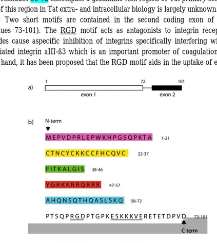
Tat is a small nuclear protein of 86 to 101 amino acids (depending on the viral strain). It is synthesized from an mRNA joined from two coding exons. The first exon encodes amino acids 1-72, the second exon encodes amino acids 73-101 (Fig. 1.2.a). Tat sequence can be subdivided into several distinct regions on the basis of its amino acid composition 3 (Fig. 1.2.b):
(i) The region 1-21, corresponding to the N-terminal region, has a static structure with secondary profiles indicative of the presence of two ß-turns 4. This domain is able to bind the T-cell activation marker CD26 and the T-cell receptor CCR2 in its extra-cellular form, facilitating T-cell activation and apoptosis 5.
(ii) The region spanning residues 22-37 has a highly-conserved 7 cystein motif. This region of Tat is of great importance for the formation of active structural domains in the protein 6.
(iii) Residues in the core region (38-46) form a highly-conserved rigid α-helix structure which enhances Tat binding to the transactivation-responsive region (TAR) 7. Chemotactic effects on monocytes are also exerted by the core region of Tat.
(iv) The basic region (residues 47-57, sequence: YGRKKRRQRRR) corresponds to the presumed nuclear localization signal (NLS) 8, 9 and the trans-activation responsive region binding domain of Tat protein 10. This domain is also involved in Tat-TAR interaction (see next section for details), owing to the high content of arginine residues 11, 12. The Tat basic domain exhibits an additional important property: it is readily taken up by cells through interactions with heparan sulfate proteoglycans displayed on the cell membrane 13. By this mechanism this Tat-derived peptide can determine cellular uptake of nonpermeant molecules 14. It is a highly immunoreactive domain, interacting with dendritic cells and monocytes in particular 15, 16.
HIV-1 Transactivator Protein (Tat) 7
(v) Residues 58-72 encompass a glutamine-rich region of Tat primary sequence. The role of this region in Tat extra- and intracellular biology is largely unknown.
(vi) Two short motifs are contained in the second coding exon of HIV-1 Tat (residues 73-101). The RGD motif acts as antagonists to integrin receptors. RGD peptides cause aspecific inhibition of integrins specifically interfering with platelet-associated integrin aIII-ß3 which is an important promoter of coagulation 17. On the other hand, it has been proposed that the RGD motif aids in the uptake of extracellular Tat.
Fig. 1.2 Primary structure of HIV-1 Tat protein. (a) Tat is synthesized from an mRNA joined from two coding exons. The first exon encodes amino acids 1-72, the second exon encodes amino acids 73-101. (b) The Tat primary sequence is here sub-divided in functional domains (described in the text).
The second one is an ESKKKVE-motif. Despite its being highly conserved throughout group M subtypes, little is known about the functional or conformational importance of this motif.
1.1.2 Intracellular Tat
The present knowledge demonstrates the pivotal role played by Tat in the viral replication cycle and some fundamental details of Tat activity, mostly concerning Tat interaction with its cellular partners inside living cells. As mentioned above, Tat plays
Vector-based delivery strategies: from HIV-1 Tat protein to cell penetrating peptides
8
its trans-activating role at the LTR promoter. This sequence was first described by Sodroski and collaborators 18 who noted that the synthesis of a reporter gene placed under the control of LTR was stimulated up to 200-300 folds in cells previously infected with the virus. Cells transfected by plasmids carrying the HIV LTR characteristically produce two distinct population of transcripts: a set of short, non-polyadenilated RNAs and a set of full-length non-polyadenilated RNAs 19. Tat is able to shift the balance from the predominant short transcripts to the full-length ones participating in a positive feedback mechanism that ensures high levels of HIV transcription 19-21. The shift from short to long viral transcripts implies that Tat activates transcription elongation.
The Tat/TAR interaction and the mechanism of Tat transactivation: the genetic
element responsible for Tat activity, TAR, is located downstream of the initiation site for transcription 22. Tat is able to specially recognize TAR RNA 23 through its basic domain 11, 12. Tat recognition of TAR induces conformational changes in the RNA structure so that it can be contacted by other basic residues of Tat, ensuring high binding affinity 24. Through the interaction with TAR, Tat promotes the assembly of transcriptionally active complexes at the LTR site by multiple protein-protein interactions 25.
1.1.3 Extracellular Tat
In addition to playing its critical role as a transcription factor Tat contributes to HIV disease as an extracellular protein. Several groups showed that Tat can be secreted by infected cells 26, 27. Furthermore, extracellular Tat is readily taken up by cells through interactions with heparan-sulfate proteoglycans displayed on the cell surface 13. Subsequently, Tat can reach the nucleus and then activate a variety of cellular genes including cytokines such as interlukin 6 (IL-6), IL-2 and tumor necrosis factor α 28, 29 and the CXCR4 and CCR5 cytokine co-receptors for HIV infection 30. Another important target for exogenous Tat is the vascular endothelium where it is able to induce angiogenesis and inflammation 31. Furthermore, Tat plays a role in the development of AIDS-related dementia 32, and also acts on different targets of the immune system contributing to the immune suppression in AIDS patients 33. Finally, it is able to transactivate the genome of other viruses favoring secondary infections 34.
Tat as a target in AIDS therapy: Tat is considered one of the candidates for an
anti-AIDS vaccine since, among viral proteins, reaction against Tat is the quickest 35 and high level of both humoral and cellular immune response against Tat correlate with
Intracellular cargo delivery using Tat-derived peptides 9
better prognosis 35-37. Furthermore, studies on HIV transactivation are providing a molecular basis for drug discovery. Several groups have identified promising “hits” that inhibit HIV replication by interfering with Tat, TAR, and cellular co-factors required for HIV transcription.
Tat as a vector for intracellular delivery: Tat is also widely studied as a powerful
tool to deliver various biological molecules within cellular compartments, bypassing the cell membrane. Frankel and Pabo 38 first demonstrated the ability of HIV-1 Tat protein to enter cells, localize in the nucleus and transactivate the HIV-1 promoter when simply added to the cell-culture medium. Internalization was observed in the absence of specific receptors and this raised the possibility of using Tat as a vector to deliver selected biomolecules into cells. Following this work, extensive molecular analysis of the Tat protein identified a short arginine-rich domain called the Tat protein-transduction domain (Tat-PTD), as the one responsible for the transduction process across the membrane 39.
1.2 Intracellular cargo delivery using Tat-derived peptides
Intracellular delivery of many therapeutic and diagnostic agents can be challenging since the plasma membrane forms a formidable barrier for biomolecules. The discovery of cell penetrating peptides (CPPs) has made it possible to transduce a broad range of physiologically- and therapeutically-active agents into living cells. Among peptides with membrane translocating activities one should mention Antennapedia homeodomain-derived peptide 40, Transportan 41, HSV-1 VP22 42, and HIV-1 Tat-derived peptide (Tat47–57) 39, 43. HIV Tat peptide and its derivatives have recently received much attention, primarily because of their efficiency and short sequence. 1.2.1 The implication of the basic cluster
The first publication showing the ability of a Tat-protein-derived peptide to deliver heterologous molecules into cells was authored by Fawell et al.14 years ago 44. In this pioneering study, the covalent coupling of a 36-aminoacid peptide (Tat37–72) to large proteins (β-galactosidase, RNAse or peroxidase) was performed through a heterolinker. Four to five peptide molecules per protein unit were sufficient to mediate the cellular delivery of the covalently bound protein 44. This CPP encompassed a highly cationic cluster composed of 6 arginine and 2 lysine residues in the very middle of the peptide sequence and an α-helical structure on the N-terminal part 45. Concomitantly, an extensive structure-activity relationship study was performed to define which feature
Vector-based delivery strategies: from HIV-1 Tat protein to cell penetrating peptides
10
within the Tat peptide was responsible for the cell-membrane translocating property 39. Several peptides carrying either partial or total deletion within the α-helical structure and a peptide harboring a deletion within the cationic domain of the original peptide were synthesized 39. It was shown, primarily by fluorescence microscopy, that the main determinant responsible for the translocating activity is the cationic cluster of amino acids. From these observations, the translocating property of the initial peptide (Tat37–72 44) could be reduced down to the sequence encompassing the cationic cluster containing 8 basic charges within the amino acid linear sequence (GRKKRRQRRR, charged residues underlined).
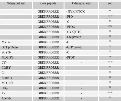
Tab. 1.1 Tat-derived peptides used for the cellular delivery of cargoes
N-terminal end Core peptide C-terminal end ref
- GRKKRRQRRR - GYK(FITC)C 46 - GRKKRRQRRR -PPQ 47, 48 - GRKKRRQRRR -G 49 - GRKKRRQRRR -PPQC 50-52 - GRKKRRQRRR -GYK(FITC) 53 - GRKKRRQRRR -Cre protein 54 MYG- GRKKRRQRRR -G 55 GST protein- GRKKRRQRRR -GFP protein 56 SGYG- GRKKRRQRRR -C 57 MLGISY- GRKKRRQRRR -PPQT 58 CY- GRKKRRQRRR - 48, 49 CGISY- GRKKRRQRRR - 59 C- GRKKRRQRRR - 60 Biotin-Y GRKKRRQRRR - 61 MLGISY- GRKKRRQRRR - 58 His6- GRKKRRQRRR - 62 Y- GRKKRRQRRR - 63, 64 Acetyl- GRKKRRQRRR - 65
It should be noted that this sequence is the common determinant in most of the studies performed with the Tat-truncations (see Tab. 1.1).
Intracellular cargo delivery using Tat-derived peptides 11
The C-terminal part of this peptide was extended by various sequences or coupled to different moieties: these include amino acids present in the native Tat protein sequence, such as the PPQ sequence but also longer cargoes in order to induce their cellular uptake (see Ref. 14 for an exhaustive review). The N-terminal part of this minimal Tat peptide was also extended by various amino acid sequences from either the native Tat protein sequence or again fused to different entities (Tab. 1.1 and Ref. 14). From these different applications, no clear influence of the additional moiety fused either to the N-terminal or to the C-terminal end of the “core” Tat peptide could be related to a variable ability of the basic Tat peptide to mediate cellular uptake.
Tat peptide is rich in cationic residues and in particular, arginines. Interestingly, the multiplicity of arginines is a common feature among many other membrane-permeable peptides. The structural aspects of arginine within a polymeric chain were investigated using several chemical analogues: these studies focused on amino acid chirality, the guanidine head group, alkyl side chain, and peptide backbone. In essence, the transmembrane activity of Tat peptide has been largely attributed to the high density of guanidine groups, whereas the remaining structural elements were found to play minor or no role 66, 67.
1.2.2 Tat peptide and cell surface interactions
The Tat protein binds to cell surface heparan sulfate (HS) and heparin 68, and consistent with its binding properties, Tat can be purified to homogeneity by heparin-affinity chromatography 69. The group of Presta and co-workers first examined the role of the basic domain in the binding of Tat to heparin 16. They found that neutralization of the positive charges in the basic domain of Tat significantly reduces its interaction with the sulfate glycosaminoglycans (GAGs) of the outer cell membrane. Given the homology of heparin to GAGs, the observed Tat-heparin interaction could reflect the initial cell surface interactions of Tat with exposed surface HS proteoglycans. GAGs, such as HS, are distributed ubiquitously on cell surfaces as the carbohydrate component of proteoglycans 70.
1.2.3 Tat-peptide-mediated cargo delivery
Tat peptide binding to cell membrane is followed by a rapid translocation to the cytosolic side within vesicle-like structures, of which some at least are acidified according to colocalization studies with pH-markers 52. The fluorescence of labeled Tat peptides when observed in live cells is often described as being punctate or vesicular and only rarely as diffuse or cytosolic (see Ref. 14 for a detailed review). Labeled Tat
Vector-based delivery strategies: from HIV-1 Tat protein to cell penetrating peptides
12
peptides were observed to be close to the membrane at early time points, progressing to larger aggregations with a more perinuclear-type pattern at later time points 44, 52, 56.
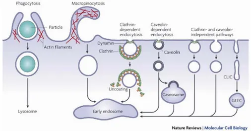
There are multiple proposals for the mechanism of internalization via such proteoglycans, ranging from classical endocytic pathways (e.g. clathrin-coated pits or caveolae) to alternative routes (e.g. direct translocation through the plasma membrane). Taken together, the data on Tat and Tat-cargoes do not support the dominance of one exclusive cell entry pathway. The lack of complete inhibition by selective drugs or complete co-localization with known markers consistently suggests a multiplicity of entry pathways for this basic peptide. Aside from clathrin and caveolae, other mechanisms of crossing the plasma membrane include the non-clathrin/noncaveolin-type pathway(s) (lipid rafts or microdomains, e.g., IL-2 receptor), macropinocytosis (platelet derived growth factor), potocytosis (folate receptor) and phagocytosis (specialized cells only) (Fig. 1.4).
Fig. 1.4 Pathways of entry into cells. Taken from Mayor et al. 71
It must be kept in mind that the Tat peptide can adhere to any negative offerings on the cell surface simply because of its charge and then be internalized through natural cell membrane recycling on regions or microdomains, presumably captured by any type of endocytic vesicle. Cell plasma membrane turnover continues constitutively at an estimated rate of ~2-5%/min 72, 73, meaning 100% of the cells surface is internalized
Intracellular cargo delivery using Tat-derived peptides 13
nonspecifically in less than an hour, notwithstanding the faster receptor-mediated or stimulated routes of uptake.
1.2.4 Intracellular fate and transport properties of Tat-peptide-cargoes
Several data indicate that the Tat peptide can determine cellular uptake of nonpermeant molecules and lead to the expected biological response, consistently with the internalization of intact peptide-cargo fusions 74, 75. Despite the variety of biotechnological applications of the Tat peptide as a delivery vector, little is known about the intracellular behavior and fate of Tat-peptide-based chimeras.
As mentioned above, the Tat peptide is considered a putative NLS, on the basis of in
vitro assays 8, 9. So far, two distinct classes of NLSs were identified: a single stretchof basic residues, exemplified by the NLS of the simian virus SV40 large tumor/antigen (T-ag) (PKKKRKV), or two clusters of basic residues separated by aspacer region of 10-12 amino acids, as typified by Xenopus laevis nucleoplasmin (KRPAAIKKAGQAKKKK)76, 77. These signals are usually recognized by the cytoplasmic receptor-proteins Importin-α (Imp-α) and Importin-β (Imp-β) and actively translocated through the pore channel 78. Briefly, Imp-α recognizes the NLS, and Imp-β is responsible for docking to the NPC and translocation through the pore (for a more detailed description of this mechanism, see chapter 5).
Because the NLS sequence present in Tat is arginine-rich, and therefore basic, it might be assumed that it would mediate nuclear import via the same import pathway utilized by the above-mentioned prototypical basic NLSs. Truant and Cullen, however, proposed an active nuclear-import mechanism involving the cytosolic factor Imp-β, but not Imp-α, and suggested the existence of a novel class of basic NLS sequences that function independently of Imp-α 9. This is not a novel pathway, since many viral and non viral proteins are known to be delivered to the nucleus by specific binding to the Imp-β carrier 79-81.
In contrast, Efthymiadis et al. reported that the Tat peptide can target a large heterologous protein (β-galactosidase from E.coli, 120kd) to the nucleus in the absence of both Imp-β and Imp-α, that nuclear import of Tat-NLS-substrate is not inhibited by an excess of an SV40 T antigen NLS peptide, and that the Tat-NLS fails to bind to either Imp-α or Imp-β. An entirely novel pathway was invoked that involves energy consumption and binding to nuclear components 8.
These controversies prompted us to further investigate Tat-peptide trasport biology in living cells. In the following chapter I shall present a novel metodological approach
Vector-based delivery strategies: from HIV-1 Tat protein to cell penetrating peptides
14
that allowed us to define the ct ual Tat-peptide transport propetifes in living cells, without the need to disrupt the integrity of the cytoplasmic or nuclear membranes.
Chapter
2
In vivo study of HIV-1 Tat arginine-rich
motif (I): nuclear import properties
In this Chapter we shall address Tat-peptide intracellular transport properties in living cells. The presented results impact this field both from the methodological point of view and from that of the specific outcome of our research with the demonstration of the actual Tat-peptide nuclear-import properties. From the point of view of the first issue here we show an in vivo approach that allows us to investigate Tat-peptide intracellular-transport properties without the need to perturbe the integrity of the cytoplasmic or nuclear membranes. More in detail, we compare Tat-peptide-induced transport properties to well-established passive-diffusion and active-import benchmarks of increasing molecular weight (MW) both below and above the threshold for passive diffusion through the nuclear pore.
Thus, we show that, contrary to previous in vitro experiments that suggested the relevance of active processes (albeit of different detailed molecular origin 8, 9), in physiological conditions Tat-cargo nuclear permeation is not an energy-consuming process. Thus, we discuss the limitations of Tat-cargo passive-diffusion route in terms of acceptable cargo size.
2.1 Materials and Methods
We constructed a prototypical passive-diffusion system composed by a small-size protein able to freely diffuse between nucleus and cytoplasm (single GFP, 27 kd) and a
In vivo study of HIV-1 Tat arginine-rich motif (I): nuclear import properties
16
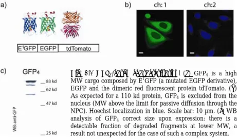
larger complex of 55 kd consisting of an EBFP linked to an EGFP (hereafter GFP2). In addition we introduced a third fusion protein, consisting of a higher MW cargo composed by E1GFP (a mutated EGFP derivative), EGFP and the dimeric red fluorescent protein tdTomato (in the following, GFP4) (Fig.2.1), whose size (110 kd) makes it unable to diffuse across the NPC 82.
Fig. 2.1 GFP4-cargo characterization. (a) GFP4 is a high
MW cargo composed by E1GFP (a mutated EGFP derivative),
EGFP and the dimeric red fluorescent protein tdTomato. (b) As expected for a 110 kd protein, GFP4 is excluded from the
nucleus (MW above the limit for passive diffusion through the NPC). Hoechst localization in blue. Scale bar: 10 µm. (c) WB analysis of GFP4 correct size upon expression: there is a
detectable fraction of degraded fragments at lower MW, a result not unexpected for the case of such a complex system.
2.1.1 Engineering of expression vectors
The expression vector encoding for EGFP protein was generated by PCR amplification of EGFP template and was inserted into HindIII-EcoRI sites of pcDNA3.1(+) vector from Invitrogen.
For the construction of the plasmid encoding for EBFP-EGFP proteins, a first fragment generated from PCR amplification of the EBFP template (p-EBFP C1 Clontech) was inserted into HindIII-BamHI sites of pcDNA3.1(+) vector. The second EGFP was cloned by two steps of PCR amplification into BamHI and XbaI sites introducing a spacer linker of 17 aminoacids (ASGGGGGLVPRGSASGA) between the two EGFP sequences. The template used for PCR derived from p-EGFP N1 (Clontech). The NLSSV40-EGFP plasmid was constructed by two rounds of multiple point mutations onto the sequence of Tat11-EGFP plasmid (see Chapter 3) using QuickChange Kit (Stratagene). The two primers used to transform the Tat11 sequence into the NLS of SV40 are reported as ‘primer 1-2’ in Appendix D. Antisense primers
Materials and Methods 17
had reverse complementary sequences. The NLSSV40-EBFP-EGFP plasmid was constructed by replacing the EGFP sequence of NLSSV40-EGFP plasmid with EBFP-EGFP derived from p-EBFP-GFP, both digested with HindIII and XbaI restrictions enzymes.
To obtain the construct p-E1GFP-EGFP-tdTomato the E1GFP template (EGFP containing mutations T65S T203Y) was inserted into HindIII and BamHI sites of pcDNA3.1(+) including a streptavidin tag (MASWSHPQFEKGA) at the amino-terminal of the E1GFP. A second EGFP (template p-EGFP N1 Clontech) was introduced by PCR amplification in BamHI and EcoRI sites. Finally, the tdTomato template, obtained from Roger Y.Tsien Laboratory 83, was amplified by PCR introducing EcoRI site in 5’ and XbaI site in 3’ extremity. To obtain the corresponding active import benchmark, NLSSV40-E1GFP-EGFP-tdTomato plasmid was constructed by replacing the EGFP sequence of NLSSV40-EGFP plasmid with E1 GFP-EGFP-tdTomato derived from E1GFP-EGFP-tdTomato, digested with HindIII and XbaI restrictions enzymes.
Here the behavior of these two classes of constructs will be compared to that of the same GFP-cargoes fused to the Tat11-sequence. Constructs with Tat11-sequence used in this study were based on the Tat11-EGFP sequence cloned in pGEX2T plasmids described in Ferrari et al. 56. The fragment encoding for Tat
11-EGFP protein was cut
BamHI and EcoRI and subcloned in pcDNA3.0 version (Invitrogen). Subsequently, a HindIII restriction site in the MCS was destroyed by mutagenesis using QuickChange
Kit (Stratagene). The primer used to remove HindIII site is reported as ‘primer 3’ in Appendix D. The unique HindIII restriction site between Tat11 and EGFP sequences, was used to engineer the following cargo constructs. Tat11-EBFP-EGFP and Tat11 -E1GFP-EGFP-tdTomato constructs were obtained by subcloning EBFP-EGFP and E1GFP-EGFP-tdTomato in Tat
11-EGFP plasmid cut by HindIII-XbaI in order to remove the EGFP sequence.
2.1.2 Cell culture, transfection, and immunoblotting analysis
CHO-K1 were provided from the American Type Culture Collection (CCL-61 ATCC) and were grown in Ham’s F12K medium supplemented with 10% of fetal Bovine Serum at 37ºC and in 5% CO2. Transfection of all constructs was carried out using Lipofectamine Reagent (Invitrogen) according to the manufacturer’s instructions. For live imaging cells were plated 24 h before transfection onto a 35-mm glass bottom dish (WillCo-dish GWSt-3522).
In vivo study of HIV-1 Tat arginine-rich motif (I): nuclear import properties
18
Expression of cargo constructs was confirmed by Western blotting. Lysis was performed in a buffer containing 1% Triton X-100, 50 mM HEPES (pH=7.5), 150 mM NaCl, 10% glycerol, 1.5 mM MgCl2, 5 mM EGTA, 10 µg/ml aprotinin, 10 µg/ml leupeptin. Lysates were then analyzed by SDS-PAGE and transferred onto nitrocellulose filters. For Western blotting procedures, filters were incubated with anti-GFP monoclonal antibody (JL-8, Clontech) and immunodetected with anti-mouse HRP-conjugated secondary antibodies (Amersham Biosciences).
2.1.3 Fluorescence imaging by confocal laser scanning microscopy (CLSM) Cell fluorescence was analyzed using a Leica TCS SP2 inverted confocal microscope (Leica Microsystems AG, Wetzlar, Germany) equipped with an Ar-laser for excitation at 458, 476, 488 and 514 nm, and with a HeNe-laser for excitation at 633 nm. A diode laser (Hamamatzu Photonics, Japan) was used for excitation at 403 nm (Hoechst imaging). Glass bottom Petri dishes containing transfected cells were mounted in a thermostated chamber (Leica Microsystems) and viewed with a 40x 1.25 NA oil-immersion objective (Leica Microsystems). Live-cell imaging was always performed at 37ºC. Images were collected using 10-20 µW excitation power at the sample and monitoring the emission by means of the AOBS-based built-in detectors of the confocal microscope. The following collection ranges were adopted: 410-450 nm (Hoechst), 500-550 nm (GFP), and 650-750 nm (Mitotracker®). All images were corrected by subtracting the background signal. Data were analyzed by Leica Imaging package version 2.61 and by an implementation of Igor-Pro software package.
2.2 Demonstration of Tat-peptide actual nuclear import properties
2.2.1 Cargo-dependent sub-cellular localization of constructsAs a first test, Chinese hamster ovary (CHO) K1 cells were transfected with the constructs encoding for GFP, GFP2, and GFP4. After 20 hours, cells were examined by confocal microscopy. As expected for a purely diffusive molecule, GFP displayed a uniform distribution throughout the cell with no sub-cellular organelle-specific localization 84 (Fig. 2.2.a). We obtained the same result in the case of GFP
2 (Fig. 2.2.b): the cytoplasm and the nucleus of cells were uniformly stained by this protein. In contrast, GFP4 was detected in the cytoplasm but excluded from the nuclear compartment in the analyzed cells (nuclear staining by Hoechst, as in Fig. 2.1.b, channel 2), as expected from the expression of a 110 kd protein lacking an NLS (Fig. 2.1 and Fig. 2.2.c). A small (~10%) fraction of the cells, however, showed detectable
Demonstration of Tat-peptide actual nuclear import properties 19
nuclear staining (data not shown) that can be linked to degradation fragments of the GFP4 protein, as suggested by immunoblotting analysis (GFP4 lane in Fig. 2.1.c). We used GFP4 to study the NLS capability of peptides, since only in the presence of active (energy-dependent) nuclear import, GFP4 signal can be detected in the nuclear compartment. In fact, a markedly different behavior was observed for NLSSV40-tagged constructs (Fig. 2.2.d-f): all GFP cargoes were successfully imported and, consequently, under physiological conditions, were predominantly detected in the nucleus (nuclear/cytoplasmic fluorescence ratio, Keq > 1). It is worth noting that the nuclear accumulation of NLSSV40-based constructs was even more marked with increasing cargo MW (compare sub-cellular localizations in Fig. 2.2.d-f).
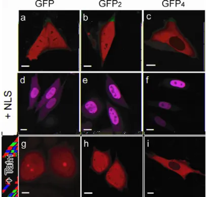
Fig. 2.2 Cargo-dependent subcellular localization of constructs. (a-b-c) Confocal images of expressed untagged GFP-cargoes. (d-e-f) The NLS-tagged cargoes are predominantly localized in the nucleus of expressing cells. In particular, the nuclear accumulation of these constructs is proportional to the increasing MW of the cargo. (g-h-i) Tat11-GFP and Tat11-GFP2 are uniformly distributed between compartments (cargo
MW is under the limit for passive diffusion), while Tat11-GFP4 is excluded from the
nucleus (cargo MW above the limit for passive diffusion). Scale bars: 10µm
In vivo study of HIV-1 Tat arginine-rich motif (I): nuclear import properties
20
This peculiar behavior can be linked to the fact that while both large and small proteins are rapidly imported thanks to the NLS-specific active processes, their nucleus-to-cytoplasm passive diffusion is increasingly impaired by the cargo size.
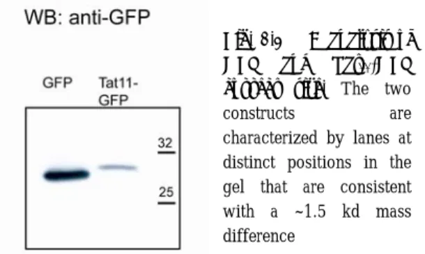
Finally, we transfected CHO K1 cells with Tat11-tagged GFP, GFP2 and GFP4 cargoes and analyzed their sub-cellular distribution at the confocal microscope. Independent of the level of transfection, Tat11-GFP sub-cellular distribution was rather uniform between cytoplasm and nucleoplasm with a concomitant detectable enrichment in nucleolar fluorescence (Fig. 2.2.g). In an analogous manner, Tat11-GFP2 preserved the homogeneous distribution between nucleus and cytoplasm (Fig. 2.2.h). Remarkably, Tat11-GFP4 was almost exclusively localized within the cytoplasm of expressing cells (Fig. 2.2.i), indicating that the Tat11-sequence was not able to determine nuclear import of a cargo that is above the limit for passive diffusion across the nuclear pore complex. In short, the cargo-dependent sub-cellular localization of Tat11-based constructs closely resembles that of their corresponding passive-diffusion benchmarks discussed above (Fig. 2.2.a-c). The actual size of GFP and Tat11-GFP constructs was verified by Western Blot analysis using an anti-GFP monoclonal antibody. Figure 2.3 shows that Tat11-GFP and GFP are characterized by lanes at distinct positions in the gel that are consistent with an approximate mass difference of 1.5 kd. The accuracy of this measurement is not sufficient to provide the exact quantitative determination of the construct, but beyond the mentioned consistency it must be noted that the gel shows no additional components or smearing of the lane as would be expected in the presence of degradation of the fused peptide.
Fig. 2.3 WB analysis of GFP and Tat11-GFP correct size. The two
constructs are characterized by lanes at
distinct positions in the gel that are consistent with a ~1.5 kd mass difference
A further check on the possibility of peptide degradation was performed by testing the effect of inhibitors such as chloroquine and MG132. These affect degradation in the
Demonstration of Tat-peptide actual nuclear import properties 21
lysosome and the proteosome, respectively. No change in Tat11-GFP subcellular distribution was observed (data not shown).
2.2.2 Nuclear permeation upon energy depletion
In order to selectively monitor the role of active processes in determining the nuclear permeation of the tested proteins, we analyzed their sub-cellular localization in response to energy depletion.
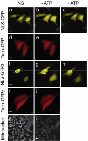
Fig. 2.4 Nuclear permeation on energy depletion. The functional NLS of SV40 is able to accumulate GFP and GFP2 cargoes into the nuclear
compartment under physiological conditions (NG, normally growing cells: a, f). After 30 min of energy depletion treatment, both constructs equilibrated between nucleus and cytoplasm (-ATP: b, g). Within 10-15 min after shifting the same cell back to physiological conditions, active nuclear import was completely restored (+ATP: c, h). Similar energy depletion assays did not alter the characteristic sub-cellular localization of Tat11-GFP
(d, e) and Tat11-GFP2 (i, j). Scale bar:
10 µm. Under physiological
conditions, Mitotracker® is selectively
sequestered by active mitochondria (k). Within 30 min of treatment with 2-deoxy-D-glucose and sodium azide, a 60-70% decreased in Mitotracker staining is observed, indicating mitochondrial failure (l). Scale bar: 10 µm.
Intracellular ATP/GTP stores were depleted by incubating cells with 2-deoxy-D-glucose and sodium azide in 2-deoxy-D-glucose-free medium. Within 30 min of this treatment, most cells lost Mitotracker® staining (Fig. 2.4.k-l), thus indicating that their mitochondria were no longer active 85, 86. We found that when cells expressing
In vivo study of HIV-1 Tat arginine-rich motif (I): nuclear import properties
22
NLSSV40-GFP were incubated in the energy-depleting medium fluorescence rapidly equilibrated between nucleus and cytoplasm (Fig. 2.4.b). This behavior is consistent with the dominant NLS-mediated nuclear import: when the latter is blocked by energy depletion, NLSSV40-GFP passive diffusion to the cytoplasm can successfully restore a homogeneous intracellular distribution. Notably, this behavior is reversible: within few minutes after shifting cells back to physiological conditions fluorescence was again predominantly localized in the nucleus (Fig. 2.4.c). The same treatment showed no effect on Tat11-GFP sub-cellular localization (Fig. 2.4.d-e), thus providing a first evidence that Tat11-cargo trafficking is an energy-independent mechanism. We performed analogous measurements on Tat11-GFP2 and NLSSV40-GFP2, as shown in Fig. 2.4.f-h and i-j, respectively. NLSSV40-GFP2 equilibrated between nucleus and cytoplasm under energy depletion conditions and promptly accumulated in the nucleus when active import was restored. Once again, however, the same treatment showed no effect on Tat11-GFP2 sub-cellular localization. As expected, GFP4-based constructs did not change their subcellular localization in response to energy-depletion treatment, as they can not permeate across the NE (data not shown). Analogously to Tat-peptide constructs, isolated GFP and GFP2 showed no energy-dependent sub-cellular distribution (data not shown), as expected for these passive-diffusion benchmark proteins. Comparison of the behavior displayed by Tat11-based constructs and the two benchmarks provides clear indication of a dominant passive-diffusion mechanism for Tat-cargo shuttling across the nuclear envelope.
2.3 Limitations of wild-type Tat-peptide as a delivery vector
So far we addressed the intracellular localization and transport properties of intact Tat-peptide-cargos in living cells. We demonstrated that Tat-cargo shuttling across the nuclear envelope is dominated by passive diffusion. Accordingly, we identified the potential and the limitations of Tat as a molecular carrier from the point of view of cargo size. Our results are in contrast with previously reported in vitro studies that highlighted the relevance of active processes in Tat-peptide-driven nuclear import 8, 9 and prompted us to consider the detailed experimental conditions of those studies. In particular, we believe that the impact of the non-physiological conditions characteristics of import assays in permeabilized cells should be carefully evaluated. For example, one important issue in such experiments is the absence of cofactors that can influence Tat-peptide properties. More in detail, although we can not exclude that
Limitations of wild-type Tat-peptide as a delivery vector 23
import carriers may be interacting partners of Tat-peptide, our data demonstrate that, if at all present, this interaction does not lead to a detectable energy-dependent route of nuclear import. A likely explanation of this scenario is that the interaction between an import carrier and Tat-peptide is efficiently inhibited by other cellular substrate molecules. In keeping with this hypothesis, Friedler et al. reported a strong inhibition of Tat-peptide nuclear import by the addition of cytosolic factors containing, among others, RNA molecules 87, that usually leak out of the cells during cell permeabilization procedures. It is well-established, in fact, that the arginine-rich motif has a strong affinity for RNA molecules. For instance, it was demonstrated that continued ribosomial RNA (rRNA) synthesis is involved in the nucleolar accumulation of Rev, a protein from HIV-1 containing an arginine-rich motif 88. Moreover, it was reported that the arginine-rich motif of Rev has a much higher affinity for RNA molecules than it does for the Importins 88, 89. On the contrary, our in vivo assays largely preserve physiological interactions of the Tat peptide and thus truly show that Tat-peptide intracellular properties are not dominated by binding to import carriers. Our results provide useful insight into the nature of target molecules available for Tat-peptide-driven nuclear delivery. Firstly, our analysis shows that protein cargoes exceeding the size-threshold value for passive diffusion through the NPC will not gain access into the nuclear compartment. This is confirmed by the data published by Nitin et al. 90: these authors reported evidence that a Tat peptide-based molecular beacon containing Streptavidin (~60 kd) is efficiently targeted to the cell cytoplasm but substantially excluded from the nucleus 90. Moreover, results reported by Neumann et al. show that peptides containing the relevant Rev and Tat motives conjugated with fluorescently labelled BSA (66 kd) are not able to perform nuclear import upon cytoplasmic microinjection, in contrast with the nuclear accumulation observed using the NLS of SV40 91. Nonetheless, it must be noted that molecular cargoes up to 55 kd (namely, GFP2 size) can successfully reach the nucleus through the passive route unveiled here.
Chapter
3
In vivo study of HIV-1 Tat arginine-rich
motif (II): intracellular trafficking and
interactions
Here the quantitative aspects of Tat11-nuclear/cytoplasm exchange will be addressed by means of a set of time-resolved experiments that were analyzed thanks to a dynamic model that includes both reversible passive diffusion between the two compartments and active nuclear import of proteins from the cytoplasm (see Appendix A for more details). The passive-diffusion flux plays a role in the case of proteins that are characterized by a MW less than the nuclear-pore exclusion limit. Active-transport flux must be included when NLS properties come into play. Nucleus-to-cytoplasm active-transport processes were not included in the dynamic model. Each contribution can be characterized by one transport parameter that must be experimentally determined by studying the fluorescence recovery after photobleaching (FRAP) of nuclear and cytoplasmic compartments. In the last part of this chapter I shall analyze Tat peptide interactions with cellular components by means of a combination of in vitro and in vivo assays
3.1 Dynamic model for nuclear/cytoplasmic exchange
In a typical FRAP measurement, nuclear or cytoplasmic fluorescence was photobleached by high laser beam power and the following recovery process was
In vivo study of HIV-1 Tat arginine-rich motif (II): intracellular trafficking and interactions
26
monitored by time-lapse imaging. The mathematical analysis of the system (see Appendix A for details) leads to equations:
(
)
( t/τ) C 0 C C C F F F F = ∞+ − ∞ ⋅e− (3.1)(
)
( t/τ) N 0 N N N F F F F = ∞+ − ∞ ⋅e− , (3.2) where FC0 and FC∞, and FN0 and FN∞ label the concentrations at time zero and at equilibrium in the cytoplasm and in the nucleoplasm, respectively.By fitting the fluorescence recovery curves to the monoexponential equations reported above, it is possible to derive the characteristic transport parameters of the diffusing molecule. Although a detailed description of the mathematical model used for FRAP data analysis will be presented in Appendix A, it is useful to define here some parameters that will be used in the following. In particular:
i) kDC, and kDN quantify the permeability of nuclear envelope to nucleus-to-cytoplasm and nucleus-to-cytoplasm-to-nucleus passive diffusion, respectively;
ii) kAT quantifies the flux of actively imported molecules from cytoplasm to nucleus a.
Experimentally, each FRAP experiment started with a line-averaged image of the cell followed by a single-point bleach (nonscanning) near the center of the nucleus with laser light at full power for the minimum time required to photobleach most of nuclear fluorescence. Fluorescence of the cytoplasm was photobleached by performing repeated scans of the whole cytoplasmic region with laser at full power. In either case recovery was measured by starting a time-lapse acquisition within few milliseconds after the end of bleaching (sampling rate was tailored to the speed of fluorescence recovery of the tested protein). Each image of the recovery was 4-time line-averaged. Image size was 512x512 pixels and scan speed was usually set to 400 Hz. The pinhole size was set to the optimal value of 1.0 Airy (corresponding to a 81.44 µm confocal aperture). Before fitting, the experimental values of fluorescence were normalized by the fluorescence of the entire cell measured at the same time point, in order to minimize the effect of cell motility and defocusing on the recovery curves and to
a
The kAT values reported in the following sections refer to the minimum value of the active transport parameter (kATm, see Appendix A) since in our system the NLS intracellular concentration is higher than that of the active import carrier molecules (cytoplasmic carriers are all in the bound form).
Dynamic model for nuclear/cytoplasmic exchange 27
correct for bleaching caused by imaging. Moreover, data were normalized by pre-bleach fluorescence values in order to verify the presence of an immobile fraction of fluorescent molecules within the bleached compartment.
3.1.1 In vivo applications: FRAP analysis of passive diffusion
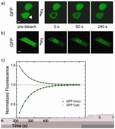
Fig. 3.1 FRAP analysis of GFP nucleus/cytoplasm shuttling. (a) Fluorescence recovery after nuclear photobleaching as GFP diffuses from cytoplasm to nucleus. Nuclear fluorescence (pre-bleach panel, white arrow) was photobleached by irradiating for 8 s a single-point with high laser power. Recovery was recorded by time-lapse imaging: selected images are reported with the corresponding time of acquisition. Scale bar: 10 µm. (b) Fluorescence of the cytoplasm was photobleached by performing repeated scans of the whole cytoplasmic region with laser at full power. Recovery was recorded by time-lapse imaging. Scale bar: 10 µm. (c) Time course of nucleoplasmic fluorescence recovery (full green circles) for the cell shown in (a). Cytoplasmic fluorescence concomitantly decreases as GFP diffuses from cytoplasm to nucleus (empty green circles). This symmetric process shows the same kinetics yielding a time constant (τ) around 70 s for the analysed cell.
In vivo study of HIV-1 Tat arginine-rich motif (II): intracellular trafficking and interactions
28
The fluorescence of the nuclear compartment of CHO K1 cells transfected with GFP was photobleached by irradiating for 8 seconds a single point at high laser power.The subsequent fluorescence recovery was recorded by time-lapse acquisition at 20-s intervals (Fig. 3.1.a). The fluorescence recovery in the nucleus is attributable to the influx of unbleached GFP molecules from the cytoplasm, in equilibrium with the efflux of bleached molecules from nucleus to cytoplasm (compare green curves on empty and full circles in Fig. 3.1.c: they show the same kinetics).Indeed, the fluorescence in the cytoplasm decreased as GFP diffused from cytoplasm into the nucleus. As expected, after cytoplasm photobleaching, cytoplasm fluorescence recovered together with a decrease in nuclear fluorescence (Fig. 3.1.b).
Since we are dealing with restricted exchange between two well-mixed compartments separated by a porous membrane, the post-bleach concentration dynamics is described by exponential decays 92, 93. Indeed, the kinetics of fluorescence recovery in all measured cells was accurately fitted by a single exponential function whose time constant (τ) expresses the rate of the protein shuttling across the nuclear membrane. Results from the whole population of analyzed cells yielded a τ value of 77 ± 19 s (Tab. 3.1) for GFP shuttling. The asymptotic value of 1 of the fluorescence recovery traces shown in Fig. 3.1.c indicates that the pool of nuclear proteins was completely exchangeable with the cytoplasm, as expected for a protein that does not interact with any sub-cellular substrates (see Materials and Methods for further details). By means of our dynamics model we derived kDN = 13.6 ± 3.7 µm3/s and kAT = 0 µm3/s (the latter value indicates the absence of any nuclear targeting property for isolated GFP proteins). Analogous FRAP measurements performed on GFP2 yielded an overall τ-value of 1178 ± 292 s (Tab. 3.1). This slower recovery rate of passively diffusing GFP2 (compared to GFP) is expected, since its molecular weight is close to the estimated molecular cutoff size for passive diffusion through the NPC (i.e., ∼60-70 kd) 94, 95 and is in agreement with recently reported in vivo analyses 96.
3.1.2 In vivo applications: FRAP analysis of active import
Next, we quantitatively investigated NLS-driven transport in order to determine the rates of active import compared to passive diffusion. NLS-GFP nuclear fluorescence was photobleached and the subsequent recovery was monitored by time-lapse imaging (Fig. 3.2). From the kinetic analysis of the whole FRAP dataset we found that active nuclear import is characterized by a recovery time constant of 60 ± 3 s (Tab. 3.1), a value not significantly faster than that of isolated GFP. From the whole population of
Dynamic model for nuclear/cytoplasmic exchange 29
examined cells we derived a characteristic kAT range of 10 - 17.1 µm3/s for NLS-GFP active import and a kDN-value of 5.2 ± 0.6 µm3/s for its concomitant reversible passive diffusion (Tab. 3.1). As for the case of single-GFP cargo, we tested the influence of molecular size on the NLS-driven nuclear-import kinetics. Notably, NLS-GFP2 active import is about five times faster than isolated GFP2 shuttling across the NE (Fig. 3.2.b-c).
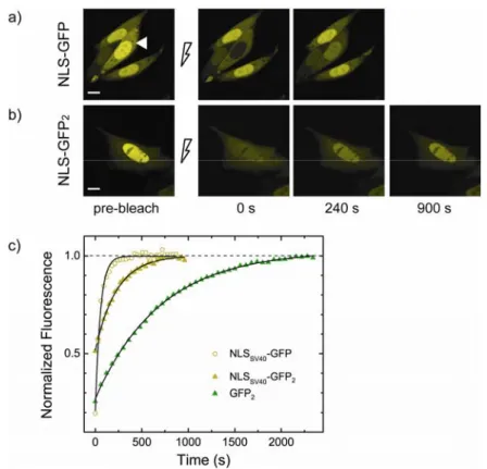
Fig. 3.2 FRAP analysis of active import. (a) FRAP analysis of actively imported NLSSV40
-GFP. Nuclear fluorescence (pre-bleach panel) was photobleached by irradiating for 10 s a single-point with high laser power and subsequent recovery was monitored by time-lapse imaging: selected images are reported with the corresponding time of acquisition. Scale bar: 10 µm. (b) FRAP analysis of NLSSV40-GFP2 (18-s-bleach). Intensity of post-bleach images
has been adjusted to better see fluorescence recovery signal. (c) Kinetics of nucleoplasmic fluorescence recovery after phothobleaching for cells shown in (a), and (b). τ-values (mean ± SD) are 704 ± 8 s (GFP2, green triangles), 230 ± 5 s (NLSSV40-GFP2, yellow triangles), 54
± 2 s (NLSSV40-GFP, yellow empty circles). The asymptotic value of 1 of recovery curves,
indicates that no immobile fraction of molecules is present in the nuclear compartment. Transport parameters for the whole population of analysed cells are reported in Tab. 3.1.
In vivo study of HIV-1 Tat arginine-rich motif (II): intracellular trafficking and interactions