R E S E A R C H A R T I C L E
Open Access
The surgical treatment of non-metastatic
melanoma in a Clinical National Melanoma
Registry Study Group (CNMR): a
retrospective cohort quality improvement
study to reduce the morbidity rates
Antonella Vecchiato
1†, Simone Mocellin
1,2, Paolo Del Fiore
1*†, Giulio Tosti
3, Paolo A. Ascierto
4,
Maria Teresa Corradin
5, Vincenzo De Giorgi
6, Giuseppe Giudice
7, Paola Queirolo
8, Caterina Ferreli
9,
Marcella Occelli
10, Monica Giordano
11, Giusto Trevisan
12, Luigi Mascheroni
13, Alessandro Testori
14, Romina Spina
1,
Alessandra Buja
15, Francesco Cavallin
16†, Corrado Caracò
17, Antonio Sommariva
18†, Carlo Riccardo Rossi
1,2on
behalf of the Clinical National Melanoma Registry Study Group at the Italian Melanoma Intergroup (IMI)
Abstract
Background: Reproducible, high-quality surgery is a key point in the management of cancer patients. Quality indicators for surgical treatment of melanoma has been presented with benchmarks but data on morbidity are still limited. This study presents the quality indicators on morbidity after surgical treatment for non-metastatic skin melanoma in an Italian registry.
Methods: Data were extracted from the Central National Melanoma Registry (CNMR) promoted by the Italian Melanoma Intergroup (IMI). All surgical procedures (WE, SNLB or LFND) for non-metastatic skin melanoma between January 2011 and February 2017 were evaluated for inclusion in the study. Only centers with adequate
completeness of information (> 80%) were included in the study. Short-term complications (wound infection, dehiscence, skin graft failure and seroma) were investigated.
Results: Wound infection rate was 1.1% (0.4 to 2.7%) in WE, 1.3% (0.7 to 2.5%) in SLNB and 4.1% (2.1 to 8.0%) in LFND. Wound dehiscence rate was 2.0% (0.8 to 5.1%) in WE, 0.9% (0.2 to 3.0%) in SLNB and 2.8% (0.9 to 8.6%) in LFND. Seroma rate was 4.2% (1.5 to 11.1%) in SLNB and 15.1% (4.6 to 39.9%) in LFND. Unreliable information was found on skin graft failure.
(Continued on next page)
© The Author(s). 2021 Open Access This article is licensed under a Creative Commons Attribution 4.0 International License, which permits use, sharing, adaptation, distribution and reproduction in any medium or format, as long as you give appropriate credit to the original author(s) and the source, provide a link to the Creative Commons licence, and indicate if changes were made. The images or other third party material in this article are included in the article's Creative Commons licence, unless indicated otherwise in a credit line to the material. If material is not included in the article's Creative Commons licence and your intended use is not permitted by statutory regulation or exceeds the permitted use, you will need to obtain permission directly from the copyright holder. To view a copy of this licence, visithttp://creativecommons.org/licenses/by/4.0/. The Creative Commons Public Domain Dedication waiver (http://creativecommons.org/publicdomain/zero/1.0/) applies to the data made available in this article, unless otherwise stated in a credit line to the data.
* Correspondence:[email protected]
†Antonella Vecchiato, Paolo Del Fiore, Francesco Cavallin and Antonio
Sommariva contributed equally to this work.
1Surgical Oncology Unit, Veneto Institute of Oncology IOV– IRCCS, Padova,
Italy
(Continued from previous page)
Conclusions: Our findings contribute to available literature in setting up the recommended standards for melanoma centers, thus improving the quality of surgery offered to patients. A consensus on the core issues around surgical morbidity is needed to provide practical guidance on morbidity prevention and management. Keywords: Melanoma, Melanoma morbidity, Skin Cancer, Melanoma morbidity, Melanoma surgical treatment, Melanoma quality improvement
Background
Worldwide, 287,723 new cases of cutaneous melanoma are diagnosed each year, causing the death of 60,712 patients [1]. In non-metastatic melanoma (i.e. without clinically evident regional lymph node or distant metas-tasis), surgery is the mainstay of treatment [2]. The sur-gical strategy involves a combination of wide excision (WE), sentinel lymph node biopsy (SLNB) and radical lymph node dissection (LFND), according to cancer sta-ging. WE can be virtually part of the surgical manage-ment of all patients with skin melanoma, SNLB is recommended based on the primary tumor thickness and LFND may be required in patients with positive sentinel node [3].
Reproducible, high-quality surgery is a key point in the management and prognosis of cancer patients [4, 5]. When a standardized surgical procedure is established, a cyclic audit (including collection, analysis and feedback of both procedural and outcome data) allows for moni-toring and improving the quality of surgery. However, cancer surgery is not always standardized, thus limiting the application of such approach [6].
While quality of surgery has been already investigated in other cancers, the use of measures of quality assur-ance for surgery is less established in melanoma [6]. Available literature offers substantial heterogeneity in surgical procedures among melanoma centers or even
among surgeons within the same center [7]. Although
adherence to current standards is part of a quality assur-ance process, the spreading of clinical practice guidelines is not sufficient per se to warrant homogeneity and qual-ity of surgical treatment [8]. There is a growing interest in the implementation of a quality assurance program that includes a quantitative analysis of a set of quality in-dicators, such as those that can be extracted from elec-tronic medical records [6].
Since 2014, the Italian Melanoma Intergroup (IMI) has been promoting the standardization and quality control of surgical treatment of stage I-III melanoma in Italy [9]. The IMI achieved an expert consensus on surgical treat-ment of melanoma, defining a list of quality indicators (detection rate, false negative rate, minimum number of excised lymph nodes, postoperative morbidity and local recurrence) with reference to benchmark values, which could be the basis for a standardized quality assurance
program in Italy [10]. While quality indicators for detec-tion rate, false negative rate and minimum number of excised lymph nodes were presented for the Central Na-tional Melanoma Registry (CNMR), data on morbidity were still pending and data on recurrence are waiting for adequate follow-up [10].
The present study focuses on evaluation of the quality indicators about morbidity after surgical treatment for non-metastatic skin melanoma.
Methods
This is a prospective multicenter study on postoperative morbidity after surgical treatment for non-metastatic skin melanoma. Data were extracted from the Central National Melanoma Registry (CNMR), which is a pro-spectively maintained national database for melanoma treatment promoted by the IMI in 43 melanoma centers
in Italy [9]. The study was conducted according to
Helsinki Declaration principles and was approved by the Ethics Committee of the Central National Melanoma Registry (CNMR). All patients gave their consent to have their data collected for scientific purpose.
The following short-term (within 30 days after sur-gery) complications were investigated: infection, dehis-cence, skin necrosis after WE; infection, dehiscence and seroma after SLNB or LFND. Complications were de-fined using the National Cancer Institute Common
Terminology Criteria for Adverse Events v4.0 [11].
Wound infection was defined as a disorder character-ized by an infectious process involving the wound. Wound dehiscence was defined as a finding of separ-ation of the approximated margins of a surgical wound. Skin necrosis was defined as a death of cells in living tissue caused by external factors such as infection, trauma, or bacterial agents. Seroma was defined as find-ing of a collection of serum in the tissues. Patient demographics were also collected.
All surgical procedures (WE, SNLB or LFND) for non-metastatic skin melanoma between January 2011 and February 2017 were evaluated for inclusion in the study. Only centers with adequate completeness of information (> 80%) were included in the study (11 centers for ana-lysis of morbidity after WE; 14 centers for anaana-lysis of morbidity after SNLB; 11 centers for analysis of morbid-ity after LFND). Within selected centers, patients with
pending information on morbidity and those with un-filled form were excluded from the analysis. Patient se-lection is shown in Supplementary Figs.1,2and3.
Statistical analysis was conducted using R 3.5 (R Foun-dation for Statistical Computing, Vienna, Austria) [12]. Categorical data were expressed as number and percent-age, and continuous data as median and interquartile range (IQR). The morbidity rate was pooled among cen-ters using a generalized linear mixed-effects model (with the center as random effect). The I2value was used to investigate heterogeneity, and meta-regression was used to explore the role of center size and groin site on het-erogeneity. A p-value less than 0.05 was considered sta-tistically significant.
Results
Wide excision
The analysis included 3118 patients (1647 males and 1471 females) who underwent WE in 11 centers (median 246 patients/center; min 90 patients/center, max 650 pa-tients/center). Median age was 57 years (IQR 45–70) and
median BMI was 24.8 kg/m2 (IQR 22.4–27.5).
Comor-bidities were present in 459 patients (15.1%), while the information was not available in 80 patients. Overall, median Charlson Comorbidity Index was 2 (IQR 0–3).
Wound infection occurred in 44 patients and dehis-cence in 100. The information on skin graft failure was not reliable due to the non-negligible number of unfilled forms (21%).
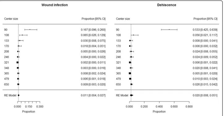
The proportion of patients developing wound infection ranged from 0.002 to 0.167 among centers, with a pooled proportion of 0.011 (95% CI 0.004 to 0.027) (Fig. 1). Heterogeneity was high (I2 = 87%) and center size (> 200 vs. < 200 patients) was found to have a sig-nificant contribution to heterogeneity (p < 0.0001). Sum-mary estimates of proportion of patients developing wound infection were 0.051 (95% CI 0.021 to 0.121; I2= 80%) in centers with low size (< 200 patients) and 0.006 (95% CI 0.003 to 0.009; I2 = 0%) in centers with large size (> 200 patients).
The proportion of patients developing dehiscence ranged from 0.005 to 0.533 among centers, with a pooled proportion of 0.020 (95% CI 0.008 to 0.051) (Fig.1). Het-erogeneity was high (I2= 93%) and center size (> 100 vs. < 100 patients) was found to have a significant contribution to heterogeneity (p < 0.0001). Summary estimate of pro-portion of patients developing dehiscence was 0.016 (95% CI 0.010 to 0.024; I2= 54%) in centers with large size (> 100 patients) and 0.533 (95% CI 0.425 to 0.639) in the only center with low size (< 100 patients).
Sentinel lymph node biopsy
The analysis included 1853 patients (993 males and 860 females) who underwent SLNB in 14 centers (median 98
patients/center; min 24 patients/center, max 338 pa-tients/center). SLNB site was axilla (1058 patients, 57.1%), neck (163 patients, 8.8%) or groin (632 patients, 34.1%). Median age was 57 years (IQR 45–69) and
me-dian BMI was 25.1 kg/m2 (IQR 22.7–28.0).
Comorbidi-ties were present in 304 patients (16.7%), while the information was not available in 40 patients. Overall, median Charlson Comorbidity Index was 1 (IQR 0–3).
Early wound complications occurred in 176 patients, including 29 wound infections, 39 dehiscences and 139 seromas (not mutually exclusive).
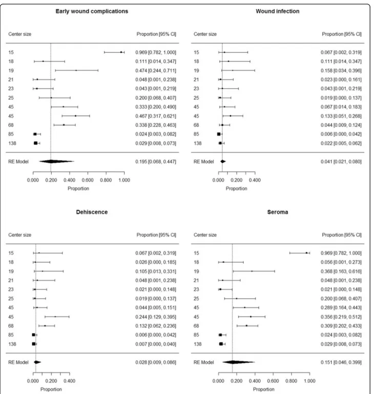
The proportion of patients developing early wound complications ranged from 0.007 to 0.831 among cen-ters. Summary estimate of proportion of patients devel-oping early wound complications after SLNB was 0.069 (95% CI 0.030 to 0.150) (Fig. 2). Heterogeneity was high (I2= 94%); neither center size (p = 0.11) or rate of groin sites among SLNBs (p = 0.84) were not found to have a significant contribution to heterogeneity.
The proportion of patients developing wound infection ranged from 0.005 to 0.104 among centers. Summary estimate of proportion of patients developing wound in-fection after SLNB was 0.013 (95% CI 0.007 to 0.025) (Fig. 2). Heterogeneity was moderate (I2= 60%); neither center size (p = 0.25) or rate of groin sites among SLNBs (p = 0.60) were not found to have a significant contribu-tion to heterogeneity.
The proportion of patients developing dehiscence ranged from 0.005 to 0.101 among centers. Summary es-timate of proportion of patients developing dehiscence after SLNB was 0.009 (95% CI 0.002 to 0.030) (Fig. 2). Heterogeneity was high (I2 = 88%); neither center size (p = 0.98) or rate of groin sites among SLNBs (p = 0.89) were not found to have a significant contribution to heterogeneity.
The proportion of patients developing seroma ranged from 0.004 to 0.792 among centers. Summary estimate of proportion of patients developing seroma after SLNB was 0.042 (95% CI 0.015 to 0.111) (Fig.2). Heterogeneity was high (I2= 95%); neither center size (p = 0.10) or rate of groin sites among SLNBs (p = 0.45) were not found to have a significant contribution to heterogeneity.
Radical lymph node dissection
The analysis included 502 patients (301 males and 201 females) who underwent LFND in 11 centers (median 25 patients/center; min 15 patients/center, max 138 pa-tients/center). LFND site was axilla (276 patients, 55.0%), neck (60 patients, 12.0%) or groin (166 patients, 33.0%). Median age was 59 years (IQR 47–70) and
me-dian BMI was 25.5 kg/m2 (IQR 22.9–28.6).
Comorbidi-ties were present in 93 patients (19.0%), while the information was not available in 12 patients. Overall, median Charlson Comorbidity Index was 2 (IQR 0–3).
Early wound complications occurred in 98 patients, in-cluding 22 wound infections, 27 dehiscences and 85 ser-omas (not mutually exclusive).
The proportion of patients developing early wound complications ranged from 0.024 to 0.969 among centers. Summary estimate of proportion of patients developing early wound complications after LFND
was 0.195 (95% CI 0.068 to 0.447) (Fig. 3).
Hetero-geneity was high (I2 = 94%); neither center size (p = 0.08) or rate of groin sites among LFNDs (p = 0.41) were not found to have a significant contribution to heterogeneity.
The proportion of patients developing wound infection ranged from 0.006 to 0.58 among centers. Summary esti-mate of proportion of patients developing wound infec-tion after SLNB was 0.041 (95% CI 0.021 to 0.080) (Fig.
3). Heterogeneity was moderate (I2= 54%); neither cen-ter size (p = 0.07) or rate of groin sites among SLNBs (p = 0.53) were not found to have a significant contribu-tion to heterogeneity.
The proportion of patients developing dehiscence ranged from 0.006 to 0.244 among centers. Summary es-timate of proportion of patients developing dehiscence after SLNB was 0.028 (95% CI 0.009 to 0.086) (Fig. 3). Heterogeneity was high (I2 = 80%); neither center size (p = 0.38) or rate of groin sites among SLNBs (p = 0.14) were not found to have a significant contribution to heterogeneity.
The proportion of patients developing seroma ranged from 0.021 to 0.969 among centers. Summary
estimate of proportion of patients developing seroma after SLNB was 0.151 (95% CI 0.046 to 0.399) (Fig.
3). Heterogeneity was high (I2 = 95%); neither center size (p = 0.18) or rate of groin sites among SLNBs (p = 0.29) were not found to have a significant con-tribution to heterogeneity.
Discussion
This study reported the morbidity rates in the largest series of non-metastatic melanoma patients in Italy and one of the largest series worldwide. The present work allowed to evaluate standardization and quality of surgi-cal treatment of cutaneous melanoma within the frame of the Italian Melanoma Intergroup, which is the largest Italian scientific organization dedicated to the manage-ment of patients with this disease [10].
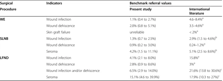
The occurrence of wound dehiscence and seroma after SLNB or LFND were in broad agreement with previous studies [13–15] (Table 1), thus suggesting limited opportunities for further improvements now. Such rates can be used as morbidity benchmarks in addition to available quality indicators for surgical treatment of melanoma [10].
Interestingly, we found lower occurrence of wound in-fection after WE or LFND when compared to available literature [13] (Table 1). On one hand, this difference might be partially due to different definitions of wound infection, i.e. presence of fever, only skin redness, sup-puration versus cellulitis, isolation of bacteria from wound. On the other hand, some factors might have led
to such difference, including: i) recording of infections occurring only during hospital stay or also at the time of outpatient department visits; ii) different frequency/type of antibiotic prophylaxis; iii) different surgical techniques (i.e., use, type and duration of permanence of drainages in the surgical wound).
Postoperative infections account for around one out of four complications associated with
hospital-related health care procedures, and can impair
patient prognosis [16]. Beyond clinical importance, a low infection rate can also have an economic impact, since the management of hospital-related infections requires about 0.8% of gross domestic product (GDP) in Italy [17].
Unfortunately, the information on skin graft failure was not reliable because one out of five forms did not report such complication. This situation likely occurred because skin graft failure required a reconstructive
surgical management that several centers demanded to a different surgical unit. This situation may be addressed by improving the information exchange among surgical centers involved in patient care.
Of note, morbidity rates showed high heterogeneity across melanoma centers, underlying the role of the cen-ter itself on this matcen-ter. Our data suggested an associ-ation between higher morbidity rate and small-volume
centers, thus confirming the relationship between pa-tient outcome and hospital surgical volume [18].
The present study contributes to the definition of quality indicators for surgical treatment for non-metastatic skin melanoma, by adding morbidity indi-cators that can be used as the basis for a standardized
quality assurance program [10]. The importance of
this topic relies on the large number of surgical
procedures for non-metastatic skin melanoma [2], thus patient management and prognosis can benefit from quality control and standardization of such pro-cedures [4, 5].
The strengths of this study included the prospective collection of data in a national registry, the multicen-ter design and the standardized definitions of the
complications [11]. The study has some limitations.
First, a considerable number of centers were excluded due to poor completeness of data. Although this ap-proach allowed limiting the impact of low-quality data on the study results, the representativeness of the in-cluded centers may be limited. Future developments will aim to achieve adequate completeness of data in the excluded centers and will implement regular audits. Second, the occurrence on skin graft failure after WE could not be evaluated due to the non-negligible number of unfilled forms. This limitation can be addressed by future improvements regarding information exchange among surgical centers involved in patient care.
Conclusion
Our findings contribute to available literature in set-ting up the recommended standards for melanoma centers, thus improving the quality of surgery of-fered to patients. Such quality indicators can be used by other hospitals to direct quality improvement efforts.
Supplementary Information
The online version contains supplementary material available athttps://doi. org/10.1186/s12885-020-07705-4.
Additional file 1: Supplementary Figure 1. Flow-chart of patient in-clusion in wide excision (WE) analysis. Supplementary Figure 2. Flow-chart of patient inclusion in sentinel lymph node biopsy (SLNB) analysis. Supplementary Figure 3. Flow-chart of patient inclusion in radical lymph node dissection (LFND) analysis.
Abbreviations
WE:Wide excision; SLNB: Sentinel lymph node biopsy; LFND: Radical lymph node dissection; IMI: Italian Melanoma Intergroup; CNMR: Clinical National Melanoma Registry; GDP: Gross domestic product
Acknowledgements
The authors would like to thank the Intergruppo Melanoma Italiano (IMI) and the Clinical Research Technology (CRT).
^ Clinical National Melanoma Registry Study Group at the Italian Melanoma Intergroup:
ASERO SALVATORE: Azienda Ospedaliera di Rilievo Nazionale e di Alta Specializzazione Garibaldi-Nesima, Catania;Italy.
BARBATI ROSANNA: ASL Roma C - Ospedale S. Eugenio, Roma, Italy. BIANCHI LUCA: Azienda Ospedaliera Universitaria Policlinico Tor Vergata, Roma, Italy.
BRUDER FRANCESCA: Ospedale Oncologico Cagliari, Cagliari, Italy. CATRICALÀ CATERINA: Istituto Dermatologico San Gallicano IRCCS– IFO, Roma;Italy.
CINIERI SAVERIO: Presidio Ospedaliero Antonio Perrino, Brindisi, Italy. DEL VECCHIO MICHELE: Fondazione I.R.C.C.S. Istituto Nazionale dei Tumori, Milano,Italy.
DI FILIPPO FRANCO: Istituto Nazionale Tumori Regina Elena IRCCS– IFO, Roma, Italy.
FARGNOLI MARIA CONCETTA: Presidio Ospedaliero San Salvatore, L’ Aquila, Italy.
FIERRO MARIA TERESA: A.O.U. Città della Salute e della Scienza - P.O. San Lazzaro, Torino, Italy.
FORCIGNANÒ ROSACHIARA: Azienda Ospedaliera Vito Fazzi, Lecce, Italy.
Table 1 Referral values for morbidity rate in the IMI-CNMR study and in the international
Surgical Indicators Benchmark referral values
Procedure Present study International
literature
WE Wound infection 1.1% (0.4 to 2.7%) 4.6–8.4%a
Wound dehiscence 2.0% (0.8 to 5.1%) 3.5–4.6%a
Skin graft failure unreliable < 2%a
SLNB Wound Infection 1.3% (0.7 to 2.5%) 2.9% (1.5 to 4.6%)b
Wound dehiscence 0.9% (0.2 to 3.0%) 0.24–1.2%a
Seroma 4.2% (1.5 to 11.1%) 5.1% (2.5 to 8.6%)b
LFND Wound infection 4.1% (2.1 to 8.0%) 15.8%a
Wound dehiscence 2.8% (0.9 to 8.6%) 3%a
Wound infection and/or dehiscence 6.5% (2.9 to 14.0%) 21.6% (13.8 to 30.6%)c
Seroma 15.1% (4.6 to 39.9%) 17.9% (10.3 to 27%)c
Data expressed as percentage with 95% confidence interval in parentheses
a
Morton DL, Cochran AJ, Thompson JF, Elashoff R, Essner R, Glass EC, Mozzillo N, Nieweg OE, Roses DF, Hoekstra HJ, Karakousis CP, Reintgen DS, Coventry BJ, Wang HJ; Multicenter Selective Lymphadenectomy Trial Group. Sentinel node biopsy for early-stage melanoma: accuracy and morbidity in MSLT-I, an international multicenter trial. Ann Surg. 2005 Sep;242 (3):302–11; discussion 311–3
b
Moody JA, Ali RF, Carbone AC, Singh S, Hardwicke JT. Complications of sentinel lymph node biopsy for melanoma-A systematic review of the literature. Eur J Surg Oncol. 2017;43:270–277
c
Moody JA, Botham SJ, Dahill KE, Wallace DL, Hardwicke JT. Complications following completion lymphadenectomy versus therapeutic lymphadenectomy for melanoma - A systematic review of the literature. Eur J Surg Oncol. 2017;43:1760–1767
GUIDOBONI MASSIMO: I.R.S.T. Istituto Scientifico Romagnolo per lo Studio e la Cura dei Tumori, Meldola,Italy.
IACONO CARMELO: Azienda Ospedaliera Sanitaria 7 Ragusa - Ospedale Maria Paternò, Arezzo, Italy.
LOSPALLUTI LUCIA: Azienda Sanitaria Locale BA - Ospedale di Venere, Bari,Italy.
MAIO MICHELE: Azienda Ospedaliera Universitaria Senese Ospedale Le Scotte, Siena;Italy.
MILESI LAURA: Azienda Ospedaliera Papa Giovanni XXIII, Bergamo, Italy. MOISE GIANMICHELE: Azienda per i Servizi Sanitari n°2 Isontina Ospedale di Gorizia Dipartimento di Medicina, Gorizia, Italy.
MORETTI GIOVANNA: Azienda Ospedaliera Ospedali Riuniti Papardo-Piemonte, Messina, Italy.
PELLICANO RICCARDO: IRCCS Casa Sollievo della Sofferenza, San Giovanni Rotondo: Italy.
PIZZICHETTA MARIA ANTONIETTA: Centro di Riferimento Oncologico, Istituto Nazionale Tumori, Aviano,Italy.
RINALDI GAETANA: Azienda Ospedaliera Universitaria Policlinico `Paolo Giaccone, Palermo, Italy.
SANNA GIOVANNI: Azienda Ospedaliero-Universitaria di Sassari, Sassari, Italy. STAIBANO STEFANIA: Azienda Ospedaliera Universitaria Federico II di Napoli, Napoli;Italy.
VISINI MARILENA: A.O. di Lecco Presidio Ospedaliero Alessandro Manzoni, Lecco,Italy.
ZANNETTI GUIDO: Azienda Ospedaliero-Universitaria di Bologna Policlinico S. Orsola -Malpighi, Bologna,Italy.
ZICHICHI LEONARDO: Azienda Sanitaria Provinciale - Presidio Ospedaliero di Trapani, Trapani, Italy.
Authors’ contributions
Study concepts: PDF, FC, SM, AV. Study design: PDF, FC, SM, AV, CRR. Data acquisition: PDF, FC, RS, MTC, GT, VDG, GG, PQ, MG, CF, GT, LM, MO, AT, IS, CC, PA. Quality control of data and algorithms: PDF, FC. Data analysis and interpretation: PDF, FC, SM, AS. Statistical analysis: FC. Manuscript preparation: PDF, FC, SM, AS. Manuscript editing: PDF, FC, SM. Manuscript review: SM, AB, CRR. All authors contributed to manuscript revision, read, and approved the submitted version.
Funding
The CNMR was supported by the grants received from Bristol Myers Squibb (New York, NY, USA), GlaxoSmithKline (Brentford, UK) and Pierre Fabre Pharma.
Availability of data and materials
The datasets generated and analysed during the current study are available on Mendeley Data.
Del Fiore, Paolo (2020),“Morbidity melanoma data”, Mendeley Data, V2, doi:https://doi.org/10.17632/vt2n4kwb25.2 https://doi.org/10.17632/ vt2n4kwb25.2
Ethics approval and consent to participate
The study was approved by the Ethics Committee of Veneto Institute of Oncology (approval No. CESC-IOV 2011/17) on 03 February 2015. All proce-dures performed in studies involving human participants were in accordance with the 1964 Helsinki declaration and its later amendments or comparable ethical standards. Written informed consent was obtained from all individual participants included in the study.
Consent for publication Not applicable. Competing interests
The authors declare that they have no competing interests. Author details
1Surgical Oncology Unit, Veneto Institute of Oncology IOV– IRCCS, Padova,
Italy.2Department of Surgery, Oncology and Gastroenterology (DISCOG),
University of Padua, Padova, Italy.3Division of Melanoma, Sarcoma and Rare Tumors, IRCCS, European Institute of Oncology, Milan, Italy.4Department of
Melanoma and Cancer Immunotherapy, Istituto Nazionale Tumori IRCCS Fondazione Pascale, Naples, Italy.5Department of Dermatology, Santa Maria
degli Angeli Hospital, Pordenone, Italy.6Section of Dermatology, Department
of Health Sciences, University of Florence, Florence, Italy.7Division of Plastic
and Reconstructive Surgery and Burn Unit, University of Bari, Bari, Italy.
8
Division of Medical Oncology for Melanoma, Sarcoma, and Rare Tumors, IEO, European Institute of Oncology IRCCS, Milan, Italy.9Section of
Dermatology, Department of Medical Sciences and Public Health, University of Cagliari, Cagliari, Italy.10Medical Oncology Unit, Santa Croce and Carle
Teaching Hospital, Cuneo, Italy.11Pathology, ASST-Lariana, Ospedale Sant’Anna, Como, Italy.12DSM-Department of Medical Sciences, University of
Trieste, Trieste, Italy.13Unit of General Surgery, San Pio X Hospital, Milan, Italy. 14Department of Dermatology, Fondazione IRCCS Policlinico San Matteo,
27100 Pavia, Italy.15Department of Cardiological, Thoracic and Vascular Sciences, and Public Health, University of Padova, Padova, Italy.
16Independent Statistician, Solagna, Italy.17National Cancer Institute
Fondazione G. Pascale, SC Chirurgia Melanoma e dei Tumori Cutanei, Naples, Italy.18Unit of Surgical Oncology of the Esophagus and Digestive Tract, Veneto Institute of Oncology IOV-IRCCS, Padua, Italy.
Received: 15 June 2020 Accepted: 1 December 2020
References
1. Bray F, Ferlay J, Soerjomataram I, Siegel RL, Torre LA, Jemal A. Global cancer statistics 2018: GLOBOCAN estimates of incidence and mortality worldwide for 36 cancers in 185 countries. CA Cancer J Clin. 2018;68:394–424.https:// doi.org/10.3322/caac.21492.
2. Ross MI, Gershenwald JE. Evidence-based treatment of early stage melanoma. J Surg Oncol. 2011;104:341–53.https://doi.org/10.1002/jso.21962. 3. Swetter SM, Tsao H, Bichakjian CK, Curiel-Lewandrowski C, Elder DE,
Gershenwald JE, Guild V, Grant-Kels JM, Halpern AC, Johnson TM, Sober AJ, Thompson JA, Wisco OJ, Wyatt S, Hu S, Lamina T. Guidelines of care for the management of primary cutaneous melanoma. J Am Acad Dermatol. 2019; 80:208–50.https://doi.org/10.1016/j.jaad.2018.08.055.
4. Sullivan R, Alatise OI, Anderson BO, et al. Global cancer surgery: delivering safe, affordable, and timely cancer surgery. Lancet Oncol. 2015;16:1193–224.
https://doi.org/10.1016/S1470-2045(15)00223-5.
5. Merkow RP, Bilimoria KY, Ko CY. Surgical quality measurement: an evolving science. JAMA Surg. 2013;148:586–7.https://doi.org/10.1001/jamasurg.2013. 128.
6. Pasquali S, Sommariva A, Spillane AJ, Bilimoria KY, Rossi CR. Measuring the quality of melanoma surgery - highlighting issues with standardization and quality assurance of care in surgical oncology. Eur J Surg Oncol. 2017;43: 561–71.https://doi.org/10.1016/j.ejso.2016.06.397.
7. Pasquali S, Spillane A. Contemporary controversies and perspectives in the staging and treatment of patients with lymph node metastasis from melanoma, especially with regards positive sentinel lymph node biopsy. Cancer Treat Rev. 2014;40:893–9.https://doi.org/10.1016/j.ctrv. 2014.06.008.
8. Bilimoria KY. Moving beyond guidelines to ensure high-quality cancer care in the United States. J Oncol Pract. 2012;8:e67–8.https://doi.org/10.1200/ JOP.2012.000686.
9. Rossi CR, Mozzillo N, Maurichi A, Pasquali S, Macripò G, Borgognoni L, Solari N, Piazzalunga D, Mascheroni L, Giudice G, Mocellin S, Patuzzo R, Caracò C, Ribero S, Marone U, Santinami M. Number of excised lymph nodes as a quality assurance measure for lymphadenectomy in melanoma. JAMA Surg. 2014;149:700–6.https://doi.org/10.1001/jamasurg.2013.5676.
10. Sommariva A, Clemente C, Rossi CR. Standardization and quality control of surgical treatment of cutaneous melanoma: looking for consensus of the Italian melanoma intergroup. Eur J Surg Oncol. 2015;41:148–56.https://doi. org/10.1016/j.ejso.2014.07.035.
11. U.S. Department of Health and Human Services (2010) Common terminology criteria for adverse events v4.03 (CTCAE).http://evs.nci.nih.gov/ ftp1/CTCAE/CTCAE_4.03_2010-06-14_QuickReference_5x7.pdf.
12. R Core Team. R: a language and environment for statistical computing. In: R Foundation for statistical computing. Vienna; 2018. URL https://www.R-project.org/.
13. Morton DL, Cochran AJ, Thompson JF, Elashoff R, Essner R, Glass EC, Mozzillo N, Nieweg OE, Roses DF, Hoekstra HJ, Karakousis CP, Reintgen DS, Coventry BJ, Wang HJ, Multicenter Selective Lymphadenectomy Trial Group. Sentinel node biopsy for early-stage melanoma: accuracy and morbidity in
MSLT-I, an international multicenter trial. Ann Surg. 2005;242(3):302–11; discussion 311-3.https://doi.org/10.1097/01.sla.0000181092.50141.fa. 14. Moody JA, Ali RF, Carbone AC, Singh S, Hardwicke JT. Complications of
sentinel lymph node biopsy for melanoma - a systematic review of the literature. Eur J Surg Oncol. 2017;43:270–7.https://doi.org/10.1016/j.ejso. 2016.06.407.
15. Moody JA, Botham SJ, Dahill KE, Wallace DL, Hardwicke JT. Complications following completion lymphadenectomy versus therapeutic
lymphadenectomy for melanoma - a systematic review of the literature. Eur J Surg Oncol. 2017;43:1760–7.https://doi.org/10.1016/j.ejso.2017.07.003. 16. Gheorghe A, Moran G, Duffy H, Roberts T, Pinkney T, Calvert M. Health
utility values associated with surgical site infection: a systematic review. Value Health. 2015;18(8):1126–37.
17. Brusaferro S, Arnoldo L, Finzi G, Mura I, Auxilia F, Pasquarella C, Agodi A, Board; Group. Hospital Hygiene and Infection Prevention and Control in Italy: state of the art and perspectives. Ann Ig. 2018;30(5 Supple 2):1–6.
https://doi.org/10.7416/ai.2018.2245.
18. Hernandez-Boussard T, Downey JR, McDonald K, Morton JM. Relationship between patient safety and hospital surgical volume. Health Serv Res. 2012; 47(2):756–69.https://doi.org/10.1111/j.1475-6773.2011.01310.xEpub 2011 Aug 30.
Publisher’s Note
Springer Nature remains neutral with regard to jurisdictional claims in published maps and institutional affiliations.