Dipartimento di Chimica e Tecnologie del Farmaco PhD Thesis
Facing antibiotic resistance
Presented by
Silvia Corradi
Supervisor:
Prof. Dr. Bruno Botta
“We are not going in circles, we are
going upwards. The path is a spiral; we
have already climbed many steps.”
Hermann Hesse (Siddhartha)
Curriculum Vitae
Date of Birth: April 14th1991
SILVIA CORRADI
Place of Birth: Rome, RM, Italy
Hometown: Rome, RM, Italy
Nationality: Italian
Scientific Experience
PhD Thesis with Prof. Dr. Bruno Botta, Sapienza Università di Roma, Italy 11/2016-10/2019: identification and synthesis of biologically active natural substances as novel colistin adjuvants in treatment of MDR Gram-negative infections.
Visiting PhD student in Prof. Dr. Filippo Mancia laboratory at the Department of Physiology and Cellular
Biophysics, Columbia University, New York (NY, USA): structural and functional elucidation by cryogenic electron microscopy (Cryo-EM) of RodA-PBP2 protein complex.
Education
Pharmacist Habilitation, 12/2016
Master degree in Medicinal Chemistry at Sapienza Università di Roma, Italy 11/2009-7/2016: “Study of Ring Opening Metathesis Polymerization’s mechanism of resorc[4]areni”.
High school diploma at Liceo- Ginnasio “Augusto”, Rome, Italy, 7/2009.
List of Publications
Corradi, S. et al., Synthesis of Bromoundecyl Resorc[4]arenes and Applications of the Cone Stereoisomer as Selector for Liquid Chromatography J. Org. Chem. 2018, 83 (15), 7683-7693; DOI: 10.1021/acs.joc.8b00488
Ghirga, F.; Stefanelli, R.; Cavinato, L.; Lo Sciuto, A.;
Corradi, S..; Quaglio, D.; Calcaterra, A.; Casciaro, B.; Loffredo,
M.R.; Cappiello, F.; Morelli, P.; Mangoni, M.L.; Mancone, C.; Botta, B.; Mori, M.; Ascenzioni, F.; Imperi, F., A novel colistin adjuvant identified by virtual screening for ArnT inhibitors submitted to J. Antimicrob. Chemother., 2019.
List of Oral and Poster Presentations
Silvia Corradi, Elisa De Paolis, Cinzia Ingallina, Simone Berardozzi, Francesca Ghirga, Deborah Quaglio, Mattia Mori, Lucia Di Marcotullio, Paola Infante, Romina Alfonsi, Bruno Botta: “Gli1/DNA interaction is a druggable target for Hedgehog-dependent tumors”. "A. Corbella" XLII International Summer School on Organic Synthesis. (Gargnano (BS), Italy, 18-22 June 2017)
Silvia Corradi, Francesca Ghirga, Deborah Quaglio, Bruno Botta, Marco Pierini, Luisa Mannina, Cinzia Ingallina, Elisa De Paolis: “Synthesis of basket recorc[4]arene 3b by intramolecular ring-closing-metathesis andinvestigation of its
aggregation propensity”. 22nd International Symposium on Olefin Metathesis and Related Chemistry- ISOM XXII (Zurich, Switzerland, 9-12 July 2017).
Silvia Corradi, Francesca Ghirga, Mattia Mori, Cinzia Ingallina, Simone Berardozzi, Elisa De Paolis, Lucia di Marcotullio, Romina Alfonsi, Bruno Botta, Deborah Quaglio: “Identification of novel natural products chemotypes of Hedgehog-dependent tumors inhibitors”. COST ACTION CM1407 2ndTraining School in Synthesis, isolation and structural elucidation of bioactive compounds (Lisbon, Portugal, 18-20 September 2017); 4th COST Action “Challenging organic syntheses inspired by nature- from natural products chemistry to drug discovery” (Lisbon, Portugal, 21-22 September 2017).
Silvia Corradi, Deborah Quaglio, Mattia Mori, Luca Tottone, Cinzia Ingallina, Elisa De Paolis, Isabella Screpanti, Bruno Botta, Rocco Palermo, Francesca Ghirga: “Inhibition of Notch signaling pathway in T-cell acute lymphoblastic leukemia: a challenge faced with a novel chalcone derivative”.
8th BeMM PhD Symposium 2017 (Rome, Italy, 20 November 2017).
Elisa De Paolis, Francesca Ghirga, Cinzia Ingallina, Cristina Tortolini, Laura Mangiardi, Franco Mazzei, Bruno Botta, Deborah Quaglio, Silvia Corradi: “Synthesis of a new artificial linker resorc[4]arene-based system for immunosensors development”. Nanotech Middle East 2017 Conference (Dubai, UAE, 4-6 December 2017).
Silvia Corradi, Francesco Imperi, Mattia Mori, Fiorentina Ascenzioni, Bruno Botta, Francesca Ghirga: “Synthesis of Diterpenoid Derivatives as Putative Arnt Inhibitors”. VII EWDSy - European Workshop in Drug Synthesis (Siena, Italy, 20-24 May 2018).
Silvia Corradi, Francesca Ghirga,Deborah Quaglio, Cinzia Ingallina, Elisa De Paolis, Silvia Balducci, Laura Mangiardi,Simone Berardozzi,Mattia Moriand Bruno Botta: “From macrocyclic architectures to small biologically active organic compounds”. Second Workshop on Research at the Department of Chemistry and Technologies of Drug (Rome, Italy, 13 July 2018).
Abstract
i
Abstract
This PhD thesis is focused on two main topics:
Part A. A novel colistin adjuvant
identified by virtual screening for ArnT
inhibitors
The spread of multidrug resistance (MDR) Gram-negative bacterial pathogens and the paucity of new drugs prompted the medical community to re-use the old polymyxin antibiotic colistin.
Unfortunately, reintroduction of colistin in clinical practice led inevitably to the emergence of colistin-resistant isolates (Jeannot, K. et al. 2017), such as P.
aeruginosa.
Gram-negative bacteria acquire colistin resistance mostly through mutations of genes responsible for remodeling of the lipopolysaccharide (LPS), primarily via
Abstract
ii
the enzymatic addition of 4-amino-4-deoxy-L-arabinose (L-Ara4N) to lipid A by the aminoarabinose transferase ArnT. The resulting positive charge reduces LPS affinity for colistin, leading to resistance (Olaitan, A.O. et al., 2014; Baron, S. et al., 2016). Accordingly, the pharmacological inhibition of L-Ara4N biosynthetic pathway could represent a suitable approach to extend the clinical lifetime of colistin for the treatment of P.
aeruginosa infections.
Here, in the attempt to identify potential inhibitors of L-Ara4N-dependent colistin resistance, a docking-based virtual screening of a unique in house library of natural products was carried out within the catalytic site of ArnT (Petrou, V.I. et al., 2016). This led to the identification of a natural diterpene (14) able to potentiate colistin activity against colistin-resistant P. aeruginosa isolates.
A series of semi-synthetic analogs were further synthesized and tested in vitro aiming at outlining the
Abstract
iii
structure-activity relationship (SARs) and improving their activity. Currently, all these compounds are covered by Italian patent.
Part B. A molecular level investigation of
the protein machinery essential for bacterial
cell wall biosynthesis
The cell wall of most negative and Gram-positive bacteria is composed of peptidoglycan (PG), a mesh-like structure of repeating glycan chains cross-linked by small peptides. Peptidoglycan is essential for growth, division and viability of the microorganism. Any disruption of its biosynthesis results in bacterial cell lysis or cessation of growth, making it a major target for antibiotics.
It was suggested that many proteins involved in PG synthesis, from the cytoplasmic enzymes that synthesize
Abstract
iv
the precursor Lipid II to the extracellular enzymes that are responsible for its polymerization, function in vivo as part of a multi- protein complex “machinery”. In particular, recent evidence suggests that the core of the PG biosynthetic complex consists of the class B penicillin binding proteins (PBPs) such as PBP2 and PBP3 that work together with membrane inserted shape, elongation, division and sporulation (SEDs) proteins, FtsW and RodA, respectively. These synthetic machines provide for cell division (FtsW-PBP3) and cell elongation (RodA-PBP2) and are also the key targets of most clinically used ß-lactam compounds.
Though structures of both RodA (a SEDS protein involved in bacterial growth and elongation) and type b PBPs are available, the interaction between these proteins and their joint enzymatic activity is poorly characterized. Here, the preliminary structural characterization of a RodA-PBP2 protein complex by single-particle cryogenic electron microscopy (cryo-EM) is presented,
Abstract
v
aiming at a better understanding of these incredibly important enzymes that could enlighten the future of antibiotics research and development.
Table of contents
vii
TABLE OF CONTENTS
List of compounds ix
List of Abbreviations, Acronyms and Symbols xi
General Introduction 1
Part A. A novel colistin adjuvant identified by virtual
screening for arnt inhibitors 15
Chapter A1
Introduction 17
Chapter A2
Identification of a natural diterpene as a novel colistin adjuvant 47
Chapter A3
Design, synthesis and biological activity evaluation of diterpene ent-beyer-15-en-18-o-oxalate analogs 79
Table of contents viii Chapter A4 General Methods 113 Chapter A5 Bibliography 153
Part B. A molecular level investigation of the protein
machinary essential for bacterial cell wall biosynthesis
179
Chapter B1
Introduction 181
Chapter B2
Structural and functional investigation of RodA-PBP2 fusion protein 201
Table of contents
ix
General Methods 233
Chapter B4
List of compounds, abbreviations, acronyms and symbols ix
List of Compounds
H N N 8 O O HO OH O OH 7 1 O OH OH HO OH O HO OH O O O O O HO OH OH HO O OH HO OH OH OH OH O OH HO OH 4 O OH O OH HO 5 OH O O O O OH O OH 6 2 3 O OH O OH HO OH 12 O O O O 11 OH O O HO 10 O O O OH H H O O OH HO HO OH 9 OH O OH O O 15 O HO O O 14 O O HO OH 13List of compounds, abbreviations, acronyms and symbols x O O 18 O HO 17 O O OH OH OH O 16 HO 34 O O O HO 35 HO 19 OH O HO 33 20 21 O O O O OAc AcO AcO AcO 24 25 26 27 28 29 30 O OH O OH HO O HO O O O OCH3 O O O O OH HO HO HO 31 32 OH O OH OH O O O HO HO HO O O OH HO HO HO O O O OH HO HO HO O O HO HO HO O O OH HO HO HO 23 22 OR2 O OR1
List of compounds, abbreviations, acronyms and symbols
xi
List of Abbreviations, Acronyms and Symbols
A
[a]D: specific optical rotation; Å: Ångstrom;
Ac: acetyl;
aPBP: class A penicillin-binding protein; Arg: arginine;
ArnT: aminoarabinose transferase; ATP: adenosine triphosphate;
B
BD: binding domain
BL-BLI: β-lactam/β-lactamase inhibitors; bPBP: class B penicillin-binding protein; br: broad signal;
List of compounds, abbreviations, acronyms and symbols
xii
C
°C: Celsius degree;
13C-NMR: carbon-13 nuclear magnetic resonance; Calcd: calculated;
CDCl3: deuterated chloroform;
CDDM: conformational dependent domain movement; CF: cystic fibrosis;
CH2Cl2: dichloromethane; CH3COOH: acetic acid; CH3OH: methanol;
CMS: colistimethate sodium;
CRAB: carbapenem-resistant Acinetobacter baumannii; CRE: carbapenem-resistant Enterobacterales
CRPA: carbapenem-resistant Pseudomonas aeruginosa; cryo-EM: cryogenic electron microscopy;
List of compounds, abbreviations, acronyms and symbols
xiii CTF: contrast transfer function;
D d: doublet; d: doublets or days; Da: Dalton; dd: doublet of doublets; DDM:n-Dodecyl β-D-maltoside
DIBMA: diisobutyl-maleic acid copolymer; dm: doublet of multiplets;
DMF: N,N-dimethylformamide; DMSO: dimethyl sulfoxide; dt: doublet of triplets
δ: NMR chemical shift in ppm;
E
List of compounds, abbreviations, acronyms and symbols
xiv
EARS-Net: European Antimicrobial Surveillance Network; EC-MR: coupling-enabled molecular replacement;
EI: electron ionization; EM: electron microscopy;
ent-CPP: ent-copalyl diphosphate precursor ent-CPP;
equiv. equivalent;
ESI: electrospray ionization; Et: ethyl;
Et2O: diethyl ether; Et3N: triethylamine;
F
FC: flash chromatography;
FT-ICR: Fourier transform ion cyclotron resonance;
G
List of compounds, abbreviations, acronyms and symbols
xv GlcNAc; N-acetylglucosamine; GPU: graphic processing unit; GTase: glycosyltransferase;
H
1H-NMR: proton nuclear magnetic resonance; H-bond: hydrogen bond;
H: histidine; h: hour;
H2NNH2xH2O: hydrazine hydrate; H2O: water;
HCOOH: formic acid
HEPES: 4-(2-hydroxyethyl)-1-piperazineethanesulfonic acid HMM-PBP: high molecular mass penicillin-binding protein; HMM: high molecular mass;
List of compounds, abbreviations, acronyms and symbols
xvi HRMS: high resolution mass spectrometry; HTS: high throughput screening;
Hz: Hertz (s–1);
I
IPTG: Isopropyl β-d-1-thiogalactopyranoside; IR: infrared; J J: coupling constant; JM: juxtamembrane; K K: lysine; K2CO3: potassium carbonate; KDa: KiloDaltons;
KOH: potassium hydroxide
List of compounds, abbreviations, acronyms and symbols
xvii L-Ara4N:
L-Ara4N: 4-amino-4-deoxy-L-arabinose; L-Dab: L-α,γ-diaminobutyric acid; LiAlH4: lithium aluminum hydride;
LMM-PBP: low molecular mass penicillin-binding protein; LMM: low molecular mass;
LPS: lipopolysaccharide Lys: lysine; M M: molar; m: multiplet; m: multiplet;
m/z: mass to charge ratio; M+: molecular ion;
List of compounds, abbreviations, acronyms and symbols
xviii Me: methyl;
MeOH-d4: deuterated methanol;
MeOH: methanol; MeOH: methanol; mg: milligram; Mg2+: Magnesium cation; MH: Mueller-Hinton; MHz: Megahertz;
MIC: minimum inhibitory concentration; min: minute;
mL: milliliter;
mM: millimole per liter; mmol: millimole Mp: melting point; MS: mass spectrometry;
List of compounds, abbreviations, acronyms and symbols
xix MSP: membrane scaffold protein;
MTT: 3-(4,5-dimethylthiazol-2-yl)-2,5-diphenyltetrazolium bromide
MurNAc: N-acetylmuramic acid; μ: micro;
μL: microliter;
N
Na2SO4: sodium sulfate NaCl: sodium chloride; NaIO4: sodium periodate;
NMR: nuclear magnetic resonance;
O
OD600: optical density at a wavelength of 600 nm OprH: outer membrane protein H1
List of compounds, abbreviations, acronyms and symbols
xx
PA14 colR 5: evolved colistin-resistant isolate of P. aeruginosa PA14; PBP: penicillin-binding protein; PD: pharmacodynamic; Pd/C: palladium on carbon; PG: peptidoglycan PhD: doctor of philosophy; PK: pharmacokinetic; PL: periplasmic loops; PMSF: Phenylmethylsulfonyl fluoride; ppm: parts per million;
Q
q: quartet;
R
List of compounds, abbreviations, acronyms and symbols
xxi R: arginine;
Rf: retention factor; RNA: ribonucleic acid; rt: room temperature;
S
s: second or singlet;
SAR: structure-activity relationship; sas/ss: saturated solution
SD: standard deviation;
SDS-PAGE: sodium dodecyl sulphate- polyacrylamide gel electrophoresis;
SEDS: shape, elongation, division and sporulation SEMC: Simons Electron Microscopy Center; SENTRY: Antimicrobial Surveillance Program; SMA: styrene-maleic acid copolymer;
List of compounds, abbreviations, acronyms and symbols
xxii SNR: signal to noise ratio;
SOCl2: thionyl chloride;
T
t: triplet;
TBAB: tetrabutylammonium bromide; TCEP: tris(2-carboxyethyl)phosphine TCs: two-component systems;
td: triples of doublet; TEG: triethylene glycol; Th: threonine;
THF: tetrahydrofuran;
TLC: thin layer chromatography; TM: transmembrane;
List of compounds, abbreviations, acronyms and symbols xxiii TMS: tetramethylsilane; TPase: transpeptidases; Tyr: tyrosine; U
UDP: uridine diphosphate; UndP: undecaprenyl phosphate; UV: ultraviolet;
V
VAP: ventilator-associated pneumonia
W
WHO: World Health Organization; WT: wild type;
X
List of compounds, abbreviations, acronyms and symbols xxiv Y - Z Zn2+: Zinc cation;
1
General introduction
3
Bacterial cell wall: a unique feature
of prokaryotic cells
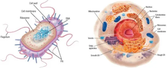
Bacteria are one of the three taxonomic domains of cellular organisms. They are prokaryotic cells and differ mainly from the eukaryotic ones for: the dimension; the lack of a membrane-bound nucleus and other intracellular membranous organelles, such as mitochondria; the reproductive strategies; and the presence of distinctive features, such as motility structures and cell wall (Figure 1) (Rogers, K. and Kadner, R.J., 2019; Parker, J., 2001).
General introduction
4
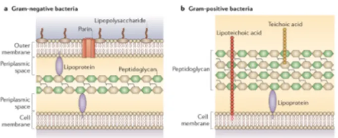
Unlike cells of higher organisms, bacteria face unpredictable and hostile environments. Thus, to survive, they evolved a unique cell envelope, which acts as the primary protective barrier. The cell wall varies between types of bacteria giving them distinctive structural characteristics and pathogen-host interaction profiles (Messner, P. et al., 2013). Based on the structure of this feature, bacterial cells were first classified by Hans Christian Gram in two main groups: Gram- positive and Gram-negative bacteria (Cabeen, M.T. and Jacobs-Wagner, C., 2005). Thus, Gram-positive cell walls are composed of a thick, multilayered peptidoglycan (PG) coat that includes embedded teichoic and lipoteichoic acids. While Gram-negative cell walls consists of an outer membrane (OM) that surrounds a thin PG layer, and a periplasmic space between the inner and outer membranes (Figure 2).
General introduction
5
Figure 2 Cell wall structure of Gram-negative bacteria (a)
and Gram-positive bacteria (b) (Brown, L., et al., 2015).
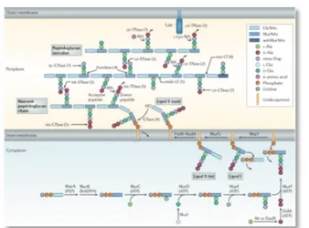
The major conserved component of the cell wall in almost all Gram-negative and Gram-positive bacteria is peptidoglycan (Vollmer, W. et al., 2008).
PG is a mesh-like structure made up of strands of repeating sugar units, N-acetylmuramic acid (MurNAc) and N-acetylglucosamine (GlcNAc), which are cross-linked by small peptides. The biosynthesis of PG starts in the cytosol, where the hydrophilic PG precursor (UDP-MurNAc-pentapeptide) is attached to the lipid carrier undecaprenyl phosphate (UndP). The lipid-linked precursor
(undecaprenyl-pyrophosphoryl-MurNAc-General introduction
6
pentapeptide or Lipid I) is modified further to undecaprenyl-pyrophosphoryl-MurNAc-(pentapeptide)-GlcNAc (Lipid II) by addition of a undecaprenyl-pyrophosphoryl-MurNAc-(pentapeptide)-GlcNAc moiety. Lipid II is then flipped across the membrane to the periplasm where its sugars are polymerized to form the glycan strands of the peptidoglycan mesh (Figure.3).
Figure 3 Peptidoglycan biosynthesis and cleavage (Typas, A. et al., 2012).
PG acts to stabilize the membrane and overall structure of the bacterial cell. It is also essential for growth,
General introduction
7
division and viability of bacterial organisms. Thus, enzymes contributing to the peptidoglycan biosynthetic pathway are attractive antibiotic targets (Silhavy, T.J. et
General introduction
8
Antibiotic resistance: a rising threat
to global health
Antibiotics revolutionized medicine in many aspects and improved significantly the quality of life, reducing childhood mortality, increasing life expectancy and saving numerous lives (Li, B. and Webster T.J., 2018; Gould, K., 2016; Fernandes, P. and Martens, E., 2017; Aminov, R.I, 2014).
The birth of the antibiotic era comes from the work of several pioneering scientists. In 1929, the isolation of penicillin from Penicillium mold and its potential use in surgical dressings were reported by Alexander Fleming. In 1935, the synthesis of sulfanilamide, the prototype for all sulfa drugs, was presented by Gerhard Domagk. In 1939, the isolation of tyrothricin, which was the first antibiotic established as a therapeutic substance, was reported by René Dubos.
General introduction
9
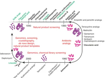
In the early 1940s, thanks to that and the industrialized production of penicillin numerous antibiotics were isolated and developed. The so-called “Golden Age” of antibiotic discovery began and the major classes of antibiotics in use, even nowadays, were produced (Fernandes, P., 2006).
From the late 1960s, resistance to existing antibiotics emerged powerfully and became the driving force for searching for new drugs. The antibiotic discovery largely abandoned natural product sources and redirected efforts on synthetic and high-tech approaches (Lewis, K., 2017). Unfortunately, the output of novel antibiotic agents fell dramatically, and only few drugs were putted on market in the last years (Figure 4) (Silver, L.L., 2011; O’Neill, J., 2016).
General introduction
10
Figure 4 A timeline for antibiotic research. (Fernandes, P.,
2006).
The decreased interest in antibiotic research of pharmaceutical companies is probably related to difficulties in clinical development and regulatory, and economic issues. However, the stimulation of antibiotic research has a pivotal role in finding new strategies to address the global threat of antibiotic- resistance (Tacconelli, E. et al., 2018).
General introduction
11
Resistance to antibiotics is a natural process that was observed since they were discovered (Fleming, A., 1945). In recent times, it is causing a public health crisis. Indeed, as a direct evolutionary consequence of widespread antibiotic use and overuse, bacteria resistance against all available antibiotics is rapidly outpacing the speed of human innovation (Wright, G.D., 2016; Tyer, M. and Wright, G.D., 2019; Hernando-Amado, S. et al., 2019). Nowadays, about 700,000 people die every year from drug- resistant strains of common bacterial infections and the burden of deaths could rise to 10 million lives each year by 2050, unless action is taken (O’Neill, J., 2016).
Bacteria can be intrinsically resistant to certain antibiotics. For example, an individual species can be resistant due to absence of a specific antibiotic susceptible target. In addition, they can acquire or develop resistance to antibiotics through the mutation of genes coding for drug targets and other compensatory proteins, or by the introduction of new genes by horizontal gene transfer
General introduction
12
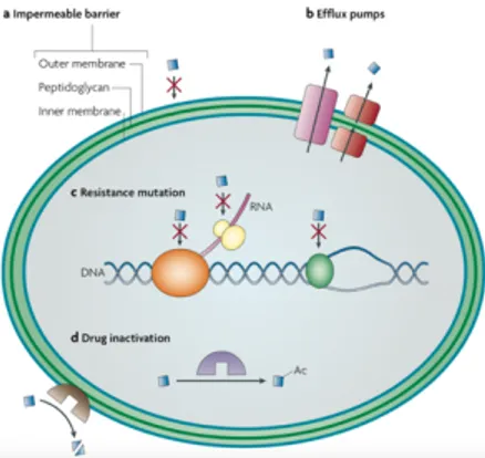
(Blair, J.M. et al., 2015; Frieri, M. et al., 2016). The acquired resistance can be mediated by several mechanisms, which fall into three main groups (Figure 5): 1. prevention of access of antibiotics to target, as a
result of poor penetration into the bacterium or of antibiotic efflux;
2. changes of antibiotic targets via genetic mutations or post-translational modifications;
3.
inactivation of antibiotic through their hydrolysis or modification.
Figure 5 Mechanisms of antibiotic resistance (Allen, H.K. et al., 2010).
General introduction
13
The first group of resistant mechanisms minimize the intracellular concentrations of the antibiotic. As example, in Gram- negative and many Gram-positive bacteria, efflux pumps are major contributors to the resistance. They can have narrow substrate specificity or a wide range of structurally dissimilar substrates (known as multidrug resistance efflux pumps); and when overexpressed, they can confer high levels of resistance to previously clinically useful antibiotics. The second group, usually, requires a mutational change in the genes encoding the antibiotic target. It comprises alterations to the target structure that prevent efficient antibiotic binding, but that still enable the target to carry out its normal function. The third one can degrade or modify antibiotics of different classes, using enzymes that catalyse their hydrolysis or add chemical groups (acyl, phosphate, nucleotidyl and ribitoyl groups) to vulnerable sites on the antibiotic molecule preventing the drug from binding to its target.
General introduction
14
In summary, as a result of the widespread use of antibiotics in several fields the emergence of antibiotic resistance is great. Thus, the current challenge is to develop novel drugs investigating the mechanisms of resistance, and exploiting available technologies and expertise (Blair, J.M. et al., 2015).
15
Part A
A novel colistin adjuvant
identified by virtual
screening for ArnT
inhibitors
The present part of the thesis deals with a research activity carried out at the Dipartimento di Chimica e Tecnologie del Farmaco at Sapienza Università di Roma, under the supervision of Prof. Bruno Botta.
17
CHAPTER A1
Introduction
A1.1 Current treatment options for multidrug-resistant
gram-negative bacteria infections 19
A1.2 Resistance to polymyxins in gram-negative bacteria 27 A1.2.1 Resistance mechanisms involving LPS structure
modifications: addition of L-ara4N to lipid A 31 A1.2.3 Aminoarabinose transferase ArnT structure and
catalytic mechanism 38
Chapter A1
19
A1.1 Current treatment options for
multidrug-resistant Gram-negative bacteria
infections
Global resistance to most available antibiotics is threatening human health and making antibiotic choices for infection control limited and more expensive (Li, B. and Webster, T.J., 2018). In particular, the emergence of multidrug-resistant (MDR) mechanisms in opportunistic Gram-negative bacterial pathogens such as Pseudomonas
aeruginosa, Acinetobacter baumannii and
Enterobacteriaceae is alarming. Hence, in 2017 the World
Health Organization (WHO) included these pathogens in the list of bacteria for which antibiotics are urgently needed as first priority (https://www.who.int/en/news- room/detail/27-02-2017-who-publishes-list-of-bacteria-for-which-new-antibiotics-are-urgently-needed). In the past few years, new treatment options for MDR
Gram-Chapter A1
20
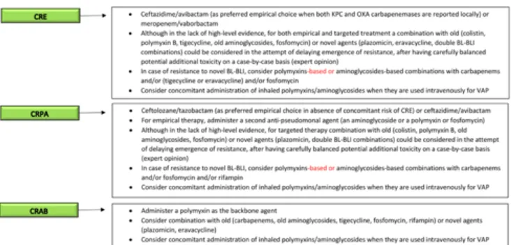
negative bacteria infections in critically ill patients became available (Figure A1.1), even though pharmacokinetic (PK) and pharmacodynamic (PD) optimization of their dosages and treatment has still some areas of uncertainty (Bassetti, M. et al., 2019).
Figure A1.1 Current clinical reasoning for the treatment of
serious MDR Gram-negative bacteria infections in critically ill patients. CRE, carbapenem-resistant Enterobacterales; CRPA, resistant Pseudomonas aeruginosa; CRAB, carbapenem-resistant Acinetobacter baumannii; BL-BLI, β-lactam/β-lactamase inhibitors; VAP, ventilator-associated pneumonia (Bassetti, M. et al., 2019).
Chapter A1
21
However, resistance to these treatments already started to emerge. Thus, because of the emergence of MDR Gram-negative organisms and lack of new drugs, medical community re-evaluated the use of the old antibiotics, such as polymyxins (Giamarellou, H., 2016; Poirel, L. et al., 2017; Velkov, T. et al., 2013).
Polymyxins were discovered more than 50 years ago (Velkov, T., 2010). They derive from various species of Paenibacillus (Bacillus) polymyxa and their chemical structure is similar to that of cationic antimicrobial peptides (defensins and gramicidins), which represent the first line of defense against bacterial colonization in eukaryotic cells. Polymyxins are cationic polypeptides that consist of a cyclic heptapeptide possessing a tripeptide side chain acylated at the N-terminus by a fatty acid tail, which accounts for most of the polymyxins antimicrobial activity and toxicity as well (Poirel, L. et al., 2017).
The antibacterial mechanism of action (Velkov, T.
Chapter A1
22
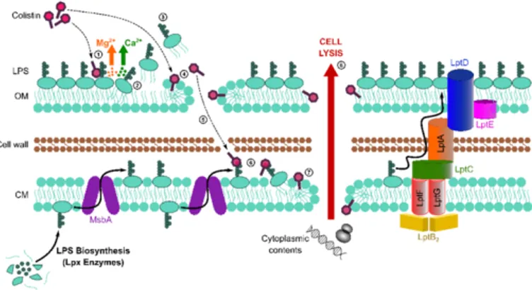
2019; Kaye, K.S. et al., 2016; Sabnis, A. et al., 2019) of polymyxins is not yet completely understood. However, they are believed to target the lipopolysaccharide in Gram-negative outer and cytoplasmic membranes, leading to disruption of the cell envelopeand bacterial lysis (Sabnis, A. et al., 2019) (Figure A1.2).
Figure A1.2 Schematic representation of antibacterial
mechanism of action of colistin (Sabnis, A. et al., 2019).
Thus, the electrostatic interaction of polymyxin cationic L-α,γ-diaminobutyric acid (L-Dab) side-chains with the phosphate groups of lipid A moiety of LPS
Chapter A1
23
displaces stabilizing cations Ca2+ and Mg2+, that bridge adjacent LPS molecules. The polymyxin molecules, then, enter into the fatty acyl chain layer of the lipid A molecules, causing derangement of outer and cytoplasmic membranes (Li, Z. and Velkov, T., 2019). This in turn results in increased membrane permeability, leakage of cell content and ultimately cell death (Storm, D.R. et al., 1977).
Based on available evidences, bactericidal activity of polymyxins also involves a secondary mode of action, which is responsible for the inhibition of bacterial respiration. In particular, it seems that the ability of polymyxins to disrupt the inner membrane structure is coincident with their inhibition of the alternative type 2 nicotinamide adenine dinucleotide dehydrogenase: the inner membrane respiratory enzyme of a number of pathogenic Gram- negative bacteria. Indeed, the downstream secondary target-polymyxin interactions trigger redox-related physiological alterations that result
Chapter A1
24
in the formation of toxic reactive oxygen species as well as stimulating repairing responses, which further contribute to cellular damage, mutations and death (Li, Z. and Velkov, T., 2019).
To date, several distinct groups of polymyxins (polymyxins A to E) were isolated and structurally identified from Paenibacillus polymyxa. Of these different groups, only two are used in the clinical setting (Velkov, T. et al., 2013; Landman, D., 2008): polymyxin B and colistin (Figure A1.3).
Figure A1.3 Chemical structures of polymyxin B and
Chapter A1
25
In particular, colistin is the last-resort therapeutic option in infections by recalcitrant Gram-negative bacteria when no other less toxic or effective antibiotics are suitable. It is a multicomponent polypeptide antibiotic, comprised mainly of colistin A and B (Li, J. et al., 2006; Li, J. and Nation, R.L., 2009). Colistin was isolated from the soil bacterium Paenibacillus polymyxa subsp.
Colistinus in 1947 (Poirel, L. et al., 2017; Vaara, M.,
2019) and it became available for clinical use in the 1960s. However, it was used in human medicine only for few years. During the 1970s, colistin was replaced by more potent and less toxic drugs due to severe side effects such as neuro- and nephrotoxicity. Currently, it is re-assessed as a critically important antibiotic in humans due to its efficacy against MDR Gram-negative bacteria (Apostolakos, I. and Piccirillo, A., 2018). There are two forms of colistin commercially available (Li, J. et al., 2006): colistin sulfate and colistimethate sodium (CMS or colistin methanesulfonate). Because of toxicity, colistin
Chapter A1
26
sulfate is used only for topical therapy. While, CMS is a less toxic formulation for parenteral use than colistin sulfate. It is the polyanionic colistin prodrug, which is converted into colistin and several inactive compounds in aqueous media and biological (Poirel, L. et al., 2017). However, based upon recent studies it is evident that colistin monotherapy is not likely to be reliably efficacious with currently recommended daily doses of CMS (Velkov, T. et al., 2013).
Furthermore, worrisome trends of increasing resistance to this antibiotic were reported in some countries. Thus, the use of colistin should be optimized as much as possible in terms of dosages and indications improving effectiveness and minimizing the emergence of further resistance (Bassetti, M. et al., 2019; Tsuji, B.T. et
Chapter A1
27
A1.2 Resistance to polymyxins in
Gram-negative bacteria
Resistance to polymyxins by bacilli that are normally susceptible to these drugs was reported. There are also reports of increases in infections caused by naturally polymyxin-resistant bacteria, such as Proteus, Providencia, Morganella, and Serratia (Olaitan, A.O.,
2014). Colistin resistance in Klebsiella pneumoniae, P.
aeruginosa and A. baumannii appears in less than 10% of
the isolates, although higher rates (50%) were reported (Jeannot, K. et al. 2017; Poirel, L. et al., 2017; Kaye, K.S.
et. al., 2016; Giamarellou, H., 2016). In a recent study by
SENTRY Antimicrobial Surveillance Program on isolates collected worldwide in 2017, the frequency of colistin resistance was still very rare. In 2015, the European Antimicrobial Surveillance Network (EARS-Net) registered 33,100 deaths due to infections caused by antibiotic-resistant bacteria. Among them, only 0.5% was
Chapter A1
28
caused by colistin-resistant A. baumannii or P. aeruginosa (Vaara, M., 2019). However, it should be noted that colistin resistance epidemiology may be underestimated due to the absence of powerful automated methods in clinical setting (Jayol, A. et al., 2018). Thus, identifying new strategies in order to decrease polymyxin resistance development and preserve the activity of these antibiotics gets imperative.
Gram-negative bacteria employ several means to protect themselves from adverse environmental stimuli, including exposure to cationic antimicrobial peptides, such as polymyxin B and colistin. Some bacteria, such as
K. pneumoniae, P. aeruginosa and A. baumannii, develop
resistance to polymyxins; whereas other bacteria, such as
Proteus spp., Serratia spp., and Burkholderia spp., are
naturally resistant to these drugs. This intrinsic resistance is mainly due to L-Ara4N modified lipopolysaccharides (Olaitan, A.O. et al., 2014), whereas in some species it was associated with defects in UDP-glucose dehydrogenase
Chapter A1
29
and UDP-glucose phosphorylase, enzymes involved in the biosynthesis of the LPS precursor (Velkov, T. et al., 2013). The acquired resistance is made of different strategies, including a variety of LPS modifications or its complete loss from the bacterial surface; the production of capsular polysaccharides; the expression of efflux pump systems (Moffatt, J.H. et. al., 2019); the expression of the outer membrane proteins such as OprH, which stabilizes the membrane under conditions of Mg2+ lack; and presence hopanoids, sterol-like compounds that are believed to perform a barrier function in the outer membrane of certain bacterial species (Velkov, T. et al., 2013) (Figure A1.4).
Chapter A1
30
Figure A1.4 Key mechanisms of polymyxin resistance in
Chapter A1
31
A1.2.1 Resistance Mechanisms involving
LPS structure modifications: addition of
L-Ara4N to lipid A
The first step in the action of polymyxin antibiotics on the Gram-negative bacterial outer membrane involves an electrostatic interaction between the positive charge of the five L-Dab residues of the polymyxin molecule and the negatively charged phosphate groups on lipid A (Velkov, T. et al., 2013). Thus, the majority of resistance mechanisms involves modifications that alter structure and charge of LPS (Moffatt, J.H. et. al., 2019). Indeed, by decreasing the net negative charge of LPS phosphate residues, these alterations tend to prevent the binding of polymyxin molecules to the bacterial surface and their further penetration into the cell interior, where they are supposed to exert the bactericidal activity (Jeannot, K. et
Chapter A1
32
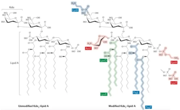
LPS is composed of three domains: the lipid A hydrophobic anchor, the core oligosaccharide and the O antigen (Needham, B.D. and Trent, M.S., 2013). Lipid A is the endotoxic portion of LPS and the site also for many LPS modifications. The various possible modifications include the addition of 4-amino-4-deoxy-L-arabinose (L-Ara4N) moieties and phosphoethanolamine moieties, as well as phosphorylation, deacylation, acylation and hydroxylation (Figure A1.5) (Nowicki, E.M. et al., 2015).
Figure A1.5 Unmodified lipid A and inner core (Kdo or 3‐
deoxy‐d-manno‐octulosonic acid) portion of LPS; Modified lipid A and inner core (Needham, B.D. and Trent, M.S., 2013).
Chapter A1
33
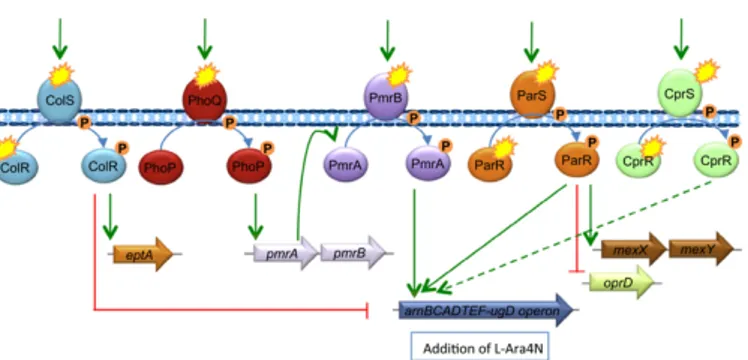
All of these alterations are subjected both transcriptional and post-translational regulation by many enzymes. Two-component systems (TCs), which are typically composed of a sensor kinase and a response regulator, small RNAs, peptide feedback loops and substrate availability are all involved in directing the activity of these enzymes. To date, widespread TCs are PhoPQ and PmrAB systems, and the ParRS, ColRS and CprRS systems in P. aeruginosa (Needham, B.D. and Trent, M.S., 2013).
In Gram negative bacteria, the most common LPS modification mainly occurs by covalent addition of L-Ara4N to lipid A moiety. Environmental stimuli and specific mutations within the TCSs trigger the activation of phosphorelay systems PhoPQ and PmrAB, which overexpress LPS-modifying genes (Figure A1.6). For example, the activation of the PmrAB TCS leads to the upregulation of the pmrCAB and arnBCADTEF-pmrE operons that mediate the synthesis and transfer of
L-Chapter A1
34
Ara4N to lipid A. (Olaitan, A.O. et al., 2014; Baron, S. et
al., 2016).
Figure A1.6 Activation of lipopolysaccharide-modifying genes
involved in polymyxin resistance in Gram-negative bacteria (Olaitan, A.O. et al., 2014).
In particular, P. aeruginosa colistin resistance correlates with mutations in PhoPQ two-component system leading to activation of the arn operon and increased production of L-Ara4N. The arn operon is also controlled by PmrA/PmrB, ParR/ParS, ColR/ColS and CprR/CprS (Figure A1.7). The absolute requirement of Arn-mediated lipid A aminoarabinosylation for acquired
Chapter A1
35
colistin resistance in P. aeruginosa was definitely demonstrated by showing that L-Ara4N defective mutants are unable to develop colistin resistance (Lo Sciuto, A. and Imperi, F., 2018).
Figure A1.7 Schematic representation of regulation of genes
contributing to colistin resistance in clinical strains of P. aeruginosa (Jeannot, K. et al., 2017).
This evidence strongly supports the notion that pharmacological inhibition of L-Ara4N biosynthetic enzymes is a proper approach to extend the anti- P.
Chapter A1
36
The biosynthesis of L-Ara4N starts with the conversion of UDP-glucose to UDP-glucuronic acid. The ArnA enzyme then catalyzes the oxidative decarboxylation of UDP-glucuronic acid to generate a novel UDP-4-ketopentose (Yan, A. et al., 2007). The resulting UDP-4-ketopentose is transaminated by ArnB to generate UDP-L-Ara4N, which is then formylated by the
N- terminal domain of ArnA. ArnC enzyme then transfers
the N-formylated L-Ara4N moiety to undecaprenyl phosphate. The resulting product is further rapidly deformylated by ArnD enzyme, generating undecaprenyl phosphate L-Ara4N. After transport of undecaprenyl phosphate-L-Ara4N to the outer surface of the inner membrane, ArnT transfers the L-Ara4N residue to the 4′-phosphate group of lipid A (Raetz, C.R.H. et al., 2007) (Figure A1.8).
Chapter A1
37
Figure A1.8 Biosynthesis of the L-Ara4N unit and its
Chapter A1
38
A1.2.3 Aminoarabinose transferase ArnT
structure and catalytic mechanism
ArnT enzyme is a member of the GT-C family of glycosyltransferases (Lairson, L.L. et al., 2008). In 2016, the ArnT structure from Cupriavidus metallidurans CH34 (ArnTCm) was determined alone and in complex with its natural lipid carrier undecaprenyl phosphate (UndP), yielding crystals in lipid cubic phase (LCP).
The structure of ArnTCm was determined to 2.8 Å resolution and appears as a monomer, consisting of (Figure A1.9):
• a transmembrane domain (TMD), which shows 13 TM helices in an intricate arrangement;
• a soluble periplasmic domain (PD) positioned above it;
• three juxtamembrane (JM) helices (JM1, JM2, JM3), which are parts of periplasmic loops (PL).
Chapter A1
39
Figure A1.9 Schematic representation of the connectivity and
structural elements of ArnTCm (Petrou, V.I. et al., 2016).
The structure of ArnTCm shows, also, three cavities that differ in their electrostatic nature (Figure A1.10):
• cavity 1 is the largest one. It is amphipathic with a lower hydrophobic portion located below the level of the membrane and an upper hydrophilic one;
• cavity 2 is the smallest one. It is connected to cavity 1 through a narrow passage and it is primarily hydrophilic;
Chapter A1
40
• cavity 3 is entirely hydrophobic and located close to the cytoplasmic side of the molecule;
Figure A1.10 Ribbon representation of ArnTCm showing the
cavities in the structure and close-up of the metal coordination site in ArnTCm (Petrou, V.I. et al., 2016).
Cavity 1 hydrophobic portion is directly accessible from the outer leaflet of the inner membrane, and it seems to be where lipid A binds to ArnT. Whereas, both cavity 2
Chapter A1
41
and cavity 3 are involved in binding of lipid carrier UndP to ArnTCm. This enzyme binds also between JM1 and PL4 a Zn2+ ion, which is coordinated by glutamic acid at position 84 (E84), histidines at positions 265 and 267 (H265 and H267, respectively), and likely by a water molecule. All the three metal-coordinating residues appear to be important for the enzymatic function (Petrou, V.I. et
al., 2016; Hall, B.E. et al., 2002).
To investigate how ArnT interacts with its lipid substrates, ArnTCm was cocrystallized with UndP (ArnTCm-UndP) and its structure was determined to 3.2 Å resolution (Figure A1.11).
Chapter A1
42
Figure A1.11 Binding of undecaprenyl phosphate (UndP) to
ArnTCm (Petrou, V.I. et al., 2016).
Overall, the structures of ArnTCm and ArnTCm-UndP suggest that the binding of substrates may be sequential, and the active site is completed by elements of the extended JM3 helix only with UndP bound (Figure A1.12).
Chapter A1
43
Figure A1.12 Schematic representation of substrate-binding–
induced conformational changes and catalytic cycle of ArnTCm
(Petrou, V.I. et al., 2016).
According with the structures reported above, ArnT catalytic mechanism involves the lipid A phosphate group, which is coordinated with two conserved aspartic acid residues (D55 and D158, respectively). Thus, the nucleophilic attack of the phosphate group on the anomeric carbon of L-Ara4N donor pushes off the lipid
Chapter A1
44
carrier UndP and leads to the formation of lipid A-L-Ara4N bond (Figure A1.13) (Petrou, V.I. et al., 2016).
Figure A1.13 Putative catalytic mechanism of ArnTCm (Petrou,
Chapter A1
45
A1.3 Aim of the work
Colistin resistance is expected to critically impair the treatment options against MDR Gram-negative infections. Epidemiology studies and experimental evidences support the hypothesis that the prevalent molecular mechanism conferring colistin resistance in Gram-negative bacteria is the enzymatic transfer of Ara4N to lipid A, by the aminoarabinose transferase ArnT (PmrK).
If this mechanism of lipid A modification could be disabled, colistin-resistant bacteria might be returned to a susceptible state. Thus, an agent capable of this restoration could enhance colistin activity, expand its repertoire of utility and allow also the durable therapeutic utility of the newer antibiotics. Up to now, only one molecule was reported interfering with Lipid A modification. It is a 4-deoxy-L-arabinose analog and it was designed to impair the L-Ara4N unit biosynthesis, mimicking the N-formylation transition state of 4-amino function of
Chapter A1
46
aminoarabinose or serving as surrogate for the formylated amine (Kline, T. et al., 2008). Accordingly, it was a goal of this PhD thesis to identify small natural molecules that could disable this lipid A modification and use as novel adjuvants to colistin activity to treat MDR Gram-negative infections.
47
CHAPTER A2
Identification of a natural diterpene
as a novel colistin adjuvant
A2.1 Natural products in drug discovery: a unique in house
library of natural compounds 49
A2.2 In silico screening of an in house library of natural
products: identification of putative arntinhibitors 52 A2.3 In vitro screening of compounds 1-18 as colistin
adjuvants 60
A2.4 A diterpene from Fabiana densa var. Ramulosa
endowed with promising biological activity 64 A2.5 Range of activity, specificity and cytotoxicity of
compound 14 70
Chapter A2
49
A2.1 Natural Products in drug
discovery: a unique in house library of Natural
Compounds
Natural products are mostly plant secondary metabolites, which are involved in physiological responses during plants interactions with their environments. They represent an important source of remedies since ancient times (Newman, D.J., 2008; Newman, D. J. and Cragg, G. M., 2007; Cragg, G.M, and Newman, D.J., 2013; Newman, D.J. and Cragg, G.M., 2016; Thomford, N.E. et al., 2018). Thus, the use of plants and preparations derived from them was the basis for medical treatments in Oriental civilizations and ancient Western world for many thousands of years, and even today, herbal and traditional medicines constitute primary health care for ∼80% of the world population (Kingston,
Chapter A2
50
D.G.I and Newman, D.J., 2012). Natural products play also a fundamental role in modern drug discovery due to their high structural variety and chemical diversity.Since the late 20th century, however, many pharmaceutical companies scaled significantly down or abandoned their natural product programs, because of the advances in both high throughput screening (HTS) and combinatorial synthesis, and the development of huge synthetic libraries of small molecules (Shen, B., 2015). Despite it, this trend is changing and a renewed interest in nature is emerging. Indeed, natural products are evolutionary refined as drug-like molecules and they possess enormous structural and chemical diversity that cannot be matched by any synthetic libraries of small molecules. Thus, natural compounds continue to remain the most abundant source of leads as valued platforms to identify and develop novel drugs
Chapter A2
51
(Casciaro, B. et al., 2019; Shen, B., 2015; Harvey, A.L. et
al., 2015; Brown, D.G, 2014).
In this scenario, a unique in house library of about one thousand natural compounds, mostly isolated from plants used in the traditional medicine of South America and collected in the years, is available at the Organic Chemistry Laboratory of the Department of Chemistry and Technology of Drugs (Sapienza University of Rome, Italy). This library consists of natural products from different classes, previously published and fully characterized, and it was also enlarged with natural compounds from commercially sources and synthetic and semisynthetic compounds. The library is characterized by a satisfactory chemical diversity, offering a unique chance to identify unexpected new scaffolds for the development of therapeutically relevant molecules. Accordingly, it is yet successfully screened in silico and in vitro for the
Chapter A2
52
identification of hit and lead compounds in previous early-stage drug discovery projects (Mascarello, A. et al., 2013; Infante, P. et al. 2015 and 2016; Casciaro, B. et al., 2019).
A2.2 In silico screening of an in house
Library of Natural Products: identification of
putative ArnT inhibitors
In the attempt to identify potential inhibitors of Ara4N-dependent colistin resistance, we carried out a docking-based virtual screening of our in house library of natural products within the catalytic site of ArnT, whose crystal structure was recently solved (Petrou, V.I. et al., 2016). By virtual screening, compounds were ranked based on their predicted binding mode and theoretical affinity within the ArnT catalytic site, and further filtered by chemical diversity criteria, leading to the final selection
Chapter A2
53
of 18 candidate hits (Table A2.1), that were submitted to biological investigations.
Chapter A2
54
Mol Chemical structure MW Molecula
r formula Ref. 1 492.60 C30H36O6 Delle Monache F. et al., 1979 2 418.39 C21H22O9 Peng C. et al., 2019 3 354.31 C16H18O9 Leitão S.G. et al., 2008
Chapter A2
55
4 622.62 C30H38O14 Scarpati M.L.,
Delle Monache, F., 1963
5 274.27 C15H14O5 Hu, Q. et al., 2017
6 384.38 C21H20O7 Tahara S. et al., 1992 O OH O OH HO OH O
Chapter A2 56 7 392.41 C23H20O6 Delle Monache F. et al., 1981 8 396.44 C23H24O6 Delle Monache F. et al., 1981
9 390.39 C17H26O10 Garaev E.E.
et al., 2014
10 194.19 C10H10O4 Gießel J.M.
Chapter A2 57 11 328.41 C20H24O4 Torres R. et al., 1979 12 356.37 C20H20O6 Harbore J.B., 1993 13 364.44 C23H24O4 Delle Monach e G. et al., 1980 14 360.49 C22H32O4 Erazo S. et al., 2002
Chapter A2 58 15 284.27 C16H12O5 Camele G. et.al, 1982 16 284.22 C15H8O6 Lee J.H. et al., 2002 17 294.39 C20H22O2 Delle Monach e F. 1977 18 360.50 C25H28O2 Gugliel mi P. et al., 2019
Table A2.1 List of compounds 1-18 selected and tested. O
HO
O O
Chapter A2
59
The potential ArnT inhibitors 1-18 identified in
silico show a remarkable range of chemical diversity and
differ also for their source, which is either natural or synthetic. They are anthranoids (namely, 1, 2, 15 and 16), chalcones (namely, 5 and 18), xanthones (namely, 7 and
8), flavonoids (namely, 6 and 12), cinnamic acid
derivatives (namely, 3 and 10), terpenoids (namely, 9, 13 and 14), a phenylpropanoid glycoside (namely, 4), a coumarin (namely, 11) and a stilbene (namely, 17).
The chemical identity of compounds 1-18 was assessed by re-running nuclear magnetic resonance (NMR) experiments and proved to be in agreement with the literature data reported for each compound. The purity of all compounds was, also, checked by reversed- phase HPLC and it proved to be always higher than 95%.
Chapter A2
60
A2.3 In vitro screening of compounds
1-18 as colistin adjuvants
Compounds identified by virtual screening were subjected to biological validation by using an in vitro evolved colistin-resistant isolate of P. aeruginosa PA14 (PA14 colR 5, colistin minimum inhibitory concentration (MIC) = 64 μg/mL), which overexpresses the arn operon responsible for lipid A aminoarabinosylation resistance (Lo Sciuto, A. and Imperi, F., 2018). First, in order to verify whether the in silico selected compounds were able to reduce or abrogate the growth of the reference strain PA14 colR 5, they were tested at a fixed concentration (50 μM) in the presence of a sub-inhibitory concentration of colistin (8 μg/mL).
Three compounds (namely, 8, 14 and 17) showed significant inhibition (>60%) of PA14 colR 5 growth in the
Chapter A2
61
presence of colistin, without notable effects on growth in the absence of the antibiotic (Figure A2.1 A). None of the other compounds showed relevant inhibitory activity on either colistin treated or untreated cells. For the three hit compounds, we further determined the dose-dependent effect on PA14 colR 5 growth in the presence of 8 μg/mL colistin.
Chapter A2
62
Compounds 14 and 17 caused a significant growth reduction at concentrations ≥ 8 μM, although they did not completely inhibit bacterial growth at any concentrations tested. In contrast, compound 14 significantly inhibited PA14 colR 5 growth at 8-16 μM and completely abrogated it at ≥ 31 μM (Figure A2.1 B). In agreement with the screening results, we confirmed that 14 has no inhibitory activity per se, as demonstrated by its marginal effect on PA14 colR 5 cultured in the absence of colistin, even at the highest concentration tested (125 μM). The potentiating effect of 14 on colistin activity was also corroborated by checkerboard assays, that showed a dose-dependent reduction of colistin MIC for PA14 colR 5 in the presence of increasing concentrations of 14 (Figure A2.1 C). Colistin MIC decreased to the lowest level (8 μg/mL, corresponding to an 8-fold reduction) with 31 μM of 14,
Chapter A2
63
and remained stable at higher concentrations of 14 (up to 125 μM).
Overall, these results demonstrated that one of the compounds identified by in silico docking of ArnT, namely 14, actually potentiates colistin activity against a colistin-resistant P. aeruginosa strain.
Chapter A2
64
A2.4 A diterpene from Fabiana densa
var. ramulosa endowed with promising
biological activity
Compound 14 is the diterpene “ent-beyer-15-en-18-O-oxalate” (Figure A2.2).
Figure A2.2 Structure of ent-beyer-15-en-18-O-oxalate (14).
It was isolated from the leaves of Fabiana densa var. ramulosa (Solanaceae) (Figure A2.3), which is a native shrub of Chile that grows from the Altiplano region
O O O HO 15 16 18 19 17 20 14 4
Chapter A2
65
to the Puna (Erazo, S. et al., 2002). This plant was botanically classified by the French botanist Esprit Alexandre Remy in 1847 and it is commonly called "checal" "tolilla" or "tola-checal". In 2002 the chemical composition of resinous exudate from the leaves of Fabiana densa var. ramulosa and its biological properties were carried out by Erazo, S. et al. at Faculty of Chemical and Pharmaceutical Sciences at University of Chile.
Chapter A2
66
In particular, a “novel” diterpenoid, the 18-O-oxalate (14), and the know ent-beyer-15-en-18-ol (19) (Figure A2.4) (San Martìn, A. et al. in 19839), were isolated and characterized. Currently, these diterpenoids occur in our library of natural compounds.
Figure A2.4 Structure of ent-beyer-15-en-18-ol (19).
They demonstrated, also, an interesting antimicrobial activity of the resinous exudate and its components against S. aureus, which could explain its use in traditional medicine to immobilize fractured limbs and
HO 15 16 18 19 17 20 19 4
Chapter A2
67
as an infusion for the cough and illness of the lungs (Erazo, S. et. al., 2002).
The chemical structure of 14 was assessed by 1H NMR, 13C NMR and high-resolution mass spectrometry (HRMS) experiments. The 1H NMR (CDCl
3) spectrum of compound 14 shows the peculiar signals of the ent-beyerene scaffold. In particular, it reveals a pair of doublets at 5.45 ppm and 5.69 ppm for the two olefinic protons (H-15 and H-16), and three intense singlets for the three methyl groups at 0.99 ppm (CH3-17), 0.91 ppm (CH3-19) and 0.99 ppm (CH3-17). The presence of oxalate group at C-18 was confirmed by comparing 1H NMR and 13C NMR spectra of ent-beyer-15-en-18-O-oxalate (14) with those of ent-beyer-15-en-18-ol (19). Accordingly, the 1H NMR spectral data are almost coincident with those of the ent-beyerene scaffold of 19, except for the signals (a pair of doublets) of H-18a and H-18b which were
Chapter A2
68
downfield at 3.90 ppm and 4.10 ppm (Figure A2.5 A). Comparison of the 13C NMR spectral data of 14, with those of alcohol diterpene 19, evidence two signals at 158.65 ppm and 157.77 ppm, which were assigned to the carbonyl groups at C-21 and C-22 of oxalate function of
14, respectively (Figure A2.5 B).
Furthermore, the purity of 14 was assessed by reversed- phase HPLC (Wellsow, J. et al., 2006; Wölwer-Rieck, U., 2012) and it proved to be higher than 95%
Chapter A2
69
Figure A2.5 Comparison of the NMR spectral data of
ent-beyer-15-en-18-O-oxalate (14) and ent-beyer-15-en-18-ol (19). (A) 1H NMR
(CDCl3, 400MHz) spectra of 14 and 19; (B) 13C NMR (CDCl3,
Chapter A2
70
A2.5 Range of activity, specificity and
cytotoxicity of compound 14
To verify the specificity of 14 towards colistin resistance, MIC of colistin for the colistin-sensitive parental strain PA14 was determined in the presence and absence of a concentration of 14 active against PA14 colR 5 (30 μM) (Table A2.2).
Species Strain MIC (µg/mL)
14 DMSO
P. aeruginosa PA14 colR 5 8 64
PA14 1 0.5 KK1 colR 1 8 128 KK1 1 0.5 KK27 colR 6 4 64 KK27 1 0.5 TR1 colR 6 4 16 TR1 1 0.5 ND76 4 32 MG75 8 32
Chapter A2 71 K. pneumoniae KP-Mo-3 16 128 KP-Mo-5 8 128 KP-Mo-6 4 32 KP-Mo-11 8 64 KP-Mo-16 8 64
Table A2.2 Colistin MIC for P. aeruginosa and K. pneumoniae strains
in the presence of 30 µM of 14 or 0.3% DMSO as the control.
Notably, compound 14 did not decrease colistin MIC for PA14, suggesting that the compound may exert adjuvant activity only on colistin-resistant isolates. This hypothesis was confirmed by testing three other in vitro evolved colistin resistant mutants of P. aeruginosa together with their parental cystic fibrosis (CF) isolates (Lo Sciuto, A. and Imperi, F., 2018). While 14 efficiently reduced the MIC of colistin for all resistant strains, it did not enhance colistin activity against their colistin-sensitive parental isolates. Indeed, for all colistin-sensitive isolates (PA14
Chapter A2
72
and the three CF isolates) we observed a slight (2-fold) increase in colistin MIC in the presence of 14, which however remained below the breakpoint for colistin-resistant P. aeruginosa (4 μg/mL, Clinical and Laboratory Standards Institute, 2016).
Finally, the potentiating effect of 14 on colistin activity was also observed in two colistin-resistant clinical isolates, namely ND76 and MG75, directly isolated from the sputum of chronically infected CF patients, indicating that 14 is also effective against colistin resistance strains evolved in vivo during the infection. These strains were kindly provided by Dr. P. Morelli from Istituto Giannina Gaslini, Genoa, Italy.
To rule out that the adjuvant activity of 14 could be due to a general destabilizing effect on the P.
aeruginosa cell envelope, we assessed whether this compound also affects the MIC of other antibiotics with