DOCTORAL SCHOOL IN BIOLOGY Section: “Biology Applied to Human Health”
XXIV cycle
INVOLVEMENT OF PEROXISOMES AT THE ONSET AND DURING THE PROGRESSION OF ALZHEIMER’S DISEASE IN A TRANSGENIC
MOUSE MODEL.
COINVOLGIMENTO DEI PEROSSISOMI ALL’ESORDIO E DURANTE LA PROGRESSIONE DELLA MALATTIA DI ALZHEIMER IN UN MODELLO
MURINO TRANSGENICO.
Candidate: Francesca FANELLI Tutor: Dr. Sandra MORENO
Coordinator: Prof. Paolo VISCA
C
ONTENTSABSTRACT I
RIASSUNTO IV
SECTION I:INTRODUCTION AND OBJECTIVES
Chapter 1. Alzheimer’s Disease 1
1.1 Forms of AD 1
1.1.1 EOAD 1
1.1.2 LOAD 2
1.2 Mild Cognitive Impairment (MCI) 2
1.3 AD Histopathology 3
1.3.1 Neurofibrillary tangles and tau protein 3 1.3.2 Amyloid plaques and APP processing 4 1.4 Role of oxidative stress in A-mediated neurotoxicity 5
1.5 Transgenic mouse models of AD 6
1.5.1 Tg2576 model 7
Chapter 2. Peroxisomes 9
2.1 Biogenesis of peroxisomes 10
2.1.1 Peroxisomal matrix protein import 11
2.1.2 Peroxisomal membrane protein import 12
2.2 Metabolic functions of peroxisomes 13
2.2.1 ROS metabolism 13
2.2.2 Lipid metabolism 14
Chapter 3. Aims of the research project 16
SECTION II:RESULTS
Chapter 4. Age-related changes in the distribution of neuronal and glial cells in Tg2576 mouse cerebral cortex and hippocampal formation 20 Chapter 5. Age-related variations in peroxisome-related proteins in the
Tg2576 hippocampal formation 27
5.1 Peroxisomal biogenesis markers: Pex5p, PMP70, and Pex14p 27
5.2 Peroxisomal -oxidation enzymes 33
5.3 Expression of the peroxisome proliferator receptor alpha at the onset and at
advanced stages of the disease 34
5.4 Oxidative stress markers at early and advanced AD stages 35 5.5 Age-related variations in ROS-scavenging enzymes expression in the
Chapter 6. Age-related variations in peroxisome-related proteins in the
Tg2576 cerebral cortex 39
6.1 Expression of peroxisomal biogenesis markers in the Tg2576 neocortex 39
6.2 Peroxisomal -oxidation enzymes 41
6.3 Expression of PPAR in the first stages of the disease 42 6.4 Expression of ROS-scavenging enzymes in the Tg2576 cerebral cortex 43 SECTION III:DISCUSSION AND CONCLUSIONS
Chapter 7. Discussion and Conclusions 46
A
BSTRACTAlzheimer’s Disease (AD) is the most common form of dementia, characterized by progressive neurodegeneration. Histopathological changes, including formation of beta-amyloid (Aβ) plaques, neurofibrillary tangles, followed by inflammation and neuronal loss, are especially prominent in the hippocampus and the neocortex. The classical hypothesis to explain the pathogenic events occurring in AD, the so-called amyloid cascade, involves a critical and early role of A peptide, involving production of reactive oxygen species (ROS) and consequent oxidative stress (Sayre et al., 2008). Even though Atoxicity is still widely accepted, this hypothesis has been recently challenged by Nunomura (2010) who proposed oxidative stress as the primary culprit in AD pathogenesis.
In this scenario, peroxisomes may play an important role, as they are involved in a wide variety of metabolic processes, including ROS and lipid metabolism (Schrader and Fahimi, 2008). It known that loss of peroxisomes makes neuronal cells vulnerable to oxidative stress and leads to degeneration (Stamer et al., 2002). On the other hand, peroxisomal proliferation attenuates Aβ-dependent toxicity in hippocampal neurons (Santos et al., 2005).
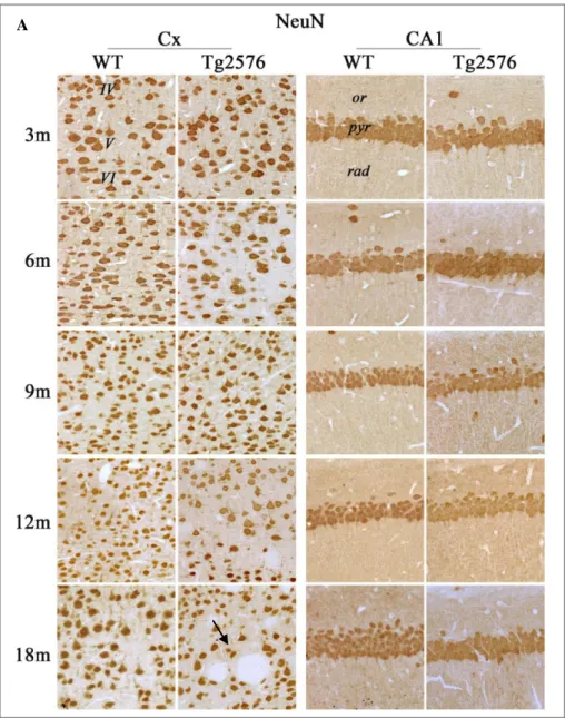
This PhD project aims to investigating the possible involvement of peroxisomes in AD onset and progression, with special reference to their role in oxidative stress. To this purpose we examined the transgenic mouse strain Tg2576, which recapitulates human pathological phenotype and is especially suited to the study of early stages, for it is characterized by slow progression (Jacobsen et al., 2006, D’Amelio et al., 2011). Our study was performed on early, advanced, and late stages of the disease (3, 6, 9, 12, 18 months of age), focusing on the hippocampus and neocortex, i.e., the areas where primary neuronal injury is known to occur. First, we analyzed the distribution of neuronal and glial markers, in WT and Tg2576 ageing/diseased mice, in order to detect possible alterations in brain organization. Our data show that the overall cytoarchitecture is conserved during normal aging, while in the pathological genotype, neuronal layering gradually changes starting from 9 months and appears dramatically altered at 18 months, when hyperproliferated and hypertrophic astrocytes and microglia cells are detected in both the neocortex and hippocampus. Consistently with the observed cytoarchitectural alterations, Congo Red staining demonstrates that small amyloid plaques are already present in the neocortex at 9 months, while their first appearance in the hippocampal formation occurs at 18 months.
Main objective of this PhD project was to investigate age-dependent variations of several peroxisome-related proteins in Tg2576 and WT hippocampus and neocortex in the course of the disease. Specifically, we characterized the peroxisomal population from a molecular and morphological point of view, examining the expression of biogenesis markers, membrane proteins, and matrix enzymes.
Our biochemical, morphological and ultrastructural data concur to demonstrate a significant peroxisomal induction in 3-month-old diseased hippocampus, as assessed by the peroxin Pex5p, the peroxisomal membrane protein PMP70, and acyl-CoA-oxidase (AOX, the first enzyme of the fatty acid peroxisomal -oxidation pathway), all of which show higher levels in Tg2576, compared to controls. This increase, paralleled by detectable oxidative modifications to biomolecules and by PPAR activation in Tg2576 hippocampus, suggests an early response to redox imbalance, mediated by PPAR, which is known to be induced by oxidized lipids.
Strikingly, the other crucial enzyme for the peroxisomal -oxidation pathway, i.e., thiolase (THL), is unchanged at 3 months, indicating possible inefficiency of the -oxidation pathway in the Tg2576 than in the WT. This would imply accumulation of VLCFA-derived intermediates in the brain, in line with the recent findings on the brain of AD patients (Kou et al., 2011). Even more remarkably, at this early stage, catalase (CAT) levels show no genotype-based differences, suggesting that increased H2O2 production by AOX is not accompanied by its efficient removal. These data, together with the low expression levels shown by another major H2O2 -scavenging enzyme, glutathione peroxidase (GPX1), strongly favour the idea that oxidative stress occurs early in AD. To this respect, it is also worth noting that mitochondrial superoxide dismutase (SOD2) is increased in the hippocampus of 3-month-old Tg2576 mice. Despite its commonly referred role as a ROS scavenger, this enzyme could even act as a pro-oxidant in this context. Indeed, the augmentation of SOD2, converting superoxide ion to H2O2, in the absence of parallel increase in any of the H2O2 scavengers, likely contributes itself to redox imbalance. Therefore, our data prefigures an early oxidative stress condition occurring in the hippocampus, strongly supporting the idea that this is the primary culprit in AD pathogenesis (Nunomura et al., 2010). Indeed, our data on the oxidative damage markers 8-OHG/OHdG and acrolein confirm this view.
During the progression of AD pathology, peroxisomal population undergoes dramatic changes, in that several proteins, including PMP70, Pex14p, and AOX, are decreased in 6-month-old Tg2576 hippocampus. This may reflect an impaired peroxisomal biogenesis or autophagic removal of excess organelles. Interestingly, in the hippocampus relatively high levels of CAT are detected at this age in the pathological genotype, suggesting a late response to peroxisomal induction, in agreement with what described in the liver following peroxisomal agonists administration (Reddy et al., 1986).
At 9 and 12 months, hippocampal levels of PMP70, AOX, and THL are relatively stable in both conditions. Other antioxidant enzymes show interesting changes at more advanced stages of the disease. Indeed, GPX1 and SOD1 increase from 6 to 9 months of age in the Tg2576 hippocampus, suggesting a late response, not involving the peroxisomes. However, at 12 months, when amyloid plaques start to form, GPX1, SOD1 and SOD2 show significantly lower levels in the Tg2576 mice, with respect to WT. Accordingly, decreased activities of SOD1 and GPX have
been reported in patients with symptomatic AD by Casado et al. (2008). The situation observed in 12-month-old Tg2576 hippocampus, closely resembling that of SOD1 and of GPX1, may reflect a decreased ability of neurons of this region to respond to A-mediated insult, possibly involving ROS generation.
A striking result regarding peroxisomes was obtained on 18-month-old mouse hippocampus. In fact, a peak of PMP70 expression is observed irrespective of the genotype, allowing to hypothesize a change in peroxisomal population related to the ageing process. Consistently, its main regulator, PPAR, shows intense immunoreactivity in the aged hippocampus, in both WT and Tg2576 animals. This late increase of peroxisomal number, not accompanied by induction of any of the peroxisomal matrix enzymes, may indicate the occurrence in the hippocampus of an attempt to counteract cellular damage, which however fails to result in improved efficiency of the organelles, either as ROS scavengers, or as lipid metabolizing sites. Indeed, AOX levels remain stably low at 18 months, while THL is dramatically decreased at the same age, allowing to hypothesize that inefficient -oxidation is associated with normal and pathological aging. Antioxidant enzymes show protein-specific variations in the aged hippocampus. Overall, it is conceivable that a pro-oxidant environment is still present in the cytosol, rather than in peroxisomes, since decreased levels of GPX1 and SOD1 are observed in Tg2576 mice, while CAT is unchanged.
Concerning the characterization of the peroxisomal population in the neocortex, our data show a delayed response in this brain area, with respect to the hippocampus, as well as substantial region-based differences in the susceptibility to A-mediated damage. The reason for this behaviour may relate to the relatively high anti-oxidant enzyme levels, which likely make the neocortex less susceptible to oxidative stress than the hippocampal formation (Cimini et al., 2009). In fact, at 3 months of age we failed to detect any variations in the expression levels of peroxisomal proteins. Nevertheless, catalase showed a delocalization, as assessed by immunoelectron microscopy, being found at extraperoxisomal sites, including the nucleus, cytosol, and post-synaptic densities. This evidence leads us to hypothesize that the presence of catalase to sensitive sites may contribute to enhancing protection against oxidative damage. The most relevant quantitative change in the neocortex are observed in 6-month-old Tg2576 mice, where significantly lower levels of peroxisomal proteins (PMP70, CAT) are detected, with respect to WT. Molecular data concerning peroxisomal -oxidation enzymes show no statistically significant variations during aging or disease progression, indicating that no perturbation of this pathway occur in the neocortex.
In conclusion, while 3 months of age is an especially promising time point for Tg2576 mice for devising therapies aimed at delaying or even preventing AD onset (Cimini et al., 2009; D’Amelio et al., 2011), the 6-month-old time point seems a more critical period, in which different approaches could be designed to counteract AD-like neurodegeneration. Based on our results, we suggest that potential therapies using PPAR agonists may be beneficial around 6 months of age.
R
IASSUNTOLa malattia di Alzheimer (AD) è la più comune forma di demenza dell’età adulta, caratterizzata da un progressivo e devastante disturbo neurodegenerativo che comporta un globale declino cognitivo, disturbi della memoria e del comportamento. Dal punto di vista istopatologico, fenomeni di gliosi e atrofia tissutale interessano inizialmente la corteccia temporale per poi estendersi anche a quella frontale. A livello microscopico le lesioni caratteristiche sono rappresentate da depositi proteici presenti sia nei compartimenti extracellulari, noti come placche amiloidi dovute all’aggregazione del peptide beta-amiloide (Aβ), sia in quelli intracellulari, noti come grovigli neurofibrillari, contenenti la proteina tau fosforilata.
È stato ampiamente dimostrato che il peptide Aβ, prodotto dalla proteolisi della proteina precursore dell’amiloide (APP), gioca un ruolo chiave nell’eziologia e nella patogenesi dell’AD. Una delle teorie più accreditate, nota come ipotesi della cascata amiloidea, pone l’accumulo di Aβ come principale causa della malattia. Secondo questa ipotesi, uno sbilanciamento tra la produzione di Aβ e il suo smaltimento porterebbe ad una progressiva degenerazione neuronale e a demenza. E’ noto come il peptide A eserciti un’azione neurotossica portando alla produzione di specie reattive dell’ossigeno (ROS) con conseguente stress ossidativo (Sayre et al., 2008).
Sebbene la tossicità indotta dall’Asia ancora ampiamente riconosciuta, l’ipotesi della cascata amiloidea è stata recentemente messa in discussione da Nunomura (2010) il quale propone lo stress ossidativo come evento precoce nella patogenesi dell’AD.
L’importanza dei perossisomi nel funzionamento del sistema nervoso è nota sin dalla loro scoperta ed è rappresentata dal fatto che questi organelli sono coinvolti in un’ampia varietà di processi anabolici e catabolici, tra i quali il metabolismo delle ROS e dei lipidi (Schrader e Fahimi, 2008). E’ stato dimostrato che la perdita dei perossisomi rende le cellule neuronali vulnerabili allo stresso ossidativo portando a degenerazione (Stamer et al., 2002); inoltre, la proliferazione perossisomiale attenua la tossicità Aβ-dipendente in neuroni ippocampali (Santos et al., 2005).
Queste evidenze ci hanno dunque spinto a pensare che i perossisomi giocassero un ruolo importante nella malattia di Alzheimer. Pertanto, lo scopo di questo progetto di Dottorato è stato quello di indagare il possibile coinvolgimento dei perossisomi all’esordio e durante la progressione della malattia, con particolare riferimento al loro ruolo nello stress ossidativo.
A questo proposito abbiamo scelto di analizzare una linea murina transgenica, Tg2576, che sovra-esprime un’isoforma Alzheimer-associata del precursore umano della proteina amiloide (APP). Questo modello ricapitola il fenotipo patologico tipico della malattia umana, pertanto rappresenta uno straordinario strumento per lo studio dei diversi meccanismi cellulari, biochimici, comportamentali ed
elettrofisiologici che caratterizzano in modo specifico le fasi precoci della malattia umana (Jacobsen et al., 2006, D’Amelio et al., 2011).
Lo studio è stato condotto su animali WT e Tg2576 di diverse età (3, 6, 9, 12, 18 mesi) al fine di analizzare le fasi precoci, avanzate e tardive della malattia. In particolare abbiamo focalizzato l’attenzione sulle due aree primariamente colpite dal danno neuronale, l’ippocampo e la corteccia cerebrale.
In una prima fase del progetto di ricerca abbiamo condotto un’analisi morfologica analizzando la distribuzione tissutale di marker neuronali e gliali in animali WT e Tg2576 di tutte le età considerate, al fine di individuare possibili alterazioni nell’organizzazione cerebrale. I nostri dati mostrano che la citoarchitettura è conservata durante l’invecchiamento fisiologico mentre, nel genotipo patologico, la stratificazione neuronale gradualmente cambia a partire dai 9 mesi risultando poi drammaticamente alterata a 18 mesi, età in cui sia l’ippocampo che la neocorteccia appaiono ricchi di astrociti iperproliferati ed ipertrofici e di cellule microgliali attivate.
L’analisi immunoistochimica condotta su sezioni incluse in paraffina di cervelli WT e Tg2576 a 18 mesi di età, con anticorpi diretti contro NeuN, GFAP e IbaI, rispettivamentente utilizzati come marker neuronali, astrocitari e microgliali, ha messo in luce le caratteristiche tipiche delle placche senili. In particolare, queste si estendono a tutti gli strati della corteccia da quelli più superficiali a quelli più profondi, causando imponenti alterazioni nell’organizzazione degli strati cellulari. Infatti i neuroni appaiono disorganizzati e orientati in maniera anomala a causa della presenza di estese placche amiloidi. Differentemente, nella formazione ippocampale, le placche sono preferenzialmente localizzate nello strato radiato del CA1 e del CA3, in siti distanti dai corpi cellulari delle cellule piramidali, così che il layering rimane conservato, anche se sono comunque evidenti alterazioni distrofiche dei prolungamenti neuritici. L’analisi morfologica ha inoltre evidenziato la presenza di numerosi astrociti fortemente positivi per la GFAP, i cui corpi cellulari sono disposti intorno alle placche, come a formare una barriera di contenimento, mentre i loro prolungamenti si inseriscono all’interno arricchendo il core della placca. Cellule microgliali attivate e immunopositive per Iba1 sono state inoltre trovate intorno e all’interno delle placche amiloidi.
I dati relativi al pattern di espressione di NeuN, non dimostrano alcuna variazione età- e genotipo-dipendente nel numero di cellule positive nonché nei livelli di immunocolorazione. Questa evidenza, in accordo con dati riportati da altri Autori (Jacobsen et al., 2006), suggerisce che non si verifica una massiva morte neuronale né durante il fisiologico invecchiamento né durante il progredire della malattia. Coerentemente con queste osservazioni riguardo alle alterazioni della citoarchitettura cerebrale, la colorazione con Rosso Congo ha dimostrato la presenza di piccole placche nella neocorteccia già a 9 mesi di età, mentre la prima comparsa a livello della formazione ippocampale si verifica a 18 mesi.
Il principale obiettivo di questo progetto di Dottorato è stato quello di analizzare possibili variazioni genotipo-dipendenti di varie proteine perossisomiali nell’ippocampo e nella neocorteccia di animali WT e Tg2576 all’esordio e durante la progressione della malattia. In particolare, è stata caratterizzata la popolazione perossisomiale da un punto di vista morfologico e molecolare, esaminando l’espressione di marcatori di biogenesi, proteine di membrana ed enzimi di matrice. Le analisi di western blot condotte su estratti proteici ippocampali di animali WT e Tg2576 alle diverse età considerate, hanno dimostrato una precoce e significativa induzione perossisomiale nell’ippocampo di animali Tg2576 di 3 mesi di età. Infatti, alti livelli della proteina perossisomiale di membrana PMP70 sono stati osservati nel genotipo patologico rispetto a quello normale. Per confermare questa induzione dei perossisomi in una fase così precoce della malattia, abbiamo condotto un’indagine di immunolocalizzazione ultrastrutturale “pre-embedding” per la PMP70. L’osservazione al microscopio elettronico ha confermato tale induzione poichè è stato osservato un più alto numero di perossisomi nei neuroni ippocampali di animali Tg2576 rispetto a quelli WT.
Dal momento che questi organuli svolgono un ruolo importante nel metabolismo lipidico, è stata esaminata l’espressione di due enzimi di matrice coinvolti nel pathway di -ossidazione perossisomiale, l’acil-CoA ossidasi (AOX) e la tiolasi (THL), che catalizzano rispettivamente la prima e l’ultima reazione del ciclo. I nostri dati biochimici e morfologici dimostrano che il pattern di espressione dell’AOX è conforme a quello della PMP70, in quanto più alti livelli di AOX sono presenti nell’ippocampo di animali Tg2576 rispetto alla controparte WT.
Sorprendentemente, la tiolasi non mostra variazioni genotipo-dipendenti a 3 mesi, indicando una possibile inefficienza della via di -ossidazione negli animali Tg2576. Questo potrebbe implicare un accumulo cerebrale di intermedi lipidici derivati dai VLCFA, cosa che è stata recentemente dimostrata verificarsi nel cervello di pazienti affetti da AD (Kou et al., 2011).
L’induzione di AOX osservata nella fase di esordio della malattia, potrebbe causare un accumulo di perossido di idrogeno, dal momento che l’AOX è un’ossidasi che produce H2O2. Pertanto abbiamo analizzato l’espressione di alcuni enzimi ROS-scavenger, focalizzando in primo luogo l’attenzione sulla catalasi (CAT), l’enzima perossisomiale di matrice per eccellenza. Tale proteina non mostra alcuna differenza genotipo-dipendente nell’ippocampo di 3 mesi, suggerendo quindi che l’eccessiva produzione di H2O2 da parte dell’AOX non sia accompagnata da una sua efficiente rimozione.
Abbiamo poi esteso l’analisi ad altri enzimi coinvolti nella difesa antiossidante, quali la glutatione perossidasi (GPX1), e le superossido dismutasi (SOD1 e SOD2). E’ da notare che i suddetti enzimi, pur essendo principalmente localizzati in altri distretti cellulari (la GPX1 e la SOD1 nel citosol, la SOD2 nei mitocondri), sono stati recentemente trovati anche all’interno dei perossisomi (Schrader e Fahimi, 2006).
I nostri risultati riguardanti l’espressione di catalasi, GPX1 e SOD1, che mostrano livelli relativamente bassi nell’ippocampo di animali Tg2576, supportano fortemente l’idea che condizioni di stress ossidativo si instaurino precocemente, rappresentando forse il primum movens nella cascata di eventi patogenetici che caratterizzano l’AD (Nunomura et al., 2010). A tal proposito, vale la pena sottolineare come la SOD2 sia incrementata nell’ippocampo di animali Tg2576 di 3 mesi di età. In contrasto con il suo comune ruolo di scavenger, l’enzima potrebbe, in questo contesto, agire come molecola pro-ossidante, dal momento che questo enzima converte lo ione superossido in H2O2. In assenza di un concomitante incremento degli enzimi che smaltiscono l’H2O2, la SOD2 potrebbe essa stessa contribuire ad uno sbilanciamento redox.
Per verificare l’esistenza di uno stress ossidativo precoce, abbiamo condotto analisi immunoistochimiche utilizzando due marker di danno ossidativo, l’acroleina e l’8-idrossiguanosina, per individuare rispettivamente modificazioni ossidative a carico dei lipidi e degli acidi nucleici. Abbiamo così potuto dimostrare la presenza di alti livelli di immunoreattività negli animali Tg2576 rispetto a quelli WT già a 3 mesi di età.
La presenza di lipidi ossidati nel tessuto ippocampale transgenico porta ad ipotizzare che questi comprendano ligandi del PPAR, in accordo con quanto riportato in letteratura (Yeldandi et al., 2000). Pertanto, si può ragionevolmente supporre che la proliferazione perossisomiale sia indotta dall’attivazione del PPAR da parte di specie lipidiche ossidate, generate in seguito ad uno squilibrio redox che presumibilmente coinvolge primariamente il mitocondrio. L’intervento del PPAR è supportato dai dati di immunoistochimica che mostrano elevati livelli del recettore nell’ippocampo di topo Tg2576 di 3 mesi. Inoltre, la microscopia elettronica dimostra la presenza del recettore nel compartimento nucleare di neuroni ippocampali transgenici, dove svolge la sua funzione di fattore di trascrizione. In questo contesto, i perossisomi proliferati, più ricchi di ossidasi che di catalasi, non sarebbero però in grado di sopperire alla richiesta di degradazione di ROS, ma piuttosto, contribuirebbero alla loro formazione, determinando così un danno ossidativo auto-sostenuto.
Durante la progressione della malattia, la popolazione perossisomiale subisce drammatici cambiamenti, infatti i livelli di espressione della PMP70, Pex14p e AOX sono fortemente diminuiti nell’ippocampo di animali Tg2576 di 6 mesi di età. Questa diminuzione potrebbe essere dovuta sia ad una danneggiata biogenesi perossisomiale, sia ad una rimozione mediante processi autofagici degli organelli in eccesso.
E’ interessante notare come, nell’ippocampo, alti livelli di CAT sono osservati a questa età nel genotipo patologico, suggerendo una risposta tardiva di questa specifica proteina all’induzione perossisomiale. Questa evidenza è in accordo con dati presenti in letteratura su quanto si verifica nel fegato in seguito a somministrazione di agonisti dei proliferatori perossisomiali (Reddy et al., 1986).
A 9 e 12 mesi, i livelli ippocampali di PMP70, AOX e THL sono relativamente stabili in entrambi le condizioni genetiche mentre gli enzimi antiossidanti mostrano interessanti cambiamenti a stadi avanzati della malattia. Infatti, a 12 mesi, età in cui le prime placche iniziano a formarsi a livello della formazione ippocampale, GPX1, SOD1 and SOD2 mostrano livelli significativamente più bassi di espressione negli animali Tg2576, rispetto a quelli WT. I nostri dati sono in accordo con quanto dimostrato da Casado e coll. (2008) riguardo al fatto che pazienti con una patologia AD conclamata mostrano una diminuita attività di SOD1 e GPX1 rispetto a soggetti di controllo. Pertanto, la situazione riscontrata a 12 mesi nell’ippocampo di animali Tg2576 può riflettere una diminuita capacità dei neuroni ippocampali di rispondere all’insulto mediato dal peptide A, portando alla produzione di ROS.
Un sorprendente risultato riguardante i perossisomi riguarda l’analisi degli estratti proteici ippocampali di animali a 18 mesi, età in cui un picco di espressione di PMP70 è stato osservato in entrambi i genotipi, suggerendo un cambiamento nella popolazione perossisomiale correlato al processo di invecchiamento. Coerentemente, anche il PPAR mostra un’intensa immunoreattività nell’ippocampo di 18 mesi, sia negli animali WT che in quelli Tg2576. Questo tardivo incremento del numero dei perossisomi non accompagnato da alcuna induzione degli enzimi di matrice, potrebbe indicare un tentativo di contrastare il danno cellulare, cui però non corrisponde una migliorata funzionalità degli organelli. Infatti a 18 mesi i livelli di AOX rimangono stabilmente bassi mentre la THL è drammaticamente diminuita, dimostrando che un inefficiente metabolismo lipidico era associato sia al fisiologico che al patologico invecchiamento.
A questa età le difese antiossidanti mostrano delle variazioni proteina-specifiche facendoci supporre che un ambiente pro-ossidante sia ancora presente nel citosol piuttosto che nei perossisomi, da momento che i livelli di GPX1 e di SOD1 sono diminuiti negli animali Tg2576 mentre la CAT rimane invariata.
La suscettibilità al danno A-mediato appare fortemente correlata all’area cerebrale considerata. Infatti i nostri risultati sulla corteccia cerebrale, mostrano una risposta ritardata di quest’area rispetto a quanto si verifica nell’ippocampo. La ragione di questo diverso comportamento potrebbe risiedere nel fatto che la neocorteccia ha relativamente più alti livelli di enzimi anti-ossidanti che la rendono meno suscettibile al danno ossidativo rispetto all’ippocampo (Cimini et al., 2009). A 3 mesi non osserviamo nessuna variazione nei livelli di espressione delle proteine perossisomiali. In maniera rilevante, l’analisi immunoistochimica per la CAT su sezioni di corteccia cerebrale ha evidenziato una delocalizzazione della proteina nei neuroni degli animali Tg2576. Questa è stata verificata con esperimenti di microscopia elettronica che hanno dimostrato la presenza di immunoprecipitati anche in siti extraperossisomiali come il nucleo, il citosol e le densità post-sinaptiche. Questa evidenza ci ha spinto a ipotizzare che la presenza di catalasi in siti sensibili possa contribuire ad incrementare la protezione contro il danno ossidativo.
I cambiamenti più rilevanti da un punto di vista quantitativo che coinvolgono la neocorteccia, si osservano a 6 mesi, quando livelli significativamente bassi di proteine perossisomiali (PMP70, CAT) sono presenti negli animali Tg2576 rispetto ai controlli WT.
Dati molecolari riguardanti gli enzimi coinvolti nel pathway di -ossidazione perossisomiale non mostrano differenze statisticamente significative durante il normale invecchiamento o durante la progressione della malattia, dimostrando che nessuna perturbazione di questo ciclo interessa la neocorteccia.
Riguardo l’espressione degli enzimi antiossidanti, la GPX1 mostra un picco di espressione nella corteccia di animali transgenici di 9 mesi. Questa evidenza, insieme all’incremento dei livelli di SOD1 e SOD2 a 12 mesi, fa supporre una risposta antiossidante compensatoria nella neocorteccia, concomitante con la deposizione delle placche senili, in accordo con dati di altri Autori (Papolla et al., 1998; Smith et al., 1998; Apelt et al., 2004). In particolare, si suppone un ruolo specifico dello ione superossido nel mediare l’azione neurotossica dell’A (Keller et al., 1998; Celsi et al., 2004).
In conclusione, i risultati ottenuti nel corso del mio progetto di Dottorato dimostrano che cambiamenti molecolari che coinvolgono i perossisomi, avvengono precocemente a livello della formazione ippocampale negli animali Tg2576. I 3 mesi di età rappresentano un time-point particolarmente promettente per disegnare terapie farmacologiche volte a prevenire o ritardare l’esordio della malattia (Cimini et al., 2009; D’Amelio et al., 2011). Noi crediamo che a questa età sarebbero opportuni trattamenti con molecole antiossidanti che avessero come target primario lo stress ossidativo. Molteplici sono i lavori presenti in letteratura che prendono in esame gli effetti di agonisti dei PPAR in diversi modelli murini di AD (Mandrekar-Colucci e Landreth, 2011). Queste molecole sono in grado di agire contrastando i processi infiammatori e il danno ossidativo attraverso differenti vie di segnalazione. Tuttavia, l’uso di ligandi del PPARa nostro avviso, dovrebbe essere considerato con qualche caveat riguardo il periodo di trattamento. Infatti, l’induzione perossisomiale potrebbe risultare dannosa a 3 mesi di età negli animali Tg2576 dal momento che potrebbe esacerbare il danno neuronale mediato dall’H2O2. Sulla base dei nostri risultati noi suggeriamo che la somministrazione di agonisti del PPAR debba avvenire intorno ai 6 mesi di età, al fine di revertire i sintomi iniziali tra cui le disfunzioni del metabolismo lipidico.
Chapter 1
Alzheimer’s Disease
Alzheimer’s disease (AD) is the most common form of dementia, accounting for 60-80% of all cases. The prevalence of dementia is below 1% in individuals aged 60-64 years, but shows an almost exponential increase with age, so that in people aged 85 years or older the prevalence is between 25% and 50% in the Western world (Finder, 2010).
This neurodegenerative disorder is characterized by a progressive decline in cognitive function, which typically begins with deterioration in memory and behavioural deficits. The gradual loss of independence of the patient leads to a heavy personal and financial toll on the family, resulting as major public-health problem that afflicts an estimated 24 million people in the world, with an expected increase to over 81 million people by the year 2040, mainly due to increased life expectancy (Ferri et al., 2005).
Besides ageing, which is a major risk factor for the disease, epidemiological studies have suggested several tentative associations. Reduced brain size, low educational and occupational attainment, low mental ability in early life, and reduced mental and physical activity during late life can be linked to AD (Mayeux, 2003; Ballard et al., 2011). Other risk factors are associated with vascular disease, including hypercholesterolaemia, hypertension, atherosclerosis, smoking, obesity, and diabetes. Nevertheless, while vascular risk factors and cerebrovascular disease clearly underlie vascular dementia, an etiological role for vascular changes in amyloid (A) deposition and, hence, AD remains unclear (Reitz et al., 2011).
1.1 Forms of AD
Alzheimer’s disease is a heterogeneous disorder that is usually classified according to its age of manifestation in: early-onset AD (EOAD) and late-onset AD (LOAD), called also sporadic form. These two forms are clinically indistinguishable; however, the former is generally more severe than the latter and it is associated with a more rapid progression. Moreover, the two forms of AD are associated with different patterns of genetic epidemiology (Bekris et al., 2010).
1.1.1 EOAD
EOAD is an autosomal dominant disorder which accounts for 1 to 6% of all cases, ranging roughly from 30 to 65 years. The only identified deterministic factors causing EOAD are the presence of mutations in the amyloid precursor protein gene (APP), located on chromosome 21, or in the presenilin genes (PSEN1 and PSEN2), located respectively on chromosome 14 and 1 (Harvey et al., 2003).
AD-linked missense mutations in APP affect the processing of the encoded protein, since the mutations are positioned in or near the A-coding exons 16 and 17. At present, 182 different AD-related mutations have been identified in PSEN1, while only 14 AD-linked mutations have been detected in PSEN2 (Alzheimer Disease
Mutation Database, 2010). To summarize, all three AD genes lend support to a common pathogenic AD pathway, with a pivotal role for A.
1.1.2 LOAD
LOAD, or sporadic form, is the most common form of AD and the majority (>95%) of the patients who develop this disease are aged more than 65 years. Although environmental factors can increase the risk of developing sporadic form of AD, behind it there is a significant genetic background. An association between the apolipoprotein E (APOE) 4 allele and AD has been reported by Poirier et al. (1993). APOE 4 allele advances the clinical onset of the disease by almost 10 years (Blennow et al., 2006) and its presence is associated with memory impairment, Mild Cognitive Impairment (MCI), and progression from MCI to dementia (Farlow et al., 2004).
The molecular mechanism by which ApoE would act is not completely clear. It is known that this lipoprotein acts as a cholesterol transporter in the brain with 4 isoform being less efficient than the other variations (APOE 2, and APOE 3), resulting in a low recovery of membrane lipids and neuronal repair (Poirier et al., 1993). On the other hand, ApoE is essential for A deposition, promoting plaques formation possibly by acting as a pathological chaperone (Holtzman et al., 2000). Therefore, the APOE 4 allele has been calculated to account for most of the genetic risk in LOAD (Raber et al., 2004).
1.2 Mild Cognitive Impairment (MCI)
In most cases, AD begins insidiously with cognitive and memory deficits that can be confused with the reduction of the ability to learn new information, that is physiological during brain aging(Forlenza et al., 2010).
Therefore, it is a difficult task to clinically differentiate incipient AD from normal cognitive aging and from the subtle cognitive changes that arise in other forms of dementia. Individuals in predementia stage of AD have been most commonly categorized according to the definition of Mild Cognitive Impaimernt (MCI) (Petersen et al., 1999) (Fig. 1.1).
This idea was introduced to define an intermediate stage between normal Fig. 1.1 Progression of cognitive and functional impairment and of neuropathological events in the transition from asymptomatic AD to MCI and clinically defined dementia of the AD type
aging and clinical dementia and it is widely accepted today that the diagnosis of MCI selects a clinically and biologically heterogeneous group of patients. It is reasonable to assume that most patients who are prone to become demented will present at early stages symptoms compatible with the definition of MCI.
1.3 AD Histopathology
The major pathological hallmarks in the brain of AD patients are neurofibrillary tangles (NFTs), formed by hyperphosphorylated tau protein, and amyloid plaques, consisting of the A peptide (Fig. 1.2). These lesions occur in brain regions involved in learning and memory, i.e. the hippocampus, the amigdala, and the cortical areas of the frontal, temporal and parietal lobes (Finder, 2010).
1.3.1 Neurofibrillary tangles and tau protein
Neurofibrillary tangles, which are intracellular filamentous inclusions, occur in AD and in other neurodegenerative disorders called tauopathies (Lee et al., 2001). The major component of the tangles is an abnormally hyperphosphorylated and aggregated form of tau.
Tau phosphorylation is regulated by the balance between multiple kinases, including glycogen synthase kinase 3 (GSK3) and cyclin-dependent kinase 5 (CDK5), and serine/threonine phosphatases (PP-1 and PP-2A) (Iqbal et al., 2005). Tau hyperphosphorylation in AD starts intracellularly and leads to sequestration of normal tau, causing disassembly of microtubules and compromised axonal transport and synaptic function (Fig. 1.3).
There has been controversy over whether and how neurofibrillary tangles and amyloid plaques are pathogenically related to each other and to the neuronal and synaptic losses that characterise the disease. However, experimental evidence indicates that A accumulation enhances tau aggregation (Lewis et al., 2001) and that A-induced neurodegeneration and cognitive deficits requires the presence of endogenous tau (Rapoport et al., 2002; Roberson et al., 2007), thus suggesting a strong relationship between the two pathological events.
Fig. 1.2 Amyloid plaques and neurofibrillary tangles in the cerebral cortex in Alzheimer’s disease. Plaques are extracellular deposits of Asurrounded by dystrophic neurites, reactive astrocytes, and microglia, whereas tangles are intracellular aggregates composed of a hyperphosphorylated form of the tau protein (Blennow et al., 2006). Tangles
1.3.2 Amyloid plaques and APP processing
The amyloid cascade hypothesis, which was formulated in the early 1990s, proposes that Aβ aggregation is upstream of all other pathological events (Hardy and Higgins, 1992; Hardy and Selkoe, 2002).
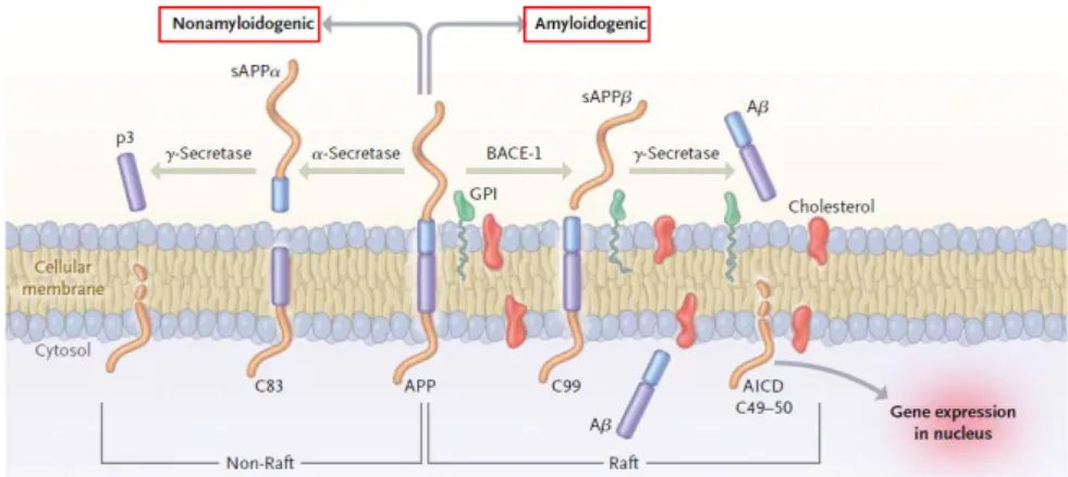
Aβ peptides originate from proteolysis of the APP by the sequential enzymatic action of two aspartyl proteases referred to as β-secretase (-site amyloid precursor protein–cleaving enzyme 1, BACE-1), and γ-secretase, a protein complex with presenilin 1 at its catalytic core (Querfurth and LaFerla, 2010). This processing of APP is called amyloidogenic pathway, as opposed to the nonamyloidogenic pathway (Fig. 1.4).
Levels of different Aβ species can be elevated by enhanced production and/or reduced clearance; in particular, the Aβ42/Aβ40 ratio can be augmented, leading to a relative increase of soluble Aβ42 species that enhance oligomer formation, which causes subtle but permanent changes of synaptic function. In parallel, local inflammatory responses (microgliosis and astrocytosis) are observed, and synaptic spine loss and neuritic dystrophy also occur.
Over time, these events result in oxidative stress and altered neuronal ionic homeostasis, followed by hyperphosphorylation of tau protein. The cascade
Fig. 1.3 Hyperphosphorylation and pathological assembly of tau protein. Tau protein normally promotes assembly and stability of microtubules and vesicle transport, but when it is hyperphosphorylated lacks affinity for microtubules and self-associates into paired helical filament structures. Destabilized microtubules lead to impaired axonal transport, compromising neuronal and synaptic function (modified from
culminates in widespread synaptic/neuronal dysfunction and cell death, leading to progressive dementia associated with extensive Aβ and tau pathology (Haass and Selkoe, 2007). This idea is also based on studies of genetic forms of AD, including Down’s syndrome (Glenner and Wong, 1984; Busciglio et al., 2002), and by evidence that soluble Aβ42 oligomers and intermediate amyloid fibrils are neurotoxic in vitro and in vivo (Walsh and Selkoe, 2007).
It is worth mentioning that prior to the appearance of extracellular deposits, intraneuronal accumulation of amyloid oligomers and fibrils occurs in the hippocampus (Shie et al., 2003; Wirths et al., 2004). Takahashi et al. (2004) using immunoelectron microscopy showed intraneuronal A42 in APP-transgenic mice in multivesicular bodies within dystrophic neurites. Recently, a 3D reconstruction study revealed fibrillar A within individual synaptic compartments, in association with abnormal morphology (Capetillo-Zarate et al., 2011).
1.4 Role of oxidative stress in A-mediated neurotoxicity
The mechanisms involved in A toxicity are unknown, but there is evidence suggesting that oxidative stress plays a key role in AD pathogenesis, being considered as the common effecter of the cascade of degenerative events in this disorder (Sayre et al., 2008; Gella and Durany, 2009; Sultana and Butterfield, 2010). Oxidative stress occurs due to an imbalance in radical production of reactive oxygen species (ROS) and antioxidant defences. Evidence of oxidative
Fig. 1.4 Processing of APP: nonamyloidogenic and amyloidogenic pathways. APP is a transmembrane protein with a large N-terminal extracellular tail. In the first pathway, cleavage by
-secretase releases the soluble APP fragment (sAPP). An 83-residue carboxy-terminal fragment (C83) is then digested by γ-secretase, liberating extracellular p3 and the amyloid intracellular domain (AICD) that is metabolised in the cytoplasm. Amyloidogenic processing is initiated by BACE-1, releasing a shortened sAPP. The retained C99 is also cleaved by γ-secretase complex, resulting in the production of several major Aβ species (Aβ38, Aβ40, Aβ42). A small tail of 50 amino acids (AICD fragment) is also released into the cytoplasm and targeted to the nucleus
stress in AD is manifested through high levels of oxidised proteins, advanced glycation end products, lipid peroxides, and oxidative modifications to nuclear and mitochondrial DNA, as well as to cytoplasmic RNA (Lovell et al., 1995; Praticò et al., 1998; Nunomura et al., 2001). Consistently, impairment of cognitive and memory functions in preclinical AD correlate with decreased antioxidant defence mechanisms (Smith et al., 2010; Jomova et al., 2010).
It was demonstrated that intraneuronal A has a causal role in neuronal oxidative damage because higher concentrations of Alead to oxidative stress in various biological systems (Behl et al., 1994;Tabner et al., 2005). In fact, A peptide itself is a source of hydrogen peroxide (H2O2) through metal ion reduction, with concomitant release of thiobarbituric acid-reactive substances (TBARS), a process probably mediated by formation of hydroxyl radicals (Miranda et al., 2000). However, another line of experiments suggests a primary role for oxidative stress in the production and accumulation of Ain vitro and in vivo (Shen et al., 2008; Dumont et al., 2009). Indeed, a long period of gradual oxidative stress precedes and leads to the pathological AD symptoms, including A deposition, neurofibrillary tangle formation, metabolic dysfunction, and cognitive decline (Bonda et al., 2010). In this view, it is worth noting that Apeptide can act as an antioxidant, suggesting a scenario in which intraneuronal A accumulation represents a compensatory response to neuronal oxidative stress (Nunomura et al., 2010).
1.5 Transgenic mouse models of AD
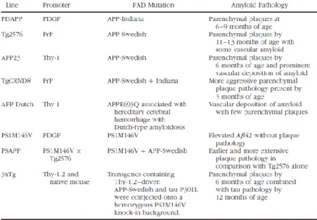
Several animal models of AD offer the possibility to study at the molecular level some of the typical aspects of human disease (Fig. 1.5) (Elder et al., 2010). Since mutations in the presenilin genes (PS1 and PS2) are the most commonly recognized causes of early-onset AD, the respective mutant transgenic lines have been generated. These strains show impaired -secretase-mediated proteolytic cleavage of APP, resulting in increased A/A ratio.While singly transgenic PS1 or PS2 mice do not develop plaques, when crossed with plaque-forming APP lines, earlier and more extensive plaque formation is found.
Different transgenic mouse lines were generated relying on strong promoters to drive expression of APP transgenes containing single or multiple familial AD mutations. Among these, APP23 transgenic mice, carry the double mutation Lys670 Asn, Met671 Leu (K670N,M67IL), which was found in a large Swedish family with early-onset AD, under the control of the mouse Thy-1 promoter. This strain develops cerebral amyloid angiopathy in addition to amyloid plaques, that appear at 6 months of age (Calhoun et al., 1999). TgCRND8 mice, in which a prion protein (PrP) promoter is used to drive expression of multiple APP mutations (Swedish and Indiana), are characterized by earlier and more dramatic amyloid deposition (Chishti et al., 2001). Another interesting model is represented by Tg2576 mouse strain, produced by Hsiao et al. (1996), the distinctive features of which are described below (1.5.1).
1.5.1 Tg2576 model
In the Tg2576 mouse line, human APP695 cDNA containing the Swedish double mutation was inserted into a hamster prion protein (PrP) cosmid vector (Hsiao et al., 1996). This was then introduced into individual cells by microinjection of embryos C57BL/6 X SJL/N F2, which have given rise to the founders. The resulting transgenic mice express the mutant amyloid precursor protein (APPSwe) leading to high Aβ levels in the neocortex and hippocampus (Kawarabayashi et al., 2001).
Differently from other mouse models, the Tg2576 strain is a slowly progressive AD model, offering the opportunity to study even subtle age-dependent alterations. Tg2576 mice are characterized by: i) behavioural deficits (Ashe, 2001; Westerman et al., 2002); ii) neuritic dystrophy and altered synaptic function, in the absence of massive neuronal loss (Irizarry et al., 1997); iii) astrogliosis, microgliosis and activation of inflammatory processes in relation to amyloidosis (Tehranian et al., 2001).
A detailed analysis of Tg2576 mice, including morphological, electrophysiological and behavioural characterization has been accomplished by Jacobsen et al. (2006), who demonstrated that neuronal deficits in Tg2576 mice are established in a time-dependent manner. The earliest changes include a decrease in spine density, deficits in hippocampal neurotransmission and in vivo memory impairments as measured by contextual fear conditioning (CFC). Slower onset deficits include an increase in amyloid load, reactive astrocytes, and microglia (Fig. 1.6).
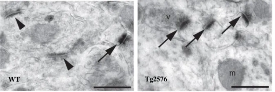
Recently, the group I collaborate with has shown that the earliest signs of synaptic dysfunction in Tg2576 mice are detectable as early as 3 months of age (D’Amelio et al., 2011). In particular, 3-month-old Tg2576 mice display enhanced caspase-3 activity in hippocampal CA1 synapses (Fig. 1.7), resulting in a downregulation of the surface expression of GluR1-containing AMPA receptor through proteolytic activation of calcineurin. GluR1 removal from the postsynaptic sites causes functional and structural synaptic alterations, resulting in glutamatergic transmission deficits, enhanced long-term depression (LTD), and reduced spine size and density.
In conclusion, D’Amelio’s work provides strong evidence that caspase-3 plays a crucial non-apoptotic role for the early synaptic dysfunction, leading to impairment of memory performance, associated with Alzheimer-like pathology (D’Amelio et al., 2011).
Fig. 1.6 Temporal progression of morphological and functional deficits in Tg2576 mice (Jacobsen et al., 2006).
Fig. 1.7 Immunoelectron microscopy for cleaved caspase-3. Immunoreactive dendritic spines (arrows) are more numerous in Tg2576 hippocampus, compared to its WT counterpart. Arrowheads indicate caspase-3 negative post-synaptic densities (D’Amelio et al., 2011).
Tg2576 WT
Chapter 2
Peroxisomes
Peroxisomes were discovered in 1954 in mouse kidney by an electron microscopist (Rhodin, 1954). In 1966, De Duve and Baudhuin were the first to isolate peroxisomes from rat liver, and their biochemical studies led to the discovery of the colocalization of several H2O2-producing oxidases with the H2O2-degrading enzyme catalase in the matrix of peroxisomes. De Duve then proposed the functional term “peroxisome”, which gradually replaced the former morphological designation, “microbody”, coined by Rhodin.
Subsequent morphological studies revealed that peroxisomes are ubiquitous cytoplasmic organelles present in a wide variety of eukaryotic cells, from yeast to humans (Hruban et al., 1972). They display roughly spherical shape (0.1 - 1 m in diameter), and a single-limiting membrane surrounding a finely granular matrix, which may contain crystalline inclusions (Fig. 2.1).
Peroxisomes show a high heterogeneity with respect to morphology, protein content, and abundance in diverse tissues and during developmental processes, including aging (Stefanini et al., 1995; Terlecky et al., 2006). In the nervous tissue, they are especially small (0,1-0,2 m in diameter), and their presence in neuronal and glial cells strongly relates to the brain region and cell population (McKenna et al., 1976; Arnold et al., 1979; Cimini et al., 1993; Moreno et al., 1995, 1999; Farioli-Vecchioli et al., 2001; Schad et al., 2003).
Peroxisomes are also able to respond to environmental changes and extracellular stimuli by altering their enzyme content, morphology and abundance. Pharmacological studies have allowed identification of a class of chemically unrelated substances, collectively known as peroxisome proliferators, capable of
Fig. 2.1 Morphology and enzymatic content of mammalian peroxisomes. A) Immunoelectron microscopy of catalase in rat kidney, showing colloidal gold-labelled peroxisomes (arrows), with a finely granular matrix in the absence of a crystalline core. (Moreno et al., 1995). B) Immunoelectron microscopy of urate oxidase in rat liver, showing peroxisomes (PO), with a colloidal gold-labelled crystalline core. (Volkl et al., 1988).
inducing remarkable increase in number and size of the organelles, especially in the liver (Reddy and Rao, 1987). Peroxisome proliferation is also accompanied by selective induction of peroxisomal proteins, mediated by peroxisome proliferator activated receptors (PPARs). These nuclear transcription factors (Issemann and Green, 1990) act as heterodimeric partners of other members of the same nuclear hormone receptor family, namely retinoid X receptors, and bind to the peroxisome proliferator response elements (PPREs) present on target genes.
The central role of peroxisomes in cell metabolism has emerged since their discovery (De Duve and Baudhuin, 1966), since they are involved in a wide range of anabolic and catabolic functions.This implies the presence of a large number of enzymes in the peroxisomal matrix. Indeed, about 50 metabolic enzymes have been identified in mammalian peroxisomes over the years. Moreover, some 32 proteins/genes, so-called peroxins (Pex), have been discovered, which are required for the biogenesis, assembly, and maintenance of functional peroxisomes (Platta and Erdmann 2007). Dysfunction of peroxins causes fatal human peroxisome biogenesis disorders (PBDs), which include the Zellweger syndrome spectrum (ZSS) disorders, neonatal adrenoleukodystrophy, infantile Refsum’s disease and rhizomelic chondrodysplasia puntata (RCDP) type I (Fujiki, 2000; Steinberg et al., 2006). Interestingly, all the above human pathologies, as well as the knockout mouse models produced to mimic them, have severe neurological implications, thus emphasising the importance of peroxisomes in the differentiation and homeostasis of the nervous system (Baes, 2000; Depreter et al., 2003; Baes and Van Veldhoven, 2006).
2.1 Biogenesis of peroxisomes
Peroxisome formation, proliferation and maintenance have been debated for a long time, and the debate is not over.
The classic view considers peroxisomes as autonomous organelles, which form out of pre-existing peroxisomes, like mitochondria and chloroplasts (Lazarow and Fujiki, 1985). The “growth and division” model was supported by the discovery of the synthesis of peroxisomal proteins on free ribosomes, their post-translational transport into peroxisomes, and the observations of interconnections between peroxisomes. Notably, according to this model endoplasmic reticulum (ER) was deemed only to be a source of membrane lipids for the enlargement of pre-existing peroxisomes. Recent discoveries have challenged what has been considered for most of the past two decades the paradigm for peroxisome biogenesis.
According to the novel concept, ER contributes to peroxisome formation (Titorenko and Mullen, 2006). This model received strong support by the observation that loss of the peroxins Pex3p, Pex16p, or Pex19p, which are required for peroxisomal membrane protein (PMP) targeting/insertion, resulted in the absence of detectable peroxisomes/peroxisomal membranes, whereas reintroduction of the missing genes led to a de novo formation of peroxisomes from the ER.The Pex3p and Pex19p have been observed to initially localize to the
ER before maturing into import-competent peroxisomes, indicating that the ER is indeed the source of the newly synthesized membrane and organelle (Hoepfner et al., 2005). However, the physiological significance of the mechanism of de novo formation in comparison to the classical pathway of growth and division is still controversially discussed.
2.1.1 Peroxisomal matrix protein import
The protein import into peroxisomes is a complex process, which differs substantially from the import mechanisms into the ER, mitochondria or chloroplasts. The peroxisomes import fully folded, co-factor bound and even oligomeric proteins by shuttling receptors (Leon et al., 2006).
The import of most peroxisomal matrix proteins is mediated by two types of cis-acting peroxisomal targeting signals (PTSs): the C-terminal uncleavable tripeptide PTS1, and the nonapeptide presequence PTS2, located at the N-terminus. The PTS1- or PTS2-containing matrix proteins are recognized in the cytosol by soluble receptors (PTS1 by Pex5p, a tetra-tricopeptide repeat (TPR) domain protein, PTS2 by Pex7p, a WD40 domain protein), which guide them to a docking site at the peroxisomal membrane. After translocation of the receptor–cargo complex to the luminal side of the peroxisomal membrane, the cargo is released and the receptors shuttle back to the cytosol (Fujiki, 2000; Ma et al., 2011).
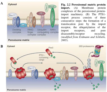
Fig. 2.2 Peroxisomal matrix protein import. (A) Membrane protein complexes of the peroxisomal protein-import machinery. (B) The PTS1-import process consists of three consecutive steps: the formation of a translocation pore by the import receptor, the ubiquitylation of the import receptors, and pore disassembly/receptor recycling.
(modified from Erdmann and Schliebs, 2005).
A
12 In particular, upon the binding of PTS1 proteins, Pex5p (a tetrameric complex) disaggregates into dimers and is transported, in a currently unknown manner, to the peroxisome where it interacts with Pex14p and Pex13p. At the peroxisomal membrane, Pex14p is associated with Pex17p and at least temporarily with Pex13p. The putative peroxisomal import complex (importomer) is formed by the RING-finger subcomplex containing Pex2p, Pex10p and Pex12p, and the docking complex comprising Pex13p, Pex14p and Pex17p (Fig. 2.2). Both subcomplexes are linked via Pex8p, which contains both targeting sequences for peroxisomal matrix protein import (PTS1 and PTS2) (Meinecke et al., 2010).
How the cargo or the cargo–receptor complex is translocated across the peroxisomal membrane is completely unknown. Pex8p triggers the association of the docking and the RING-finger complex and might contribute to cargo release. At the end of the pathway, Pex5p is recycled back to the cytosol in an ATP-dependent manner (Heiland and Erdmann, 2005).
2.1.2 Peroxisomal membrane protein import
The targeting and insertion of PMPs require other components than those involved in peroxisomal matrix protein import and is less well understood.
Only three of the 32 peroxins so far identified– Pex3p, Pex16p and Pex19p – are demonstrably involved in this process. It has been suggested that there are at least two distinct classes of peroxisomal membrane proteins. Class I PMPs are post-translationally inserted into the peroxisomal membrane in a Pex19p- and Pex3p-dependent manner, while class II PMPs, such as Pex3p and tail-anchored PMPs, are independent of Pex19p. Accumulating evidence suggests that class II PMPs might be targeted to the ER prior to their transport to peroxisomes (Heiland and Erdmann 2005; Schrader and Fahimi, 2008).
Fig. 2.3 Model of peroxisomal membrane biogenesis. Class I PMPs are recognized by Pex19p that directs them to the peroxisomal membrane. Membrane association of the Pex19p receptor–cargo complex is mediated by Pex3p. Topogenesis of class II PMPs is independent of Pex19p. These might be targeted to the ER prior to their transport to peroxisomes. How these proteins reach the ER and their final destination in the peroxisomal membrane is unknown (Heiland and
2.2 Metabolic functions of peroxisomes
Peroxisomes are involved in a wide range of anabolic and catabolic functions, including ROS metabolism (Schrader and Fahimi, 2006; Bonekamp et al., 2009),
-oxidation of very long chain fatty acids (VLCFAs) (Poirier et al., 2006; Wanders and Waterham, 2006), biosynthesis of polyunsaturated fatty acids (PUFAs) (Sprecher et al., 1995) and plasmalogens (Wanders 2004), cholesterol (Singh, 1997) and dolichol biosynthesis, and calcium homeostasis (Drago et al., 2008). The most relevant functions to the nervous tissue are described below.
2.2.1 ROS metabolism
Although mitochondria have been described as the major source of endogenous ROS generation (Rigoulet et al., 2011), peroxisomes have emerged as central organelles that play a key role in both production and scavenging of ROS (Schrader and Fahimi, 2006). This dual action derives from the fact that peroxisomes harbour both several H2O2-generating oxidases and antioxidant enzymes, including catalase (Fig. 2.3).
Peroxisomal oxidases include: (i) acyl-CoA oxidase (AOX, see below for pathway details); (ii) urate oxidase, involved in the final step of metabolic degradation of purines; (iii) xanthine oxidase (XOx) also involved in purine catabolism; (iv) D -amino acid oxidase; (v) D-aspartate oxidase; (vi) pipecolic acid oxidase; (vii) sarcosine oxidase; (viii) L--hydroxy acid oxidase; (ix) polyamine oxidase (Schrader and Fahimi, 2006).
Peroxisomal ROS-scavenging enzymes include: (i) catalase (CAT), which converts H2O2 into water through either the catalatic or the peroxidatic reaction; (ii) glutathione peroxidase (GPx), catalysing H2O2 removal with concomitant conversion of reduced glutathione (GSH) to glutathione disulfide (GSSG); (iii) copper zinc superoxide dismutase (CuZnSOD, SOD1) and manganese superoxide dismutase (MnSOD, SOD2), catalysing superoxide anion conversion into H2O2. While CAT is bona fide peroxisomal, all the other ROS-detoxifying enzymes are also present in other cell compartments. Oxidative balance within peroxisomes needs to be tightly regulated; shifting this equilibrium via endo- or exogenous Fig. 2.3 Overview of peroxisomal ROS metabolism.
H2O2 is produced by several peroxisomal
oxidases including XOx, and decomposed by catalase and GPx or converted to hydroxyl radicals (·OH). Hydroxyl radicals can damage the peroxisomal membrane by lipid peroxidation of unsaturated fatty acids. Peroxisomal oxidases also generate superoxide anions (O2·−) that are scavenged by MnSOD and CuZnSOD (Schrader and Fahimi, 2006).
14 factors, aging or disease conditions leads to a deregulation of the system, causing oxidative stress (Bonekamp et al., 2009).
2.2.2 Lipid metabolism
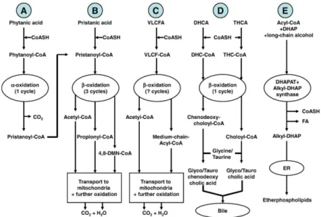
The main peroxisomal functions related to lipid metabolism have been recently reviewed by Wanders and coll. (2010). Fig. 2.4 summarizes the peroxisomal pathways of fatty acid -oxidation (A), fatty acid -oxidation (B, C, D), and plasmalogen biosynthesis (E).
Fig. 2.4 A schematic representation of lipid metabolic pathways in peroxisomes.
(A) Peroxisomal -oxidation pathway starts with activation of phytanic acid by hydroxylation to phytanoyl-CoA via phytanoyl-CoA hydroxylase. Then, phytanoyl-CoA is converted to pristanoyl-CoA and further oxidized as described in (B).
(B, C, D) Peroxisomal -oxidation systems are involved in the oxidation of pristanic acid, very long chain fatty acids (VLCFA), and bile acid synthesis intermediates (dihydroxycholestanoic acid, DHCA, and trihydroxycholestanoic acid, THCA). In particular, pristanic acid undergoes three cycles of -oxidation in peroxisomes to produce acetyl-CoA, propionyl-CoA, and 4,8-dimethylnonanoyl-CoA (4,8-DMN-4,8-dimethylnonanoyl-CoA). Subsequently, these molecules are transported to the mitochondria for full oxidation to CO2 and H2O (B). For VLCFA -oxidation, it has not been established yet how many
cycles of -oxidation take place in the peroxisomes (C). In liver, the bile acid intermediates DHCA and THCA undergo one cycle of -oxidation in peroxisomes, with chenodeoxycholoyl-CoA and choloyl-CoA as end products, respectively. These two CoA-esters are then conjugated with either taurine or glycine within peroxisomes. Subsequently, the taurine- and glycine-esters are transported out of the peroxisome into the cytosolic space, to end up in bile (D).
(E) Etherphospholipid biosynthesis from acyl-CoA and dihydroxyacetone phosphate (DHAP) is catalyzed by dihydroxyacetone phosphate acyltransferase (DHAPAT) and alkyl-dihydroxyacetone phosphate synthase (alkyl-DHAP synthase), either localized in peroxisomes. Subsequently, the reduction of alkyl-DHAP to etherphospholipids can take place at the endoplasmic reticulum (ER)
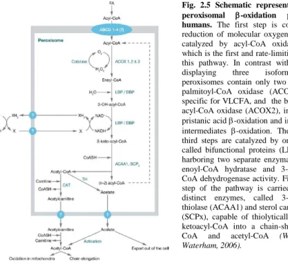
Among these lipid-related functions, peroxisomal fatty acid -oxidation, the details of which are illustrated in Fig. 2.5, is the most extensively characterized and attracts most attention, playing a central role in both catabolic and anabolic processes. In fact, it is not only essential to the degradation of several lipid species, but also participates in the biosynthetic pathway to polyunsaturated fatty acids (PUFAs).
The architecture of the peroxisomal -oxidation system involves a set of four consecutive reactions: (1) dehydrogenation; (2) hydration (of the double bond); (3) dehydrogenation again; and (4) thiolytic cleavage. Through this 4-step pathway a 2-carbon unit is split from each fatty acid in the form of an acetyl-CoA unit, which can then be degraded in the citric acid (Krebs) cycle to produce CO2 and H2O (Fig. 2.5) (Wanders, 2004).
Interestingly, the transcriptional activation of genes involved in fatty acid oxidation in livers of rats and mice is regulated by isotype of the peroxisome proliferator activated receptor (PPAR). Consistently, PPAR-null mice show deficiencies in peroxisomal fatty acyl-CoA oxidase, and in some of the other enzymes of the -oxidation pathway, thus emphasizing the critical importance and prominent role of PPAR in lipid catabolism (Reddy and Hashimoto, 2001).
Fig. 2.5 Schematic representation of the peroxisomal -oxidation pathway in humans. The first step is coupled to the reduction of molecular oxygen to H2O2 and
catalyzed by acyl-CoA oxidase (ACOX), which is the first and rate-limiting enzyme of this pathway. In contrast with rat species, displaying three isoforms, human peroxisomes contain only two oxidases: the palmitoyl-CoA oxidase (ACOX1), that is specific for VLCFA, and the branched-chain acyl-CoA oxidase (ACOX2), involved in the pristanic acid -oxidation and in the bile acid intermediates -oxidation. The second and third steps are catalyzed by one of two so-called bifunctional proteins (LBP and DBP) harboring two separate enzymatic functions: enoyl-CoA hydratase and 3-hydroxy-acyl-CoA dehydrogenase activity. Finally, the last step of the pathway is carried out by two distinct enzymes, called 3-ketoacyl-CoA thiolase (ACAA1) and sterol carrier protein X (SCPx), capable of thiolytically cleaving 3-ketoCoA into a chain-shortened acyl-CoA and acetyl-CoA (Wanders and Waterham, 2006).
Chapter 3
Aims of the research project
The importance of peroxisomes for nervous system functioning is represented by their involvement in a wide variety of metabolic processes, including ROS and lipid metabolism. Concerning the maintenance of cellular redox status, peroxisomes participate in both ROS generation and removal. When this balance is disrupted towards ROS production, oxidative stress is generated.
The brain is prone to oxidative stress, and is inadequately equipped with antioxidant defence systems to prevent oxidative damage, leading to neurodegeneration. Notably, the nervous tissue is especially rich in PUFAs and plasmalogens, i.e., molecules especially susceptible to oxidative modifications (Lukiw and Bazan, 2008; Lessig and Fuchs, 2009). These lipids, whose biosynthesis partially occurs in peroxisomes, participate in several physiological processes in neural cells, ranging from regulation of membrane fluidity, to synaptic remodelling, to neuroprotection (Wanders and Waterham, 2006). Interestingly, decreases in plasmalogen content have been found in several neuropathological conditions, including Alzheimer’s disease, dementia and ischemia and it has been proposed that plasmalogens may modulate the severity and progression of the disease (Goodenowe et al., 2007). Among PUFAs, docosahexaenoic acid (DHA; 22:63) seems particularly relevant, since low dietary DHA is a factor increasing the risk of age-related cognitive decline and AD (Cunnane et al., 2009). However, the existing data favor a role of -3 PUFA supplementation in slowing cognitive decline in elderly individuals without dementia, but not for the prevention or treatment of dementia (including AD) (Fotuhi et al., 2009).
Normal peroxisomal metabolism is also required for several aspects of development of the nervous system. Indeed, peroxisomal dysfunctions in humans or mouse mutants are associated with white matter abnormalities, such as defects in myelination and axon degeneration (Faust et al., 2005; Hulshagen et al., 2008). Consistent with these findings, deficiency in PPAR/, which is the most highly expressed PPAR isoform in oligodendrocytes, neurons, and astrocytes, results in brain developmental defects, including altered myelination and neuronal functioning in the CNS (Peters et al., 2000; Hall et al., 2008).
Moreover, peroxisomes play a key role in neuronal migration (Grabenbauer et al., 2001). For instance, peroxisome biogenesis disorders, among which Zellweger syndrome is the most severe, result in dramatically altered CNS neuronal migrations, causing heterotopias and defective cortical layering. Studies on PEX KO mice have shown that defects in neuronal differentiation, proliferation and survival may also contribute to CNS malformations (Baes and Van Veldhoven, 2006).
Consistent with their essential role in CNS development, peroxisomes are generally more abundant in differentiating neurons than in mature neurons and are