Studi sperimentali
Morphovolumetric changes after EMDR treatment in drug-naïve
PTSD patients
Modifiche morfo-volumetriche dopo trattamento EMDR in pazienti drug-naïve
affetti da DPTS
LETIZIA BOSSINI
1*, EMILIANO SANTARNECCHI
2, ILARIA CASOLARO
1, DESPOINA KOUKOUNA
1,
CLAUDIA CATERINI
1, FEDERICA CECCHINI
1, VALENTINA FORTINI
1, GIAMPAOLO VATTI
2,
DANIELA MARINO
2, ISABEL FERNANDEZ
3, ALESSANDRO ROSSI
2, ANDREA FAGIOLINI
1 *E-mail: [email protected]1Department of Molecular Medicine and Development, Psychiatry Section, University of Siena, Italy 2Department of Medicine, Surgery and Neuroscience, University of Siena, Italy
3Private Practice, Milan, Italy
INTRODUCTION
Eye Movement Desensitization and Reprocessing (EM-DR) is a psychotherapy that has been found to effectively
re-solve the effects of traumatic experiences1and following
nu-merous randomized clinical trials2 in patients with
post-trau-matic stress disorder (PTSD) it has been recognized as a first-line treatment for PTSD3.
SUMMARY. Introduction. Few studies have investigated the effects of efficacious psychotherapy on structural alterations of discrete brain
regions associated with posttraumatic stress disorder (PTSD). We therefore proposed to evaluate the neurobiological effects of eye move-ment desensitization and reprocessing (EMDR) on 19 patients with drug-naïve PTSD without comorbidity, matched with 19 untreated healthy controls. Methods. We administered the Clinician Administered PTSD Scale (CAPS) and conducted brain MRI measurements (with Optimized Voxel-Based Morphometry). Patients received 12 EMDR sessions over three months. Then patients and controls were reassessed.
Results. At baseline, grey matter volume (GMV) differed significantly between patients and controls (F 1,35 =3.674; p=.008; η2=.298). Analy-ses of 3-month scans showed no changes for controls, while significant changes were highlighted for patients post-EMDR, with a significant increase in GMV in left parahippocampal gyrus, and a significant decrease in GMV in the left thalamus region. The diagnosis of PTSD was effectively eliminated in 16 of 19 patients, reflected in a significant improvement on the CAPS (t(35)=2.132, p<.004). Discussion and
con-clusions. Results indicated post-EMDR changes for patients in brain morphology. We discuss whether EMDR’s mechanism of action may
work at the level of the thalamus, an area implicated in PTSD pathology.
KEY WORDS: PTSD, EMDR, morphovolumetric.
RIASSUNTO. Introduzione. Esistono pochi studi che hanno indagato gli effetti di una psicoterapia ritenuta efficace sulle alterazioni
strut-turali delle regioni cerebrali associate al disturbo da stress post-traumatico (PTSD). Ci siamo, pertanto, proposti di valutare gli effetti neuro-biologici della terapia EMDR su un gruppo di 19 pazienti con PTSD, senza cura farmacologica e senza alcuna comorbidità con altri disturbi psichiatrici, a confronto con un gruppo di controllo costituito da 19 soggetti sani non trattati. Metodi. Abbiamo somministrato la CAPS e sot-toposto ciascun soggetto a misurazioni MRI del cervello (condotte con Optimized Voxel-Based Morphometry). I pazienti hanno ricevuto 12 sedute di EMDR nell’arco di tre mesi. Sia i pazienti che i controlli sono stati successivamente rivalutati al termine della terapia. Risultati. Alla misurazione baseline, il volume della materia grigia (GMV) differiva significativamente tra pazienti e controlli (F 1,35=3.674; p=,008; η2=0,298). Le analisi delle scansioni ottenute a 3 mesi non hanno mostrato variazioni per i controlli, mentre hanno messo in evidenza cam-biamenti significativi per pazienti che sono stati sottoposti a terapia EMDR, con un aumento significativo della GMV nel giro paraippo-campale sinistro, e una diminuzione significativa della GMV nella regione del talamo sinistro. A seguito del trattamento EMDR 16 pazienti su 19 non soddisfacevano più criteri per una diagnosi di PTSD, dato che si riflette in un miglioramento significativo ottenuto alle CAPS (t(35)=2.132, p<,004). Discussione e conclusioni. I risultati hanno indicato cambiamenti nella morfologia del cervello per i pazienti sotto-posti a terapia EMDR. Nell’articolo verrà discusso il meccanismo di azione del trattamento EMDR, con l’obiettivo di comprendere se esso possa agire a livello del talamo, un’area implicata nel PTSD.
PTSD is characterized by dysfunction and structural al-teration of several discrete brain regions. Neurobiological in-vestigations of PTSD have shown that it may be character-ized by lower density in limbic and paralimbic cortices4, with
changes in gray and white matter volume and concentration (GMV and GMC, respectively) in hippocampus, parahip-pocampal gyrus and cingulum4,5. However possibly due to
the high heterogeneity of traumatic events causing PTSD and of patients symptoms (i.e. hyperarousal vs dissociation) as well as of cohort sizes a surprisingly large variance across studies has been reported.
Most Magnetic Resonance Imaging (MRI) studies on PTSD have measured volumetric changes in discrete brain regions or small brain structures. Karl et al. in a meta-analy-sis of structural brain MRI in PTSD6 concluded that the
dis-order is associated with abnormalities in multiple frontal-limbic system structures, notably in hippocampi, amygdala, and anterior cingulate cortex. Similarly, a recent meta-analy-sis by Woon et al on 39 hippocampal volumetric studies iden-tified significant hippocampal volume reduction in individu-als with PTSD7.
Furthermore, investigating the changes in GMC in pa-tients with and without PTSD, Zhang et al.5found those with
PTSD showing significantly decreased GMC in left anterior hippocampus and left parahippocampal gyrus and Nardo et al.4showed a lower grey matter density in limbic and
paral-imbic cortices to be associated with PTSD diagnosis. Studies investigating the effect of Cognitive behavioural therapy (CBT) on hippocampal volume in PTSD patients have reported conflicting results8,9. Recently functional
stud-ies have reported EMDR-related neurobiological changes10,11
and our group has investigated the structural changes after successful treatment of PTSD with EMDR showing an aver-age increase of 6% in hippocampal volume following remis-sion of diagnosis after three months of EMDR therapy12-14.
The aim of the present study was to extend such investi-gation beyond the regional assessment computing in PTSD patients and healthy controls a voxel-wise analysis on the whole brain assessing the anatomical changes occurring fol-lowing EMDR therapy.
METHOD Participants
Thirty-eight participants were studied: 19 drug-naive patients with PTSD (10 men and 9 female) and 19 age matched healthy controls (15 men and 4 women). The patient group was recruited at the Center for the Diagnosis and Treatment of Post-Traumatic Stress Disorder, Department of Psychiatry, University of Siena, between September 2010 to May 2012 and largely overlapped the cohort recruited for a previous study14. Patient inclusion criteria were: age between 18 and 65 years and the drug-naïve status. Ex-clusion criteria were: a history of current and/or lifetime comorbid psychiatric diagnoses as determined by the SCID; previous or cur-rent use of any psychotropic medications; history of head trauma; presence of neurological, endocrine, or degenerative disorders. Healthy controls were recruited at the hospital “Le Scotte” in Siena, Italy, and matched for age, education, handedness, weight and height. Exclusion criteria for controls were: a history of meningitis, traumatic brain injury, presence of neurological,
en-docrine and degenerative disorders, use of drugs and previous or current use of any psychotropic medications, neurological or psy-chiatric problems, as shown by clinical history and psypsy-chiatric evaluation. All participants consented to participate after having been informed about the purpose of the research and none of them received economic compensation for participating in the study. The study was approved by the Institutional Ethical Com-mittee of Siena University, and the study adhered to the tenets of the Declaration of Helsinki.
Procedure
Patients and control participants underwent a complete Psy-chiatric evaluation and a MRI at baseline (T1). Patients received 3 months of EMDR treatment, and then were evaluated post-treatment (T2) with MRI and CAPS. Healthy controls were re-evaluated by MRI at 3 months (T2) after baseline acquisition.
Psychiatric evaluation
A comprehensive psychiatric evaluation was conducted at baseline. Psychiatric diagnoses based on DSM-IV15and on the Structured Clinical Interview for DSM-IV (SCID)16,17 were deter-mined by a consensus of two psychiatrists not otherwise involved in the study. Healthy controls were assessed with the SCID - Non-Patient version18. Patients were evaluated with the SCID for DSM-IV Axis I (SCID-I/P) and Axis II (SCID-II/P) disorders in order to determine a single diagnosis of PTSD, and were assessed with the Clinician Administered PTSD Scale (CAPS) Italian Ver-sion19, which is known to be a reliable measure of PTSD symp-toms severity with subcomponents for the individual symptom clusters. We administered the Davidson Trauma Scale (DTS), a di-mensional measure of PTSD with 17 items (with scores ranging from 0-136) for PTSD severity. An evaluation for the presence of overlapping symptoms between PTSD, Major Depressive Disor-der, and state of anxiety was also performed respectively with the Hamilton Depression Rating Scale (HAM-D)20, and the Hamil-ton Anxiety Rating Scale (HAM-A)21. At post-treatment, the CAPS was administered to patients again.
EMDR treatment
The treatment followed the guidelines by Shapiro1. In brief, as the EMDR session begins the worst image of the traumatic mem-ory is recalled as well as negative beliefs, disturbing emotions and body location of the disturbance. Then, the patient focuses on these memories while the therapist performs for about 30s a bi-lateral stimulation guiding attention from right to left with sets of 30s. At the end of each set the patient reports what she noticed and the procedure is repeated until memory is reprocessed and adapted. At this stage the patient recalls the traumatic experience without disturbing emotions, improving her self-belief and being free of body tension. A successful treatment implies that the client visualizes himself in a situation where he will face the same trau-matic events without emotional disturbance. EMDR desensitizes past, present and future issues related to traumatic events repro-cessing them and reaching symptom remission.
Patients were randomly assigned to one of three trained psy-chotherapists. Duration of the treatment was 3 months, with 12 90-minute EMDR sessions provided on a weekly basis. The BLS in-cluded eye movements (patient following the therapist’s finger) or
RESULTS
Patients and controls did not statistically differ for demo-graphic data, as reported in Table 1. Patients’ diagnoses of PTSD at baseline (T1) were confirmed by clinical evaluation and by the fulfilment of all the criteria at CAPS. All patients had experienced a one-time adult trauma: natural disaster (n=3), sudden death of a family member (n=5), car accident (n=2), as-sault/robbery (n=6), and terrorist attack (n=4). One patient dropped out because of a depressive episode onset and conse-quently we removed a matched healthy control participant.
Baseline comparisons between patients and controls: Grey Matter Volume
The GMV comparison between patients and healthy par-ticipants at baseline showed significant differences (F 1,35=3.674; p=.008; η2=.298). Analyses revealed a region of
significantly decreased GMV in patients’ left
parahippocam-tactile (therapist tapping the patient’s hand). Tactile taps were al-ternated to eye movements during some of the sessions only if the patient could not concentrate during eye movements or com-plained about not being able to follow the fingers. All sessions were videotaped; the fidelity to the treatment protocol was as-sessed by an independent evaluator, a psychologist and licensed psychotherapist, who was an EMDR European-approved consult-ant and supervisor and not involved in the present study.
Neuroradiological acquisition
MRI examinations of all participants were performed at a 1.5 Tesla Philips Intera scanner (Philips Medical Systems, Best, The Netherlands). Morphovolumetric analysis were run onto T1-weighted Fast Field Echo (FFE) 1-mm thick images of the entire brain (TR/TE=30.00/4.6 ms, flip angle=30.00, FOV=250 mm, matrix 256x256, slice number=150), acquired in the axial plane parallel to the anterior and posterior commissures. Neuroradiological exami-nation also included FFE 1-mm thick coronal images (TR=30.00/4.6 ms, flip angle=30.00, FOV=250 mm, matrix 512x512, slice number=75), T2-weighted Turbo Fluid Attenuated Inversion Recovery (FLAIR) 3-mm-thick axial images (TR/TE=9000/110ms, IR delay=2500ms, FOV=230mm, matrix 512x512, slice number=40).
Optimized voxel-based morphometry
An optimized voxel-based morphometry (VBM) protocol was performed (i) at baseline (Time 1), to determine any abnormality of grey matter concentration (GMC) and volume (GMV) in patients compared to healthy participants, and (ii) at post-EMDR (Time 2) to evaluate longitudinal changes in regional brain volumes of patients. For image pre-processing we used the freely available SPM8 software package (Statistical Parametric Mapping software: http://www.fil.ion.ucl.ac.uk/spm/) implemented in Matlab 7.11 (Math-Works Inc., Sherborn, MA). Pre-processing included “unified” seg-mentation and spatial registration as implemented in the tool “new segment” followed by diffeomorphic registration22of the grey and white matter probability maps derived from the previous step and affine-only registration to the standardized Montreal Neurological Institute - MNI space (ICBM 152, Montreal Neurological Institute standard T1-weighted template). Hidden Markov Random Field model was applied in all segmentation processes in order to remove isolated voxels. The customized prior images and T1-weighted tem-plate were smoothed using an 8 mm Full-Width at Half-Maximum Isotropic Gaussian Kernel (FWHM IGK). Modulated gray matter images were smoothed using an 8-mm FWHM IGK for gray matter volume analysis, unmodulated gray matter images were smoothed us-ing a 12 mm FWHM IGK for GMC analysis. Differently from cross-sectional analysis, where data images can be processed independent-ly for each participant, for treatment effect evaluation we adopted special analysis parameters23-25. Each participant image was regis-tered to mean baseline image and spatial normalization process was applied only for the baseline image and then applied to all images.
Statistical analysis
Cross-sectional comparisons between PTSD and control groups at baseline
One-way Analysis of CoVariance (ANCOVA) models as im-plemented in SPM8 were applied. We compared GMV changes
across groups covarying for age, gender and Total Intracranial Volume (sum of gray and white matter tissues maps, TIV). GMC group differences were assessed using age and gender as covari-ates. Multiple comparison corrections were performed using Montecarlo simulation (corrected p<0.05), taking into account both the individual voxel probability threshold and voxel cluster size in order to establish the probability of false-positive detection (cluster connection radius 4 mm, individual voxel threshold p=0.01, iterations=1000, FWHM=8 mm, inclusive masks obtained by averaging participants grey matter tissue maps). All results were reported using MNI coordinate system. Anatomical localiza-tion of significant clusters was performed using ANATOMY tool-box for SPM8 (http://www.fz-juelich.de/inm/inm-/DE/Forschung/ docs/SPMAnatomyToolbox).
Longitudinal evaluation of brain morphology changes We compared the T1 and T2 images of patients and controls, for both GMV and GMC applying the same statistical thresholds. All analyses were calculated using MNI coordinate system. Sig-nificant clusters anatomical mapping was performed using ANATOMY toolbox for SPM8.
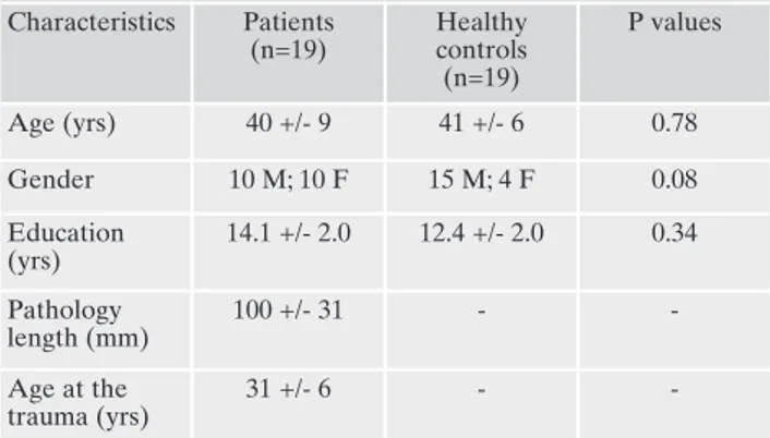
Table 1. Demographic and clinical characteristics of participants. Characteristics Patients (n=19) Healthy controls (n=19) P values Age (yrs) 40 +/- 9 41 +/- 6 0.78 Gender 10 M; 10 F 15 M; 4 F 0.08 Education (yrs) 14.1 +/- 2.0 12.4 +/- 2.0 0.34 Pathology length (mm) 100 +/- 31 - -Age at the trauma (yrs) 31 +/- 6 -
-Note: P-values are for two-tailed t-test for two independent sam-ples. Means ± standard deviation are reported.
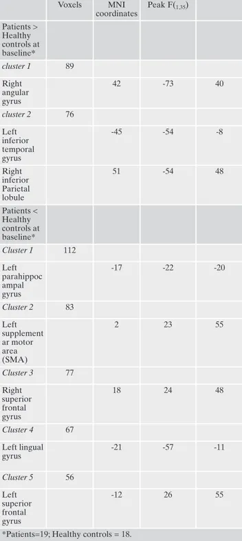
pal region, supplementary motor area, lingual gyrus, and both left and right superior frontal gyrus. Patients with PTSD also showed a significant increase in GMV corresponding to right angular gyrus, inferior parietal lobule and left inferior temporal gyrus. MNI coordinates of each significant cluster, F-values and clusters dimension are reported in Table 2.
Baseline comparisons between patients and controls: Grey Matter Concentration
The GMC comparison between patients and healthy par-ticipants at baseline did not show any significant differences (F 1,35=0.984; p=.332).
Longitudinal comparisons for patients’ clinical PTSD symptom scales pre and post EMDR
During the baseline assessment, patients showed a mod-erate to severe PTSD symptom severity, as highlighted by the DTS values: DTS total score was 99 +/- 9 with mean scores for each subscale of Intrusion 32 +/- 9, Avoidance 40 +/- 14 and Hypervigilance 27 +/- 9. At pre-treatment, the mean CAPS total score was 75.8 (+/- 21.8), with mean score for re-experiencing subscale of 17.0 8, avoidance 20.5 +/-9.0; and hyperarousal 18.5 +/- 9.8. After 12 sessions of EM-DR (Time 2), there was a significant pre-post decrease on the mean CAPS total score (19.3 +/- 15.5) (t (35)=2.132, p<.004)
and hyperarousal subscale (4.1 +/- 9.8; p<.001) (t (35)=1.347,
p<.008), and a non-significant trend to decrease on the re-ex-periencing (6.8 +/- 8.0) and avoidance (9.8 +/- 9.0) subscales. All 19 patients completed EMDR therapy and reported im-provements in their PTSD symptoms, with 16 patients no longer satisfying necessary criteria for PTSD diagnosis.
Longitudinal comparisons between patients and controls: Grey Matter Volume
Group-time interactions for GMV maps were significant (F (1,35)=4.324; p=.006; η2=.398), indicating a larger increase
in GMV in patients as compared to healthy controls, specifi-cally for left parahippocampal gyrus (F (1, 35)=11.237; p=.001, MNI x=-24, y=-21, z=-29; voxels=246), where patients had showed a significantly smaller GMV compared to con-trols before the EMDR treatment (Figure 1). Additionally, in comparison to healthy controls, a cluster of decreased GMV was found in patients’ left thalamus region after EMDR treatment (F (1, 35)=9.432; p=.002, MNI x=-9, y=-24, z=6; voxels=168) (Figure 2). No differences between first and sec-ond MRI acquisition were highlighted for healthy control participants (F 1,35=01.346; p=.389).
Longitudinal comparisons between patients and controls: Grey Matter Concentration
The ANCOVA comparing baseline and post-EMDR GMC and VCBT did not show any significant differences (F 1,35=0.989; p=.421).
DISCUSSION
In this study brain MRI measurements with Optimized Voxel-Based Morphometry was used to investigate the neu-robiological effects of EMDR treatment in drug-naïve PTSD without comorbidity. Consistent with other volumetric
find-ings26,27, when we compared patients with PTSD to healthy
Table 2. Significant GMV Differences at Baseline between pa-tients with PTSD and healthy controls.
Voxels MNI coordinates Peak F(1,35) Patients > Healthy controls at baseline* cluster 1 89 Right angular gyrus 42 -73 40 cluster 2 76 Left inferior temporal gyrus -45 -54 -8 Right inferior Parietal lobule 51 -54 48 Patients < Healthy controls at baseline* Cluster 1 112 Left parahippoc ampal gyrus -17 -22 -20 Cluster 2 83 Left supplement ar motor area (SMA) 2 23 55 Cluster 3 77 Right superior frontal gyrus 18 24 48 Cluster 4 67 Left lingual gyrus -21 -57 -11 Cluster 5 56 Left superior frontal gyrus -12 26 55
controls at baseline, we found significantly smaller GMV in the patients’ parahippocampal, parietal and frontal regions, and significantly larger GMV in temporal and parietal areas (Table 2) all regions involved in processing and storing mechanism of traumatic events27. Furthermore, after
treat-ment completion comparisons with baseline showed in pa-tients a significant increase in GMV in left parahippocampal gyrus and a significant GMV decrease in left thalamus. The implementation of VBM has allowed to extend the structur-al anstructur-alysis to the entire brain overcoming the limitation of our previous investigations restricting the assessment of the effect of EMDR to the hippocampal region. Structural eval-uation provides understanding of a disorder’s neurobiologi-cal substrate and allows to anatomineurobiologi-cally identify and meas-ure changes which have clinical implications. Although to date we are still far from matching symptoms and single al-terations, several works investigating PTSD suggested that many symptoms and/or psychopathological
characteriza-tions appear to be closely related to some specific neurobio-logical alterations. In the present study hippocampus, the main site for short-term memory processing, was found at baseline significantly smaller than in healthy controls and its volume increased following successful EMDR therapy. Hip-pocampus is involved in encoding, consolidating and retriev-ing declarative memories28,29 and receives extensive inputs
from several regions of the neocortex30,31. Hippocampal
dys-function has been claimed to play a key role in the memory disturbances considered to be the core component in PTSD5-7
and it is known by long that PTSD is associated with abnor-malities in activity and volume of the hippocampus32, as is it
true in the symptomatic phase for our patients. It has been speculated that in PTSD emotional information is retained in amygdala and hippocampus and this pathological condi-tion might be related to hippocampal volume reduccondi-tion, pos-sibly due to the effect of chronic release of cortisol, affecting specifically this brain region. Moreover, a failure in the
func-Figure 1. Significant increased GMV post-EM-DR in patients’ left parahippocampal gyrus. Panel A shows coronal and axial views of in-creased grey matter volume in left hippocam-pus area of patients with PTSD (p<0.001 un-corrected; p<.0.05 using MonteCarlo correc-tion for multiple comparisons).
The figures are reproduced in color in the online edition.
Figure 2. Significant decreased GMV post-EM-DR in patients’ bilateral thalamus regions. Panel A shows coronal and axial views of de-creased grey matter volume in left thalamus of PTSD patients.
tionality of this structure reduces the ability to recover from PTSD following EMDR treatment implying an impairment in extinguishing conditioned trauma responses by modifying or integrating traumatic memories4. Our results not only
replicate at whole brain level the ones reported when only hippocampi where analyzed in the same cohort of patients before and after EMDR14but are also in accordance with
other investigations in which significant hippocampal atro-phy was found in patients not responding to EMDR therapy4
and hippocampal grey matter volume was reported to in-crease following successful psychotherapies9,33. Such increase
in volume might not only be due to the decreased cortisol levels during the asymptomatic phase9, but also likely
re-flects an increase in synapses and dendritic connections due to a better networking after psychotherapy34. At this stage
when clinical PTSD scores decrease and the symptoms of hy-perarousal disappear hippocampus regain its capability to properly project to cortical regions and its activity normal-izes. There is little doubt the thalamus plays an important role in the etiology of PTSD; however, the data related to thalamic alterations are difficult to understand, and the liter-ature expresses disparate views. There are conflicting data relating to the structure and volumetry of the thalamus in re-lation to the clinical pattern but it is widely accepted that this brain structure is implicated in the pathology of PTSD. Thal-amus is deactivated in patients with PTSD35and the
alter-ations in thalamo-cortical connectivity may be implicated in excessive fear recall, failure of expression and maintenance of extinction memory, and heightened traumatic remem-brance36. We observed a cluster of decreased GMV in
pa-tients’ left thalamus region after EMDR treatment. Decreas-es in thalamic volume27 thalamic hyperactivation and
hyper-function37 were correlated with PTSD re-experiencing
symp-toms37. Thalamus was also shown by fMRI to be
hyperacti-vated in stress-induced analgesia38, non-conscious fear39and
alexithymia40. A possible explanation for our findings is that
post-EMDR decrease in re-experiencing symptoms, also if a large but not significant trend, has resettled the normal func-tioning of the thalamus and somehow diminishes its hyper-activation resulting in parallel changes at a structural level. On the other hand it has been reported that emotional pro-cessing is linked to anterior and posterior cingulate cortex co-activation mediated by the thalamus. The clinical efficacy of EMDR reducing the emotional valence of the event might have also contributed to a decrease of thalamic involvement in trauma re-living and hence to a volume decrease as com-pared to the symptomatic phase. The structural changes found after EMDR therapy as compared to the symptomatic phase paralled the significant decrease in clinical (CAPS) ones, mainly due to the hyperarousal sub-scale but also de-termined by the sub-threshold decreases in the re-experienc-ing and avoidance sub-scales. The latter results showre-experienc-ing a mere trend to significant changes could be due to the small sample size increasing the likelihood not only of type I (false positive) but also type II (false negative) errors. EMDR is widely recognized as an efficacious treatment for PTSD3
rap-idly effective, significantly reducing PTSD symptoms, and re-sulting in a high rate of elimination of the diagnosis of PTSD. Beside, its neurobiological correlates have been recently in-vestigated and reported4,12-14,41. According to our results the
clinical effectiveness of EMDR may lay in the restoration of hippocampus structure. Following EMDR treatment, we
ob-served in this region an increase in GMV speaking in favour of a parallel recovering in hippocampal functions and corti-cal networking, with successful processing of the traumatic memory and resolution of current triggers. Also if its mecha-nism of action is beyond the purposes of this paper our re-sults suggest that the EMDR leads to a better hippocampal functioning favoring neuronal integration between subcorti-cal areas and cerebral cortex. This is in agreement with pre-vious investigations in which the memory retention of the traumatic event was shown to move from emotional limbic subcortical regions including parahippocampal gyrus to an explicit cortical ones and elaborated at higher cognitive
lev-el10,42. On the other hand working memory processed by
hip-pocampus has been hypothesized to be a determinant of EMDR effectiveness. The reduced vividness of emotional memory during bilateral stimulation might be due to limited working memory resources and this would be in accordance with the reduced hippocampal volume found in PTSD pa-tients at baseline. In this respect we provided preliminary ev-idence that effective treatment may be reflected in anatomi-cal and functional changes in some critianatomi-cal areas altered by PTSD and this has in perspective a great utility, producing more objective measurement than the self-report measures typically used in treatment outcome studies. Such insights could be used to select treatments which could target specif-ic neurobiologspecif-ical alterations, with the goal of achieving res-olution of biological damage and, subsequently, the disorder.
LIMITATIONS AND RECOMMENDATIONS
One limitations of this study is the small sample size pos-sibly overestimating the number of foci showing significant differences43. On the other hand the relative high costs of the
methodology, makes the recruitment of an inadeguate num-ber of subjects to be investigated a common limitation in neuroimaging studies. For this reason in our study as in oth-er ones in the past patients recruitment and charactoth-erization suffer of the presence of different trauma types and of dcrepancies about the number of previous traumas, both is-sues potentially biasing the results. We also acknowledge that the recruitment of PTSD patients without comorbidity and of non-traumatized control subjects might render the results of the present investigation not directly comparable to other studies in the same field. However, the with-in subject analy-sis strengthened, along with the objective decrease of PTSD clinical scores, the reliability of the pre- to post-therapy changes and in the most of the control subjects mix lifetime traumas, even if not causing symptoms have certainly hap-pened10. Furthermore, the absence of follow-up to evaluate
the maintenance of symptomatic improvement and the volu-metric changes does not allow to draw conclusion on the long-term effectiveness of EMDR therapy. Future research might benefit of optimized voxel based morphometry and by the use of diffusion weighted images acquisition aimed at white matter fiber tracts changes detection, to examine the possible impact of psychotherapies on brain structural con-nectivity.
Acknowledgements: Louise Maxfield, Ph.D. professionally edited
this paper. She is a Psychologist in London Ontario Canada, affiliat-ed with London Health Sciences Centre, the Departments of
Psy-chiatry and Psychology at University of Western Ontario, and the Department of Psychology, Lakehead University, Canada. She is ed-itor in chief of the Journal of EMDR Practice and Research. Contact information: email: [email protected]; mail: 523 Upper Spring St. Port Stanley ON, Canada, N5L1G7. She has no conflict of interest.
Conflicts of interest: the authors declare that no source of funding
was received to prepare this article.
REFERENCES
Shapiro F. Eye Movement Desensitization and Reprocessing: 1.
Basic principles, protocols, and procedures. New York: Guilford Press, 1995.
Bisson J, Andrew M. Psychological treatment of post-traumatic 2.
stress disorder (PTSD). Cochrane Database of Systematic Re-views 2007; 3.
National Institute for Clinical Excellence (NICE). Post-traumat-3.
ic stress disorder (PTSD). The management of PTSD in adults and children in primary and secondary care. Clinical Guideline 26. 2005.
Nardo D, Högberg G, Looi JCL, Larsson S, Hällström T, Pagani 4.
M. Gray matter density in limbic and paralimbic cortices is asso-ciated with trauma load and EMDR outcome in PTSD patients. J Psychiatr Res 2010; 44: 477-85.
Zhang J, Tan Q, Yin H, et al. Decreased gray matter volume in 5.
the left hippocampus and bilateral calcarine cortex in coalmine flood disaster survivors with recent onset PTSD. Psychiatry Re-search: Neuroimaging 2011; 192: 84-90.
Karl A, Schaefer M, Malta LS, Dörfel D, Rohleder N, Werner A. 6.
A meta-analysis of structural brain abnormalities in PTSD. Neu-rosci Biobehav Rev 2006; 30: 1004-31.
Woon FL, Sood S, Hedges DW. Hippocampal volume deficits as-7.
sociated with exposure to psychological trauma and posttrau-matic stress disorder in adults: a meta-analysis. Prog Neuropsy-chopharmacol Biol Psychiatry 2010; 34: 1181-8.
Lindauer RJ, Booij J, Habraken JB, et al. Effects of psychothera-8.
py on regional cerebral blood flow during trauma imagery in pa-tients with post-traumatic stress disorder: a randomized clinical trial. Psychol Med 2008; 38: 543-54.
Levy-Gigi E, Szabo C, Kelemen O, Keri S. Association among 9.
clinical response, hippocampal volume, and FKBP5 gene expres-sion in individuals with posttraumatic stress disorder receiving cognitive behavioral therapy. Biol Psychiatry 2013; 74: 793-800. Pagani M, Di Lorenzo G, Verardo AR, et al. Neurobiological cor-10.
relates of EMDR monitoring: an EEG study. PLoS ONE 2012; 7: e45753.
Pagani M, Di Lorenzo G, Verardo AR, et al. Substrato neurobio-11.
logico della terapia con EMDR. (Neurobiological correlates of EMDR therapy.) Riv Psichiatr 2012; 47: 16-8.
Bossini L, Casolaro I, Santarnecchi E, et al. Studio di valutazio-12.
ne dell’efficacia clinica e neurobiologica dell’EMDR in pazienti affetti da disturbo da stress post-traumatic. [Evaluation study of clinical and neurobiological efficacy of EMDR in patients suf-fering from Post-Traumatic Stress Disorder]. Riv Psichiatr 2012; 47: 12-5.
Bossini L, Fagiolini A, Castrogiovanni P. Neuroanatomical 13.
changes after eye movement desensitization and reprocessing (EMDR) treatment in posttraumatic stress disorder. J Neu-ropsychiatry Clin Neurosci 2007; 19: 475-6.
Bossini L, Tavanti M, Calossi S, et al. EMDR treatment for post-14.
traumatic stress disorder, with focus on hippocampal volumes: a pilot study. J Neuropsychiatry Clin Neurosci 2011; 23: E1-2. American Psychiatric Association. Diagnostic and Statistical 15.
Manual of Mental Disorders. 4th ed., text rev. American Psychi-atric Publishing, 2000.
First MB, Spitzer RL, Gibbon M, Williams JB. Structured clinical 16.
interview for Axis I DSM-IV disorders-patient Edition (SCID-I/P, version 2.0). New York: Biometrics Research Department, New York State Psychiatric Institute, 1996.
First MB, Spitzer RL, Gibbon M, Williams JB. Structured clinical 17.
interview for axis II DSM-IV disorders (SCID-II/P). New York: Biometrics Research Department, New York State Psychiatric Institute, 1994.
First MB, Spitzer RL, Gibbon M, Williams JB. Structured clinical 18.
interview for Axis I DSM-IV disorders, Non-patient edition (SCID-I/NP, version 2.0). New York, Biometrics Research De-partment, New York State Psychiatric Institute, 1995.
Blake DD, Weathers FW, Nagy LM, et al. The development of a 19.
Clinician-Administered PTSD Scale. J Trauma Stress 1995; 8: 75-90. Hamilton M. A rating scale for depression. J Neurol Neurosurg 20.
Psychiatry 1960; 23: 56-61.
Hamilton M. The assessment of anxiety states by rating. Br J 21.
Med Psychol 1959; 32: 50-5.
Ashburner J. A fast diffeomorphic image registration algorithm. 22.
Neuroimage 2007; 38: 95-113.
Hayasaka S, Phan K, Liberzon I, Worsley KJ, Nichols TE. Non-23.
stationary cluster-size inference with random field and permuta-tion methods. Neuroimage 2004; 22: 676-87.
Draganski B, Gaser C, Busch V, Schuierer G, Bogdahn U, May A. 24.
Neuroplasticity: changes in grey matter induced by training. Na-ture 2004; 22: 311-2.
Draganski B, Gaser C, Kempermann G, et al. Temporal and spa-25.
tial dynamics of brain structure changes during extensive learn-ing. J Neurosci 2006; 26: 6314-7.
Chen Y, Fu K, Feng C, et al. Different regional gray matter loss 26.
in recent onset PTSD and non PTSD after a single prolonged trauma exposure. PLoS One 2012; 7(11).
Shucard JL, Cox J, Shucard DW, et al. Symptoms of posttrau-27.
matic stress disorder and exposure to traumatic stressors are re-lated to brain structural volumes and behavioral measures of af-fective stimulus processing in police officers. Psychiatry Re-search 2012; 204: 25-31.
Squire LR, Zola-Morgan S. The medial temporal lobe memory 28.
system. Science 1991; 253: 1380-6.
van Strien NM, Cappaert NL, Witter MP. The anatomy of mem-29.
ory: an interactive overview of the parahippocampal-hippocam-pal network. Nat Rev Neurosci 2009; 10: 272-82.
Lavenex P, Amaral DG. Hippocampal-neocortical interaction: a 30.
hierarchy of associativity. Hippocampus 2000; 10: 420-30. Furtak SC, Wei SM, Agster KL, Burwell RD. Functional neu-31.
roanatomy of the parahippocampal region in the rat: the perirhi-nal and postrhiperirhi-nal cortices. Hippocampus 2007; 17: 709-22. Gilbertson MW, Shenton ME, Ciszewski A, et al. Smaller hip-32.
pocampal volume predicts pathological vulnerability to psycho-logical trauma. Nat Neurosci 2002; 5: 1242-7.
Hölzel BK, Carmody J, Vangel M, et al. Mindfulness practice 33.
leads to increases in regional brain gray matter density. Psychia-try Res 2011; 191: 36-43.
Kircher TV, Arolt A, Jansen M, et al. Effect of cognitive behav-34.
ioral therapy on neural correlates of fear conditioning in panic disorder. Biol Psychiatry 2013; 73: 93-101.
Lanius RA, Williamson PC, Densmore M, et al. The nature of 35.
traumatic memories: a 4-T FMRI functional connectivity analy-sis. Am J Psychiatry 2004; 161: 36-44.
Yin Y, Jin C, Hu X, et al. Altered resting-state functional connec-36.
tivity of thalamus in earthquake-induced posttraumatic stress disorder: a functional magnetic resonance imaging study. Brain Res 2011; 1411: 98-107.
Kim SJ, Lyoo IK, Lee YS, et al. Decreased cerebral blood flow of 37.
thalamus in PTSD patients as a strategy to reduce re-experience symptoms. Acta Psychiatr Scand 2007; 116: 145-53.
Lanius RA, Bluhm RL, Frewen PA. How understanding the neu-38.
robiology of complex posttraumatic stress disorder can inform clinical practice: a social cognitive and affective neuroscience ap-proach. Acta Psychiatr Scand 2011; 124: 331-48.
Felmingham K, Kemp A, Williams L, et al. Changes in anterior 39.
cingulate and amygdala after cognitive behavior therapy of post-traumatic stress disorder. Psychol Sci 2007; 18: 127-9.
Frewen PA, Lanius RA. Toward a psychobiology of posttrau-40.
matic self-dysregulation: Reexperiencing, hyperarousal, dissocia-tion, and emotional numbing. Ann N Y Acad Sci 2006; 1071: 110-24.
Lansing K, Amen DG, Hanks C, Rudy L. High-resolution brain 41.
SPECT imaging and eye movement desensitization and
repro-cessing in police officers with PTSD. J Neuropsychiatry Clin Neurosci 2005; 17: 526-32.
Pagani M, Di Lorenzo G, Monaco L, et al. Neurobiological re-42.
sponse to EMDR therapy in clients with different psychological traumas. Front Psychol 2015; 27.
Fusar-Poli P, Radua J, Frascarelli M, et al. Evidence of Reporting 43.
Biases in Voxel-Based Morphometry (VBM) studies of psychi-atric and neurological disorders. Hum Brain Mapp 2014; 35: 3052-65.