University of Naples Federico II
Department of Chemical Sciences
PhD in Chemical Sciences
AMPs: Rational Design, Synthesis and
Biophysical Studies of the Interaction
Process with Model Membranes
Rosario Oliva
Tutors: Prof. Luigi Petraccone
Prof. Pompea Del Vecchio
Advisor: Prof. Flavia Nastri
Contents
List of Abbreviations……….…..1
Preface……….3
Chapter 1 – Antimicrobial Peptides (AMPs)
1.1 Introduction………..………61.2 AMPs Properties………..……….7
1.3 Classification of AMPs………...………12
1.4 AMPs Action Mechanisms……….………13
1.5 AMPs in Clinical Trials………..……18
Chapter 2 – Biological Membranes
2.1 Introduction………...……….202.2 Membrane Functions: a Brief Overview……….………22
2.3 The Membrane Composition………..24
2.3.1 Lipids………..……….24
2.3.2 Membrane Proteins……….……….29
2.3.3 Membrane Carbohydrates………31
2.4 A Comparison between Eukaryotic and Prokaryotic (Bacterial) Membranes...33
2.5 Lipids Self-Assembly………...………..35
2.6 Lipid Structures: Lamellar and Non-Lamellar Structures (Phases)…...………..37
2.7 Lamellar States and Phase Transitions………..………..40
2.8 Model Membranes: Liposomes………..43
Chapter 3 - Synthesis, Biological and Biophysical Studies of Unnatural
Amino Acids Containing Peptides
3.1 Introduction………473.2 Peptides’ Design……….48
3.4 Results: Biological Study………54
3.4.1 Evaluation of Antimicrobial Activity, Serum Stability and Cytotoxicity…….55
3.5 Results: Biophysical Study of the Interaction of the P9Nal(SS) Peptide with Model Membranes………58
3.5.1 The Conformation of P9Nal(SS) Peptide……….58
3.5.2 The Effect of P9Nal(SS) on Bilayer Stability………...60
3.5.3 P9Nal(SS) Induces the Formation of Lipid Domains………...63
3.5.4 P9Nal(SS) Inserts in the Hydrophobic Core of Bacterial-like Membranes but not in the Eukaryotic Model Membranes………..64
3.6 Discussion………..67
Chapter 4 - The Interaction of Two P9Nal(SS)-derived Peptides with
Bacterial Model Membranes
4.1 Introduction………714.2 Materials and Methods………72
4.3 Results………74
4.3.1 The Conformational Behavior of P9Nal(SR) and P9Trp(SS) Peptides………74
4.3.2 The Effects on the Lipid Bilayer Stability………76
4.3.3 Abilities of Peptides to Penetrate in the Membrane………..79
4.4 Discussion…..………..………..………81
Chapter 5 - The Cytotoxic and Antimicrobial Activities of the Human
Thrombin-derived Peptide (P)GKY20: A Biophysical Study
5.1 Introduction………....845.2 Materials and Methods………....85
5.3 Results………90
5.3.1 The Interaction and the Conformational Behavior of (P)GKY20 with Model Membranes………...90
5.3.2 The Effects of (P)GKY20 on Stability of Eukaryotic and Bacterial Model Membranes………...93
5.3.3 (P)GKY20 Clusters Anionic Lipids: Formation of Lipid Domains…………..97
5.3.4 The Localization of (P)GKY20 Upon Interaction with the Membrane……..100
5.3.5 Visualizing the Effect of (P)GKY20 on Bacterial Model Membrane: Atomic Force Microscopy………...102
5.3.6 The Effects of (P)GKY20 on Size and Morphology of Lipid Vesicles……...104
5.4 Discussion..………..106
Chapter 6 - The Complexation of (P)GKY20 Peptide with Cyclodextrins
6.1 Introduction………..1136.2 Materials and Methods………..115
6.3 Results………..118
6.3.1 The Interaction of (P)GKY20 with CDs: SBE-β-CD versus HP-β-CD……..118
6.3.2 The (P)GKY20 Peptide Forms a 1:1 Complex with SBE-β-CD……….119
6.3.3 (P)GKY20 Secondary Structure upon Interaction with SBE-β-CD………...121
6.3.4 Thermodynamics of Interaction between (P)GKY20 and SBE-β-CD………122
6.3.5 The Effect of (P)GKY20/SBE-β-CD Complex on the Thermotropic Properties of DPPC/DPPG Liposomes………123
6.4Discussion……...………..125
References………...129
List of Publications………..149
1
List of Abbreviations
2Nal = 2-naphthyl-L-alanine AFM = Atomic Force Microscopy AMPs = Antimicrobial Peptides CD = Circular Dichroism CDs = Cyclodextrins Chol = Cholesterol CL = Cardiolipin
cmc = Critical Micellar Concentration Cys(StBu) = S-(tert-butylthio)-L-cysteine
Cys(tBu) = S-(tert-butyl)-L-cysteine
DLS = Dynamic Light Scattering
DMPC = 1,2-dimyristoyl-sn-glycero-3-phosphocholine DOPC = 1,2-dioleoyl-sn-glycero-3-phosphocholine DPH = 1,6-diphenyl-1,3,5-hexatriene DPPC = 1,2-dipalmitoyl-sn-glycero-3-phosphocholine DPPE = 1,2-dipalmitoyl-sn-glycero-3-phosphoethanolamine DPPG = 1,2-dipalmitoyl-sn-glycero-3-phospho-1′-rac-glycerol DSC = Differential Scanning Calorimetry
εAhx = 6-aminohexanoic acid
FRET = Fluorescence Resonance Energy Transfer GP = Generalized Polarization
GUVs = Giant Unilamellar Vesicles HII = Inverted Hexagonal Phase
HP-β-CD = Hydroxypropyl-β-cyclodextrin ITC = Isothermal Titration Calorimetry
2
Laurdan = 6-dodecanoyl-2-dimethylaminonaphtalene Lα = Lamellar Liquid Crystalline Phase
Lo = Lamellar Liquid Ordered Phase
Lβ = Lamellar Gel Phase
Lβ’ = Lamellar Gel Phase with Tilted Hydrocarbon Chains
LPS = Lipopolysaccharide LTA = Lipoteichoic acid
LUVs = Large Unilamellar Vesicles L/P = Lipid-to-peptide Ratio
MIC = Minimum Inhibitory Concentration MLVs = Multilamellar Vesicles
N-Rh-DHPE = N-(Lissamine rhodamine B sulfonyl) phosphatidylethanolamine PA = Phosphatidic Acids PC = Phosphatidylcholine PE = Phosphatidylethanolamine PG = Phosphatidylglycerol PI = Phosphatidylinositol POPC = 1-palmitoyl-2-oleoyl-sn-glycero-3-phosphocholine POPG = 1-palmitoyl-2-oleoyl-sn-glycero-3-phospho-1′-rac-glycerol PS = Phosphatidylserine SBE-β-CD = Sulfobutylether-β-cyclodextrin SM = Sphingomyelin
SUVs = Small Unilamellar Vesicles TA = Teichoic acid
3
Preface
The emergence of resistance from bacteria to the conventional antibiotics has become a serious global problem during the last years. The onset of resistance is mainly due to the massive and out-of-control use of these drugs in our community. In fact, microorganisms have developed a series of mechanism which render antibiotics ineffective. Therefore, seeking for new anti-infective agents is becoming increasingly necessary. Among these antimicrobial peptides (AMPs) have been proposed.
AMPs are a class of peptides with a broad spectrum of activity against different pathogenic organisms (e.g. bacteria, fungi, viruses and cancer cells) and they are part of the innate immune system of virtually all forms of life. Usually, they are composed by 10-50 amino acids and are enriched of positively charged (e.g. lysine and arginine) and hydrophobic residues. Even if the exact action mechanism is still under debate, it is widely accepted that the primary target is represented by the lipid matrix of the pathogens’ membranes. Thus, AMPs interact with them in a non-specific way, inducing membrane destabilization and finally cell death. It is believed that, due to the unique mode of action, AMPs could overcome the problem of resistance to conventional antibiotics in multi-drug resistant bacteria and even to currently used anticancer drugs for the treatment of tumor cells. For these reasons, AMPs have attracted great attention as drugs of the future. Thus, the final goal in studying AMPs is their use as drugs. However, AMPs pharmacological application is not straightforward. Many aspects must be considered. A good AMP should interact selectively with the bacterial membrane, should not be toxic to eukaryotic cells and should be resistant to proteases degradation. To develop AMPs with these improved features it is of fundamental importance to understand the role played by lipid composition and peptide physico-chemical properties in determining peptides activity. To this aim, in my PhD thesis I studied the molecular details, at level of peptide-lipid interaction, of the action mechanism of several antimicrobial peptides by a series of biophysical techniques. In particular, I characterized the interaction of natural amino acids-containing peptide as well as of synthetic peptides composed by unnatural amino acids with model biomembranes.
In the first part of the project, I carried out the synthesis of the 9-residue peptide P9Nal(SS) which contains unnatural amino acids. It was designed in order to obtain a peptide with a good antimicrobial activity, low cytotoxicity and high resistance to proteases. Then, biophysical studies of its interaction with eukaryotic and bacterial model membranes were carried out. The obtained information can be very useful in
4
developing antimicrobial agents for biomedical applications. This study is described in detail in the chapter 3.
In the chapter 4 is reported the biophysical characterization of the interaction of two P9Nal(SS)-derived peptides, named P9Nal(SR) and P9Trp(SS), with liposomes mimicking bacterial membrane. The two peptides were obtained by replacing some residues in the primary sequence of P9Nal(SS). As shown in chapter 4, the peptides are less hydrophobic than the parent peptide. The obtained data reveal how these substitutions can modulate the membranotropic activity of peptides.
In the second part of the project, I faced the problem to understand the molecular details of the action mechanism of (P)GKY20, a natural amino acids-containing peptide modelled on the C-terminus region of the human thrombin. Its good antimicrobial activity and low cytotoxicity is well known. However, its action mechanism has never been studied before. Thus, I performed an extensive biophysical characterization of the interaction of the (P)GKY20 with model membranes. The obtained results elucidated its mechanism of action against bacterial model membrane and, at the same time, allow to understand its low cytotoxicity. All the results concerning this AMP are presented in the chapter 5.
The last part of this thesis is devoted to an idea developed during the third year: the encapsulation of AMPs with cyclodextrins (CDs) as a way to protect the peptides from degradation and improve their pharmacological properties. Indeed, antimicrobial peptides containing natural L amino acids are, unfortunately, prone to degradation which limits seriously AMPs applications as drugs. To verify the ability of CDs to encapsulate AMPs without altering their antimicrobial properties, I studied the interaction of (P)GKY20 peptide with sulfobutylether-β-CD and of the obtained complex with bacterial-like liposomes.
These preliminary results reveal that (P)GKY20 form a 1:1 stable complex with sulfobutylether-β-CD. Further, the obtained complex is able to perturb the stability of bacterial-like liposomes. Thus, the sulfobutylether CD could represent a suitable encapsulating agent for (P)GKY20 peptide which could improve its pharmacological profile. The obtained results are presented in the chapter 6.
Since the peptide interaction with model membranes is quite complex being composed by many steps, a strategy which involves different biophysical techniques was adopted to study a particular aspect of the interaction process. Calorimetric, spectroscopic and microscopic techniques were applied (where possible) and by combining the data, it was possible to depict a possible action mechanism highlighting the key steps. Differential Scanning Calorimetry (DSC) was employed to study the effect of peptide interaction on the liposomes thermotropic properties. Moreover, using an appropriate lipid system, information about lipid self-organization (e.g. lipid domains formation) can be obtained. Steady-State
5
Fluorescence Spectroscopy and Fluorescence Anisotropy were extensively employed to obtain information on both AMPs (e.g. determination of binding constants, degree of insertion into the lipid bilayer) and lipids (e.g. changes in membrane viscosity due to changes in lipid packing). Liposomes morphological changes were monitored by Dynamic Light Scattering (DLS). Instead, changes in peptide secondary structure upon binding were followed by means of Circular Dichroism (CD) and an estimation of secondary structure content was obtained by the spectra deconvolution. Atomic Force Microscopy (AFM) and Confocal Fluorescence Microscopy instruments were employed to directly visualize the effect of peptides on bacterial model membranes.
6
Chapter 1
Antimicrobial Peptides (AMPs)
1.1 Introduction
The increasing spread of bacteria resistant to conventional antibiotics has become a worldwide emergence [1,2]. In fact, due to massive and out-of-control use of these drugs in humans and animals, bacteria have developed a series of mechanisms [3] which render antibiotics ineffective. For example, about 80% of bacterial strains of Staphylococcus aureus are immune to the penicillin [4]. Moreover, the number of new discovered (or synthetized) antibiotics is decreasing [5]. Thus, there is an urgent need of a new class of antimicrobial agents.
Antimicrobial peptides (AMPs) are an interesting class of small peptides which can overcome this problem. It is believed that, due their unique and non-specific action mechanisms which involve a direct interaction with the lipid matrix of pathogens’ membranes, they could help in fighting infections caused by multidrug-resistant (MDR) bacteria [6,7]. For this reason, AMPs represent serious candidates as alternative to conventional antibiotics and have attracted much attention in the scientific community.
AMPs are primary known for their antibacterial activity, but they are also active against other microorganisms such as fungi (antifungal peptides), viruses (antiviral peptides), parasites (anti-parasite peptides) and even cancer cells (anticancer peptides) [8–10]. AMPs are part of the defense system of virtually of form of life, from prokaryotic bacteria to eukaryotic mammals and plants [11]. Some of them may be expressed constitutively. Others are inducible and are expressed only in response to the presence of a foreign microorganism [12]. AMPs can be modified after their expression (post-translational modification). The most common modifications are: amidation, glycosylation, acylation and halogenation [13,14]. They represent the first defense line against invading microorganisms. In fact, they are particularly abundant in all tissues that are in some way in contact with external world. For example, in humans AMPs are found in the skin, intestinal and oral mucosa [15]. The era of amino-acids based antimicrobials started in 1922, when Fleming discovered lysozyme [16]. This small globular protein can hydrolyze the glycosidic bond between the N-acetylglucosamine and N-acetylmuramic acid residues in the peptidoglycan, the constituent of the bacterial cell wall [17]. This discovery was overshadowed when the same scientist discovered penicillin in 1928 [18] starting the antibiotic era. Due to this important discovery, the interest on AMPs drastically diminished. In the 1960s, the development of multi-drug resistant bacteria brought
7
the attention again to AMPs as alternative to antibiotics. The first antimicrobial agent discovered was the gramicidin in 1939 in Bacillus strains [19,20]. Despite, its cytotoxicity, gramicidin was found effective for topical treatments of wounds and ulcers. Probably, the first eukaryotic AMPs was found in plants. From the plant Triticumaestivum, an AMP named purothionin was isolated. Purothionin showed a good activity against some bacterial strains [21]. In the 1960s, the first AMP from animals was reported. It was the brombinin from frogs [22]. In the following years, a series of new AMPs were reported thus increaseing our knowledge on the subject. Just to cite two important studies, it is possible to report the discovery of two well studied peptides: cecropins and magainins (a brief and comprehensive review about history of AMPs can be found in [23]). In the 1980s, Boman reported the discovery of the potent AMPs cecropins in Hyalophora cecropia after bacteria injection in the insect. It was the first report of an α-helix antimicrobial peptide [24,25]. In 1987, Zasloff reported the isolation and characterization of cationic AMPs magainins from the African frog Xenopus laevis, pointing out its defensive role in the organism [26]. Today, more than 3000 antimicrobial peptides are known. Information about them can be found in an online database [27] at http://aps.unmc.edu/AP/main.php which is constantly update. It is probably that this database represents only a small fraction of existing AMPs and others will be discovered in the future.
1.2 AMPs Properties
Antimicrobial peptides are a heterogeneous class of small peptides composed by 10-50 amino acids [12]. The primary target of AMPs is represented by the lipid matrix of the cytoplasmic bacterial membrane that they destabilize leading to the pathogen death [28]. As stated in the previous section, more than 3000 different peptide sequences are known. Despite this, all AMPs share common physico-chemical properties which are fundamental for their biological activity. The general features that must be considered are:
Sequence; Conformation; Amphipathicity; Hydrophobicity; Charge; Polar angle.
Sequence. AMPs can be composed by any combinations of amino acids. Essentially, they are enriched of hydrophobic and cationic residues. Usually the ratio between
8
the number of hydrophobic and charged residues varies from 1:1 to 2:1 [29]. At physiological pH, lysine and arginine residues are positively charge which render AMPs cationic leading to the class of cationic antimicrobial peptides (cAMPs). There are also anionic AMPs [30] which represent a small fraction of known AMPs [8] and will not be discussed here. Although there is a low sequence homology among AMPs, two features are frequently observed. The first one regards the presence of a glycine residue at the N-terminus, that is highly conserved. The glycine serves as capping residue providing resistance to aminopeptidases [31]. The second one is the amidation (a post-translational modification) at the C-terminus which provides resistance to carboxypeptidases [31].
Finally, the presence of aromatic residues such as tryptophan and tyrosine could favor the localization of peptide at the membrane-water interface [32] due to the contemporary presence of polar groups (amide in Trp and OH in Tyr) and hydrophobic aromatic rings in the side chains which render the residues nor too much hydrophilic nor too much hydrophobic.
Conformation. Despite the low sequence homology and the wide variety of primary structures, AMPs tend to adopt similar conformation upon interaction with membranes. AMPs can adopt three different conformations: α-helix, β-sheet and random structure.
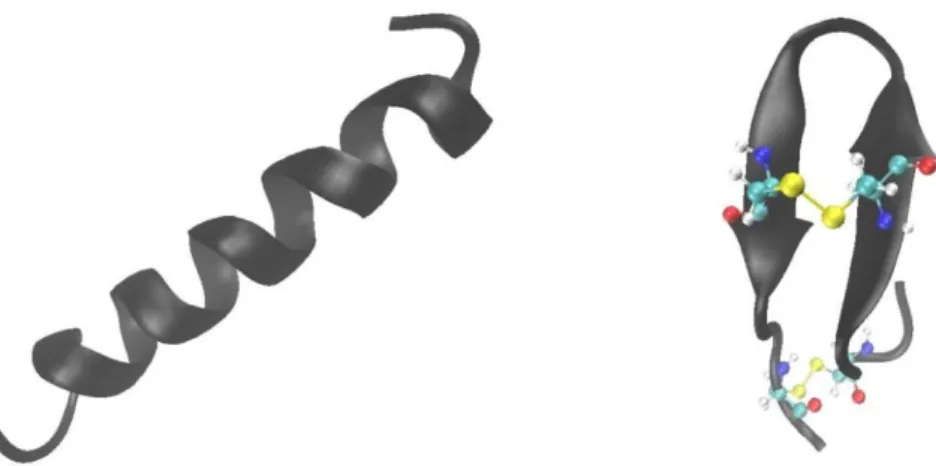
The most common secondary structure is the α-helix. Usually, AMPs have a random structure in solution and adopt an α-helix structure upon interaction with membrane. An example of helical peptide is represented by magainin [33]. The conformational change has a profound impact on the peptides’ activity. In fact, the peptides’ ability to adopt a helical structure is related to its ability to perturb the membrane integrity. Moreover, the lack of the secondary structure upon binding to eukaryotic membrane represents a way through which the peptides’ cytotoxicity could be minimized [34]. Peptides that adopt β-sheet conformation are quite different from α-helical ones. Usually, they adopt a more ordered structure in solution due to presence of cysteine residues that can form disulfide bridges thus stabilizing the three-dimensional structure. Upon interaction with the membrane, their conformation does not change. A good example is tachyplesin that possess two disulfide bonds [35]. In Fig. 1.1 are reported the structures of magainin 2 [36] and tachyplesin I [37] as examples of α-helix and β-sheet peptides, respectively.
9
Fig. 1.1 Cartoon representations of (left) magainin 2 (pdb: 2MAG) and (right) tachyplesin I (pdb: 1MA2). In tachyplesin I the two disulfide bridges are represented as yellow spheres. The picture was made with VMD (Visual Molecular Dynamics) software.
Amphipathicity. This property is directly linked to the conformation of AMPs. The α-helix or β-sheet structure adopted by AMPs are amphipathic structures. This means that they have a hydrophobic face on one side and a polar face on the other side (Fig. 1.2) where the respective residues are clustered.
Fig. 1.2 Schematic representations of amphipathic structures. (Left) β-sheet and (right) α-helix. Blue and red circles correspond to polar and non-polar residues, respectively. Adapted from [29].
This structure facilitates the peptide interaction with the membrane: the hydrophilic side interacts with the polar head groups of lipids and, at the same time, the hydrophobic side interact with the hydrophobic acyl chains. Thus, it plays an important role in the interaction with microbial membrane [38].
The peptides’ amphipathicity is quantitatively measured by the mean helical hydrophobic moment (<μH>) [39]. It is defined as the vector sum of the single amino
acids hydrophobicities, normalized to that of an ideal α-helix. High values of the hydrophobic moment mean high amphipathicity. It is possible to calculate the hydrophobic moment by using online tools, such as Heliquest at
10
http://heliquest.ipmc.cnrs.fr/cgi-bin/ComputParams.py [40]. The best way to represent the amphipathicity is by means of the helical wheel projections. An example is reported in Fig 1.3 for the designed AMP “peptide 8” [41]. From this projection, the peptide amphipathic structure is quite clear.
Fig. 1.3 The helical wheel projection of the AMP named peptide 8. Yellow circles are hydrophobic residues. Blue circles represent hydrophilic residues. The arrow represents the calculated
hydrophobic moment (0.741). Adapted from [41].
The effect of amphipathicity on the AMPs activity is not completely clear. For example, it was reported no correlation between amphipathicity and biological activity in a series of L-V13K analogs antimicrobial peptides [42]. Another study on a series of 20 synthetic peptides demonstrated that the increase of amphipathicity is correlated with hemolytic activity but in a minor extent with antimicrobial activity [43]. On the other hand, a study with magainin 2 analogs demonstrated that both bacterial and hemolytic activities increase with an increase of amphipathicity [44]. Thus, it seems that the amphipathicity plays a role in determining the antibacterial and hemolytic activities and it is peculiar of the peptide sequence.
Hydrophobicity. Peptide hydrophobicity is related to the primary structure and is defined as the percentage of hydrophobic residues within a peptide [12]. For most antimicrobial peptides it reaches 50%, meaning that half of the residues in the sequence are hydrophobic [31]. Hydrophobicity is an important parameter for the peptides’ biology activity since it determines the degree of partition inside the hydrophobic core of the membrane [45]. Several studies evidenced that an increase of hydrophobicity is related to a hemolytic activity increase but a decrease of selectivity against bacterial strains [12]. For example, a study on the peptide L-V13 K showed that the substitution of Ala with the more hydrophobic Leu residue, increases the hemolytic activity of about 62 times. At the same time, the
11
antimicrobial activity against Pseudomonas aeruginosa does not change [46]. In another study (in which the effects of different parameters on a series of 20 AMPs were explored) is reported that an increase of hydrophobicity increases the hemolytic activity slighting affecting the antimicrobial activity [43].
Charge. Antimicrobial peptides exhibit a selective interaction towards the bacterial membrane. It is believed that electrostatic interaction between the positively charged peptide and negatively charged membrane is the first step in the interaction process and it is responsible for the selectivity. For this reason, the global charge on the peptide plays an important role in the antimicrobial activity [12].
Most cationic antimicrobial peptides possess a net positive charge ranging from +2 to +9. They are enriched of lysine and arginine residues positively charged at physiological pH. At acidic conditions histidine residues can be positively charged contributing to the overall charge.
There is a relation between charge and biological activity. As general rule, an increase of the net positive charge causes an increase of the antimicrobial activity [29]. In fact, a study on a series of peptides demonstrated that an increase of the positive charge increases the peptide potency against bacteria [47]. However, it seems that the charge cannot be increased indiscriminately. In fact, for some peptides there is a threshold value above which a decrease of the antimicrobial activity was observed. For example, in a study carried out on a series on magainin 2 analogs it was shown that an increase of the charge above the threshold value of +5 leads to a decrease of antimicrobial activity [48]. The observed decrease could be due to the strong electrostatic repulsions in the packed structure of the peptide upon interaction with the membrane. Even if the increase of positive charge seems a straightforward way to improve the antimicrobial activity, the possible rise of the hemolytic activity should be considered. In fact, the hemolytic activity also depends on the net charge. There is a threshold charge value (depending on the peptide sequence), above which the undesirable hemolytic activity increases [29]. This aspect was well demonstrated in a study carried out on series of peptides analogs where the positive charge of a peptide was varied holding constant the other peptide features [47].
Polar angle. This parameter measures the relative spatial proportion of polar and apolar face in amphipathic peptides [49]. In a perfect amphipathic α-helix, where one face is composed by hydrophobic residues and one face by only polar residues, the polar angle is 180°. An increase of the hydrophobic side will reduce the polar side leading to a decrease of the polar angle and vice versa. Changes in the primary sequence can alter the polar angle. Several studies have found a correlation between the polar angle and the permeabilization of the membrane. It seems that a decrease of the polar angle leads to an increase of the membrane permeabilization [49]. In a
12
study on two model peptides which only have different polar angle (100° and 180°), it was shown that the peptide with the lower polar angle induces greater permeabilization, translocation and rate of pores formation [50].
Even just from this brief description of antimicrobial peptides general features, it becomes clear that the fine interplay among these parameters defines the peptides’ activity. Moreover, they are not independent to each other. A modification of one parameter can change others. This demonstrates that the inter-relationships among these parameters are the key factors in determining the biological activity of antimicrobial peptides.
1.3 Classification of AMPs
Antimicrobial peptides are a heterogeneous class of molecules. It’s very hard to categorize them in specific classes. However, a possible classification can be attempted according to their secondary structures (adopted upon interaction with the membrane) and amino acids composition [8,51,52]:
Linear α-helical peptides; β-sheet peptides;
mixed α/β peptides
peptides enriched of a specific amino acid.
Linear α-helical peptides represent the most characterized group. These peptides adopt a random structure in solution but an amphipathic α-helix structure when interact with the membrane. Examples of AMPs belonging to this class are: magainins (Fig. 1.1), pexiganan and cecropins [53,54].
The second class is composed by peptides with a β-sheet structure. These peptides are usually enriched of cysteine residues. In solution, they can adopt a β-sheet conformation stabilized by disulfide bridges. Upon interaction with membranes, the conformation only slightly changes. β-sheet peptides can contain two or more cysteine residues. An example of AMPs with two Cys residues is tachyplesin (reported above, in Fig. 1.1). Another good example is represented by human defensins (both α- and β-defensins) which are characterized by the presence of three disulfide bridges [55].
Peptides with a mixed structure of α and β are also described. As an example, the drosomycin from the insect Phormia terranovae has a β-sheet composed by three strands and a small segment which adopts a α-helical structure. The whole structure is stabilized by the presence of four disulfide bridges [56]. From the same insect, it
13
was isolated the peptide defensin A with a β-sheet formed by two antiparallel strands and a segment in α-helix. The structure is stabilized by two disulfide bridges [56]. Finally, there are some AMPs in which a specific amino acid is overrepresented. These peptides can adopt different structures apart from the ones reported above. They can be enriched of proline, glycine, arginine, tryptophan and histidine residues. The peptide formaecin from the ant Myrmecia gulosa is a proline-rich peptide. It is composed by 16 residues: 5 of which are proline residues [57]. Plasticins are examples of glycine-enriched AMPs which adopt a β structure in solution [58]. PR-39 is an arginine enriched AMPs of PR-39 residues, 11 of which are arginines [59]. It is effective against both gram -negative and -positive bacteria and even cancer cells. Its action mechanism probably involves a direct bind to DNA [60] rather than the common membrane destabilization. In fact, it is thought that arginine-enriched peptides can translocate across the membrane interacting with intracellular components. This class of AMPs are called cell penetrating peptides (CCPs) and will not be considered here [61,62]. As an example of Trp enriched AMPs it should be cited indolicidin [63]. This 13-residues peptide with 5 Trp adopts a unique extended structure with two half turns in the presence of dodecylphosphocholine micelles [64]. Finally, the antimicrobial peptide clavanin A is an example of histidine-enriched peptide [65]. It is composed by 23 amino acids, 4 of which are histidine residues. Upon interaction with liposomes mimicking bacterial membrane, it adopts a curved helical structure [66].
1.4 AMPs Action Mechanisms
Antimicrobial peptides are a class of active molecules. They are involved in a wide variety of functions such as epithelial cell proliferation, wound healing and stimulation of the production of chemokines [52]. However, they are best known for their antimicrobial activity, which is the subject of this thesis. How do they carry out this important biological activity? It is widely accepted that the final target of AMPs is the lipid matrix of the membrane [6,12,28]. In fact, AMPs can interact with the cytoplasmic membrane of bacteria leading to its permeabilization and, finally, cell death. It is important to note that this interaction is non-specific with no receptor involved. In fact, it has been reported that the replacement of all L- amino acids in the sequence with the corresponding D- enantiomers does not affect antimicrobial activity [67]. The first step of the interaction process between peptides and the pathogens’ cell is represented by electrostatic interactions. At physiological pH, AMPs are positively charged (section 1.2) and bacterial membranes are enriched of negatively charged lipids (section 2.4) [68]. Once peptides are in contact with the cell surface, they must cross the cell wall for gram-positive bacteria, and outer (cytosolic) membrane and cell wall for gram-negative bacteria before to interact with
14
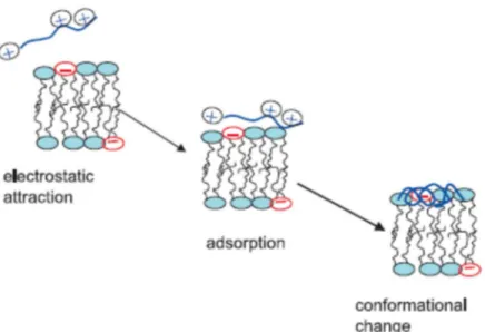
the cytoplasmic membrane, the target of AMPs [69]. For gram-negative bacteria, it was proposed that peptides displace divalent ions associated with lipopolysaccharides (LPS) in the cytosolic membrane [70]. In this way, a destabilization of the membrane occurs, and peptides can gain access to the inner membrane. The way through which AMPs cross the thick cell wall of gram-positive bacteria is not fully understood. It seems that the presence of peptidoglycan is not important in gaining the access to the cytoplasmic membrane. On the contrary, teichoic acids are involved in the translocation across the cell wall, since they could limit peptides’ availability at the surface of the cytoplasmic membrane [71,72]. Moreover, the peptides’ ability to cross the cell wall seems to depend on their secondary structures and oligomerization state. In some way, peptides reach the cytoplasmic membrane of bacteria and are adsorbed on the membrane surface where they change the conformation. The most common conformational change is from random-coil to α-helix (for linear AMPs). Together, these three steps constitute “the binding steps” of peptides to the membrane (Fig. 1.4) and is common to all AMPs.
Fig. 1.4 The three steps involved in the binding of linear AMPs to the negatively charged membrane. Adapted from [73].
After this stage, the peptide molecules remain associated on the surface of the negatively charged membrane. To exert their activity, peptides must be locally concentrated. Thus, they must reach a critical concentration called the threshold concentration [28]. This parameter is defined as the minimum peptide concentration (or lipid-to-peptide ratio, L/P, in an experiment) required to exert the biological effect [28]. Melo et al. [28] established that to observe a biological effect a very high
15
membrane coverage, close to the membrane saturation, is required [12]. Thus, it appears clear that the binding affinity of a peptide for a membrane with a specific composition is fundamental in determining how much peptide is required to kill the bacteria. A correlation between Minimum Inhibitory Concentration (MIC), the lipid to peptide ratio, L/P at saturation and the partition constants exists. The minimum inhibitory concentration is the microbiological parameter indicating the minimum peptide concentration at which bacterial growth is inhibited). As an example, in the case of melittin a partition constant of 6∙104 was determined for the interaction with
liposomes composed by phosphatidylcholines (PCs) and phosphatidylglycerols (PGs) [74]. From this partition constant a lipid-to-peptide ratio at saturation of about 2.5/1 is estimated. From these two parameters, the MIC value can be determined which reproduce the experimental value for E. coli. Thus, there is a link between membrane coverage (threshold) and effect in vivo [28].
As a consequence of membrane coverage (threshold concentration), the peptide can destabilize the membrane, but how does it take place? This is a very difficult task to address. The mechanism by which a peptide destabilizes a membrane depends on several parameters, such as lipid composition and all the physico-chemical properties of both the membrane (e.g. physical state) and peptide (charge, length, amphipathicity, hydrophobicity, conformation). However, to explain the AMPs action mechanism, different models have been proposed. Three of them are the most common invoked and are schematically represented in Fig. 1.5:
barrel-stave model; toroidal model; carpet model.
16
Fig. 1.5 The three most common invoked AMPs action mechanisms. (A) Barrel-stave model; (B) Carpet model; (C) Toroidal model. The amphipathic peptides are represented as cylinders: the red portion are polar residues, the blue one represents hydrophobic residues. Adapted from [12].
In the so-called barrel-stave model (Fig. 1.5, A), AMPs induce the permeabilization by forming pores in the membrane. These pores are formed by interacting trans-membrane peptides that face their apolar residues toward the hydrophobic core of the membrane and polar residues toward the center of the pores which are filled with water. In the initial step, peptides bind in their monomeric form to the surface, interacting with the polar head groups of lipids leading to a local membrane thinning. This favors the peptides insertion in the hydrophobic part of the outer leaflet of the membrane, since most of them are too short to span the membrane completely. At the threshold concentration, the peptides’ monomers self-assemble and insert deep in the membrane forming pores. This mechanism is characteristic of hydrophobic peptide with a low charge density. An example of peptide which it is believed to act through this mechanism is alamethicin [75]. This peptide forms a helix bundle composed by 6 peptides which lines the pore. Its orientation respect to the bilayer depends on the membrane hydration state and the lipid-to-peptide ratios underlying the importance of threshold concentration concept [76,77].
The second mechanism is called toroidal model. As in the barrel-stave, also this model predicts the formation of pores. The main difference is that in the toroidal model, peptides are always associated with the lipid polar head groups.
17
In this model, peptides partition into the membrane (assuming a trans-membrane orientation) and induce a bending in the membrane leaflets which connect to each other (Fig. 1.5, B). Thus, the pore is lined by peptides and lipid head groups and filled with water. Peptides with high charge density and not particularly hydrophobic act through this mechanism. The bending of the membrane and the association of lipid polar heads with peptides stabilizes the pore. In fact, without it, the electrostatic repulsion among peptides will be too high that a pore cannot be formed. An example of peptide that acts through this mechanism is magainin [78,79].
In the carpet mechanism (Fig. 1.5, C), proposed by Shai in 1996 [80] peptides cover the membrane like a carpet. At the threshold concentration, peptides produce a detergent-like effect which solubilize the membrane. Thus, the permeabilization does not occur through the formation of pores. In this mechanism, the peptide is not necessarily inserted in the hydrophobic core, but it can remain attached to the lipid polar head groups. In addition, peptides might form transient pores that allow to peptides to translocate to the inner leaflet of the membrane favoring the solubilization process. Examples of peptides which it is believed to act through this mechanism are ovispirin [81] and cecropin [80]. Both these peptides don’t penetrate inside the membrane hydrophobic core and remains attached to the lipid head groups with their helical axes perpendicular to the bilayer normal.
The three models described above are membranolytic mechanisms. It means that they predict the membrane permeabilization through its disruption (with stable pores or with a detergent-like effect).
Other mechanisms have been proposed in which a membrane disruption does not occurs [12]. The development of these models was necessary from the observation that some peptides are effective against bacteria, but they are not able to perturb the membrane [82,83].
One of these models is the aggregate channel model [84,85]. In this model, peptides insert into the membrane in form of unstructured aggregates. These aggregates are only transient and allow to the peptides to cross the membrane without causing a significant membrane disruption. Once inside, the peptides can interact with intracellular targets as DNA, RNA and proteins [68].
Another one is the molecular electroporation model, proposed for the peptide annexin V [12]. In this model, peptides with a high charge density generate an electric field. The electric field promotes the formation of transient pores which increase the membrane permeability without causing it any kind of damage.
Some peptides can perturb the membrane by promoting the formation of specific lipid-peptide domains [86]. In this mechanism, the preferential interaction of peptides with the negatively charged lipids (phosphatidylglycerols, cardiolipins) leads to a lateral phase segregation of anionic lipids from the zwitterionic ones. The formation of discrete domains induces a destabilization of the membrane since the
18
interface among domains act as defects which increase membrane permeability [87]. For example, it was demonstrated by means of calorimetric measurements that the small peptide Ac-RW [88] is able to induce the formation of a PE (phosphatidylethanolamine) and PG (phosphatidylglycerols) enriched domains in model membrane composed by a mixture of both lipids. It is known that PEs and PGs, in their pure forms, prefer to adopt different phases: inverted hexagonal and lamellar, respectively (see section 2.6). The interface among domains of different phases destabilizes the membrane without disrupting it. The formation of domains can have a deep impact also on the biological activity of the cell [12]. For example, it can alter the diffusion rates of lipids and membrane proteins. Moreover, the membrane curvature is also affected by the lipid segregation process. It is known that a correct curvature is required for some process such as cell division [89].
In conclusion, several peptides and membranes properties dictate the action mechanism. Often, a peptide doesn’t act through a single, well defined mechanism. Sometimes, its mechanism is unique. Find a relation between peptides’ structural properties and mechanism is a very difficult task.
1.5 AMPs in Clinical Trials
The efforts of the scientific community in studying AMPs are all devoted to a common final goal: the application of AMPs as drugs. These studies are important in revealing the relation between peptides’ properties and biological activity. In this way the design of new AMPs with high antimicrobial activity and low cytotoxicity is possible.
AMPs as drugs offer a series of advantages [90,91] among which it is possible to cite: safety, tolerability, efficacy and selectivity. A lot of peptides are characterized by low MIC values and low hemolytic activity. This automatically means that human body well tolerates them because AMPs are selective and interact preferentially with pathogens. Unfortunately, peptides are not chemically e physically stable. They are prone to hydrolysis and oxidation reactions which can modify the biological activity. Moreover, the plasma half-life is short. In fact, once inside the human body, they are subjected to the proteases action [92] that cleave the peptide at specific residue or directly at one of the two termini. This inevitably leads to an unfavorable pharmacological profile. Thus, there are some limitations in the application as drugs and several strategies have been developed to face these problems. The introduction of unnatural amino acids, β-amino acids or D- enantiomers as well as protection of both N- and C- termini contribute increasing the peptide half-life [91,93]. The attachment of fatty acids (lipopeptides) or PEG (polyethylene glycol) can also enhance serum stability [90].
19
Apart from the “scientific problems”, financial problems should be considered, as the high production cost which limit AMPs applications in medicine [94]. From this very brief description, it appears clear that the development of a peptide-based drug is not simple. However, due to selectivity, low cytotoxicity, broad spectrum of activity and no resistance development, AMPs have attracted the attention of several pharmaceutical companies [95].
Today, about 60 peptide-based drugs are available on the market and many others are at different stages of clinical trials [90,91]. Examples of AMPs in clinical trials are reported.
Pexiganan is a peptide derived from magainins and is at phase III of clinical trials. It is composed by 22 amino acid and it has a broad spectrum of activity against bacteria (both gram-positive and negative) and fungi [54]. The final goal is to obtain a pexiganan-containing formulation for topical application in the treatment of infected diabetic foot [91,96]. Omiganan, is an antimicrobial peptides derived from indolicidin, a Trp-rich peptide [97]. It is in phase III of clinical trials for the evaluation of safety and efficacy in the topical treatment of rosacea. Omiganan is also in phase II for the development of a gel against acne vulgaris. Finally, another example of AMP in development is represented by PAC-113, a peptide derived from histatin 5, enriched in histidine residues. It has shown activity against the fungus Candida albicans [98]. It is in phase III clinical trials aimed in finding the optimal dose for the treatment of candidiasis of oral cavity [91].
These examples show that application of AMPs as drugs is possible. So, AMPs have an enormous potential as future therapeutics.
20
Chapter 2
Biological Membranes
2.1 Introduction
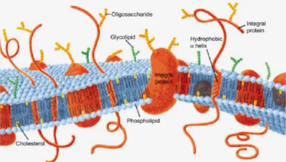
Biological membranes are fluid structures composed by lipids, proteins and carbohydrates [99]. Membranes are fundamental components of every kind of cells, from simpler prokaryotes like bacteria to the more complex higher organisms as animals [100]. Biological membranes separate the inner part (intracellular space) of a cell from the external part (extracellular space) or delimit organelles inside a cell (e.g. membrane of mitochondria). More specifically, in the first case we will refer to the membrane as “the plasma membrane” which constitute the cell boundaries. In Fig. 2.1 is reported a schematic representation of a plasma membrane highlighting its major components.
Fig. 2.1 A schematic representation of a plasma membrane. Their main constituents are lipids, proteins and carbohydrates. Adapted from [99].
The fluid mosaic model developed by Singer and Nicolson [101] is the common representation used to describe cell membrane structure and dynamics. In this model and in its update version [102] the membrane components, lipids and proteins are distributed inhomogeneously like a mosaic. But differently from a Roman mosaic in which the tesserae are fixed, the membrane tesserae (its components) are in constant motion. They can diffuse along the plane of the membrane (translational diffusion)
21
or can rotate around an axis perpendicular to the membrane plane. Occasionally, lipids can go from the inner leaflet to the outer and vice versa (transbilayer diffusion or flip-flop) [103]. Following, the general features of a membrane are reported:
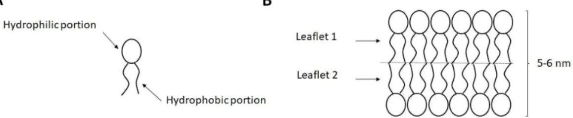
A membrane is composed by a lipid double layer (two leaflets) with a thickness of 5-6 nm. Lipids are amphipathic molecules (Fig. 2.2A). They present a polar head group (hydrophilic) and two lipid chains (hydrophobic). When exposed to aqueous environment, they organize themselves spontaneously by forming a structure (the bilayer) in which the hydrophobic portion of each lipids is hidden to the water, whereas the hydrophilic portion is exposed to the water (Fig. 2.2B);
Fig. 2.2 (A) Simplified representation of a lipid molecule. The hydrophilic and hydrophobic portion of the molecule are indicated by arrows. (B) The lipid bilayer: it is formed by two lipid monolayers (or leaflets) facing their hydrophobic portions. The hydrophilic head groups are surrounded by water molecules.
Membranes are enriched in proteins. Proteins associated with the polar head groups are called peripheral proteins. They can be found on both side of the membrane. Proteins which interact with the hydrophobic matrix of the membrane are called integral proteins. Usually, not the entire protein is embedded in the membrane, but some regions come out of the membrane (Fig. 2.1);
All membranes contain carbohydrates. Carbohydrates do not possess a hydrophobic moiety. Consequently, they are never localized in the membrane interior. Interestingly, sugars are only found on the outer surface of the plasma membrane. Carbohydrates are always associated with lipids (glycolipids) and proteins (glycoproteins);
The membrane is an asymmetric structure [104]. It means that the composition of the inner, cytosolic leaflet is different from the outer leaflet. For example, in the eukaryotic plasma membrane the lipid phosphatidylserine is almost found in the inner leaflet. The same holds for proteins: a peripheral protein which interact, for example, on the surface of
22
the outer leaflet will be found always there and never in the inner leaflet [102];
The membrane is characterized by later heterogeneity. This means that the constituents’ composition is not the same everywhere. There are small (0.1 – 1 µm in diameter) patches called “domains”, enriched of specific lipids and proteins, which exhibit characteristic functional properties. The so called “lipid rafts” are a good example of domains. The rafts are enriched of sphingolipids and cholesterol. It is believed that rafts domains are the house of a series of membrane proteins involved in the cell signaling [105]. 2.2 Membrane Functions: a Brief Overview
The most obvious function of a biological membrane is to separate two aqueous compartments. For a plasma membrane this involves separation between intracellular and extracellular space. Clearly the separation cannot be absolute. In fact, a cell must be able to uptake nutrients from the external environment and to remove molecules from its interior. In other words, a cell must communicate with the external environment. Thus, it acts as a selective permeable barrier which regulates the enormous traffic of molecules in and out of the cell. Essentially, molecule movements across the membrane can be divided in two big groups: passive diffusion and active transport [103,106]. In Fig. 2.3 are summarized these basic types of membrane transport.
Fig. 2.3 The basic types of membrane transport: passive transport (which includes also simple diffusion) does not require energy. In contrast, active transport requires energy in form of ATP. Adapted from [99].
23
Passive diffusion does not require energy, but it is driven by only solutes concentration gradient. Passive diffusion can be either simple diffusion across the membrane or “assisted” diffusion, where some carriers (proteins) are involved in the process. Potassium and Sodium Channels are examples of integral membrane proteins which facilitate the diffusion of K+ and Na+ across the membrane. In
contrast, active transport requires energy, usually in form of ATP, and it is always performed by membrane proteins. An important example of active transport is represented by Na+/K+ ATPase which uses ATP to pump Na+ out of the cell and to
pump in K+ both against their concentration gradients. These mechanisms are mainly
involved in the transportation of small molecules. For larger molecules (e.g. macromolecules) other ways are required. These ways include: 1) receptor mediated endocytosis (RME) also known as clathrin-dependent endocytosis where a membrane protein named clathrin favors the internalization of molecules by forming a small lipid vesicle; 2) pinocytosis, in which an invagination of the plasma membrane encapsulates external fluid materials. This leads to the formation of a vesicle which it is released inside the cell. Every kinds of fluid can be incorporated; thus, this mechanism is completely non-specific; 3) phagocytosis involves the uptake of large solid particles of macromolecules, parts of cell or even a whole microorganism. The solid particle is recognized by receptors on the membrane surface. Then, the formation of vesicles called phagosomes take place which internalize the solid particle. Finally, the particle will be transported to the lysosome for digestion.
In addition, the plasma membrane carries out other important functions:
It contributes in protecting the structural integrity of the interior of the cell, e.g. maintaining the cytosol pH and the right osmotic pressure;
It constitutes the point of attachment to the cytoskeleton: this helps in maintaining the cell shape;
It is involved in the cell recognition thanks to the presence of glycolipids on its surface;
It is involved in a series of biochemical and physiological functions: inter-cellular communication, cell adhesion [107] and energy transduction events, just to cite a few.
From this description it appears clear that membranes are involved in a great number of processes most of which are still unknown.
24
2.3 The Membrane Composition
The basic components of all biological membranes are lipids, proteins and carbohydrates. The composition of a plasma membrane can vary from cell to cell, depending primary on the cell function [108].
Even in a cell, the composition of plasma membrane differs from that one of organelles. For example, the myelin sheath (the plasma membrane of nervous fiber) is enriched of sphingomyelin and cholesterol and it contains only 20% by weight of proteins [109]. In contrast, the mitochondrial membrane is enriched in proteins, up to 75% by weight. This difference is primary due to the function of the membranes: the role of the myelin sheath is to insulate nerve cells (structural role), instead the mitochondrial membrane is involved in many processes such as electrons transport (active role).
2.3.1 Lipids
Lipids are fundamental components of the membrane. The principal classes of lipids found in membranes are essentially three: glycerophospholipids, sphingolipids and sterols.
For sure, glycerophospholipids are the main components of a membrane. They are composed of one glycerol molecule, a phosphate, two fatty acid chains and an alcohol. A phospholipid is an amphipathic molecule: it has a polar head group formed by glycerol, phosphate and alcohol, and a hydrophobic portion formed by the two fatty acid chains. In Fig. 2.4 is reported a representation of a glycerophospholipid.
Fig. 2.4 Chemical structure of a glycerophospholipids.
The fatty acids are esterified at C1 (sn-1 position) and C2 (sn-2 position) of the glycerol. Usually the acyl chain in position sn-1 is saturated, instead that one in position sn-2 is unsaturated. Clearly this is not a rule. Both chains can be saturated or even both with multiple unsaturations. Their length can vary from 14 to 22 carbon
25
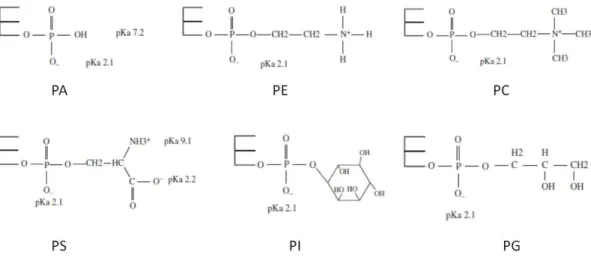
atoms. At the C3 of the glycerol, a phosphate group is esterified. Attached to phosphate, there is an alcohol. Based on the type of alcohol, it is possible to obtain different kinds of phospholipids (Fig.2.5): phosphatidylcholine (PC), phosphatidylethanolamine (PE), phosphatidylserine (PS), phosphatidylinositol (PI) and phosphatidylglycerol (PG). There are also phosphatidic acids (PA) in which there is no alcohol attached to the phosphate group.
Fig. 2.5 Chemical structures of the polar head groups of glycerophospholipids. The pKa values of the
chemical groups forming the polar heads are also reported. Adapted from [99].
A particular lipid found both in eukaryotic and prokaryotic membrane [99,110] is the cardiolipin (CL). It is formed by two PAs linked together by a glycerol (Fig. 2.6). Thus, it differs from other lipids having four fatty acid chains.
Fig. 2.6 Chemical structure of cardiolipin (CL). Adapted from [99].
All the subclasses of glycerophospholipids (Fig. 2.5 and 2.6) are amphipathic molecules. At physiological pH of 7.4 some of them are zwitterionic, and some are negatively charged. Due to the acidic character of the phosphate group, at physiological pH, it brings always a net negative charge. Thus, the net charge of a particular lipid is determined by the phosphorylated alcohol. For example, PCs have
26
not ionizable groups and a positive charge on the nitrogen atom. Thus, it is zwitterionic with no net charge. PEs instead, are slightly negative charged. This is because the amine group of PE has a pKa around 8.5. CLs, instead, have a negative
charge of about 2 dues to the presence of two phosphate groups which are not fully ionized [111].
Each subclass of phospholipids has a role in the membrane. PCs are the most abundant phospholipids in all the eukaryotic membranes. Basically, they have a structural role, determining many properties of the membrane, for example the fluidity. Another structural lipid is the PE. It is the second most abundant lipid in eukaryotic organisms, but it is the main lipid in the membrane of bacteria. This is because, in eukaryotes PEs are converted into PCs. This process is not possible in bacteria. PSs are negatively charged lipids which have a different role. In fact, they are involved in activating and anchoring proteins to the membrane. It is interesting to note that PSs are found almost exclusively in the inner leaflet of the membrane (bilayer asymmetry). This is accomplished by proteins named flipases [112]. With the ageing of the cell, PSs accumulate in the outer leaflet of the membrane and this is a signal that the cell should be recycled. PIs, another negatively charged lipids, play a central role in cell signaling and regulation [113]. PIs are present, more or less, in every cell type but they are particularly abundant in the cells of the brain. PGs, anionic lipids, are found in low abundance in the membrane of eukaryotes. An exception is represented by lung surfactant in which PGs constitute up to 10% of all lipids [114]. Moreover, PGs are among the main components of the bacterial membrane [115]. PAs are unique lipids involved in different processes such as membrane fission and fusion events. At physiological pH, they are negatively charged. Finally, CLs are abundant in the mitochondrial membrane where they are involved in electron transport stabilizing some electron transport proteins. CLs are also present in the bacterial membrane. They take part in some processes such as cell division, membrane transport and energy metabolism [116].
The second important class of lipids is represented by sphingolipids. All the sphingolipids are based on the sphingosine, an amino alcohol with a hydrophobic tail of 18 carbon atoms and a trans double bond at C4 (Fig. 2.7).
27
Fig. 2.7 The chemical structure of sphingosine, the precursor of all sphingolipids.
All the sphingolipids derive from the sphingosine by attaching an alcohol to the C1 and a fatty acid chain (saturated or not) to the its unique nitrogen atom. Depending on the alcohol on the C1 we can identify five different subclasses:
Ceramide, where the attached group is a simple hydrogen atom;
Sphingomyelin, where the attached group is a phosphatidylcholine or a phosphatidylethanolamine;
Cerebroside, with a sugar such as glucose or galactose; Globoside, with up to four sugar molecules;
Ganglioside where the attached group is a complex oligosaccharide.
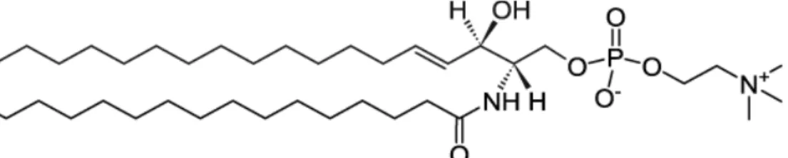
The most abundant sphingolipid in humans is sphingomyelin (SM). In this lipid, the head group is a phosphocholine. Thus, like PCs, at physiological pH it is zwitterionic. The fatty acid chain can vary in length (up to 24 C atoms) and usually it is longer and more saturated respect to the hydrocarbon chains in PCs. In Fig. 2.8 is reported a chemical structure of a sphingomyelin, where the attached fatty acid has 16 C atoms and it is saturated.
Fig. 2.8 The chemical structure of palmitoyl-sphingomyelin. Taken from https://avantilipids.com/.
Sphingomyelin is present in many mammalian cells, but it is particularly abundant in the plasma membranes of the nervous cells [117]. It has a higher affinity for cholesterol, with which forms organized microdomains termed “lipid rafts” [118,119]. These microdomains can function as signaling platforms that regulate the localization of proteins [120]. Other sphingolipids are involved in different
28
functions. For example, gangliosides are found in the outer leaflet of the membranes where are involved in cell-cell recognition [121].
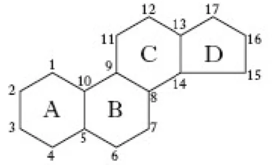
The third class of lipids commonly found in the membranes are sterols [122]. They are components of the membrane of animals, plant and fungi but it is completely absent in prokaryotes. Sterols are very water insoluble and readily partition inside the hydrophobic core of the membrane. The basic structure is formed by four rigid rings (Fig. 2.9) where at C3 an OH group is attached which form the polar head group of this lipid.
Fig. 2.9 The chemical structure of sterol which constitutes the basic structure of all sterols.
Some carbons in the rings could be also unsaturated. At the C16, a hydrocarbon tail is found. Depending on the structure of the tail, we will have different kinds of sterols. In Fig. 2.10 are reported the three main sterols found in animals (cholesterol), fungi (ergosterol) and plants (β-sitosterol).
29
Cholesterol (Chol) is found in many cell membranes. It constitutes up to 50% by weight of all lipids in the plasma membrane and it is particularly abundant in the brain. The main role of cholesterol in membranes is to regulate their fluidity [122] controlling the phase behavior of the bilayer (see section 2.7). This is accomplished thanks to its unique flat structure which permits its intercalation among phospholipid chains. Thus, cholesterol is important in supporting membrane lateral organization, stability and preventing the leakiness of solutes across the membrane. Moreover, cholesterol is a precursor of a series of steroid hormones (e.g. testosterone), vitamin D and bile salts. In conclusion, cholesterol has two functions: it has a structural role and a biochemical role as a precursor of important molecules.
2.3.2 Membrane Proteins
Proteins are an important component of the membrane. Differently from lipids, all proteins have an active role. They can serve as enzymes which catalyze some reaction at the water/membrane interface, they are involved in the active transport of solutes and they can function as receptor in the surface of the membrane. Basically, all the membranes have proteins. Their percentage by weight can vary from 20% up to 75%, depending on the function of the membrane. If the membrane has only a structural role, the % of proteins will be very low. Conversely, if a membrane must catalyze a series of reactions, its protein content will be very high.
Membrane proteins can be classified in two big groups: peripheral and integral proteins [123].
Essentially, a peripheral protein is a water-soluble protein which interact with the membrane surface through electrostatic interaction or hydrogen bonds. Some of them interact directly with the anionic lipids, others interact on the surface of integral proteins. Since the weak forces involved in this interaction, peripheral proteins can be easily removed by changing the pH or the ionic strength of the medium.
30
Fig. 2.11 A schematic representation of the two kinds of peripheral proteins found in membranes. Adapted from [99].
A good example of peripheral protein which interact directly with lipid head groups is myelin basic protein [124] whose malfunctions are involved in multiple sclerosis. Cytochrome c, instead, is an example of peripheral protein which interacts with an integral protein. In fact, cytochrome c is weakly bound to cyctchrome c oxidase that is localized in the inner membrane of mitochondria [125].
Integral proteins penetrate inside the membrane and interact directly with the hydrophobic core. Usually, these proteins span the entire membrane (they are trans-membrane protein) with some segments exposed on both side of the trans-membrane (as well represented in Fig. 2.1). The residues which interact with the lipid chains are clearly hydrophobic. Instead, the portions out of the membrane are enriched in polar amino acids. A general feature of integral proteins is that they have aromatic residues as tryptophan and tyrosine localized at membrane/water interface [32,126]. The portion of integral protein, which is embedded in the membrane hydrophobic core, adopts a specific motif. On this basis we can divide integral proteins in several classes (Fig. 2.12):
1. Single trans-membrane α-helix proteins; 2. Multiple trans-membrane α-helices proteins; 3. β-barrel proteins.
31
Fig. 2.12 The three kinds of integral proteins. From left to right: single trans-membrane α-helix, multiple trans-membrane α-helices proteins and β-barrel.
Single trans-membrane α-helix proteins have a single helix which span the entire membrane. Since the average thickness of a membrane is about 5-6 nm, a trans-membrane α-helix should have 20 amino acids. An example of this kind of integral protein is represented by glycophorin [127]. Multiple trans-membrane α-helices proteins, instead, are composed by several helical structures embedded in the membrane. An example is represented by the 7 α-helix trans membrane protein bacteriorhodopsin [128]. The last type of structure adopted by integral proteins is the β-barrel. It is composed by 16 or more anti-parallel β-strands arranged like a cylinder (the barrel) inside the membrane. β-barrels are very common in the outer membrane of gram-negative bacteria. The bacterial porins are an example [129]. They are composed by 18 strands connected by turns on cytoplasmic side and loops on the extracellular side.
Many integral proteins are also attached to the membrane through lipid anchors. The most important lipid chains involved are: myristic acid, palmitic acid, prenylated hydrophobic chains and glycosylphosphatidylinositol (GPI).
2.3.3 Membrane Carbohydrates
Carbohydrates (or sugars) are the third component found in biological membranes. Being water-soluble molecules, they do not partition inside the hydrophobic core of the membrane. Sugars are exclusively found on the outer surface of the membrane and always attached to lipids (glycolipids) or proteins (glycoproteins). This aspect suggest that sugars are involved in the interaction of the cell with the external environment. Among carbohydrates diverse function, it is possible to mention:
cell-32
cell recognition, membrane receptors, membrane proteins protection from degradation, chaperones, proteins stability [99]. Carbohydrates have also a protecting role in protein-derived antimicrobial peptides. Moreover, sugars are fundamentals for the activity of some AMPs [130]. Carbohydrates can also contribute to the membrane physical state, modulating its fluidity [131]. Commonly, in membranes are found about nine different sugars: D-glucose, D-mannose, α-D-galactose, α-L-fucose, α-D-xylose, α-L-arabinose, acetylglucosamine, N-acetylgalactosamine and sialic acid. It is possible to find attached to proteins and/or lipids a single sugar molecule or more, up to 15.
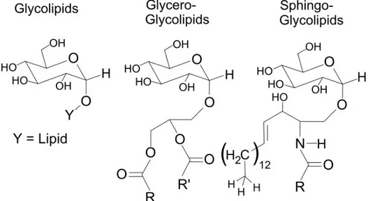
In glycolipids, carbohydrates can be covalently linked (through a glycosidic bond) directly to a fatty acid (simple glycolipids) or to the glycerol of the lipid head group of glycerolipids, forming the class of glycerol-glycolipids. On the other hand, carbohydrates linked to the oxygen atom on C1 of sphingolipids form the class of sphingo-glycolipids (Fig. 2.13).
Fig. 2.13 The chemical structure of glycolipids (left), glycero-glycolipids (middle) and sphingo-glycolipids (right). Adapted from https://en.wikipedia.org/wiki/Glycolipid.
In animals, the major glycolipids are sphingo-glycolipids. They can accumulate into lipid rafts where they are involved in the cell signaling. Cerebrosides are sphingo-glycolipids with only one sugar molecules (usually a glucose or a galactose). Globosides can have two, three or four sugar molecules (in the form of di-, tri- or tetra-saccharide) linked to the sphingolipid. Finally, gangliosides have a complex oligosaccharide. A special mention merits a class of glycolipid localized in the outer membrane of gram-negative bacteria: the lipopolysaccharide (LPS) [132]. Briefly, it is composed by a disaccharide of N-acetylglucosaimne with multiple fatty acid
33
chains. The hydrophobic tails can be linked to the disaccharide free OH group or to the acetyl group. Attached to the disaccharide moiety, there is a complex polysaccharide whose composition varies from bacterium to bacterium.
Carbohydrates can also be found attached to membrane proteins. The combination of proteins and sugars forms glycoproteins. The process through which sugars are added to proteins is known as glycosylation. There are two types of glycoproteins: glycosylated and O-glycosylated (Fig. 2.14). Intuitively, in the case of N-glycosylated proteins, the glycosylation occurs at the nitrogen atom of asparagine. The first sugar attached is always a N-acetylglucosamine. From this unit, other sugars can be attached. In O-glycosylated proteins, carbohydrates are linked to the oxygen atom of serine and threonine residues.
Fig. 2.14 The two types of proteins glycosylation. (Left) glycosylation always starts with a N-acetylglucosamine. (Right) O-glycosylation involving a residue of serine. Adapted from [99].
Usually, the sugar chain of O-glycosylated proteins is shorter respect to N-glycosylated proteins. Moreover, the N-glycosylation is common for all the membrane proteins which have an active role (enzymatic), whereas O-sugars are predominant in structural proteins. For example, in the plasma membrane (a very active membrane) about 90% of protein are glycosylated. The number of N-linked sugars is more than the O-linked ones [99].
2.4 A Comparison between Eukaryotic and Prokaryotic (Bacterial) Membranes All the living organisms are formed by cells [100]. Essentially, there are two cell types: prokaryotic cells and the more complex eukaryotic ones. They differ in a wide variety of aspects which can be found in every biology text. Here we focus our attention to a specific part of the cell: the membrane. Eukaryotic and prokaryotic membranes are both formed by a lipid bilayer. However, there are some important differences between them, both structurally and compositionally.