Vol. 19, 1670 –1679, April 2008
Activation of Srk1 by the Mitogen-activated Protein
Kinase Sty1/Spc1 Precedes Its Dissociation from the
Kinase and Signals Its Degradation
Sandra Lo´pez-Avile´s,*
†Eva Lambea,*
†Alberto Moldo´n,
‡Maribel Grande,*
§Alba Fajardo,* Miguel A. Rodrı´guez-Gabriel,
储Elena Hidalgo,
‡and Rosa Aligue*
*Departament de Biologia Cellular, Universitat de Barcelona. Institut d’Investigacions Biome`diques August Pi
i Sunyer, 08036 Barcelona, Catalunya, Spain;
‡Departament de Cie`ncies Experimentals i de la Salut,
Universitat Pompeu Fabra. 08003 Barcelona, Catalunya, Spain; and
储Departamento de Microbiologia II.
Universidad Complutense, 28040 Madrid, Spain
Submitted July 5, 2007; Revised January 9, 2008; Accepted February 1, 2008 Monitoring Editor: Daniel Lew
Control of cell cycle progression by stress-activated protein kinases (SAPKs) is essential for cell adaptation to extracellular stimuli. The Schizosaccharomyces pombe SAPK Sty1/Spc1 orchestrates general changes in gene expression in response to diverse forms of cytotoxic stress. Here we show that Sty1/Spc1 is bound to its target, the Srk1 kinase, when the signaling pathway is inactive. In response to stress, Sty1/Spc1 phosphorylates Srk1 at threonine 463 of the regulatory domain, inducing both activation of Srk1 kinase, which negatively regulates cell cycle progression by inhibiting Cdc25, and dissociation of Srk1 from the SAPK, which leads to Srk1 degradation by the proteasome.
INTRODUCTION
In response to extracellular stimuli, cells induce an elaborated program that includes changes in transcription and transla-tion as well as cell cycle progression to allow cells to adapt. Activation of the stress-activated protein kinases (SAPKs) is essential to this response. In the fission yeast,
Schizosaccha-romyces pombe, the main elements of the SAPK pathway are
the mitogen-activated protein kinase (MAPK) Sty1/Spc1, the MAPKK Wis1, and the MAPKKKs Wak1 and Win1 (reviewed in Hohmann, 2002). Several of the SAPK pathway components were isolated in screenings designed to identify new genes regulating cell cycle division (Millar et al., 1995; Shiozaki and Russell, 1995; Shiozaki et al., 1997), signifying the link between environmental stress response and cell cycle control. In S. pombe spc1/sty1 mutants were identified in two different genetic analyses, spc1 mutant as a recessive suppressor of lethality caused by loss of phosphatase 2C (Shiozaki and Russell, 1995) and sty1 mutant as a recessive suppressor of lethality caused by the simultaneous inactiva-tion of pyp1 and pyp2 phosphatases (Millar et al., 1995).
sty1/spc1 mutants have a G2 delay that is greatly exacerbated
by growth in high osmolarity. In addition, a lethal
interac-tion of spc1/sty1 and cdc25 mutainterac-tions shows that Spc1/Sty1 promotes the onset of mitosis (Shiozaki and Russell, 1995). A number of effectors of Sty1/Spc1 MAP kinase have been identified, including the Atf1 transcription factor, which is homologue to mammalian ATF-2 and c-Jun (Shiozaki and Russell, 1996; Wilkinson et al., 1996). In addition, Sty1/Spc1 binds and phosphorylates two downstream kinases, Cmk2 and Srk1 (Sty1-regulated kinase), which are related to the mammalian calmodulin-dependent kinases, MAPKAP ki-nases and to the budding yeast RCK1 and RCK2 kiki-nases, which were identified as suppressors of a checkpoint mutant (Dahlkvist et al., 1995; Alemany et al., 2002; Sanchez-Piris et
al., 2002; Smith et al., 2002).
Recently, a molecular link between the SAPK pathway and the G2-M transition was described through the Srk1 kinase (Lopez-Aviles et al., 2005). The Srk1 kinase was ini-tially identified because it is transcriptionally up-regulated after activation of the Sty1 MAPK pathway (Smith et al., 2002). It is present in a complex with the Sty1 MAPK and is directly phosphorylated by Sty1 (Smith et al., 2002). In addition, it is also shown that Srk1 controls mitotic entry by directly phosphorylating and inhibiting Cdc25 during normal cell cycle (Lopez-Aviles et al., 2005). Here we show that, in response to stress, Sty1 phosphorylates Srk1 and induces various effects; Srk1 is rapidly activated and dissociated from Sty1, which leads to the inactivation of mitosis entry by trans-locating Cdc25 from the nucleus to the cytoplasm; in addition, Srk1 released from its interaction to Sty1 is targeted to degra-dation by the proteasome, as a negative feedback.
MATERIALS AND METHODS
Fission Yeast Strains, Media, and Techniques
The strains used are listed in Table 1. Media and genetic methods for studying
S. pombe were performed as described by Moreno et al. (1991). Where
indi-cated hydroxyurea (HU; 10 mM final concentration, Sigma, St. Louis, MO),
This article was published online ahead of print in MBC in Press (http://www.molbiolcell.org/cgi/doi/10.1091/mbc.E07– 07– 0639) on February 13, 2008.
†These authors contributed equally to this work.
§Present address: Institut de Biologia Molecular de Barcelona
(IBMB-CSIC), Parc Cientı´fic de Barcelona, C/Joseph Samitier 1-5, 08028 Barcelona, Catalunya, Spain.
Address correspondence to: Rosa Aligue ([email protected]). Abbreviations used: GST, glutathione S-transferase.
MG132 (50M final concentration, StressGen, San Diego, CA), and cyclohex-imide (100g/ml final concentration, Sigma) were added to liquid cultures.
Plasmid and Strain Construction
Strain construction, Srk1 tagging, and mutagenesis of residue lysine 153 to alanine to obtain Srk1-KA were as described elsewhere (Lopez-Aviles et al., 2005). Plasmid pINV-DEGRON-HA-Sty1 was constructed as described by Iaconovi et al. (1999). The fragments GST-Srk1⌬30-420and GST-Srk11-403-KA
were made by restriction enzyme digestion. GST-Srk1⌬30-420was obtained by
deleting from GST-Srk1-KA the sequence between amino acid 30 and 420 by digesting with NcoI enzyme. GST-Srk11-403-KA was obtained by deleting
from GST-Srk1-KA the sequence between amino acid 403 and 573 by digest-ing with SalI enzyme. Mutagenesis of residue threonine 463 to alanine and to aspartic acid was performed by using the QuickChange site-directed mu-tagenesis kit according to the manufacturer’s protocol (Stratagene, Amster-dam, The Netherlands). Finally, the GST-Srk1KA plasmid was used as template to obtain GST-Srk1-KA-T463A, the GST-Srk1 was used to obtain GST-Srk1-T463A and GST-Srk1-T463D and pREP1/81-Srk1 were used to obtain pREP1/81-Srk1-T463A and pREP1/81-Srk1-T463D.
Immunoprecipitation and Western Blotting
The Srk1-HA protein was immunoprecipitated from cell extracts with 2g of monoclonal anti-HA antibody and using 30l of protein A Sepharose beads (Pierce, Rockford, IL). Western blot analysis was performed with the follow-ing primary antibodies: monoclonal anti-HA (12CA5, Roche, Indianapolis, IN; 1/1000); actin (1/2000, Santa Cruz Biotechnology, Santa Cruz, CA); anti-PSTAIR (1/1000, Upstate Biotechnology, Lake Placid, NY); anti-Hog1 (1/ 1000, Santa Cruz Biotechnology), and anti-phospho p38 (1/1000, Cell Signal-ing Technology, Beverly, MA). Horseradish peroxidase– conjugated anti-mouse or anti-rabbit antibodies (Bio-Rad, Richmond, CA) were used as secondary antibodies. Membranes were developed by enhanced chemilumi-nescence (ECL kit, Amersham-Pharmacia, Piscataway, NJ).
In Vitro Kinase Assays
Glutathione S-transferase (GST) fusion proteins were expressed and purified as described (Lopez-Aviles et al., 2005). Purified GST-Sty1 and GST-Sty1-KA were preincubated with GST-PBS2-EE in a kinase buffer (50 mM Tris-HCl, pH 8.0, 10 mM MgCl2, 5 mM-mercaptoethanol, 50 M ATP) for 20 min at 30°C.
Next, GST-Srk1 or GST-Srk1-KA was added to the reaction together with GST-Cdc2556-145when indicated, and 0.1Ci/l [␥-32P]ATP for 20 min at
30°C. Preincubation of purified GST-Sty1-KA and GST-Srk1 was performed when indicated, in a kinase buffer without ATP for 30 min at 30°C. Immu-noprecipitates from S. pombe cell lysates were incubated in kinase buffer containing 0.5g/l GST-Cdc2556-145for 30 min at 30°C. Labeled proteins
were resolved by SDS-PAGE and detected by autoradiography.
Time-Lapse Live Imaging Analysis
Wild-type and⌬srk1 cells containing endogenously tagged Cdc25-green flu-orescent protein (GFP) were grown in 10 ml of YES media in a 100-ml flask at 30°C in the dark overnight. To reduce background fluorescence for visualiza-tion of Cdc25-GFP protein, cells were grown overnight to A600⬃ 1.0 and then
diluted to a 1:10 concentration in YES media and allowed to grow for 1.0 –1.5 h before observation. For live analysis, cells were mounted on a thin layer of 2% agarose containing minimal medium, which was attached to a glass slide. Live images were viewed with a Leica TCS SP5 laser scanning confocal microscope (Leica Microsystems Heidelberg GmbH, Mannheim, Germany) equipped with a DMI6000 inverted microscope, Argon laser, and a 63⫻ oil immersion objective lens (NA 1.4). Cells were synchronized with HU, re-leased and after 60 min from the release wedged to the bottom of a micro-scope glass dish, which was then filled with 500l of minimal media with 0.6 M KCl (final concentration) when required.
For visualization of Cdc25-GFP, images were acquired using a 488-nm laser line, an acousto-optical beam splitter, with an emission detection range 500 – 600 nm, and the confocal pinhole set to 3 Airy units. Images were collected at 10-min intervals in a 512⫻ 512 format, zoom 4, and pixel size 120 ⫻ 120 nm. Simultaneously, differential interference contrast images were acquired. Im-aging and processing were performed by ImageJ software (http://rsb.info. nih.gov/ij/).
RESULTS
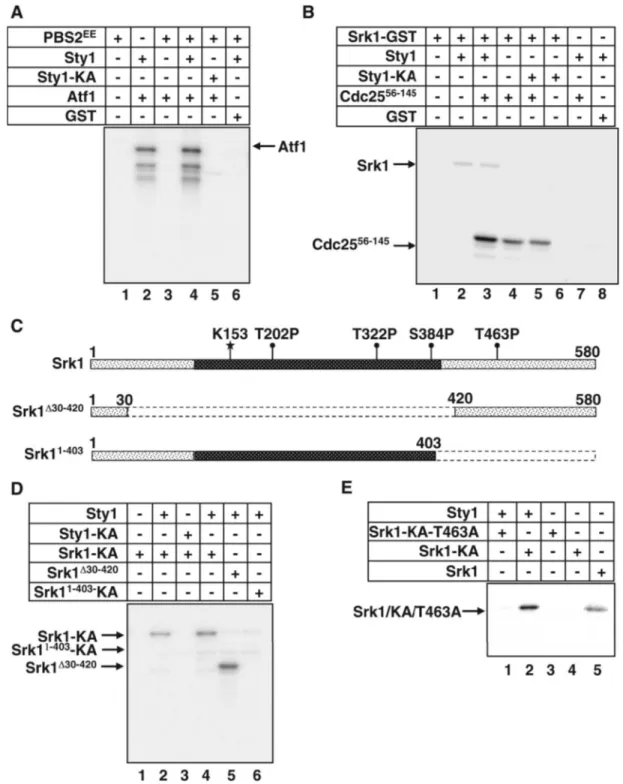
Sty1 Activates Srk1 through Phosphorylation at Threonine 463
Previous studies found that Srk1 was phosphorylated by Sty1 in response to stress (Smith et al., 2002). To investigate the effect of Sty1 phosphorylation on Srk1 activity, purified Sty1 and Srk1 proteins were used in an in vitro kinase assay. For this purpose, Sty1, Sty1-KA (Sty1 kinase dead, which has the lysine 153 mutated to alanine), and Srk1 were purified as GST fusion proteins from Escherichia coli. In the first step of the reaction, Sty1 was preactivated by phosphorylation in the presence of purified Saccharomyces cerevisiae PBS2-EE (EE denotes serine 514 and threonine 518 from the activation loop of the Pbs2 MAPKK mutated to glutamic acid) and ATP (the same reaction was performed with the catalytically inactive allele Sty1-KA, as a control). The activation of Sty1 by PBS2-EE was assayed, using Atf1 as a bona fide substrate for Sty1. It should be noted that, although the activity of Sty1 was higher when incubated with PBS2-EE (Figure 1A, lane 4), the purified Sty1 kinase was already active without pre-vious activation by PBS1-EE (Figure 1A, lane 2). Next, Srk1 and a fragment of Cdc25 (Cdc2556-145), which was shown to be phosphorylated by Srk1 (Lopez-Aviles et al., 2005), were added to the kinase reaction to assay the effect of Sty1 phos-phorylation on Srk1. As shown in Figure 1B, although Srk1 purified from E. coli was able to phosphorylate Cdc2556-145 (Figure 1B, lane 4), its activity increased when it was phosphor-ylated by Sty1 (Figure 1B, lane 3), indicating that Sty1 phos-phorylation activates Srk1.
The Srk1 protein contains four putative Sty1 phosphory-lation sites, three in the catalytic domain (Thr202, Thr322, Ser384) and one in the C-terminal regulatory domain, thre-onine 463 (Figure 1C). To map them, we initially created two truncated Srk1 forms, Srk11-403 including the three sites (Thr202, Thr322, Ser384) from the catalytic domain and Srk1⌬30-420containing Thr463 from the regulatory domain (Figure 1C). In addition, we used the Srk11-403containing the Lys 153 from the ATP binding loop mutated to alanine (Srk11-403-KA) to avoid autophosphorylation activity of Srk1. The full-length (Srk1-KA) and truncated forms of Srk1 (Srk11-403-KA and Srk1⌬30-420), expressed as GST fusion pro-teins and purified from E. coli, were subjected to in vitro phosphorylation by Sty1 kinase (as described above). We found that only the Srk1⌬30-420 fragment was phosphory-lated by Sty1 kinase (Figure 1D, lane 5), indicating that Sty1 phosphorylates T463 from the regulatory domain of Srk1.
Table 1. Schizosaccharomyces pombe strains
Strain Genotype Source
RA0104 h⫺leu1-32 ura4-D18 Lab stock JM1160 h⫺sty1::ura4 leu1-32 ura4-D18 J. Millar
RA0770 h⫺wis1::ura4 leu1-32 ura4-D18 Lab stock RA1102 h⫺srk1::kanMX6 leu1-32
ura4-D18
Lab stock RA1449 h⫺srk1::kanMX6 atf::ura4
leu1-32 ura4-D18 Lab stock RA0772 h⫺atf::ura4 leu1-32 ura4-D18 Lab stock RA1380 h⫺srk1-HA::kanMX6 leu1-32 ura4-D18 Lab stock RA1648 h⫺srk1-HA::kanMX6 sty1::ura4
leu1-32 ura4-D18 Lab stock RA1737 h⫺srk1-T463A-HA::kanMX6 leu1-32 ura4-D18 Lab stock RA1715 h⫺srk1-T463D-HA::kanMX6 leu1-32 ura4-D18 Lab stock KGY4337 h⫺cdc25-GFP:KanRura4-D18
ade6-M210 leu1-32
K. Gould RA1726 h⫺cdc25-GFP:KanRsrk1::ura4
ura4-D18 ade6-M210 leu1-32
Lab stock S1299 h⫹cdc25-9A leu1-32 P. San-Segundo
(H. Piwnica-Worms)
Furthermore, we created a point mutant version to sub-stitute threonine 463 for alanine (Srk1-T463A) and tested it
for phosphorylation by Sty1. Again, the catalytically inactive version of Srk1, Srk1-KA-T463A, was used to avoid
auto-Figure 1. Positive regulation of Srk1 by Sty1 kinase. (A) In vitro Sty1 kinase activity. Equal concentrations of purified GST-Sty1 (lanes 2, 4, and 6) and Sty1-KA (lane 5), previously incubated with budding yeast PBS2-EE (lanes 1 and 3– 6), were assayed with Atf1 (lanes 2–5) as substrate and GST as a control (lane 6). (B) Activity of Srk1 after phosphorylation by Sty1. The purified GST-Srk1 (lanes 1– 6) was incubated with Sty1 (lanes 2 and 3) or Sty1-KA (lanes 5 and 6) and then the fragment of Cdc25 (Cdc2556-145; lanes 3–5). As a control, the fragment of
Cdc25 (Cdc2556-145) (lane 7) or GST (lane 8) were incubated with Sty1 (lanes 7 and 8). (C) Diagram of Srk1 protein kinase (top) and the
truncated forms of Srk1 (bottom). In dark, Srk1 catalytic domain where K indicates lysine from the ATP binding loop. SP and TP indicate the putative phosphorylating sites for Sty1. (D) The full-length GST-Srk1-KA (lanes 1– 4) and the two truncated forms, GST-Srk1⌬30-420(lane 5) and GST-Srk11-403–KA (lane 6), were used as substrates of purified Sty1 (lanes 2 and 4 – 6) or Sty1-KA (lane 3), as a control in the kinase
assay. (E) The GST-Srk1-KA-T463A inactive and containing a point mutation corresponding to threonine 463 to alanine (lanes 1 and 3), the inactive GST-Srk1-KA (lanes 2 and 4) and the full-length GST-Srk1 (lane 5), were assayed in a kinase reaction with purified GST-Sty1 (lanes 1 and 2) as in D.
phosphorylation. As shown in Figure 1E, phosphorylation of Srk1 was abolished in the mutant version.
Together these results show that Sty1 phosphorylates and activates Srk1 at threonine 463.
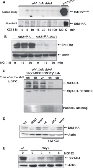
Sty1 Protein, But Not Its Activity, Is Essential for Srk1 Stability
Once established that Srk1 is activated by phosphorylation at threonine 463, we wondered whether this activation was totally dependent on Sty1. The sty1-deleted cells were ex-posed to osmotic stress and Srk1 activity was examined. Srk1 was immunoprecipitated at different times after stress exposure, and its activity was analyzed using Cdc2556-145 fragment as substrate. As expected, there was no Srk1 activ-ity detected when compared with control (Figure 2A, top). However, it was observed that no Srk1 was purified from these samples (Figure 2A, bottom). The same assay was performed again to compare in parallel the expression of Srk1 from wild-type and ⌬sty1 cells exposed to osmotic stress, and indeed, the Srk1 protein is not present in⌬sty1 cells (Figure 2B). This result suggests that Sty1 regulates Srk1 stability in addition to its activity. To confirm this observation, Srk1 protein stability was analyzed using a different approach. We analyzed Srk1 protein expression in a ⌬sty1 strain where sty1 gene was expressed under the control of the invertase (inv1) inducible promoter (Iacovoni
et al., 1999) in a plasmid bearing the “heat-inducible degron
cassette” (Sanchez-Diaz et al., 2004) fused at the N-terminal of the sty1 gene (pINV1-DEGRON:Sty1-HA). A shift from glucose- to sucrose-based culture medium leads to a very rapid induction of the inv1 promoter, and therefore of Sty1 (Iacovoni et al., 1999). In addition, the “heat-inducible de-gron cassette” caused Sty1 degradation by a specific ubiq-uitin-mediated pathway when cells were shifted from 25 to 37°C (Sanchez-Diaz et al., 2004). The ⌬sty1 cells with the endogenous srk1 gene fused to the epitope hemagglutinin (HA) containing the pINV1-DEGRON:Sty1-HA were shifted to 37°C, and both the Sty1 and Srk1 proteins were detected by Western blot at different times after the shift. We ob-served that Srk1 protein vanished in parallel with Sty1 deg-radation induced by the DEGRON cassette at 37°C (Figure 2C).
To determine whether Srk1 stability was dependent on Sty1 activity, Srk1 protein levels were analyzed from⌬wis1 cells, whose Sty1 protein is not activated under stress. As shown in Figure 2D, Srk1 protein was detected in⌬wis1 cells under both normal and stress conditions, indicating that Srk1 stability is dependent on Sty1 protein and independent on its activity.
To investigate further whether a lack of Sty1 protein pro-motes the loss of Srk1 by degradation, ⌬sty1 cells were treated with a proteasome inhibitor, MG132, and Srk1 pro-tein was analyzed at different times after treatment. As shown in Figure 2E, the Srk1 protein level was recovered when degradation was prevented, suggesting that the bind-ing of Sty1 to Srk1 avoids Srk1 degradation.
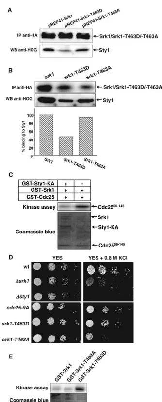
Phosphorylation of Srk1 at Threonine 463 Promotes Its Degradation
To study the in vivo effect of threonine 463 phosphorylation, we examined the level of Srk1 protein in cells in which the endogenous srk1 gene was HA tagged (srk1-HA) or replaced by srk1-T463A T463A-HA) or by srk1-T463D
(srk1-T463D-HA). As shown in Figure 3A, the levels of
Srk1-T463D protein were lower than those of Srk1 (Figure 3A, lanes 1 and 2). In contrast, the levels of Srk1-T463A protein did not change compared with Srk1 (Figure 3A, lanes 4 and
IP anti-HA
Cdc2556-145
srk1::HAΔΔsty1 srk1::HA
KCl 1 M
Srk1-HA
pINV1-DEGRON:sty1-HA Time after the shift
to 37ºC 0h 1h 2h 4h 6h srk1::HAΔsty1 Srk1-HA Sty1-HA:DEGRON Ponceau staining C A min Kinase assay 0 15 60 0 15 60 srk1::HAΔsty1 srk1::HA KCl 1 M Srk1-HA B min Cdc2 0 5 10 15 30 60 100 120 C D wt Δst y1 Δwis1wt Δsty1Δwis1 1 M KCl Srk1-HA Actin wt Δsty1 Srk1-HA 0 2 4 6 MG132 0 E Actin
Figure 2. Sty1 stabilizes Srk1 protein. (A)⌬sty1 cells containing endog-enous tagged srk1-HA were exposed to 1 M KCl and Srk1 was immuno-precipitated with anti-HA antibody from samples taken at indicated time points. In vitro kinase assay was performed with the immunoprecipitates, using Cdc25 (Cdc2556-145) as a substrate (top). The last lane corresponds to
Srk1 activity from wild-type cells. Western blots of the immunoprecipi-tated samples were probed with anti-HA to monitor the presence of Srk1 (bottom). (B) Wild-type and⌬sty1 cells, both with endogenous srk1-HA, were exposed to 1 M KCl and Western blots of protein extracts were prepared at the indicated times and probed with anti-HA to detect Srk1 protein (top). The same membrane was probed with PSTAIR anti-bodies to detect Cdc2 protein as a loading control (bottom). (C) srk1-HA ⌬sty1 cells were transformed with pINV-DEGRON-Sty1 plasmid express-ing DEGRON-Sty1 under the control of the INV1 gene promoter at 25°C. Cells were transferred to 37°C, samples were taken at the indicated time points and Western blots were probed with anti-HA to detect in parallel either Srk1-HA (top) or Sty1-HA (middle). The Western blot membrane was labeled with Ponceau staining and shown as loading control (bottom). (D) Wild-type,⌬sty1, and ⌬wis1 cells containing endogenous srk1 gene tagged with HA (srk1-HA) were exposed to 1 M KCl for 30 min. Western blots of protein extracts were prepared and probed with anti-HA antibod-ies to detect Srk1 protein (top). The same membrane was probed with actin antibody as a loading control (bottom). (E)⌬sty1 cells carrying endogenous srk1 gene tagged with HA (srk1-HA) were exposed to MG132 drug (50M final concentration) and samples were collected at the indi-cated time points. Srk1 protein levels were monitored by Western blot with anti-HA antibody (top). Control levels of Srk1 protein were shown from wild-type cells carrying endogenous srk1 gene tagged with HA (srk1-HA) (lane 1). The same membrane was probed with actin antibody as a control (bottom).
5). To ascertain whether the loss of Srk1-T463D protein levels was due to degradation, cells carrying the
srk1-T463D-HA allele were treated with MG132. In this case the
levels of Srk1-T463D were recovered (Figure 3A, lane 3), indicating that phosphorylation at threonine 463 promotes instability and degradation of Srk1.
To confirm that Srk1 phosphorylation by Sty1 promotes Srk1 instability, we analyzed Srk1 protein levels in response to stress once it is phosphorylated by Sty1. Cells carrying the endogenous srk1 gene tagged with the HA epitope (srk1-HA) were treated with 0.8 M KCl, and samples were taken at different times, analyzing the level of Srk1 protein by West-ern blot (Figure 3B). Before exposure to osmotic stress, we added cycloheximide to the culture to avoid the activation of
srk1 transcription, because it has been reported that the srk1
transcription is induced upon stress in a Sty1-dependent manner (Smith et al., 2002). As shown in Figure 3B, the level of Srk1 protein decreased in response to stress. This result supports the hypothesis that phosphorylation of Srk1 in-duces its instability and degradation.
The Srk1 Phosphorylated at Threonine 463 Dissociates from Sty1
The observations that Srk1 stability requires Sty1 binding and that the Srk1-T463D protein becomes unstable, suggest that the phosphorylation of Srk1 by Sty1 induces, in addition to activation, dissociation of both proteins. To determine whether phosphorylated Srk1 is separated from Sty1, we examined the binding between Sty1 and Srk1 phospho-mutants. The Srk1, Srk1-T463D, and Srk1-T463A proteins were overexpressed and immunoprecipitated to test their binding to Sty1 by Western blot. As shown in Figure 4A, Srk1-T463D had less affinity than Srk1 and Srk1-T463A for Sty1 protein, indicating that Srk1 phosphorylation induces
Figure 4. Srk1-T463D is active and dissociates from Sty1. (A) srk1, srk1-T463D, and srk1-T463A overexpressed under the control of nmt41 promoter were immunoprecipitated with anti-HA antibody (top) and the bound Sty1 protein was detected with anti-HOG antibody (bottom). (B) The same as in A was performed immuno-precipitating the endogenous Srk1, Srk1-T463D, and Srk1-T463A proteins expressed from the integrated srk1, T463D, and srk1-T463A genes tagged with HA (top) and the bound Sty1 protein was detected with anti-HOG antibody (bottom). Sty1 binding to Srk1 protein was normalized relatively to the Srk1 protein immunopre-cipitated (columns graphic). (C) In vitro kinase assay. Srk1 and Srk1 Figure 3. Srk1-T463D protein is unstable and degradates. (A) srk1,
srk1-T463D, and srk1-T463A genes tagged with HA were integrated and levels of Srk1 protein were detected by Western blot using anti-HA antibody (top). Cells carrying srk1-T463D and srk1-T463A were treated with MG132 drug (50M final concentration; top, lanes 3 and 6). The Cdc2 protein was detected from the same samples with anti-PSTAIR as a loading control (bottom). (B) srk1-HA cells were treated with cycloheximide (100 g/ml) and exposed to osmotic stress (0.8 M KCl). Samples were processed to monitor the level of Srk1 protein with anti-HA antibody (top) and quantified and normalized with the control level of Cdc2 protein (bottom) detected with anti-PSTAIR antibody.
Sty1 dissociation. To confirm this observation, we per-formed the same experiment immunoprecipitating the Srk1, Srk1-T463D, and Srk1-T463A proteins from cells were the endogenous srk1 gene was replaced by either HA,
srk1-T463D-HA, or srk1-T463A-HA, and their binding to Sty1 was
analyzed by Western blot. As expected, Srk1-T463A ex-pressed at physiological levels had similar affinity than Srk1 for Sty1 protein, whereas Srk1-T463D had reduced affinity for Sty1 protein than Srk1 and Srk1-T453A (Figure 4B).
The fact that Srk1 phosphorylated by Sty1 induces the activation of Srk1 and its dissociation from Sty1 suggests that the binding of Srk1 to Sty1 may inhibit Srk1 activity. We therefore investigated the activity of Srk1 when bound or not to Sty1, by incubating the purified Srk1 protein with or without the inactive purified Sty1-KA and by performing a kinase assay in vitro. As predicted, the activity of Srk1 diminished when bound to Sty1-KA, compared with the activity when operating alone (Figure 4C).
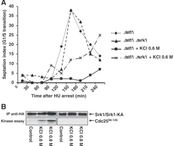
Next, we studied the response of the Srk1 phospho-mu-tants when exposed to stress. Wild-type,⌬srk1 cells and cells containing srk1-T463D allele or srk1-T463A allele were plated on 0.8 M KCl media to induce osmotic stress. As shown in Figure 4D, cells carrying srk1-T463D were viable, whereas cells carrying srk1-T463A were sensitive to stress in the same way as the⌬srk1 cells were (Figure 4D). Furthermore, the kinase activity of purified Srk1 and the phospho-mutants, Srk1-T463A and Srk1-T463D was assayed in vitro. As shown in Figure 4E, the activity of Srk1-T463D was highest, whereas Srk1-T463A was as active as the wild-type Srk1. All these results indicate that it is not sufficient for the Srk1 kinase to be catalytically active; it has to be activated by Sty1 to respond to stress.
Srk1 Activity Is Necessary for G2/M Inhibition in Stress Response
In fission yeast, Srk1 negatively regulates the onset of mito-sis (Lopez-Aviles et al., 2005). To determine whether Srk1 activation by Sty1 was to inhibit the G2/M transition after osmotic stress, we analyzed cell cycle progression of srk1-deleted cells under stress conditions. Wild-type and ⌬srk1 were synchronized in S phase with HU and released in presence of KCl (0.6 M KCl). Both wild-type and⌬srk1 cells showed the same cell cycle progression pattern. They pre-sented a temporal arrest before mitosis when exposed to osmotic stress (data not shown). We believe that the osmotic stress-signaling pathway, like the DNA damage-signaling pathway, controls two responses, one delaying the cell cycle and the other allowing response to stress. Thus, to determine the effect of⌬srk1 on the kinetics of cell cycle delay per se, experiments were done again in a genetic background that prevents adaptation to osmotic stress. Exposure of yeast to increases in external osmolarity induces synthesis and acti-vation of the Atf1 transcription factor responsible for the synthesis of several genes necessary for the response to
stress and adaptation (Shiozaki and Russell, 1996; Wilkinson
et al., 1996). Therefore, the atf1 gene was deleted to avoid
immediate adaptation. The atf1 deleted cells (⌬atf1) and the double mutant ⌬atf1⌬srk1 were synchronized in S phase with HU and released in presence of 0.6 M KCl, as described above. We observed that ⌬atf1 showed a delay in G2/M transition, as described for wild-type cells (Figure 5A), whereas the double mutant progressed throughout the cell cycle, albeit to a lesser extent than in unstressed conditions (Figure 5A). This result indicates that Srk1 is necessary for inhibiting G2/M transition in the presence of osmotic stress. Srk1 kinase activity was assayed from cells bearing an endogenously tagged Srk1 exposed to different doses of osmotic stress (0.6 and 0.8 M KCl) and using a phosphory-latable fragment of Cdc25 (Cdc2556-145) as a substrate (Fig-ure 5B). As expected, Srk1 was dose-dependent activated by osmotic stress and able to phosphorylate Cdc25. The same assay was performed in cells bearing the catalytic-inactive Srk1 (Srk1-KA) to confirm that Cdc25 phosphorylation is due to Srk1 (Figure 5B).
Next, we studied the sensitivity to osmotic stress of cells containing the cdc25-9A allele (nine consensus phosphory-latable sites for Srk1 are mutated to alanine), which express a Cdc25-9A protein unable to be phosphorylated by Srk1 (Lopez-Aviles et al., 2005). The cdc25-9A cells showed some sensitivity to grow on medium containing 0.8 M KCl than wild-type cells, but not as sensitive as cells lacking Srk1 or Sty1 MAP kinase (Figure 4D).
Cdc25 Nuclear Export Is Dependent on Srk1 under Stress Conditions
Cdc25 phosphatase is a direct substrate of Srk1 (Lopez-Aviles et al., 2005). Srk1 phosphorylates and promotes the
Figure 4 (cont). preincubated with Sty1-KA were assayed with the fragment of Cdc25 (Cdc2556-145) as a substrate (top). SDS-PAGE gels
from the kinase assay were stained with Coomassie blue (bottom). (D) Serial dilution of log phase cultures of wild-type,⌬srk1, ⌬sty1, cdc25-9A, srk1-T463D, and srk1-T463A. Cells were plated in rich medium (YES) and rich medium containing KCl 0.8 M (YES⫹ 0.8 M KCl). Colony formation was analyzed after 2 days of growth at 30°C. (E) In vitro kinase assay from Srk1 purified phospho-mutants. GST-Srk1, -Srk1-T463A, and -Srk1-T463D were assayed and auto-phosphorylation was measured (top). SDS-PAGE gels from the kinase assay were stained with Coomassie blue (bottom).
0 5 10 15 20 25 30 35 40 0 30 60 90 120 150 180 210 240 ∆atf1 ∆atf1 + KCl 0.6 M ∆atf1∆srk1 ∆atf1∆srk1 + KCl 0.6 M
Time after HU arrest (min)
Septation index (G1/S transition)
A
Control KCl 0 .6 M KCl 0 .8 M IP anti-HA Cdc2556-145B
Srk1/Srk1-KA Kinase assay Control KCl 0 .6 M KCl 0 .8 MFigure 5. The G2/M delay caused by osmotic stress needs Srk1 protein. (A) The⌬atf1 and the double mutant ⌬atf1 ⌬srk1 cells were synchronized with HU and released in presence or absence of 0.6 M KCl. The septation index, an indication of the percentage of cells that have completed mitosis, was measured at different times cor-responding to minutes after release from HU and addition of KCl. (B) Srk1 activated in response to stress phosphorylates Cdc25. Cells were exposed to different doses of KCl (0.6 or 0.8 M) for 15 min and Srk1 (left panels) or Srk1-KA (right panels) were immunoprecipi-tated with anti-HA antibody (top), and its kinase activity was as-sayed using the Cdc25 fragment (GST-Cdc2556-145) as a substrate
(bottom).
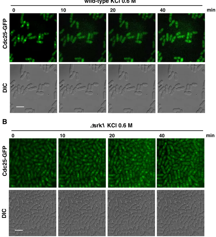
cytosolic localization of Cdc25 and consequent mitotic ar-rest. Therefore, we analyzed whether the subcellular local-ization of Cdc25 during stress response depended on Srk1. Wild-type and ⌬srk1 cells carrying the endogenous cdc25 gene tagged with the GFP, Cdc25-GFP, were synchronized in S phase with HU, released and exposed to KCl (0.6 M KCl; Supplementary Figure 2A). Cdc25-GFP localization was im-aged in living cells by time-lapse confocal microscopy. In wild-type cells, we found that Cdc25 localization was al-ready cytosolic 10 min after osmotic stress, and this was maintained in the cytosol up to 40 min after the osmotic stress exposure (Figure 6A; Movie 6A and Supplementary Figure 2B). In contrast, in srk1-deleted cells, Cdc25 was generally found in the nucleus during the entire period of osmotic stress (Figure 6B; Movie 6B and Supplementary Figure 2B). Therefore, we can conclude that Cdc25 protein is exported from the nucleus under stress conditions in a Srk1-dependent manner.
DISCUSSION
Activation of the Sty1 MAPK is a key step for the generation of adaptive responses that allow cell survival to stress. Mod-ulation of cell cycle progression is essential for adaptation to stress. Here we show the multiple effect of Srk1 phosphor-ylation by the MAPK Sty1 during stress response, and we present the Srk1 kinase as a component that coordinates stress response and cell cycle control.
Multiple Effect of Srk1 Phosphorylation by the MAPK Sty1
Srk1 kinase was identified as a protein kinase whose expres-sion is up-regulated upon exposure to stress in a Sty1-dependent manner. Srk1 is present in a complex with Sty1 and is directly phosphorylated by MAPK (Smith et al., 2002). We observed that Srk1 stability is dependent on the presence of the Sty1 protein and independent of Sty1 activity. Inter-estingly, similar observations have been made in mamma-lian cells for MK2 (the human homolog of Srk1) and p38 MAPK. Both proteins bind and form a stable complex. Elim-ination of either of the two proteins reduces the remaining partner’s level (Kotlyarov et al., 2002). The effect of Sty1 on Srk1 protein stability can also be attributed to the regulation of srk1 transcription. However, Srk1 disappears in parallel with Sty1 protein degradation, as shown in Figure 2C, and the Srk1 degradation can be reverted by the addition of a proteasome inhibitor (Figure 2E). Moreover, phosphoryla-tion at threonine 463 by activated Sty1 induces both Srk1 activation and protein instability, as observed from the low level of the endogenous Srk1-T463D protein and its recovery by the addition of a proteasome inhibitor (Figure 3A). These results indicate that Srk1 stability requires Sty1 binding and moreover, the Srk1 phosphorylated protein (Srk1-T463D) becomes unstable. Thus, the phosphorylation of Srk1 by Sty1 induces, in addition to activation, the dissociation of the Srk1-Sty1 protein complex and the destabilization of the Srk1. In support of this hypothesis, Srk1-T463D showed less affinity to Sty1 protein than was shown by unphosphory-lated Srk1 and Srk1-T463A (Figure 4A).
Sty1 phosphorylates and activates Srk1 at threonine 463 (Figures 1, B and E, and 4E). However, it seems that phos-phorylation at threonine 463 activates Srk1, because it allows Srk1 to separate from Sty1. Thus, dissociation from Sty1 is sufficient to activate Srk1. This conclusion is supported by the observation of Srk1 phospho-mutants activities. Srk1-T463A is catalytically active in a kinase assay in vitro at a level similar to that of Srk1 (Figure 4E). All the same, the in
vitro Srk1 activity can be inhibited by allowing Srk1 binding to Sty1 (Figure 4C). Nevertheless, analysis of the in vivo effect of the Srk1 phospho-mutants showed that mutation of the threonine 463 to alanine (Srk1-T463A) makes cells sen-sitive to osmotic stress similarly to srk1 deletion. This indi-cates that the catalytic activity of Srk1-T463A is not sufficient for Srk1 function in stress response and supports the fact that threonine 463 has to be phosphorylated to activate Srk1 function in stress response. Collectively, these observations point to an additional mechanism of Srk1 regulation by Sty1.
Srk1 Kinase as a Component That Coordinates Stress Response and Cell Cycle Control
Significantly, Srk1 is rapidly and highly activated in re-sponse to stress. In a previous study, we demonstrated that the Srk1 kinase is activated during G2/M transition of the cell cycle (Lopez-Aviles et al., 2005). However, the cell cycle activity of Srk1 is lower than its activity detected at stress response. This is also reflected in the phenotype of srk1-deleted cells, which showed a semi-wee phenotype (Lopez-Aviles et al., 2005). The simplest interpretation of these ob-servations is that Srk1 activity has a short requirement during a normal cell cycle, where its function is to maintain the inhibited status of Cdc25 phosphatase emerging during G2 phase. Therefore, low activity of Srk1 is sufficient to inhibit the Cdc25 protein expressed throughout the G2 phase. On the other hand, in stress response, as the function of Srk1 is essential to maintain cell integrity, its activation has to be high to guarantee the correct response.
Furthermore, upon stress, Srk1 translocates from the cy-toplasm to the nucleus in a process that is dependent on the Sty1 MAPK (Smith et al., 2002). We showed that, 10 min after osmotic stress, the Cdc25 protein translocates to the cyto-plasm, concurrent with Sty1 and Srk1 activation. Signifi-cantly, this nuclear export depends on the Srk1 kinase ac-tivity. This observation also agrees with our previous demonstration that the association between Cdc25 and Rad24 is highly induced under stress conditions (Lopez-Aviles et al., 2005). Thus, in response to stress, Srk1 controls G2/M transition by phosphorylating Cdc25 and promoting its binding to Rad24 and its subsequent translocation to the cytoplasm.
In fission yeast, stress induces a temporal arrest at the G2/M transition, which, it has been reported, was not de-pendent on the DNA-damage checkpoint or mitotic spindle checkpoint (Kawasaki et al., 2006). The results shown here indicate that the temporal G2/M arrest of the stress response is dependent on Srk1 kinase. Moreover,⌬srk1 cells are sen-sitive to osmotic stress similar to cells bearing cdc25-9A allele, which is unable to be phosphorylated by Srk1, strengthening the role of Srk1 regulating the cell cycle in stress conditions through Cdc25 protein. Of note,⌬srk1 cells are more sensitive than cdc25-9A, suggesting an additional role for Srk1 in stress response. To examine the effect of Srk1 protein on the kinetics of the cell cycle after osmotic stress, we used a mutant that was defective in its stress adaptation response,⌬atf1. The assumption behind this approach was that osmotic stress-signaling pathways control two re-sponses, one delaying the cell cycle and the other allowing response and adaptation to stress. This idea is consistent with similar studies done in S. cerevisiae (Alexander et al., 2001), where the glycerol-based osmoregulation mutants were used to analyze the role of Swe1 and Hog1 in cell cycle regulation after hyperosmotic stress. Taken together, the results indicate that Srk1 is not part of the rapid stress-adaptation response, but that it is necessary to coordinate cell cycle progression under stress.
It has been previously reported that Srk1 is regulated by Sty1 with two different mechanisms, transcriptional and posttranscriptional (Smith et al., 2002). As shown here, in response to stress, Sty1 regulates the activity of Srk1 on cell
cycle control by phosphorylation on threonine 463. More-over, the expression of srk1 gene is up-regulated in response to stress dependent of Sty1 (Smith et al., 2002). We do not yet understand the physiological meaning of Sty1 inducing Srk1
A
Cdc25-GFP
DIC
0
10
20
40
min
wild-type KCl 0.6 M
B
∆
srk1 KCl 0.6 M
Cdc25-GFP
DIC
0
10
20
40
min
Figure 6. Cytosolic localization of Cdc25 in response to stress is dependent on Srk1 kinase. Wild-type (A) and⌬srk1 (B) cells carrying endogenous Cdc25-GFP were synchronized with HU and released, and after 60 min from the release were treated with or without 0.6 M KCl (A and B and Movie 6, A and B). Cellular localization of Cdc25 was observed by time-lapse microscopy at 10-min intervals in a single focal plane (top panels, A and B). Simultaneously, differential interference contrast images were acquired (bottom panels, A and B). Scale bar, 10m.
instability and degradation in addition to activation, while concomitantly promoting srk1 transcription-activation. A simple explanation as to why Sty1 induces Srk1 degradation might be to ensure Srk1 activity in cell-cycle control, within a narrow window. Thus, the Sty1-dependent activation of
srk1 transcription might serve to reestablish Srk1 levels,
which are constant throughout the cell cycle (Lopez-Aviles
et al., 2005).
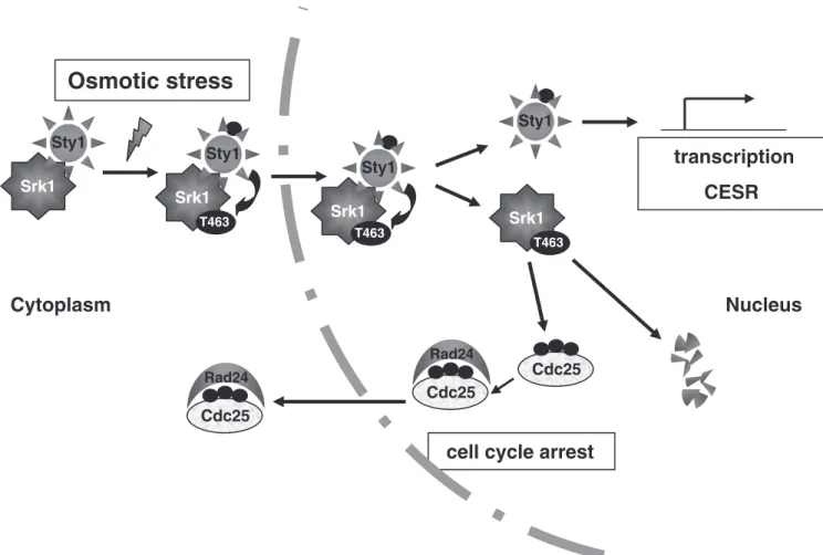
Overall, Srk1 is present in a complex with Sty1 in un-stressed conditions, whereas stress-activated Sty1 kinase phosphorylates Srk1 at threonine 463 inducing the dissoci-ation of Srk1 and Sty1 proteins, which appears to be neces-sary for the full activation of Srk1 (Figure 7). As a result, phosphorylation at threonine 463 leads to Srk1 activation and the inhibition of cell cycle progression through the Cdc25 cytoplasmic translocation. The single phosphoryla-tion of Srk1 turns the Srk1 protein unstable and promotes its degradation (Figure 7), whereas activated Sty1 induces the transcriptional activation of srk1 and the rest of CESR (Core Environmental Stress Response genes).
ACKNOWLEDGMENTS
We are grateful to Paul Russell (The Scripps Research Institute, CA) for gifts of plasmids and strains and Kathleen Gould (Howard Hughes Medical Insti-tute and Vanderbilt University School of Medicine, TN) for gift of Cdc25-GFP strain. We are also grateful to Anna Llado´ and Maria Calvo from the Confocal
Mycoscopy Unit for excellent support and advice in the time-lapse confocal microscopy. This research was supported by grants from the MEC (BFU2006-00375/BMC) and “Distincio´ de la Generalitat de Catalunya per a la Promocio´ de la Recerca Universita`ria. Joves Investigadors” (DURSI). S.L.-A. is the recipient of an F.I. predoctoral fellowship from the Generalitat de Catalunya, Spain.
REFERENCES
Alemany, V., Sanchez-Piris, M., Bachs, O., and Aligue, R. (2002). Cmk2, a novel serine/threonine kinase in fission yeast. FEBS Lett. 524, 79 – 86. Alexander, M. R., Tyers, M., Perret, M., Craig, B. M., Fang, K. S., and Gustin, M. C. (2001). Regulation of cell cycle progression by Swe1p and Hog1p following hypertonic stress. Mol. Biol. Cell 12, 53– 62.
Dahlkvist, A., Kanter-Smoler, G., and Sunnerhagen, P. (1995). The RCK1 and RCK2 protein kinase genes from Saccharomyces cerevisiae suppress cell cycle checkpoint mutations in Schizosaccharomyces pombe. Mol. Gen. Genet. 246, 316 –326.
Hohmann, S. (2002). Osmotic stress signaling and osmoadaptation in yeasts. Microbiol. Mol. Biol. Rev. 66, 300 –372.
Iacovoni, J. S., Russell, P., and Gaits, F. (1999). A new inducible protein expression system in fission yeast based on the glucose-repressed inv1 pro-moter. Gene 232, 53–58.
Kawasaki, Y., Nagao, K., Nakamura, T., and Yanagida, M. (2006). Fission yeast MAP kinase is required for the increased securin-separase interaction that rescues separase mutants under stresses. Cell Cycle 5, 1831–1839. Kotlyarov, A., Yannoni, Y., Fritz, S., Laass, K., Telliez, J., Pitman, D., Lin, L., and Gaestel, M. (2002). Distinct cellular functions of MK2. Mol. Cell. Biol. 22, 4827–4835.
Sty1 Srk1
Osmotic stress
Cdc25
Rad24
cell cycle arrest
Sty1 Srk1 T463 Sty1 Srk1 Sty1 Srk1 T463 T463 Cdc25 Rad24 Cdc25Nucleus
Cytoplasm
transcription
CESR
Figure 7. Srk1 activation and regulation during stress response. Stress-activated Sty1 phosphorylates Srk1 at threonine 463, inducing dissociation of Srk1-Sty1 complex and activation of Srk1 to inhibit cell cycle progression by promoting Cdc25 cytoplasmic localization. Subsequently, the phosphorylated Srk1 becomes unstable, leading to Srk1 degradation, whereas active Sty1 kinase activates transcrip-tion of CESR (core environmental stress response) genes.
Lopez-Aviles, S., Grande, M., Gonzalez, M., Helgesen, A. L., Alemany, V., Sanchez-Piris, M., Bachs, O., Millar, J. B., and Aligue, R. (2005). Inactivation of the Cdc25 phosphatase by the stress-activated Srk1 kinase in fission yeast. Mol. Cell 17, 49 –59.
Millar, J. B., Buck, V., and Wilkinson, M. G. (1995). Pyp1 and Pyp2 PTPases dephosphorylate an osmosensing MAP kinase controlling cell size at division in fission yeast. Genes Dev. 9, 2117–2130.
Moreno, S., Klar, A., and Nurse, P. (1991). Molecular genetic analysis of fission yeast Schizosaccharomyces pombe. Methods Enzymol. 194, 795– 823.
Sanchez-Diaz, A., Kanemaki, M., Marchesi, V., and Labib, K. (2004). Rapid depletion of budding yeast proteins by fusion to a heat-inducible degron. Sci. STKE 223, 1–20.
Sanchez-Piris, M., Posas, F., Alemany, V., Winge, I., Hidalgo, E., Bachs, O., and Aligue, R. (2002). The serine/threonine kinase Cmk2 is required for oxidative stress response in fission yeast. J. Biol. Chem. 277, 17722–17727.
Shiozaki, K., and Russell, P. (1995). Cell-cycle control linked to extracellular environment by MAP kinase pathway in fission yeast. Nature 378, 739 –743. Shiozaki, K., and Russell, P. (1996). Conjugation, meiosis, and the osmotic stress response are regulated by Spc1 kinase through Atf1 transcription factor in fission yeast. Genes Dev. 10, 2276 –2288.
Shiozaki, K., Shiozaki, M., and Russell, P. (1997). Mcs4 mitotic catastrophe suppressor regulates the fission yeast cell cycle through the Wik1-Wis1-Spc1 kinase cascade. Mol. Biol. Cell 8, 409 – 419.
Smith, D. A., Toone, W. M., Chen, D., Bahler, J., Jones, N., Morgan, B. A., and Quinn, J. (2002). The Srk1 protein kinase is a target for the Sty1 stress-activated MAPK in fission yeast. J. Biol. Chem. 277, 33411–33421.
Wilkinson, M. G., Samuels, M., Takeda, T., Toone, W. M., Shieh, J. C., Toda, T., Millar, J. B., and Jones, N. (1996). The Atf1 transcription factor is a target for the Sty1 stress-activated MAP kinase pathway in fission yeast. Genes Dev.
10, 2289 –2301.