The experiments for this PhD thesis were carried out in the
laboratories of:
Home institute:
Prof. Filippo Drago
Department of Biomedical
And Biotechnological Sciences
Section of Pharmacology
Via S. Sofia, 97
95123
University of Catania
During the 2
ndyear of my PhD program, I spent two months
working as visiting PhD student at University of Helsinki. During
this period, I worked on the project entitled: Dopamine D
3receptor-dependent ectopic expression of
α
6 GABA
Asubunit counteracts
alcohol intake by increasing GABA inhibition in the nucleus
accumbens
Guest institute:
Prof. Esa R Korpi
Department of Pharmacology,
Faculty of Medicine
Haartmaninkatu 8, 00014
Table of contents
Acknowledgements 7
List of abbreviations 9
Preface 12
Chapter I General Introduction
1. Dopamine pathways in the central nervous system 15
2. DAergic receptors 17
3. Dopamine D3 Receptor 20
4. D3R and alcohol addiction 24
5. D3R and Schizophrenia 28
6. Design of the present research 32
Chapter II 34
Chapter IV
6. General Discussion
6.1 Cross-talk between D3R and GABAA receptor in alcohol
addiction 149
6.2 D3R plays a key role in the pathophysiology of schizophrenia:
implications in MAM model 152
Concluding Remarks 155
Chapter V
References 158
Chapter VI
Acknowledgements
I would like to express my gratitude to Prof. Filippo Drago for
giving me the opportunity to join his lab during these years of PhD
Program.
I would like to thank Prof. Salvatore Salomone for his excellent
supervision, constructive comments and discussion, as well as for
welcoming me in his group and leading me working on diverse
exciting projects.
I would like to thank Prof. Gian Marco Leggio, who has supported
me during my Ph.D., he taught me how to approach “Research”. His
immense knowledge, enthusiasm and guidance, allowed me to reach
this important goal.
I sincerely thank Prof. Esa R. Korpi who gave me the chance to join
his lab in Helsinki.
I want to thank my colleagues with which I spent these years
learning and enjoying research.
List of abbreviations
5-HT 5-hydroxytryptamine
7-OH-DPAT ((+/-)-7-hydroxy-N, N-(di-n-propyl-2-aminotetralin)) AC adenilate cyclase
ADE alcohol deprivation effect ANOVA analysis of variance
BDNF brain development neurotrophic factor cAMP cyclic adenosine monophosphate CB1 cannabinoid receptor 1
CBD cannabidiol
CNS central nervous system DAG diacylglycerol
D2L dopamine D2 long D2S dopamine D2 short D3R dopamine D3 receptor
D3R-/- dopamine D3 receptor deficient mice DA dopamine
DID drinking in the dark DMSO dimethyl sulfoxide DYS dysbindin
GABA γ-aminobutyric acid
GABAA γ-aminobutyric acid receptor A GD gestional day
GIRK G-protein gated inwardly rectifying K+
channel
GPCR G protein-coupled receptors GRK GPCR kinases
HIP hippocampus
IP3 inositol 1,4,5 trisphosphate i.p. intraperitoneal injection
MAM methylazoxymethanol acetate MSN Medium Spiny Neurons NAc nucleus accumbens NMDA N-methyl-D-aspartate NP alcohol Non-Preferring rats P alcohol Preferring rats PFC prefrontal cortex PKA protein kinase A PLC phospholipase C PV parvalbumin
RGS regulators of G protein signalling SCZ schizophrenia
STR striatum VEH vehicle
VTA ventral tegmental area WT wild type mice
Preface
Dopamine (DA) modulates several essential functions of the central nervous system (CNS), including reward and cognition. The dopaminergic neurotransmission in the CNS is mediated by two different classes of G protein-coupled receptors (GPCR), the “D1R-like” receptors (D1R and D5R) and “D2 R-like” receptors (D2R, D3R and D4R, Seeman et al., 1994). Among dopamine receptors, the dopamine D3 receptor (D3R) has captured the scientific interest because of its restricted distribution in the brain, seemingly related to functions of dopamine associated with the limbic brain. So, the dopamine D3R shows a limited distribution in the limbic brain areas involved in the control of cognitive and emotional functions, and it seems to represent a target for the treatment of several neuropsychiatric disorders such as drug addiction and schizophrenia (Leggio et al., 2016). D3R, acting as autoreceptor, regulates the activity of DAergic neurons throughout the mesolimbic, mesocortical and nigrostriatal DAergic pathways (Gobert et al., 1995; Tepper et al., 1997; Diaz et al., 2000). D3 deficient mice (D3R
-/-) exhibit extracellular levels of dopamine twice as high as their wild-type (WT) littermates suggesting that D3R could play an inhibitory role in the control of basal extracellular DA levels (Koeltzow et al., 1998; Joseph et al., 2002). Moreover, Leggio and colleagues (2014) demonstrated that D3R
WT littermates, in several ethanol-drinking paradigms. Furthermore, D3R -/-
mice show a 15-fold higher expression of γ-aminobutyric acid receptor A (GABAA) α6 subunit in striatum compared to their WT littermates (Leggio et al., 2015). Based on previous data present in literature, the main hypothesis of my research project has been that the D3R, showing a main role in the control of the mesolimbic DAergic pathway, is involved in neuropsychiatric disorders linked to alteration of DAergic pathways. So, the aims of this thesis were: 1) to investigate the role of the cross-talk between GABAA/D3R in the mesolimbic DA control of ethanol consumption; 2) to assess the involvement of D3R in the pathophysiology of schizophrenia.
Chapter I
1. Dopamine pathways in the central nervous system
DA regulates important physiological brain’s functions, including reward and cognition, through four different DAergic pathways: the nigrostriatal pathway arising from the substantia nigra pars compacta (SNpc; Dahlström and Fuxe, 1964) and projecting to the dorsal striatum; the mesolimbic pathway originating in the ventral tegmantal area (VTA) and projecting to the nucleus accumbes (NAc); the mesocortical pathway that arises from the VTA and projects to the central cortex and the tuberoinfundibular pathway that connects the hypothalamus to the pituitary gland (Anden et al., 1964; Dahlstroem and Fuxe, 1964). The different DAergic pathways mediate several physiological functions, with the mesolimbic/mesocortical pathways implicated in reward and cognition. The evidence that neuropsychiatric disorders, like schizophrenia and addiction, involve a dysregulation of mesolimbic/mesocortical pathways, reinforces the hypothesis of a functional segregation of DAergic pathways. So, a key role of the mesocorticolimbic pathway has been recognized in reward, craving and aversion (Wise, 2009) and in schizophrenia (Perez-Costas et al., 2010; Yoon et al., 2013; Weinstein et al., 2017).
2. DAergic receptors
DA mediates its effect through two families of GPCR, classified into “D1 R-like” and “D2R-like” receptors. D1R-like receptors (D1R and D5R) are coupled to Gs proteins and stimulate adenylate cyclase (AC), with production of cyclic adenosine monophosphate (cAMP) and activation of cAMP-dependent pathways, including protein kinase A (PKA) and other downstream signals. D1R modulate different ionic channels, including voltage-activated Na
+ -(Nav), K+ -(Kv) and Ca 2+ (Cav) channels, Ca 2+ -activated K+
-(KCa) and G-protein gated inwardly rectifying K+
(GIRK) channels (Maurice et al., 2001; Witkowski et al., 2008; Yang et al., 2013). D2R-like receptors (D2R, D3R and D4R), by coupling to Gi proteins, induce inhibition of AC and PKA-dependent pathways, as well as activation of GIRK and closure of CaV (Missale et al., 1998, Figure 2). D2 R-like receptors genes generate variants. D2R exists in two functional isoforms, D2 long (D2L) and D2 short (D2S; Giros et al., 1989), whereas several D3R isoforms have been identified (Giros et al., 1991). Multiple D4R variants are produced, mostly having a domain repeated 2 (2R), 4 (4R) and 7 (7R) times (Van Tol et al., 1992). In addition to act as monomers, DAergic receptors constitute dimeric and/or oligomeric complexes by association of different subtypes either with DA receptors or with other GPCRs and ligand-gated channels. Furthermore, D1R-D2R dimers are linked to Gq proteins, thus modulating phospholipase C (PLC), which produces inositol 1,4,5 trisphosphate (IP) and diacylglycerol
(DAG) to regulate intracellular Ca2+ (Lee et al., 2004, Figure 2). While the prevailing belief is that DAergic receptors act through G proteins, they can also activate G independent mechanisms. A role in the G protein-independent signaling is played by arrestins, multifunctional adaptor proteins which bind DAergic receptors phosphorylated by GPCR kinases (GRKs; Gainetdinov et al., 2004). Binding of arrestins recruits several other proteins, including Akt, GSK-3, MAPK, c-Src, Mdm2 and N-ethylmaleimide-sensitive factor, thereby enhancing DA-activated pathways (Beaulieu and Gainetdinov, 2011; Figure 2). GRKs also regulate DAergic receptors by mediating their desensitization, since their phosphorylation elicits receptor endocytosis. Besides GRKs, the regulators of G protein signaling (RGS), a group of GTPase-activating proteins acting on G protein, negatively modulate DAergic receptors.
3. Dopamine D
3Receptor
The D3R was cloned in 1990 and characterized for its high sequence homology with the D2R (Sokoloff et al., 1990). The D3R subtype has an important role in the modulation of the mesolimbic DA pathway and in the control of drug- seeking behaviour (Heidbreder et al, 2005; Joyce and Millan, 2005). The D3R is located both at pre- and post-synapses, in the ventral striatum (nucleus accumbens and island of Calleja; Bouthenet et al, 1991; Murray et al, 1994); in these structures, stimulation of presynaptic D3R may modulate DA synthesis and release (Levant, 1997). Activation of D3R expressed in a transfected mesencephalic cell line inhibits dopamine release (Tang et al., 1994) and synthesis (O’Hara et al., 1996). Among DA receptors, D3R exhibits the highest affinity for DA (70-fold higher than D2R receptors) suggesting that DA may occupy D3R in vivo for extended periods of time leading to high spontaneous activation of D3R (Richtand et al., 2001; Vanhauwe et al., 2000). In rat, the largest D3R densieties have been found in granule cells of the island of Calleja and in medium spiny neurons (MSN) on the rostral and ventromedial shell of NAc (Diaz et al., 1994, 1995; Le Moine and Bloch, 1996). In addition, recent advances in technologies for the identification of specific cell types, including BAC transgenic mice expressing fluorescent reporter or the Cre recombinase, allow a more comprehensive understanding of the involvement of D3 R-expressing MSNs in various physiological and pathological conditions
(Gangarossa et al., 2013). Analysis of GFP expression in Drd3- Cre crossed with the Rosa26:loxP reporter mouse line (Genstat, Gene Expression Nervous System Atlas) makes possible the detailed characterization of the microanatomical distribution of D3R-expressing MSNs in the mouse NAc. D3R activates Gαi/o proteins to inhibit cAMP production and decrease PKA activity (Missale et al., 1998; Robinson and Caron, 1997), but D3R also regulates other
intracellular pathways, including the extracellular signal regulated kinase 1/2 and Akt cascades through G protein-dependent and/or independent mechanism, this latter involves β-arrestin (Collo et al., 2008, 2012; Cussac et al., 1999). The ability of ligands to differentially affect signaling through these pathways, referred to as biased agonism or functional selectivity, may be therapeutically exploitable. Recently, ligands that are devoid of D2R-mediated Gαi/o protein
signaling, but behave as partial agonists for D2R/β-arrestin interactions, have
been found to exert a number of effects in preclinical models of schizophrenia-like behavior while causing lower catalepsy (Park et al., 2016). As the majority of G protein-coupled receptors, D3R forms both homo and heteromers (Maggio
et al., 2015). Heteromers have been reported with D2R (Scarselli et al., 2001),
D1R (Fiorentini et al., 2008; Marcellino et al., 2008), and also with the
adenosine receptor A2AR (Torvinen et al., 2005). Immunocytochemical experiments, showing that D3R is expressed in all dopaminergic neurons (Diaz et al., 2000), support the notion of D3R functioning as autoreceptor. The higher
dopamine extracellular levels in NAc (Koeltzow et al., 1998) and striatum (Joseph et al., 2002) in D3R
compared to their WT littermates suggest a D3 R-mediated control of dopamine release. These convergent results supported the fact that D3R
-/-
mice seem to be more responsive in several physiological situations compared to their WT littermates (Le Foll et al., 2005). By contrast, it has been demonstrated that mice with a striatal overexpression of D3Rs have less marked, but still noteworthy phenotype (Simpson et al., 2014). Indeed, these mice exhibit a disrupted motivation, suggesting that targeting D3R might
have effect on motivational symptoms. Yet, D3R is important for prefrontal executive function, as pharmacological and
genetic manipulations that affect prefrontal D3Rs alter anxiety, social interaction and reversal learning. So, Clarkson and colleagues (2017) showed that D3R expression defines a novel subclass of layer 5 glutamatergic pyramidal cell in mouse prefrontal cortex. D3 receptor-expressing pyramidal neurons are electrophysiologically and anatomically separable from neighboring neurons expressing D1 and D2 receptors. Moreover, they discovered that D3R activation, within these neurons, regulates low-voltage activated Cav3.2 calcium channels localized to the axon initial segment. Thus, Clarkson and colleagues’ data indicate that D3 receptors regulate the excitability of a unique, intratelencephalic prefrontal cell population, thereby defining novel circuit and cellular action for D3Rs in PFC.
4. D
3R and alcohol addiction
Addiction is a neuropsychiatric disorder characterized by compulsive engagement in rewarding stimuli, despite adverse consequences. Modifications in the mesolimbic/mesocortical DAergic pathways represent core biological changes underlying addictive behaviors. Synthetic/natural rewards increase extracellular DA in limbic/cortical areas, besides producing other long-term modifications, including a potentiation of glutamatergic transmission in midbrain DAergic nuclei, NAc, striatum and cortex (Volkow and Morales, 2015). Moreover, long-term changes in DAergic receptor responsiveness possibly contribute to synaptic/neuronal adaptations leading to psychostimulant-induced sensitization and compulsion (Hyman et al., 2006). Actually, addictive drugs down-regulate D2R-like receptors, with a reduced expression of striatal D2R and D3R in individuals addicted to cocaine, methamphetamine, alcohol or heroin (Volkow et al., 1993, 1996, 2001). Particularly, alcohol induces an increase of DA release in the shell, but not in the core of NAc (Bassareo et al., 2003; Cadoni et al., 2000). Moreover, in rats, intravenous administration of alcohol produces an increase in the firing rate of dopamine mesolimbic neurons in a dose-dependent manner (Gessa et al., 1985). In line with this preclinical evidence, it has been reported that intoxicating doses of alcohol trigger dopamine release in the ventral striatum of humans (Boileau et al., 2003) and an activation of this brain area by alcohol-associated
cues in abstinent high-risk drinkers and alcohol-dependent individuals has been found as well (Braus et al., 2001; Kareken et al., 2004). It is well demonstrated that D3R, which is widely expressed in the shell of NAc, regulates the mesolimbic DA pathway and is involved in the neural mechanism underlying drug seeking behaviour (Heidbreder et al., 2005). Several studies have explored the involvement of D3R in ethanol-drinking paradigms (Cohen et al, 1998; Harrison and Nobrega, 2009; Heidbreder et al, 2007; Rice et al, 2012; Silvestre et al, 1996; Thanos et al, 2005), but their precise role remains unclear. Indeed, pharmacological studies generally report that D3R blockade decreases ethanol consumption (Heidbreder et al, 2007; Rice et al, 2012; Silvestre et al, 1996; Vengeliene et al, 2006). As was demonstrated by Heidbreder and colleagues in 2007, the selective D3R antagonist SB277011A reduces alcohol intake and prevents relapse to alcohol-seeking behaviour of male C57BL/6N mice exposed to oral operant self-administration. Moreover, the preferential D3R antagonist S33138 decreases the binge drinking of ethanol without significantly affect the consumption of water (Rice et al., 2012). In agreement with these preclinical evidence, the dopamine receptor agonist with reasonable selectivity for the D3R 7-OH-DPAT ((+/-)-7-hydroxy-N,N-(di-n-propyl-2-aminotetralin) enhances both ethanol intake and preference at the dose of 0.01 mg/kg (Silvestre et al., 1996). Vengeliene and colleagues (2006) reported that the selective D3R antagonist SB277011A induces a dose-dependent decrease of relapse-like
drinking in the alcohol deprivation effect (ADE) model as well as a reduction in cue-induced ethanol-seeking behaviour. Yet, SB277011A significantly decreases ethanol preference, intake and lick responses both in alcohol Preferring (P) and Non-Preferring (NP) rats tested in the two bottle choice paradigm (Thanos et al., 2005). Regarding the genetic manipulation of D3R, D3R
mice are resistant to ethanol sensitization (Harrison and Nobrega, 2009). Furthermore, Leggio and colleagues (2014) demonstrated that D3R
show significant lower levels of ethanol intake compared to their WT littermates. Despite several studies have investigated the involvement of D3R in ethanol reward, its precise role is largely unknown.
Figure 3: VTA– NAc reward circuit. The major reward circuit consists of dopaminergic fibers originating from the VTA and projecting to the NAc (in green), which release dopamine in response to reward-related stimuli. (Russo and Nestler, 2013).
5. D
3R and Schizophrenia
Schizophrenia is a disease affecting about 1% of population worldwide, characterized by abnormalities of behaviour and thinking with inability to understand reality. The ‘‘DA theory’’ for a dysfunction of DAergic transmission represents the first pathogenetic hypothesis of psychosis, being postulated following the fortuitous discovery of antipsychotics, acting as D2R antagonists. Actually, schizophrenia is characterised by a hyperactivation of DAergic mesencephalic nuclei associated to a DAergic hypofunction in prefrontal cortex (PFC; Howes and Kapur, 2009; Perez-Costas et al., 2010; Yoon et al., 2013; Weinstein et al., 2017). Besides DAergic dysfunctions, alterations in glutamatergic transmission occur, with the ‘‘DA-Glutamate hypothesis’’; this hypothesis represents the current pathogenetic theory for schizophrenia, suggesting that this condition is associated with excessive stimulation of striatal DA D2 receptors, deficient stimulation of prefrontal DA D1 receptors and, alterations in prefrontal connectivity involving glutamate transmission at N-methyl -D-aspartate (NMDA) receptors (Laruelle et al., 2003). Supersensitivity to DA, due to modified DAergic receptors expression and/or functions, might contribute to schizophrenic symptomatology (Seeman et al., 2005). The first-line pharmacological treatment for schizophrenia is represented by antipsychotics. At first, antipsychotics were considered D2R antagonists (Kapur & Mamo, 2003), and later on reconsidered as DR-like
antagonist, to indicate their low selective binding at D2R, D3R and D4R. D3R has been proposed as an available target for schizophrenia treatment, as a result of their restricted localization in limbic areas (Gurevich et al., 1997). D3R modulation could improve cognitive/negative schizophrenic symptoms, without producing extrapyramidal/motor effects as D2R antagonists (Joyce and Millan, 2005). Actually, novel antipsychotics acting as D3R partial agonists/antagonists, ameliorate cognitive/negative schizophrenic symptoms (Leggio et al., 2016). None of the antipsychotic currently available act as selective ligand for D3R (Schotte et al., 1996; McCormick et al., 2010); for example, in vivo human PET studies have shown that clozapine, olanzapine and risperidone poorly occupy D3R in the brain of patients with schizophrenia (Graff-Guerrero et al., 2009; Mizrahi et al., 2011). In contrast with human studies, a number of D3R selective ligands have recently become available for animal studies, where they have been tested, together with genetic deletion, to sort out the role of D3R in schizophrenia. Available drug treatments are effective in improving positive symptoms (delusions, hallucinations), but show limited activity on negative symptoms (anhedonia, social withdrawal, lack of motivation) and on cognitive dysfunction. Although preclinical data are conflicting, it has been suggested that blockade of D3R may impact cognitive impairment; indeed, D3R
show a better performance than WT in a step-through passive-avoidance paradigm (Micale et al., 2010), while treatment with the D3R selective antagonist
SB277011A does not improve the performance in the Morris water maze test (Tanyeri et al., 2015). On the other hand, while overexpression of D3R in striatum does not induce cognitive deficits, it disrupts motivation, suggesting that changes in D3R may be involved in the negative symptoms of schizophrenia (Simpson et al., 2014). Most antipsychotics, either first or second generation, do not display selectivity for D3R over D2R, but few compounds, including aripiprazole, blonanserin and cariprazine, show some D3R selectivity. Asenapine has higher affinity at D3R compared to D2R, but displays higher affinity at some 5-hydroxytryptamine (5-HT) receptor subtypes (Shahid et al., 2009).
Figure 4: Cartoon depicting the aberrant regulation of the dopamine system in
Schizophrenia. Hippocampal hyperactivity (suggested to be associated with a decrease in GABA transmission) results in an increased activation of the nucleus accumbens (NAc). The subsequent increase in NAc output inhibits the ventral pallidum (VP) resulting in the disinhibition of VTA dopamine neurons. (Logde and Grace, 2010).
6. Design of the present research
Based on the reviewed data present in literature, the aim of the present thesis has been to assess: (i) The GABAA/D3R interaction in the mesolimbic DA
modulation of alcohol reward; (ii) the involvement of D3R in the
pathophysiology of schizophrenia, using the methylazoxymethanol acetate
(MAM) rats, an animal model of SCZ. The following aspects were investigated:
1. Testing the hypothesis that D3R-dependent changes in GABAA α6
subunit expression, in NAc, affect the alcohol intake behavior, and, at the cell level, the electrical activity of MSN, thereby influencing the inhibitory synaptic transmission of NAc.
2. Assessing if the pharmacological blockade of D3R affect α6 GABAA
subunit expression in the NAc of WT littermates.
3. Investigating the involvement of D3R in pathophysiology of MAM-induced SCZ-like phenotype, in rats
4. Evaluating D3R expression in different brain areas of MAM rats, where alterations have been documented in SCZ.
5. Testing the hypothesis that the cannabinoid receptor 1 (CB1) pharmacological blockade controls dopaminergic alterations, via a modulation of the EC signalling.
Submitted
Dopamine D3 receptor-dependent expression of alpha6
GABAA subunit in the NAc counteracts alcohol intake
by increasing GABA inhibition
Gian Marco Leggio1#, Roberta Di Marco1#, Marcello D’Ascenzo2, Sebastiano Alfio Torrisi1, Kristiina Dahl3, Giovanni Giurdanella1, Alessandro Castorina1,4,
Teemu Aitta-aho3, Giuseppe Aceto2, Claudio Bucolo1, Claudio Grassi2, Esa R. Korpi3, Filippo Drago1 and Salvatore Salomone1
1
Department of Biomedical and Biotechnological Sciences, Section of Pharmacology, School of Medicine, University of Catania, Catania, Italy.
2
Institute of Human Physiology, Medical School, Università Cattolica, Rome, Italy.
3
Department of Pharmacology, Faculty of Medicine, University of Helsinki, Helsinki, Finland.
4
Present address Sydney University, Sidney, Australia. # Equally contributing authors
Abstract
We tested the hypothesis that dopamine D3 receptor (D3R)-dependent changes
voluntary alcohol intake, and, at the cell level, the electrical activity of medium spiny neurons (MSN), thereby influencing the inhibitory synaptic transmission of NAc. We revealed GABAA α6 activity by using Ro 15-4513. At baseline, α6
expression in NAc is negligible in wild type (WT) mice, whereas it is robust in D3R−/−. In the drinking-in-the-dark paradigm (DID), Ro 15-4513 inhibited
alcohol intake in WT, but it increased it in D3R−/−. Treatment with SB
277011A, a D3R antagonist, increased α6 expression and partially reversed the
behavioral effect of Ro 15-4513 in the DID. In situ hybridization and qPCR confirmed α6 subunit mRNA expression especially in the NAc, being very low in other forebrain areas, while other relevant GABAA subunits were not
changed in D3R-/-. Peak amplitudes of miniature inhibitory postsynaptic currents
in NAc MSN showed a significant increase in D3R-/- compared to WT.
Furthermore, Ro 15-4513 reduced the peak amplitude in the NAc of D3R-/-, but
not in that of WT. These data indicate that D3R-dependent enhanced expression
of α6 GABAA receptor subunit inhibits voluntary alcohol intake by increasing
Key words: dopamine D3 receptor, GABAA receptor, alpha6 subunit, ethanol,
Ro 15-4513
Significance
The dopamine D3 receptor (D3R), highly expressed in the nucleus accumbens
(NAc), plays an important role in reinforcement and reward mechanisms of substance abuse disorders, including alcohol addiction. CNS effects of ethanol are partly mediated by stimulation of GABAA receptor, a ligand-gated anion
channel composed of 5 subunits. Here, we show that D3R-dependent changes in
GABAA α6 subunit expression in the NAc control the alcohol intake behavior
by increasing inhibitory GABAA currents in medium spiny neurons, the major
cell population in the NAc. Thus, D3R/GABAA cross talk is operative in the
reinforcing mechanisms of alcohol, and represents a potential target for treatment of alcohol abuse.
1. Introduction
Alcohol is the most widely used and abused of all psychoactive drugs. Despite its mechanism of action being still elusive, a general consensus recognizes its major impact on the brain reward system. In fact, repeated intake of ethanol induces alterations in the nucleus accumbens (NAc), a main component of the mesolimbic reward circuit, as do several other drugs of abuse (1, 2). In this brain region more than 95% of the cells are GABAergic Medium Spiny Neurons (MSNs), whose activity is regulated by dopaminergic and glutamatergic inputs (3). MSNs comprise three distinct cell subpopulations; one expresses dopamine D1-like receptors (D1R and D5R), a second one expresses
dopamine D2-like receptors (D2R, D3R, D4R), and a small third one expresses
both D1-like and D2-like receptors (4, 5). GABAA receptors (GABAARs) in the
NAc have long been considered as a primary target for alcohol, and may be involved in voluntary alcohol consumption (6-9); moreover, chronic alcohol intake alters GABAergic function in the NAc, which sustains behavioral addictive patterns (2, 9). GABAARs are pentamers assembled from a variety of
subunits to form multiple isoforms that are likely to differ in their alcohol sensitivity (10). The GABAAR is an heteromeric chloride channel comprising
five subunits from the 19 known up to now, α1-6, β1-3, γ1-3, δ, ε, θ, π, ρ1-3 (11). This ionotropic receptor represents a major pharmacological target for many drugs, including benzodiazepines, barbiturates and ethanol. While GABA
binds to an orthosteric site, these exogenous compounds (and some endogenous modulators) bind to allosteric sites, which affect the gating of the channel and/or the response to GABA (11). In some experiments, the GABAARs
containing α6 subunit seem particularly sensitive to alcohol; indeed, rats expressing the naturally occurring R100Q allelic variation of α6 exhibit a higher sensitivity to motor incoordination induced by moderate doses of ethanol (12) and avoid alcohol consumption (13). This mutation was originally found enriched in a selectively bred, alcohol-sensitive rat line (14), which also shows reduced voluntary acceptance of alcohol solutions (15). Furthermore, the hypersensitivity to ethanol is also seen in tonic inhibitory currents mediated by the α6βδ-type GABAARs in cerebellar slices (16). GABAergic MSNs receive
dopaminergic inputs from the ventral tegmental area (VTA)(17); activation of this circuitry, the dopaminergic mesolimbic pathway, is classically considered as responsible for the reward response to physiological (e.g. food intake, sexual activity) or pathological (drug of abuse) stimuli. Activation of D3R, highly
expressed in the NAc, is involved in the control of alcohol consumption (18-20). Indeed, either D3R gene deletion or D3R pharmacological blockade inhibit
alcohol intake (18). Because DRs and GABAARs are co-localized in MSNs,
both contributing to the control of NAc output (21), we hypothesized that some cross-talk may exist between D3R and GABAARs in the regulation of reward
pharmacological blockade of D3R increases GABAA α6 subunit expression in
the ventral striatum (22).
Here, we tested the hypothesis that D3R-dependent changes in GABAA α6
subunit expression in the NAc affect the alcohol intake behavior, and, at the cell level, the electrical activity of MSNs, thereby influencing the inhibitory synaptic transmission in the NAc. To do so, we attempted to directly reveal GABAA α6 activity, by using Ro 15-4513, an imidazobenzodiazepine GABAA
ligand exerting differential effects depending on the α subunit present in the GABAAR isoform, showing negative allosteric agonism with α1,2,3 and 5, but
positive agonism with α4 and α6 (23, 24). Interestingly, based on molecular docking analysis and ligand binding interactions, Ro 15-4513 has been proposed to compete with ethanol within a binding pocket involving α6 (25-27). More importantly, Ro 15-4513 has shown efficacy in reducing alcohol drinking in rodents (28-30), but the detailed mechanisms of action have remained unknown. However, Ro 15-4513 may be considered α6-specific, since its binding is obvious in a α6 reach brain structure, such as the cerebellum, while it is hardly detectable in the very same structure in α6 null mice (31).
2. Materials and Methods
Male mice D3R-/-, D3R +/- and WT littermates (57) were used; experiments were
carried out according to the Directive 2010/63/EU and to the Institutional Animal Care and Use Committee of the Catania University. The 4-hour version of the Drinking in the dark paradigm (DID) was used, according to Rhodes et al. (58). All drugs were intraperitoneally (i.p.) injected (for dose-regimens, solubility and commercial sources see SI). Analysis of mRNA Expression was carried out by Real-Time Quantitative RT-PCR and in situ hybridization (59); for more details see SI. [3H]Ro 15-4513 autoradiography protocol is described in SI; Preparation of brain slices for electrophysiology followed the protocol by Scala et al. (60); for further details see SI. Data are expressed as means ± standard errors of the means (SEM). Statistical significance was assessed with the Student’s t test (when used, paired-t test has been indicated in the text), one or two-way analysis of variance (ANOVA). The post hoc Newman-Keuls test was used for multiple comparisons. The level of significance was set at 0.05.
3. Results
3.1 Alcohol intake inversely correlated with GABAA α6 subunit expression.
We previously reported that D3R-/- mice have low ethanol intake (18) and
exhibit higher basal ectopic expression of GABAA α6 in the ventral striatum
(22). Here, we assessed whether or not a correlation exists between alcohol consumption and GABAA α6 subunit expression in the NAc. Based on our
previous data, we compared here WT (D3R+/+), heterozygous D3R+/- and
homozygous D3R-/-. As shown in Fig. 1, D3R+/- exhibited low α6 expression,
similar to WT. In contrast, D3R-/- exhibited about 5-fold higher basal mRNA
expression of α6 subunit as compared with WT and D3R+/- [main effect of
genotype F (2, 14) = 9.447, P<0.01; post hoc: P<0.01]. As expected, WT showed obvious ethanol preference in the drinking-in-the-dark (DID) paradigm, whereas D3R-/- showed significantly lower ethanol intake [Fig. 1a, main effect
of day: F (3, 60) = 40.58, P<0.01; main effect of genotype F (2, 20) = 7.812, P<0.01; post hoc: P<0.01 and P<0.05]. D3R+/- showed alcohol intakes similar to
WT, indicating that, the partial D3R deletion in D3R+/- did not modify the
ethanol-preferring phenotype. Overall, the inverse correlation between α6 mRNA expression and alcohol intake was statistically significant (R2=0.44, P<0.01, Fig. 1G). Thus, the high level of GABAA α6 subunit expression in the
3.2 Alcohol antagonist Ro 15-4513 increased ethanol consumption in mice expressing GABAA α6 in NAc.
The imidazobenzodiazepine Ro15-4513 was earlier named “alcohol antagonist” (32), because, in some studies, it inhibited alcohol intoxication, preference and self-administration in wildtype rodents (33-35). Furthermore, Ro 15-4513 has been proposed to bind to alcohol-sensitive competitive sites at α4/6β3δ-type GABAA receptors with high affinity (KD ≈ 10 nM)(36, 37), and to exert an
effect on GABAAR currents which might depend on α6 and δ subunits (12),
while in the cerebellum, the Ro 15-4513 binding seems to be almost entirely α6 subunit-dependent (31). Therefore, we tested here the hypothesis that Ro 15-4513 differently affects ethanol intake in mice expressing different levels of α6 in the NAc. As shown in Fig. 1C, Ro 15-4513 decreased voluntary ethanol intake in WT [main effect of day F (3, 63) = 55.62, P<0.01; main effect of treatment F (1, 21) = 7.198, P < 0.05; post hoc: P<0.05], but increased voluntary ethanol intake in D3R-/- (Fig. 1D) [main effect of day F (3, 39) =
34.87, P<0.01; main effect of treatment F (1, 13) = 9.384, P<0.01; post hoc: P<0.05]. Worthy of note, D3R-/-, which normally show low preference for
alcohol (18), following Ro 15-4513–treatment reached a level of ethanol consumption similar to that of WT. These data indicate that the paradoxical response to Ro 15-4513 seen in D3R-/- is related to increased expression of
GABAAR containing α6 subunit in the NAc, and reinforce the negative
association between α6 expression and ethanol consumption mentioned above. Changes of GABAAR function induced by alterations in dopaminergic
transmission may have clinical relevance, because a number of DR ligands, including D3R selective ligands, are currently used to treat different
neuropsychiatric disorders (38). In this respect, consistent with data obtained in D3R-/- mice, we previously reported that chronic treatment with the selective
D3R antagonist SB 277011A increases α6 expression in the ventral striatum and
accelerates the appearance of tolerance to the anxiolytic effect of diazepam (22). Here, to obtain further evidence of a functionally relevant cross-talk between D3R and GABAAR containing α6 subunit in the control of voluntary
alcohol consumption, we treated WT with SB 277011A for 7 days, before testing in the DID paradigm. As shown in Fig. 1E, SB 277011A-treatment reduced ethanol voluntary intake [main effect of day F (3, 90) = 42.90 P<0.01; main effect of treatment F (2, 30) = 6.754 P<0.01; post hoc: P<0.05, P<0.01]; the inhibition by SB 277011A was partially reverted by a treatment with Ro 15-4513, that increased ethanol intake on day 4, though not attaining statistical significance. Consistent with our previous finding (18), repeated treatment with SB 277011A also robustly increased the GABAA α6 subunit expression in the
NAc of WT animals [Fig. 1F, main treatment effect F (2, 9) = 17.07 P < 0.01; post hoc: P<0.01]. Therefore, following D3R blockade, increased expression of
GABAAR containing α6 subunit in the NAc was accompanied by a tendency
toward a paradoxical effect of Ro 15-4513, similar to D3R-/-.
To precisely assess the spatial expression of α6 subunit in CNS, we carried out in situ hybridization (ISH) experiments. These experiments confirmed that, while heavily enriched in the cerebellar granule cell layer (not shown), significant α6 expression in the forebrain occurred specifically in the NAc, being very low in the other examined brain areas (Fig. 2A, B, Tab. 1, S1, S2). Furthermore, the expression of other relevant GABAA subunits was not
changed in D3R-/- (Tab. 1). Data obtained by ISH were confirmed by qPCR
(Fig. S3). Autoradiography following incubation with a high 15-nM concentration of [3H]Ro15-4513 showed a modest but statistically significant increase of [3H]Ro15-4513 binding in the NAc (Fig. 2C, D), consistent with an increase in total number of GABAA receptors in the NAc, due to increase in α6
expression.
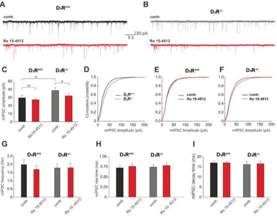
3.3 D3R-/- mice exhibited Ro 15-4513-driven decrease of mIPSC amplitude in
Medium Spiny Neurons.
To test the hypothesis that α6 subunit expression in the NAc shell, as seen in D3R-/- mice, modifies inhibitory transmission, we performed whole-cell
patch-clamp recordings on GABAergic MSNs, which represent >95% of the cell population in this brain region, and recorded miniature inhibitory postsynaptic
currents (mIPSCs). Analysis of the peak amplitudes of mIPSCs revealed a significant increase in D3R-/- compared to WT (Fig. 3; A-D; 38.58 ± 3.35 pA, n
= 19 versus 29.51 ± 2.96 pA, n = 16; P<0.05). In contrast, there was no significant difference in mIPSC frequency (D3R-/-: 1.98 ± 0.30 Hz, WT: 1.77 ±
0.26 ms) and mIPSC kinetics (Fig. 3 G, H; rise time, D3R-/-: 0.72 ± 0.06 ms;
WT: 0.72 ± 0.06 ms; decay time, D3R-/-: 16.96 ± 1.10 ms; WT: 16.14 ± 1.31
ms). Next, we tested the effects of Ro 15-4513, which preferentially acts on α4/6 subunit-containing GABAARs (36, 37) on mIPSCs in MSNs from WT and
D3R-/-. Based on ISH and qPCR data indicating that GABAARs containing α6
subunit in the NAc are almost absent in naïve WT mice and given the opposite effect of Ro 15-4513 treatment on ethanol intake observed in D3R-/- mice, we
expected that Ro 15-4513 would have differential effects on mIPSCs. For this in vitro experiment we selected the 0.3 µM Ro 15-4513 concentration, because it completely antagonizes ethanol enhancement of α4β3δ-type GABAAR
current (36). As shown in Fig. 3, bath application of 0.3 µM Ro 15-4513 did not significantly alter the frequency, rise time, decay time and amplitude of mIPSCs in WT (n = 16; paired t test). Surprisingly, 0.3 µM Ro 15-4513 induced a significant reduction of amplitude in the NAc of D3R-/- (Figure 3; B-F; 38.58 ±
3.35 pA, versus 31.93± 3.03 pA, n = 19 P<0.05; paired t test) while frequency, rise time and decay time were not affected. These results suggest that the activity of α6-subunit-containing GABAAR in D3R-/- influences inhibitory
synaptic transmission of MSN within NAc shell. Since this effect of Ro 15-4513 resulted in a negative modulation, it is possible that the induced α6 subunit expression generates a novel population of GABAA receptor with
4. Discussion
We found that D3R-dependent increased expression of α6 GABAA subunit
counteracts alcohol intake by increasing GABA inhibition in the NAc. We revealed GABAA α6 activity by using Ro 15-4513, a GABAA ligand which
appeared to exert α6-dependent effects, both in terms of behavior (ethanol intake) as well as of neuronal excitability (electrophysiology). The α6 subunit came to the attention of the alcohol addiction studies following the identification of the R100Q mutation in the Sardinian non-ethanol-preferring rat line, suggesting a possible involvement of the GABAAR containing α6 subunit
in the genetic predisposition to alcohol preference (13). This mutation is associated to hypersensitivity to motor-impairing effects of ethanol as well as to tonic inhibitory currents mediated by the α6βδ-type GABAAR, measured by
patch-clamp in cerebellar granule cells (10, 16). Worthy of note, this mutation strongly increases diazepam effect on GABA-evoked currents (14), consistent with a model where the amino acid residue at position 100 that affects ethanol sensitivity in the GABAARs is part of the benzodiazepine ligand-binding pocket
on the α6-subunit (22, 40, 41). Other studies have also described in humans α6 polymorphisms that correlate to alcohol dependence (42, 43). Our observation that genetic deletion or pharmacological blockade of D3R increases GABAA α6
subunit expression in the ventral striatum (19, the present study), a brain structure involved in voluntary ethanol intake, provided a tool to study how
increased expression of α6 subunit-containing receptors may affect alcohol intake. Several studies, in the last two decades, have tried to elucidate how the subunit composition of different GABAARs determines their
electrophysiological and pharmacological features (inhibitory currents, ligand binding), or, at the organism level, the animal behavior (anxiety, addiction, response to anxiolytics). While most studies have dealt with recombinant systems, such as Xenopus laevis oocytes injected either with cRNA coding for the different subunits (12, 36) or with cRNA coding for concatenated subunits (44), no studies had the opportunity to examine native systems, i.e. animals spontaneously and stably expressing ectopic specific subunits in defined CNS structures. Polymorphisms of α6 subunit have been found to be associated both to anxiety-related traits (45) and to benzodiazepine sensitivity in humans (46). It is not yet known whether ectopic expression of α6 subunit containing GABAAR isoforms in brain areas that normally express negligible amounts of
α6 produces different responses to GABA (i.e. different inhibitory currents) and/or to exogenous modulators, including benzodiazepines and ethanol, probably because in vivo systems with significant ectopic α6 expression are not commonly available. The early studies with α6 subunit knockout mice (31, 47) remained inconclusive as it was later discovered that the knockout construct affected the expression of neighboring subunits in the GABAA gene cluster
compete with ethanol within a binding pocket involving α6 (25). We expected a different effect of Ro 15-4513 in WT, which barely express α6 in the NAc, versus D3R-/-, which robustly express α6. Indeed, we found an opposite effect of
Ro 15-4513 in the two groups; in WT Ro 15-4513 reduced ethanol intake, presumably as a result of its action as a negative allosteric modulator in multiple GABAARs (36), where it would behave as an “ethanol antagonist” (25,
26); in contrast, in D3R-/- Ro 15-4513 paradoxically increased ethanol intake, a
surprising finding that might be explained in terms of differential modulation of the GABAAR containing α6 subunit by Ro 15-4513. Indeed, the antagonism
between Ro 15-4513 and ethanol might be more at the functional level, rather than at the binding level; in fact, while the reported affinity of Ro 15-4513 for α4 and α6 containing GABAAR is quite close, in the nanomolar range (12, 25,
36, 49), the effect on the GABA-dependent currents in cells expressing either isoform is not clear, but might be quite different, as suggested by a paradoxical activation of neurons by gaboxadol in a transgenic Thy1α6 mouse line, ectopically expressing the GABAAR α6 subunit gene under the Thy-1.2
promoter (23). We directly address this issue by measuring MSN mIPSCs in the NAc and their sensitivity to Ro 15-4513. Based on the above premises, we hypothesized that a change in GABAA α6 subunit expression would increase
spontaneous mIPSCs and that Ro 15-4513 would inhibit mIPSCs in MSN from D3R-/-, robustly expressing α6, whereas it would be ineffective in α6-deficient
MSNs from WT. The electrophysiological analysis of MSNs revealed a significant increase in mIPSC amplitude in D3R-/-, which expressed GABAAR
containing α6 subunit in NAc, compared to WT; perfusion with Ro 15-4513
induced a significant reduction of amplitude in the NAc of D3R-/-, but was
ineffective in WT. This latter observation clearly indicates that the modulation of the GABAAR channel by Ro 15-4513 depends on the presence of α6 subunit
and is consistent with the observation of opposite effects of this drug on ethanol intake in WT and D3R-/-. Systematic assessment of α6 expression in the CNS by
ISH, confirmed by qPCR, indicates that “ectopic” α6 expression in D3R-/- was
restricted to a limited brain area, corresponding to ventral striatum and including the NAc, a finding reinforced by autoradiography data obtained with [3H]Ro 15-4513. The fact that genetic or pharmacological manipulation of D3R
specifically induced changes in the NAc, leaving relatively unchanged other brain areas is not so surprisingly, when considering that, at variance with D2R,
the expression of D3R is mainly restricted to the very same structures where we
observe increased α6 expression (50). To the best of our knowledge, it is not known in detail how D3R control GABAAR subunit mRNA expression;
however, other studies have shown dynamic D3R-dependent down-regulation of
GABAergic control over lateral/basolateral amygdala neurons (51), NAc (52) and hippocampus (53, 54). A direct dynamic interplay between metabotropic dopamine receptors and ionotropic (NMDA) receptors in plasma membrane has
been documented by single-molecule detection imaging and electrophysiology in live hippocampal neurons (55). Furthermore, cell signaling downstream of D3R affects GABAARs in the NAc (52), but numerous other complex
mechanisms may impact GABAARs trafficking (56) and deserve further studies
to be elucidated. Finally, because these changes in GABAAR function can be
related to dopaminergic transmission, they may assume further relevance in clinical situations, such as schizophrenia and Parkinson’s disease, where D3R
are chronically blocked or stimulated by drug-treatments (38).
In conclusion, these data indicate that α6-containing GABAARs in the NAc play
an important role in controlling alcohol intake by increasing GABAergic-inhibition in the MSNs. Because changes in α6-containing GABAARs are
specifically induced in the NAc by D3R-blockade, the interplay between
DAergic and GABAergic transmission may present a novel relevant mechanisms in reinforcing properties of alcohol and other addictive drugs.
Acknowledgments
This work was supported by Finanziamento Ricerca di Ateneo – FIR Unict 7E646B. The Academy of Finland and the Sigrid Juselius foundation grants to Esa R. Korpi are gratefully acknowledged. Roberta Di Marco was supported by the PhD program in Neuroscience, Catania University. We thanks Dr. Daniela Puzzo for reading the manuscript and Dr. Chiara Platania for help in preparing figures.
References
1. Kauer JA, Malenka RC (2007) Synaptic plasticity and addiction. Nat
Rev Neurosci 8:844-858.
2. Olsen RW (2011) Extrasynaptic GABAA receptors in the nucleus accumbens are necessary for alcohol drinking. Proc Natl Acad Sci USA 108:4699-4700.
3. Maldve RE, Zhang TA, Ferrani-Kile K, Schreiber SS, Lippmann MJ, Snyder GL, Fienberg AA, Leslie SW, Gonzales RA, Morrisett RA (2002) DARPP-32 and regulation of the ethanol sensitivity of NMDA receptors in the nucleus accumbens. Nat Neurosci 5:641-648.
4. Lobo MK, Karsten SL, Gray M, Geschwind DH, Yang XW (2006) FACS-array profiling of striatal projection neuron subtypes in juvenile and adult mouse brains. Nat Neurosci 9:443-452.
5. Bertran-Gonzalez J, Bosch C, Maroteaux M, Matamales M, Hervé D, Valjent E, Girault JA (2008) Opposing patterns of signaling activation in dopamine D1 and D2 receptor-expressing striatal neurons in response to cocaine and haloperidol. J Neurosci 28:5671-5685.
6. Milton GV, Randall PK, & Erickson CK (1995) Low-dose effect of ethanol on locomotor activity induced by activation of the mesolimbic system. Alcohol Clin Exp Res 19:768-776.
7. Hodge CW, Alken AS (1996) Discriminative stimulus function of ethanol: role of GABAA receptors in the nucleus accumbens. Alcohol
Clin Exp Res 20:1221-1228.
8. Rewal M, Jurd R, Gill TM, He DY, Ron D, Janak PH (2009) Alpha4-containing GABAA receptors in the nucleus accumbens mediate moderate intake of alcohol. J Neurosci 29:543-549.
9. Nie H, Rewal M, Gill TM, Ron D, Janak PH (2011) Extrasynaptic delta-containing GABAA receptors in the nucleus accumbens dorsomedial shell contribute to alcohol intake. Proc Natl Acad Sci USA 108:4459-4464.
10. Olsen RW, Sieghart W (2009) GABA A receptors: subtypes provide diversity of function and pharmacology. Neuropharmacology 56:141-148.
11. Rudolph U, Knoflach F (2011) Beyond classical benzodiazepines: novel therapeutic potential of GABAA receptor subtypes. Nat Rev Drug
Discov 10:685-697.
12. Wallner M, Hanchar HJ, Olsen RW (2003) Ethanol enhances alpha 4 beta 3 delta and alpha 6 beta 3 delta gamma-aminobutyric acid type A receptors at low concentrations known to affect humans. Proc Natl Acad
Sci USA 100:15218-15223.
13. Saba L, Porcella A, Congeddu E, Colombo G, Peis M, Pistis M, Gessa GL, Pani L (2001) The R100Q mutation of the GABA(A) alpha(6) receptor subunit may contribute to voluntary aversion to ethanol in the sNP rat line. Brain Res Mol Brain Res 87:263-270.
14. Korpi ER, Kleingoor C, Kettenmann H, Seeburg PH (1993) Benzodiazepine-induced motor impairment linked to point mutation in cerebellar GABAA receptor. Nature 361:356-359.
15. Sarviharju M, Korpi ER (1993) Ethanol sensitivity and consumption in F2 hybrid crosses of ANT and AT rats. Alcohol 10:415-418.
16. Santhakumar V, Wallner M, Otis TS (2007) Ethanol acts directly on extrasynaptic subtypes of GABAA receptors to increase tonic inhibition.
Alcohol 41:211-221.
17. Morales M, Margolis EB (2017) Ventral tegmental area: cellular heterogeneity, connectivity and behaviour. Nat Rev Neurosci 18:73-85.
18. Leggio GM, Camillieri G, Platania CB, Castorina A, Marrazzo G, Torrisi SA, Nona CN, D'Agata V, Nobrega J, Stark H, Bucolo C, Le Foll B, Drago F, Salomone S (2014) Dopamine D3 receptor is necessary for ethanol consumption: an approach with buspirone.
Neuropsychopharmacology 39:2017-2028.
19. Heidbreder CA, Andreoli M, Marcon C, Hutcheson DM, Gardner EL, Ashby CR Jr (2007) Evidence for the role of dopamine D3 receptors in oral operant alcohol self-administration and reinstatement of alcohol-seeking behavior in mice. Addict Biol 12:35-50.
20. Vengeliene V, Leonardi-Essmann F, Perreau-Lenz S, Gebicke-Haerter P, Drescher K, Gross G, Spanagel R (2006) The dopamine D3 receptor plays an essential role in alcohol-seeking and relapse. FASEB J 20:2223-2233.
21. Koo JW, Lobo MK, Chaudhury D, Labonté B, Friedman A, Heller E, Peña CJ, Han MH, Nestler EJ (2014) Loss of BDNF Signaling in D1R-Expressing NAc Neurons Enhances Morphine Reward by Reducing GABA Inhibition. Neuropsychopharmacology 39:2646-2653.
22. Leggio GM, Torrisi SA, Castorina A, Platania CB, Impellizzeri AA, Fidilio A, Caraci F, Bucolo C, Drago F, Salomone S (2015) Dopamine D3 receptor-dependent changes in alpha6 GABAA subunit expression in striatum modulate anxiety-like behaviour: Responsiveness and tolerance to diazepam. Eur Neuropsychopharmacol 25:1427-1436. 23. Hellsten KS, Linden AM, Korpi ER (2015) Paradoxical widespread
c-Fos expression induced by a GABA agonist in the forebrain of transgenic mice with ectopic expression of the GABA(A) alpha6 subunit. Neuroscience 293:123-135.
24. Luddens H, Korpi ER, Seeburg PH (1995) GABAA/benzodiazepine receptor heterogeneity: neurophysiological implications.
Neuropharmacology 34:245-254.
25. Wallner M, Hanchar HJ, Olsen RW (2014) Alcohol selectivity of beta3-containing GABAA receptors: evidence for a unique extracellular alcohol/imidazobenzodiazepine Ro15-4513 binding site at the alpha+beta- subunit interface in alphabeta3delta GABAA receptors.
Neurochem Res 39:1118-1126.
26. Linden AM, Schmitt U, Leppä E, Wulff P, Wisden W, Lüddens H, Korpi ER. (2011) Ro 15-4513 Antagonizes Alcohol-Induced Sedation in Mice Through alphabetagamma2-type GABA(A) Receptors. Front
Neurosci 5:3.
27. Baur R, Kaur KH, Sigel E (2009) Structure of alpha6 beta3 delta GABA(A) receptors and their lack of ethanol sensitivity. J Neurochem 111:1172-1181.
28. McBride WJ, Murphy JM, Lumeng L, Li TK (1988) Effects of Ro 15-4513, fluoxetine and desipramine on the intake of ethanol, water and food by the alcohol-preferring (P) and -nonpreferring (NP) lines of rats.
Pharmacol Biochem Behav 30:1045-1050.
29. Wegelius K, Honkanen A, Korpi ER (1994) Benzodiazepine receptor ligands modulate ethanol drinking in alcohol-preferring rats. Eur J
Pharmacol 263:141-147.
30. Petry NM (1995) Ro 15-4513 selectively attenuates ethanol, but not sucrose, reinforced responding in a concurrent access procedure; comparison to other drugs. Psychopharmacology 121:192-203.
31. Homanics GE, Ferguson C, Quinlan JJ, Daggett J, Snyder K, Lagenaur C, Mi ZP, Wang XH, Grayson DR, Firestone LL (1997) Gene knockout of the alpha6 subunit of the gamma-aminobutyric acid type A receptor:
lack of effect on responses to ethanol, pentobarbital, and general anesthetics. Mol Pharmacol 51:588-596.
32. Suzdak PD, Glowa JR, Crawley JN, Schwartz RD, Skolnick P, Paul SM. (1986) A selective imidazobenzodiazepine antagonist of ethanol in the rat. Science 234:1243-1247.
33. Samson HH, Haraguchi M, Tolliver GA, Sadeghi KG (1989) Antagonism of ethanol-reinforced behavior by the benzodiazepine inverse agonists Ro15-4513 and FG 7142: relation to sucrose reinforcement. Pharmacol Biochem Behav 33:601-608.
34. June HL, Hughes RW, Spurlock HL, Lewis MJ (1994) Ethanol self-administration in freely feeding and drinking rats: effects of Ro15-4513 alone, and in combination with Ro15-1788 (flumazenil).
Psychopharmacology 115:332-339.
35. Melon LC, Boehm SL, 2nd (2011) GABAA receptors in the posterior, but not anterior, ventral tegmental area mediate Ro15-4513-induced attenuation of binge-like ethanol consumption in C57BL/6J female mice. Behav Brain Res 220:230-237.
36. Wallner M, Hanchar HJ, Olsen RW (2006) Low-dose alcohol actions on alpha4beta3delta GABAA receptors are reversed by the behavioral alcohol antagonist Ro15-4513. Proc Natl Acad Sci USA 103:8540-8545. 37. Hanchar HJ, Chutsrinopkun P, Meera P, Supavilai P, Sieghart W,
Wallner M, Olsen RW (2006) Ethanol potently and competitively inhibits binding of the alcohol antagonist Ro15-4513 to alpha4/6beta3delta GABAA receptors. Proc Natl Acad Sci USA 103:8546-8551.
38. Leggio GM, Bucolo C, Platania CB, Salomone S, Drago F (2016) Current drug treatments targeting dopamine D3 receptor. Pharmacol
39. Luddens H, Killisch I, Seeburg PH (1991) More than one alpha variant may exist in a GABAA/benzodiazepine receptor complex. J Recept Res 11:535-551.
40. Wieland HA, Luddens H, Seeburg PH (1992) A single histidine in GABAA receptors is essential for benzodiazepine agonist binding. J
Biol Chem 267:1426-1429.
41. Kleingoor C, Wieland HA, Korpi ER, Seeburg PH, Kettenmann H (1993) Current potentiation by diazepam but not GABA sensitivity is determined by a single histidine residue. Neuroreport 4:187-190.
42. Han DH, Bolo N, Daniels MA, Lyoo IK, Min KJ, Kim CH, Renshaw PF (2008) Craving for alcohol and food during treatment for alcohol dependence: modulation by T allele of 1519T>C GABAAalpha6.
Alcohol Clin Exp Res 32:1593-1599.
43. Radel M, Vallejo RL, Iwata N, Aragon R, Long JC, Virkkunen M, Goldman D (2005) Haplotype-based localization of an alcohol dependence gene to the 5q34 {gamma}-aminobutyric acid type A gene cluster. Arch Gen Psychiatry 62:47-55.
44. Minier F, Sigel E (2004) Positioning of the alpha-subunit isoforms confers a functional signature to gamma-aminobutyric acid type A receptors. Proc Natl Acad Sci USA 101:7769-7774.
45. Arias B, Aguilera M, Moya J, Sáiz PA, Villa H, Ibáñez MI, García-Portillo MP, Bobes J, Ortet G, Fañanás L (2012) The role of genetic variability in the SLC6A4, BDNF and GABRA6 genes in anxiety-related traits. Acta Psychiatr Scand 125:194-202.
46. Iwata N, Cowley DS, Radel M, Roy-Byrne PP, Goldman D (1999) Relationship between a GABAA alpha 6 Pro385Ser substitution and benzodiazepine sensitivity. Am J Psychiatry 156:1447-1449.
47. Jones A, Korpi ER, McKernan RM, Pelz R, Nusser Z, Mäkelä R, Mellor JR, Pollard S, Bahn S, Stephenson FA, Randall AD, Sieghart W, Somogyi P, Smith AJ, Wisden W. (1997) Ligand-gated ion channel subunit partnerships: GABAA receptor alpha6 subunit gene inactivation inhibits delta subunit expression. J Neurosci 17:1350-1362.
48. Uusi-Oukari M, Heikkilä J, Sinkkonen ST, Mäkelä R, Hauer B, Homanics GE, Sieghart W, Wisden W, Korpi ER. (2000) Long-range interactions in neuronal gene expression: evidence from gene targeting in the GABA(A) receptor beta2-alpha6-alpha1-gamma2 subunit gene cluster. Mol Cell Neurosci 16:34-41.
49. Knoflach F, Benke D, Wang Y, Scheurer L, Lüddens H, Hamilton BJ, Carter DB, Mohler H, Benson JA (1996) Pharmacological modulation of the diazepam-insensitive recombinant gamma-aminobutyric acidA receptors alpha 4 beta 2 gamma 2 and alpha 6 beta 2 gamma 2. Mol
pharmacol 50:1253-1261.
50. Guillin O, Diaz J, Carroll P, Griffon N, Schwartz JC, Sokoloff P (2001) BDNF controls dopamine D3 receptor expression and triggers behavioural sensitization. Nature 411:86-89.
51. Diaz MR, Chappell AM, Christian DT, Anderson NJ, McCool BA (2011) Dopamine D3-like receptors modulate anxiety-like behavior and regulate GABAergic transmission in the rat lateral/basolateral amygdala. Neuropsychopharmacology 36:1090-1103.
52. Chen G, Kittler JT, Moss SJ, Yan Z (2006) Dopamine D3 receptors regulate GABAA receptor function through a phospho-dependent endocytosis mechanism in nucleus accumbens. J Neurosci 26:2513-2521.
53. Hammad H, Wagner JJ (2006) Dopamine-mediated disinhibition in the CA1 region of rat hippocampus via D3 receptor activation. J Pharmacol
Exp Ther 316:113-120.
54. Swant J, Stramiello M, Wagner JJ (2008) Postsynaptic dopamine D3 receptor modulation of evoked IPSCs via GABA(A) receptor endocytosis in rat hippocampus. Hippocampus 18(5):492-502.
55. Ladepeche L, Dupuis JP, Bouchet D, Doudnikoff E, Yang L, Campagne Y, Bézard E, Hosy E, Groc L (2013) Single-molecule imaging of the functional crosstalk between surface NMDA and dopamine D1 receptors. Proc Natl Acad Sci USA 110:18005-18010.
56. Mele M, Leal G, Duarte CB (2016) Role of GABAA R trafficking in the plasticity of inhibitory synapses. J Neurochem 139:997-1018.
57. Accili D, Fishburn CS, Drago J, Steiner H, Lachowicz JE, Park BH, Gauda EB, Lee EJ, Cool MH, Sibley DR, Gerfen CR, Westphal H, Fuchs S (1996) A targeted mutation of the D3 dopamine receptor gene is associated with hyperactivity in mice. Proc Natl Acad Sci USA 93:1945-1949.
58. Rhodes JS, Best K, Belknap JK, Finn DA, Crabbe JC (2005) Evaluation of a simple model of ethanol drinking to intoxication in C57BL/6J mice.
Physiol Behav 84:53-63.
59. Sinkkonen ST, Vekovischeva OY, Möykkynen T, Ogris W, Sieghart W, Wisden W, Korpi ER (2004) Behavioural correlates of an altered balance between synaptic and extrasynaptic GABAAergic inhibition in a mouse model. Eur J Neurosci 20:2168-2178.
60. Scala F, Fusco S, Ripoli C, Piacentini R, Li Puma DD, Spinelli M, Laezza F, Grassi C, D'Ascenzo M (2015) Intraneuronal Abeta accumulation induces hippocampal neuron hyperexcitability through
A-type K(+) current inhibition mediated by activation of caspases and GSK-3. Neurobiol Aging 36:886-900.
Figure 1. Alcohol intake inversely correlated with D3R-dependent GABAA α6 subunit mRNA
expression in the NAc; effect of Ro 15-4513 on ethanol consumption in mice expressing different levels of α6. A and B, ethanol intake (in the drinking in the dark paradigm, DID) and α6 expression in wild type (D3R+/+) heterozygous (D3R+/-) and null mice (D3R-/-). DID was
measured for 4 days, in mice with limited access (2h/day for 3 days and 4h the 4th day) to ethanol solution (20%). Abundance of transcripts in the NAc was assessed by qPCR after DID; expression level is expressed as mean fold changes relative controls. C and D, ethanol intake (DID) in D3R+/+ and D3R-/- treated with vehicle (VEH) or Ro 15-4513 (5 mg/kg, i.p.). E and F,
ethanol intake (DID) and α6 expression in D3R+/+ treated with vehicle (VEH) or the selective
D3R antagonist, SB 277011A (10 mg/kg, i.p.) with or without Ro 15-4513. Each experimental
group included 8-10 mice.
*P<0.05, **P<0.01 vs. the corresponding control (D3R+/+, VEH or VEH+VEH); one- or
two-way ANOVA and Newman–Keuls post hoc test.
G, correlation between differences ethanol intake (DID) and α6 expression (R2 = 0.44, Pearson r = -0.66, p < 0.001); different symbols represent different genotypes and/or treatments, as indicated.
Figure 2. Ectopic expression of α6 GABAA subunit mRNA and [3H]-Ro 15-4513 binding in
the NAc of D3R+/+ and D3R-/- mice. A and B, In situ hybridization (ISH) detection of α6; C and
D, [3H]-Ro 15-4513 autoradiography. Representative images and averaged optical density, mean fold changes relative to control (n=6-8 per group).
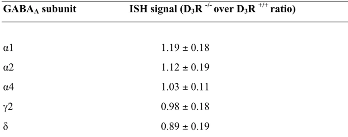
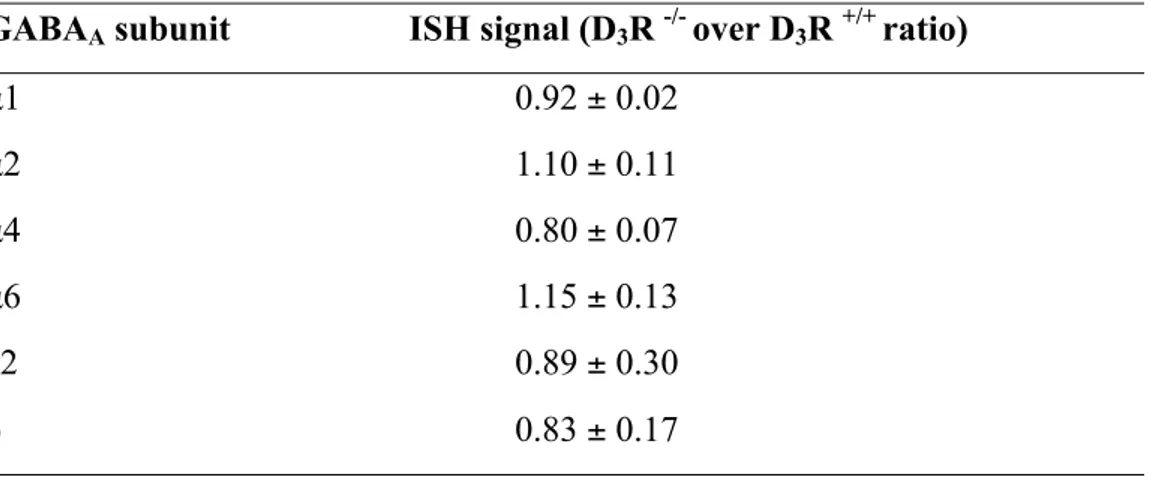
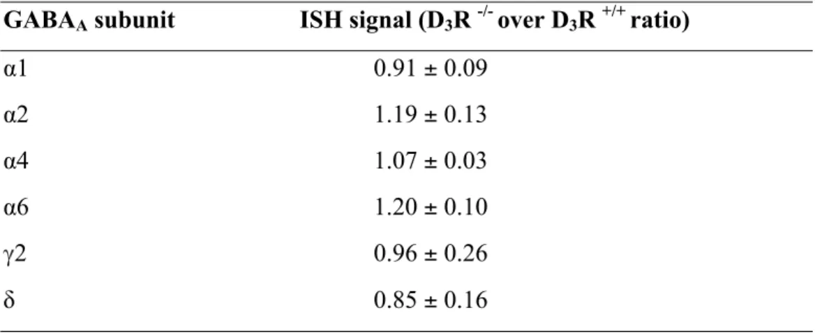
Table 1.In situ hybridization (ISH) signals for of GABAA !1, !2, !4, γ2, δ mRNA subunits
in the nucleus accumbens from D3R+/+ and D3R-/- mice.
GABAA subunit ISH signal (D3R -/- over D3R +/+ ratio)
α1 1.19 ± 0.18
α2 1.12 ± 0.19
α4 1.03 ± 0.11
γ2 0.98 ± 0.18
Figure 3. NAc medium spiny neurons from D3R-/- mice exhibited increased GABAA inhibitory
currents sensitive to Ro 15-4513. A and B, representative traces showing mIPSC recordings in slice from D3R+/+ (WT) and D3R-/- mice before and after treatment with Ro-154513 (0.3 µM; in red). C, analysis of the peak amplitudes of mIPSCs; notice an increase in D3R-/- compared to
D3R+/+ and a decrease following Ro 15-4513 application in D3R-/- only. *P<0.05, unpaired (D3R -
vs D3R+/+) or paired (pre- vs post- Ro 15-4513) t test (D3R-/-, n=19; D3R+/+, n=16). D-F,
cumulative frequency distributions for mIPSC amplitude in the experimental conditions shown in A and B. G-I, analysis of mIPSC frequency, rise time and decay time.
![Figure 2. Ectopic expression of α6 GABA A subunit mRNA and [ 3 H]-Ro 15-4513 binding in](https://thumb-eu.123doks.com/thumbv2/123dokorg/4526347.35138/64.892.208.670.105.741/figure-ectopic-expression-gaba-subunit-mrna-ro-binding.webp)