1
Abstract
In the last decades, great attention has focused on the impact of oil refinery wastewaters and discharged effluents, which are released to the environment as gases, particles, sludge, and liquid effluents, and therefore represent potential sources of pollution. In environmental biomonitoring programmes, marine mussels have been often used as bioindicator species because of their ability to bioaccumulate toxic compounds and their tolerance towards a huge variety of environmental conditions. To recover polluted sites, several remediation actions were made. In this work, two remediation techniques (BF-MBR and Soil Washing) versus wastewater and natural polluted sediments, collected from a contaminated area in Augusta (eastern Sicily, Italy), were evaluated. To this aim, in the first part of thesis, mesocosm scale-up experiments were settled in order to measure the recovery effects on mussels Mytilus galloprovincialis. Both for wastewater and sediments, three different environmental conditions were simulated: a control area (W, white group), a petrochemical polluted area (B, black group), and a polluted area subjected to the remediation actions (G, grey group). Mussels were exposed to each condition for 15 days, and then the biological effects of technological actions for the potential recovery of petrochemical contamination were evaluated by a multi-biomarker approach, including histology, metabolomics, immunohistochemistry, molecular and enzymatic investigations. Mussel gills, mainly involved in nutrient uptake and gas exchange, were chosen as target
2 organs. In respect to the W group, the gills of mussels from the B mesocosms, both for wastewater and sediments, showed marked morphological alterations with loss of cilia. Changes in serotoninergic (i.e. serotonin, 5-HT, and its receptor, 5-HT3R) and cholinergic (i.e. acetylcholinesterase, AChE, and acetylcholintransferase, ChAT) systems were observed in mussel gills from the B group by immunohistochemistry, as well as supported by the enzymatic analysis of AChE activity, metabolomics and molecular assay, which revealed changes in neurotransmitters. Contrarily, in the G mesocosms, the same battery of biomarkers indicated a general recovery trend in mussel gills. Overall, the application of a multi-biomarker panel results effective in assessing the environmental influences of petrochemical pollutants on the health of aquatic organisms. Furthermore, findings from this study confirmed that the remediation actions herein applied on wastewaters and natural polluted sediments might be good tools to recovering a petrochemical polluted ecosystem. Some of the results presented in this PhD thesis are already submitted for publication in international peer-reviewed journals.
Since in the first part of thesis it was demonstrated the high sensitivity and the extensive alterations induced to the respiratory organs (gills), in parallel a brief internship at the Swedish Toxicology Sciences Research Center (SWETOX) in Södertälje, Sweden, was done under the supervision of Dr. Ernesto Alfaro-Moreno, in order to acquire competences on the use of cell culture in toxicology. So, in the second part of Thesis, two different techniques were assessed for the
3 evaluation of tight junction disruption induced by pollutants (in our case NaClO) on human airway epithelial cells. Overall, the use of the
in vitro technique is a promising tool for future investigations on the
effects of environmental toxic compounds on human airway epithelial cells, which confirmed also the high sensitivity of this kind of cells versus pollutants.
4 CHAPTER I. Introduction 1.1. General introduction 1.2. Remediation 1.2.1. Soil Washing 1.2.2. Membrane Bioreactor (MBR)
1.3. Mesocosm: definition and application
1.4. Enviromental Biomonitoring
1.5. Mussels:Mytilidae
1.6. Neuronal gill activity regulation
CHAPTER II. Aim of the thesis
CHAPTER III. Biological approach to evaluate the efficiency of
BF-MBR treatment on wastewater
3.1. Material and Methods Set-up of mesocosms
Mixture of potential toxic compounds
Dispersant
Mussel Exposure
Measurement of the main physico-chemical parameters
Chemical Oxygen Demand (COD) measures
Quantitative analysis of hydrocarbons
Histological analysis
Immunohistochemical analysis
Metabolomics analysis
Gill metabolite extraction
1H NMR-based metabolomics analysis
Enzymatic analysis
Molecular analysis
5
5-HT3R quantitative gene expression (qPCR)
Statistical analysis
3.2. Results
Physico-chemical parameters
COD results
Quantitative analysis of hydrocarbons
Histology Immunohistochemistry Metabolomics Enzymatics Molecular results 3.3. Discussion 3.4. Conclusion
CHAPTER IV. Biological evaluation of the Soil Washing
remediation versus natural mercury-polluted sediments in a
mesocosm scale experiment
4.1. Material and methods
Set-up of mesocosms
Mussel Exposure
Measurement of the main physico-chemical parameters
Chemical Oxygen Demand (COD) measures
Quantitative analysis of metals in sediment
Histological analysis
Immunohistochemical analysis
Metabolomics analysis
Gill metabolite extraction
1H NMR-based metabolomics analysis
6
Molecular analysis
RNA extraction and cDNA synthesis
5-HT3R quantitative gene expression (qPCR)
Statistical analysis
4.2. Results
Physico-chemical parameters
COD results
Metal concentration in sediment of B and G mesocosms
Histology Immunohistochemistry Metabolomics Enzymatics Molecular results 4.3. Discussion 4.4. Conclusion
CHAPTER V. Evaluation of alteration induced by pollutants
(NaClO) on tight junction of human airway epithelial cells
5.1. General introduction
5.2. Use of Cell Culture in Toxicology
5.3. Aim of the Internship
5.4. Material and Methods Cell Culture
Pollutant
Transepithelial electrical resistance (TEER) measurement
Reagent Immunofluorescence staining of Zonula Occludens-1 (ZO-1)
Immunofluorescence staining of Zonula Occludens-1 (ZO-1)
7 5.5. Results TEER results Zo-1 immunofluorescence 5.6. Discussion 5.7. Conclusion
Chapter VI: General Conclusion
8
CHAPTER I
Introduction
1.1 General Introduction
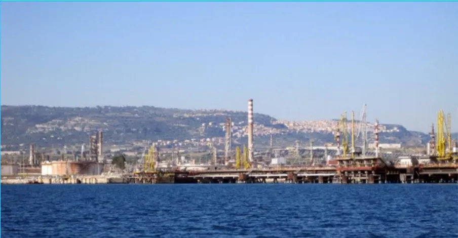
In the last years, various biomonitoring programmes have been carried out with the aim to evaluate the environmental effects of various pollutants, in particular those from oil refinery activity. The activity of petrochemical industry and oil refinery is responsible for the emission of a huge amount of pollutants in the water in form of gases, particles, sludge, and liquid effluent. In these wastes, it is possible to observe a substantial amount of polycyclic aromatic hydrocarbons (PAHs), phenols, heavy metals and their derivatives, sulphides, naphthenic acids, and other chemicals (Dorris et al., 1972). It is well known that most of these pollutants are even toxic at low concentration (Long et al., 1995). Hence, petrochemical pollutant is able to provoke serious damages to the marine environment and human health (Fasulo et al., 2012a; Sureda et al., 2011). Particularly negative effects of these pollutants are observed in harbour areas, where the quality of water, the water exchange and the biodiversity are reduced (Guerra-García and García-Gómez, 2005). Therefore, harbour areas (for instance the Augusta-Melilli-Priolo petrochemical area in Sicily, Italy) became a target for various biomonitoring programmes (Cappello et al., 2017; Fasulo et al., 2015; Maisano et al., 2017). The data collected within
9 several of these research programmes have offered interesting results which are useful not only in the evaluation of the real damage caused by these kind of wastes, but also in the development of new remediation approaches and improvement of the existing techniques. The main aim of remediation is to reduce the amount and the effects of wastes in the environment. It is possible to list various techniques of remediation, which can be based on various approaches (i.e. chemical, engineering and microbiological). The main aim of this thesis, which is part of the project ”SYSTEMS BIOLOGY” (PRIN2010-2011) (Fasulo et al., 2015), is principally evaluate efficiency of two recovery techniques, the first one versus petrochemical polluted wastewater (BF-MBR) and the second one versus petrochemical polluted sediments (Soil Washing), by assessing different biological responses elicited by marine mussels
Mytilus galloprovincialis treated in mesocosm scale experiments
10
1.2. Remediation
The recovery of polluted areas is becoming a very urgent and relevant problem. In some cases, as for instance in petrochemical areas such as the Augusta-Melilli-Priolo (Sicily, Italy), the concentration of pollutants such as Polycyclic Aromatic Hydrocarbons (PAHs) and heavy metals, like mercury, reach elevated concentrations. In fact, in the Augusta-Melilli-Priolo petrochemical area, the level of mercury in sediment exceeds the standard limit reported by national and international sediment quality guidelines (SQGs) (Ministerial Decree No. 260/2010 De Domenico et al., 2013). This contamination degree is related to the anthropogenic activities in the area, responsible to provoke a severe alteration of the environment. In light of this, it has become fundamental to develop techniques able to remove pollutants or mitigate their effects. During last years, various methods were produced based on engineering, physic-chemical and biological procedures. The various remediation actions could reduce the effect and the concentration of various organic and inorganic pollutant in a different matrix (Water and Soil).
11
1.2.1. Soil Washing
An example of remediation activity is “Soil Washing”. The term “Soil Washing” is used to describe an ex situ technique whose aim is to extract contaminant from soil. It therefore differs from the “Soil Flushing”, which is an in-situ technique. It is a very useful technique able to reduce the amount of contaminant in the soil, in particular the level of heavy metals. Various techniques of soil washing could be listed. It is possible to classify them in physical techniques, which try to use different physical properties of contaminants, and chemical techniques, where the different chemical properties of the contaminants are used for the extraction. It is also possible to observe even combined techniques. In the soil washing, the knowledge of the contaminated soil properties is very relevant in order to apply the better technique (Dermont et al., 2008). The aim of this kind of technique is to concentrate contaminants into a smaller volume of soil by employing the common physical differences with regards to characteristics (size, density, magnetism, and hydrophobic surface properties) between the pollutant particles and soil particles. The foremost physical separation techniques are:
Mechanical screening Hydrodynamic classification Gravity concentration Froth Flotation
The choice of the physical separation technology is heavily related to the soil and site types to be treated (Williford et al., 2000). These kind
12 of techniques are principally appropriate to “anthropogenic” soils placed in urban or industrial areas, and soil with a good amount of sand of 50-70% (USEPA, 1997). On the other hand, physical separation techniques are not appropriate for treating the “natural” soils or agricultural soils affected by a diffuse contamination. Hence, these soils typically have a high content of silt/clay and organic matter the metals present in soils are mostly in sorbed forms as opposed to discrete particles. For these reasons, physical separation is often associated with chemical procedures to improve pollutant removal (Dermont et al., 2008). Chemical extraction (CE) tries to remove pollutants by chemical reagents in order to transfer metals or other kind of pollutants from soils to an aqueous solution. Since it uses an aqueous solution that “wash” the soil, the chemical extraction is considered from some authors as the proper "Soil Washing. In this technique, in order to increase solubility leaching solutions in which the contaminants are dissolved can be used. Another way to increase solubility could be the conversion to soluble salts by valence change (Dermont et al., 2008). It is possible to consider different solutions able to dissolve pollutants:
Acids (FRTR 2007)
Salts and high concentration chloride solutions (Tampouris et al., 2001; Kuo et al., 2006)
Chelating Agents (Peters, 1999) Surfactant (Ehsan et al., 2006)
13 Also, in this case, it is very important to have good information about the chemical aspect of the soil because this knowledge could address versus the best technique. In various situation, chemical extraction looks better in respect to physical extraction (as for instance for sorbed metals) but, at the same time, the application of chemical reagents can increase processing cost and generate further environmental problems (Dermont et al., 2008).
1.2.2. Membrane Bioreactor (MBR)
Membrane Bioreactor (MBR) is a technology that affords biological approach with a membrane separation. The two main steps of MBR are:
- Biological degradation of organic pollutants by adapted microorganisms in a bioreactor;
- Physical separation by a membrane able to segregate
microorganisms, which permit either recycling the activated sludge and creating a clean matter.
The position of membranes in important in MBR organisation. Hence, the membrane could be located outside or inside in respect to the bioreactor. In the first case, there is a recirculated configuration with an external membrane unit. Mixed liquor is circulated outside of the reactor to the membrane module, where is filtered under pressure. The concentrated sludge recycling occurs back into the reactor. In the second one, the membrane module is submerged in the activated sludge (Submerged configuration). This last system looks more economical concerning energy expenditure (Huang et al., 2001). The MBR, in
14 respect to other technique, looks more efficient respect to the common clarifier (Ng et al., 2007), because it is possible to use a membrane module more compact. Furthermore these systems permit to work with a higher biomass concentration than other systems (Yamamoto et al., 1989; Jefferson et al., 2000) The use of a bioreactor is related to the acquired ability of different bacteria stain to biodegrade several pollutants, as for example hydrocarbons (Cappello et al., 2016). An excellent example of treatment technology is the Moving Bed Biofilm Reactor (MBBR). In this method biomass is able to develop either as suspended flocs or as biofilm, implementing a higher total biomass concentration in the reactor. Newly, MBR and MBBR have been employed together, known as Biofilm Membrane Bioreactor (BF-MBR), substituting the secondary settler by means of MBR (Di Trapani et al., 2014).In these conditions the removal concentration of hydrocarbons refractory is considerably higher. The bioreactors for bioremediation procedures are tank where living organisms are able to perform their biological reactions. A reactor should be easy to maintain and operate (Evangelho et al., 2001) and should be able to work both in aerobic and anaerobic conditions. Their effectiveness is guaranteed by the capacity of bacteria to attach on the inert material, such as granular activated carbon, to generate high biomass at interfaces (Bouwer and McCarty, 1982; Teitzel and Parsek, 2003). By the rupture of solid aggregates and the dispersion of insoluble supports, desorption of hydrocarbons and contact with the aqueous phase is raised, with consequent improvement of biodegradation. Several models of bioreactors are broadly applied in a large kind of aerobic bioprocesses
15 such as aerobic fermentation, biological wastewater and hydrocarbon impacted soil/sediments treatments (Van Hamme et al., 2003). The filtration of oil-contaminated water with a porous membrane bioreactor (MBR) is a novel improvement in wastewater discharges bioremediation. In particular marine wastewater discharges are related to operations deriving from slops, dirty ballast, sewage and bilge water management are held responsible of high oil load and consequent risk of marine pollution (Ciacci et al., 2012; Fasulo et al., 2015; Mancini et al., 2017). A membrane bioreactor couples the activated sludge process among a membrane separation method. The work of the reactor is comparable to a traditional activated sludge process but, in this case, secondary clarification and tertiary steps like sand filtration are not necessary. Especially, in MBR treatment, native, water-borne micro-organisms in a controlled habitat performed the bioremediation, so the choice and identification of microbial consortia with high capability to degrade hydrocarbons is a primary step for the optimization of the process. Nowadays, various aspects of these techniques are considered in different scientific works aimed at improving the wastewater discharges treatment (Cappello et al., 2016; Mancini et al., 2017; Pirrone et al., 2018; Yakimov et al., 2007).
16
1.3 Mesocosm: definition and application
A simple definition of mesocosm is an experimental set-up able to combine the complexity of an environmental experiment and the controllability provided by lab context. Mesocosm should be considered as a useful tool that give explanations when it is difficult to obtain results in extremely controlled oversimplified conditions or complex and largely uncontrolled natural conditions (Sagarin et al., 2016). This experimental set-up offers various opportunities in several research topics. For instance, in the ecological field, the mesocosm permits to have an easier observation and setting of several parameters in respect to nature. In addiction, the possibility to create perturbation is more pleased. For these reasons, mesocosm in the ecological field it is hugely applied. Indeed, in this field various scientific works were developed (Davis et al., 1996; Scheinin et al., 2015; Kelly et al., 2014). Another interesting context in which mesocosm is applied is the assessment of the effects of various pollutants on living organisms in an enclosure environment (Oviat et al., 1984, Vethaak et al., 1996, Sanchis et al., 2018. Oviat et al. (1984) tested the recovery capacity of an ecosystem with polluted sediments. Vethaak et al. (1996) directed their investigations on possible pathologies found on Platichthys flesus and their direct correlation to pollution. In the work of Sanchis et al. (2018), the metabolic responses of M. galloprovincialis to fullerenes were evaluated. On other interesting use of mesocosm is in scale-up experiments to better evaluate the efficiency of remediation techniques. For instance, Pirrone et al. (2018), used a microcosm approach to measure BF-MBR efficiency in mitigating the impact of
oily-17 wastewater discharge into marine environments using M. galloprovincialis. In all these cases, the experimental use of mesocosms
allowed to getting notable results.
1.4. Enviromental Biomonitoring
As mentioned earlier, the anthropogenic activity is one of the main causes, for various reasons, of adverse repercussions on the state of the wellness of the environment, and in particular, of the marine environment. Aquatic ecosystems resulted deeply disrupted by anthropogenic interferences by pollution. In particular, estuaries and coastal waters are commonly exposed to contamination because, during history, they have often been fundamental areas for human settlement and resource use (Lotze et al., 2006; Marean et al., 2007) among the biological effects of pollutants it is possible to add physiological alterations processes related to the accumulation of substances at toxic levels and movement of these substances through different trophic levels. For monitoring pollution levels It is possible applying several techniques, in relation to the specific target of the study. Direct chemical analysis of water and sediment in order to Measure the concentration of inorganic (e.g., heavy metals, radionuclides, rare earth elements) and organic pollutants (dichloro-diphenyltrichloroethane, DDT; hexachlorocyclohexanes, HCHs; polychlorinated biphenyls, PCBs; polycyclic aromatic hydrocarbons, PAHs etc.) in the environment is very common approach (Superville et al., 2014) For several years, “Mussel Watch”-like programmes (Farrington et al., 1983; Goldberg et al., 1978; 1983; Goldberg and Bertine, 2000; Mee et al., 1995) have used chemical analyses of pollutant concentrations in
18 bioindicator organisms as the tool to evaluate water quality.. Nevertheless, this kind of results contributed little data with regards to the concentration of bioavailable toxins. Bioavailable toxins are the available pollutants for uptake and accumulation by living organism and their evaluation is one the most relevant aspects in environmental toxicology (Rainbow, 1995; Soto et al., 1995). Hence they are linked to a potential health risk to humans and the food reservoirs that they depend upon (Schöne and Krause, 2016). It is a common knowledge that various contaminants cause negative effects on the aquatic organisms. On the other hand, the living organisms react to the pollutant by different biological responses (i.e. biochemical, molecular, cellular, and physiological). These biological responses are usually named as "biomarkers". Biomarker is defined as a change in a biological response starting at the sub-cellular level (e.g. interference with molecular pathways) and ultimately leading to adverse effects at higher levels of biological organization (De Coen and Janssen, 2003), which can be related to exposure to or toxic effects of environmental chemicals (Peakall, 1994). The living organisms able to produce these kinds of responses are called “biondicators”, which can be considered as “sentinel organisms” (Walker et al., 2006). The organisms belonging to the genus Mytilus are very common bioindicators. Hence, it is possible to find several research works about their use as bioindicators (Bebianno et al., 2007; Ciacci et al., 2012; Cappello et al 2013a; Cappello et al, 2017; Fasulo et al., 2008, 2012, 2015; Fernández et al., 2012; Hellou and Law, 2003; Maisano et al., 2017; Manduzio et al., 2004; Shaw et al., 2011; Sureda et al., 2011; Viarengo et al., 2007). The
19 reason of their wide use is associated to several aspects of these organisms. Mussels are filter-feeding organisms able to withstand also baseline levels of pollutant. Accordingly, they may be exposed to a large range of concentration of chemical pollutants even if these chemical compounds are found in moderately diluted concentrations. Another interesting aspect of the mussels is their ability to bioconcentrate xenobiotics up to numerous thousand times in respect to the biotope background. They are sessile species, a property particularly helpful for bioindicators since they are likely to reflect changes in the pollution status of a point sampling area (Manduzio et al., 2004). Mussels are known to accumulate high levels of metals and organic contaminants including PAHs and PCBs in their tissues with observable cellular and physiological responses (Livingstone et al., 2000; McDowell et al., 1999). Alterations in cellular metabolism are used as biomarkers for the detection of pollutant-induced cellular effects and help as early warning signals of exposure to contaminants in environmental monitoring.
Other interesting aspects of these bioindicators are: Their wide geographic distribution
They are easy to be collected They are copious in estuarine waters
They are able to endure a range of environmental conditions and to accumulate toxic chemicals in their tissues
They are suitable for caging experiments at field sites and mesocosm experiment (Andral et al., 2004; Fasulo et al., 2012a; 2015; Gornati et
20 al., 2018; Maisano et al., 2017; Pirrone et al., 2018; Romeo et al., 2003; Tsangaris et al., 2010; Viarengo et al., 2007; Wu and Shin, 1998; Sanchis et al., 2018).
1.5. Mussels: Mytilidae
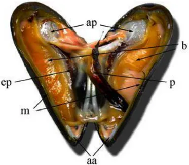
The mussel Mytilus galloprovincialis, chosen as bioindicator in the first part of thesis, is endemic to the Mediterranean coast and the Black and Adriatic Seas. Mussels belong to one of the major classes of molluscs, Bivalvia. Mussels are essentially marine bivalves. The valves are equal in size and shape with an elongated oval-triangular form. It has been able to set itself up at broadly spread points in several seas around the world. Most of these introductions have happened in temperate areas and at sites where there are extended shipping ports (Branch and Stephanni, 2004). A relevant reason of this huge spread is also related to shipping hull fouling and transport of ballast water as indicated by a number of studies and observations (Carlton, 1992; Robinson and Griffiths, 2002; Geller, 1999). The mussel tissue investigated in the present study was the gills, which is a first organ exposed to various pollutants. In Mytilus, the two shell valves are similar in size with a roughly triangular shape, connected together at the anterior of the shell (Fig. 1.2). The shell, as a skeleton, permits the attachment of muscles and defends against predators. The opening and the closing of the shell valves are set by the two muscles, the anterior and the posterior adductors. The mantle is composed of connective tissue with haemolymph vessels, nerves and muscles. This organ encloses the animal within the shell contained most of the gonads. Moreover, the
21 mantle represents the main site for storage of nutrient. Cilia on the inner surface of the mantle move particles onto the gills and pull away heavier material towards the inhalant opening, where it can be discharged (Gosling, 2003; Bayne et al., 1976a). The pair of gills (ctenidia) is made up o of ciliated filaments through which branchial blood vessels move. The opposite face is called ventral or frontal. The gills perform different functions, i.e. respiratory gas exchange, blood haematosis, the capture of blood particles, uptake of nutrients and dissolved organic particles. All these functions are mainly performed by ciliary and mucous cells of the gill epithelium (Gosling, 1992; Auffret et al., 2003). In particular, these mucociliary mechanisms are used in the mussel gills and labial palps in order to filter and ingest particles suspended in the ambient water, such as bacteria, phytoplankton, detritus, microzooplankton and dissolved organic matter (Widdows and Donkin, 1992; Gosling, 2003). This way of nutrient acquisition is known as filter feeding. Water flows into the mantle cavity through the inhalant syphon, is carried through the gill filaments where particles are captured, and exits through the exhalant syphon. The quantity of particles captured depends on the volume of water transported across the gills (pumping rate) and the efficiency with which the particles are retained on the gills (Bayne et al., 1976a). After capture, the particles are moved towards the ventral ciliated particle channels on the gill filaments, incorporated into mucus strings and further transported along ciliated grooves to the labial palps for particle sorting (Beninger and StJean, 1997). The particles are both directed towards the mouth for ingestion or discarded as pseudofaeces (Beninger et al., 1992; Foster-Smith, 1978). The ingested material
22 undergoes extracellular digestion in the gut, and selected particles are moved from the stomach to the tubules of the digestive gland for a more complete intracellular digestion (Bayne et al., 1976a). Afterwards, the material is addressed to the intestine where absorption can occur throughout its length before elimination as faeces (Reid, 1968). in the terminal intestine part is located anus, and faecal pellets are swept away through the exhalant opening of the mantle.
1.6. Neuronal gill activity regulation
The activity of gills results regulated by the sympathetic and parasympathetic innervations of the autonomic nervous system located in the connective tissue (Catapane et al., 1974). The ciliary beating is regulated by various neurotrasmitters (Stefano 1990). Serotonin, or 5-hydroxytryptamine (5-HT), involved in the serotonergic system, plays a cilio-excitatory activity in the gills of lamellibranchiates (Gosselin, 1961; Carroll and Catapane, 2007). The molluskan gills are able to produce endogenous 5-HT, which stimulate in prompt, sustained, and reversible way. A wide range of 5-HT level is able to grade the magnitude of the response (Gosselin, 1961). The exposure to toxicant compounds is responsible of impairment in the serotoninergic system as observed in gills of the mussel M. galloprovincialis exposed to chromium and copper, resulting in increase of the 5-HT-stimulated adenylate cyclase activity in vivo and over-expression of 5-HT receptors (Fabbri and Capuzzo, 2006). In efferent nervous system acetylcholine is used as neural transmitter, it is synthesized in the cytoplasm of cholinergic neurons by the enzyme choline
23 acetyltransferase (ChAT) and split into choline and acetate in cholinergic synapses and neuromuscular junctions by acetylcholinesterase (AChE) (Matozzo et al., 2005). AChE plays a fundamental role for the regular function of the central and peripheral nervous system (Lionetto et al., 2013). The measure of AChE activity is a well known biomarker of neurotoxic compounds in aquatic organisms (Cajaraville et al., 2000; Matozzo et al., 2005; Ciacci et al., 2012; D'Agata et al., 2014). In particular, Lionetto et al. (2013) showed that AChE activity is directly inhibited by pollutants like organophosphate and carbamate pesticides, but other chemicals, such as heavy metals and hydrocarbons, may be responsible of its responsiveness (Rank et al., 2007; Ciacci et al., 2012, Cappello et al., 2015).
Fig. 1.2. General anatomy of Mytilus sp. Arrows indicate (aa) anterior adductor muscle; (ap) posterior adductor muscle; (m) mantle; (ep) hepatopancreas; (b) branchial epithelium.
24
CHAPTER II
Aim of the thesis
In this thesis, it is evaluated the performance of two remediation actions, the first one versus oil wastewater and the second one versus metal-polluted sediments, particularly by mercury. To this aim, mussels
Mytilus galloprovincialis, well-known bioindicators, were used as the
sentinel organisms, exposed in mesocosms and their responses assessed by a multi-biomarker approach.
The main aims of this thesis are:
1) The evaluation of a recovery technique (BF-MBR) versus wastewater discharges by performing mesocosm scale experiments and assessing the biological responses of mussels using a multi-biomarker approach on gills;
2) The evaluation of a remediation technique (Soil Washing) versus natural mercury-polluted sediments collected from the petrochemical site “Augusta-Melilli-Priolo” (Sicily, Italy) by performing mesocosm scale experiments and assessing the biological responses of mussels using a multi-biomarker approach on gills;
3) The evaluation of two techniques for the analysis of tight junction disruption in human airway epithelial cells induced by NaClO, a well known respiratory irritant compound.
25
CHAPTER III
BIOLOGICAL APPROACH TO EVALUATE THE
EFFICIENCY OF-BF-MBR TREATMENT ON
WASTEWATER
3.1 Material and methods
Set-up of mesocosms
All experiments were carried out in the "Mesocosm Facility" of the IAMC-CNR of Messina (Italy). Animals were housed in fiberglass tanks (150 x 150 x 150 cm, volume 3375 L) filled in continuous (125 L/h) with seawater (salinity 37-38‰) directly collected, by a pipeline, from the station “Mare Sicilia” (38°12.23’N, 15°33.10’E; Messina, Italy), in order to ensure daily water turnover (Della Torre et al., 2012; Cappello et al., 2015). Natural seawater was filtered through a 300 μm nylon mesh to remove large metazoans and detritus. To ensure a constant level of water, each mesocosm was equipped with a relief valve connected by a vertical conduct (PVC-u pn10, 200 mm Ø) placed laterally of the tank to continuously discharge the excess of seawater. Water within each mesocosm was gently mixed in a continuous mode with a pump (35 L/h) placed close to the entrance of each tank to provide more homogenous conditions within each mesocosm. The measurement of pH and temperature, performed through a multi-parametric probe, Waterproof CyberScan PCD 650 (Eutech Instruments, The Netherlands), revealed values of 19.5-20.5 °C (daily
26 temperature fluctuations not exceeded 1 °C) and approximatively constant pH values (around 7). The experimental set-up was conceived as follow:
1) White (W): mesocosm supplied only with seawater (uncontaminated system) at flow rate of 2 L/h.
2) Black (B): mesocosm conceived to simulate the effect of an untreated oily wastewater discharged to the marine environment. In order to achieve this goal, the mesocosm B was constantly supplied with a mixture of seawater, commercial Diesel (300 mL) and Bioversal HC (30 mL/g). The mixture was prepared by vigorously mixing in a 50 L-volume tank, 40 mL of the commercial Diesel and 4 mL of dispersant. A peristaltic pump (flow rate of 2 L/h) was used to supply all the volume of the continuously stirred tank every 24 h. The ratio between the simulated discharge and the incoming seawater flow-rate was 1:4. 3) Grey (G): this mesocosm was conceived to measure the biological effects of a BF-MBR treatment as a strategy to mitigate the marine impact of oily wastewater discharge to the sea. In this context, the same mixture used for the mesocosm B (seawater, commercial Diesel and Bioversal HC) was prepared in the continuously-stirred 50 L tank and then pre-treated through the BF-MBR as previously described (Pirrone et al., 2018). Permeate was withdrawn by using a vacuum pump; the flow rate, adjusted by opening or closing a valve before the vacuum pump, was set at 2 L/h transferring the permeate in the mesocosm G. The oxygen required for microorganism growth and membrane aeration was supplied by two sets of air diffusers, which were placed in the biological reactor and below the membrane module respectively.
27 Permeate flow was monitored by a rotameter placed upstream the vacuum pump. (Gornati et al., 2018)
Mixture of potential toxic compounds
Commercial Diesel (ENI Technology S.p.A.), as a mixture of potential toxic compounds, was used as described in Cappello et al. (2007).
Dispersant
Bioversal HC (BIOVERSAL INTERNATIONAL GmbH), density at 20 °C 1.02 g/ml, non toxic, pH 7.5, was here used in addition to the commercial Diesel to increase the dispersion of hydrocarbons in the entire water depth of the mesocosm, reducing its accumulation at the surface (Pirrone et al., 2018). This method was used to simulate the effect of the natural sea-water turbulence, which promotes the mixing of the oil over the water column (Gornati et al., 2018).
Mussel exposure
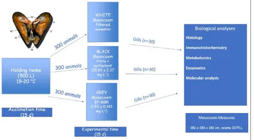
Adult Mytilus galloprovincialis Lamarck, 1819 (5.2 ± 0.4 cm shell length) were collected on March 2016 from an aquaculture plant located in the Faro lake (38°16'6.51"N; 15°38'18.57"E), one of the two brackish coastal lakes nearby Capo Peloro (Messina, Sicily). Mussels were then placed in large flow-through holding tanks (500 L) filled with natural seawater and maintained at 19-20 °C. After two weeks, almost 300 animals were transferred in each mesocosm and maintained for 15 days (experimental time) (Fig. 3.1.). At the end of the experimental time, 30
28 individuals were randomly sampled. from, each mesocosm (Gornati et al., 2018). Gills were frozen in liquid nitrogen and then stored at −80 ◦C for molecular, metabolomics and enzymatic analysis. For histological and immunohistochemical evaluation tissues were fixed in 4% paraformaldehyde in 0.1 M phosphate buffered solution (Maisano et al., 2017).
29
Measurement of the main physico-chemical parameters
The main physic-chemical parameters (pH, temperature) were measured daily in each of the three tanks through the use of a Waterproof CyberScan PCD 650 multi-parametric probe (Eutech Instruments, The Netherlands). Measurements of dissolved oxygen were likewise made by HI97196 multi-parameter probe (Hanna Instruments, Italy).
Chemical Oxygen Demand (COD) measures
The Chemical Oxygen Demand (COD) measurement was performed daily in triplicate on the three tanks by spectrophotometric measurement. This procedure provided the use of specific LCK 1014 cuvettes, that after appropriate preparation were incubated in a suitable digester (Digester HT 200 S, HACH Lange) for 15 minutes at 170 °C. The COD reading, expressed as mg/L, is obtained by the spectrophotometer (DR 3900 spectrophotometer, HACH Lange).
Quantitative analysis of hydrocarbons
Quantitative measurements of the hydrocarbons present in the experimental systems were carried out on the three mesocosms in triplicate, on days 7, 10 and 15 from the beginning of the experimentation in order to verify the BF-MBR efficiency during the experimental time. Hydrocarbon extraction and analysis was conducted
30 in accordance to the 3510 EPA (Environmental Protection Agency) method.
Histological analysis
Gills for histological assessment were fixed in 4% paraformaldehyde in 0.1 M phosphate buffered solution (pH 7.4) at 4 °C, dehydrated in a graded series of ethanol and embedded in Paraplast (Bio-Optica, Italy). Histological sections, 5 µm thick, were cut with a rotary automatic microtome (Leica Microsystems, Wetzlar, Germany), glass-slide mounted and stained with Hematoxylin/Eosin (Bio-Optica, Italy) to evaluate morphological features. Observations were made on five fields of one section for sample using a 40X oil-immersion objective with a motorized Zeiss Axio Imager Z1 microscope (Carl Zeiss AG, Werk Gottingen, Germany) equipped with an AxioCam digital camera (Zeiss, Jena, Germany)
Immunohistochemical analysis
Histological sections of mussel gills were also used for immunodetection of neurotransmission biomarkers applying an indirect immunofluorescence method (Cappello et al., 2015; Maisano et al., 2017) for localization of 5-HT and its receptor (5-HT3R), AChE and ChAT. Briefly, sections were incubated for 1 h with normal goat serum (NGS) in PBS (1:5) to blocking non-specific binding sites for immunoglobulins. The sections were then incubated o.n. at 4° C in a
31 humid chamber with the primary antisera, namely mouse anti-5-HT antibody (Product No. M0758; Dako Cytomation, Milan, IT) diluted 1:50, rabbit anti-5-HT3R antibody (Product No. S1561; Sigma-Aldrich, St. Louis, MO, USA) diluted 1:100, mouse anti-AChE antibody (Product No. MAB304; Chemicon International, Temecula, CA, USA) diluted 1:50, rabbit anti-ChAT antibody (Product No. AB6168; Abcam, Cambrige, UK) diluted 1:250. After a rinse in PBS for 10 min, sections were incubated for 2 h at room temperature with fluorescein isothiocyanate (FITC)-conjugated goat anti-rabbit IgG (Sigma) or tetramethylrodamine isothiocyanate (TRITC)-conjugated goat anti-mouse IgG (Sigma), diluted 1:50. Positive controls for labelling specificity of each peptide were performed by incubating sections with antiserum pre-absorbed with the respective antigen (10-100 g/mL). The pre-absorption procedures were carried out o.n. at 4°C. In addition, negative controls were also performed by substitution of non-immune sera (without antibodies) for the primary antisera. All observations were made on five fields of one section per sample using a 40X oil-immersion objective with a motorized Zeiss Axio Imager Z1 epifluorescence microscope (Carl Zeiss AG, Werk Gottingen, Germany), equipped with an AxioCam digital camera (Zeiss, Jena, Germany) for the acquisition of images. Sections were imaged using the appropriate filters for the excitation of FITC (480/ 525 nm) and TRITC (515/590 nm), and then processed by using AxioVision Release 4.5 software (Zeiss).
32
Metabolomics analysis
Gill metabolite extraction
Polar metabolites were extracted from gill tissues of mussels using a “two-step” methanol/chloroform/water procedure (Cappello et al., 2013b; Wu et al., 2008; Maisano et al 2017). In brief, a 100 mg sub-sample of each gills was homogenized in 4 mL/g of cold methanol and 0.85 mL/g of cold water by a TissueLyser LT bead mill (Qiagen) with 3.2 mm stainless steel beads, for 10 min at 50 vibrations/s. Homogenates were transferred into glass vials, and 4 mL/g chloroform and 2 mL/g water were added. After vortexed, samples were left on ice for 10 min for phase separation, and centrifuged for 5 min at 2000 g at 4° C. A volume of the upper methanol layer (600 µL) containing the polar metabolites were transferred into glass vials, dried in a centrifugal vacuum concentrator (Eppendorf 5301), and stored at -80 °C. Prior to Nuclear Magnetic Resonance (NMR)-based metabolomics analysis, the dried polar extracts were resuspended in 600 µL of a 0.1 M sodium phosphate buffer (pH 7.0, 10% D2O; Armar AG, Dottingen, Switzerland) containing 1 mM 2,2-dimethyl-2-silapentane-5-sulfonate (DSS) (Sigma-Aldrich Co) as internal reference. The mixture was vortexed and transferred to a 5 mm diameter NMR tube. The DSS acted as an internal standard and provided a chemical shift reference (δ = 0.0 ppm) for the NMR spectra, whereas the D2O provided a deuterium lock for the NMR spectrometer.
33
1H NMR-based metabolomics analysis
Extracts of gill tissue were analyzed on a Varian-500 NMR spectrometer operating at a spectral frequency of 499.74 MHz at 298 K. One-dimensional (1-D) 1H NMR spectra were obtained using a PRESAT pulse sequence to suppress the residual water resonance and 6983 Hz spectral width with a 2.0 s relaxation delay. A total of 256 transients were collected into 16,384 data points requiring a 19 min acquisition time. All data sets were zero filled to 32,768 data points and exponential line-broadenings of 0.5 Hz were applied before Fourier transformation. All 1H NMR spectra were manually phased, baseline-corrected, and calibrated (DSS at 0.0 ppm) using Chenomx Processor, a module of Chenomx NMR Suite (version 5.1; Chenomx Inc., Edmonton, Canada) software. The peaks of interest, namely the metabolites related to serotonergic (i.e. serotonin) and cholinergic (i.e. acetylcholine) systems, were identified within the 1H NMR spectra (Cappello et al., 2013b; Fasulo et al., 2012b) and quantified using Chenomx Profiler, another module of Chenomx NMR Suite software, which uses the concentration of a known DSS signal to determine the levels of individual metabolites (Brandao et al., 2015; Cappello et al., 2015).
34
Enzymatic analysis
Acetylcholinesterase (AChE) activity was estimated in the gills of mussels by using the colourimetric method of Ellman et al. (1961), with small changes, by UV–Vis–UV mini-1240 spectrophotometer (Shimadzu, Kyoto, Japan). In short, thiocholine derivatives are hydrolysed by acetylcholinesterase to yield thiocholine. The subsequent combination with 5,5-dithiobis-2-dinitrobenzoic acid (DTNB) forms the yellow anion 5-thio-2-nitrobenzoic acid, which absorbs strongly at 412 nm. AChE activity was expressed as µmol/min/mg (Maisano et al., 2017).
Molecular analysis
RNA extraction and cDNA synthesis
Tissue homogenization and RNA extraction from gill tissues of Mytilus
galloprovincialis collected at each sampling site were performed using
Qiazol reagent (Qiagen). RNA quantity and quality were evaluated as detailed by Giannetto et al. (2015). cDNA synthesis from 1 mg total RNA was performed by QuantiTect reverse transcription kit (Qiagen) after gDNA wipeout buffer treatment in order to remove any potential genomic DNA contamination, following manufacturer's instructions.
35
5-HT3R quantitative gene expression (qPCR)
Degenerate primers (Table 3.1.) were designed on the conserved regions of 5-HT receptor genes isolated from other mussels.
PCR product was separated on 1% agarose gels. The region containing the expected size fragment was sliced, purified using QIAquick Gel Extraction kit (Qiagen) and sequenced using ABI PRISM BigDye Terminator 3.1 Cycle Sequencing kit (PE Applied Bio-system). The sequence (340 bp) was submitted to NCBI database. Quantification of 5-HT3R gene expression in M. galloprovincialis gills was performed by real-time PCR using the Rotor-Gene Q 2plex Hrm thermocycler (Qiagen) with SYBR Green chemistry (Qiagen) as mentioned by Giannetto et al. (2014). qPCR primers, listed in Table 3.1., were designed using the Beacon Designer TM online tool (http://www.premierbiosoft.com/qOligo/ Oligo.jsp?PID=1) for the target and reference genes analysed.
The actin (act), 18S ribosomal RNA (18S rRNA) and elongation factor (ef1-), were chosen as reference genes. The primers for the reference genes are reported in Giannetto et al. (2015). Twenty-fold diluted gill cDNA samples were run in duplicate and no template and minus reverse transcriptase controls were included in each reaction The PCR efficiency determination was made by a five-point standard curve of a 5-fold dilution series (1:1 to 1:32) from pooled RNA (Fernandes et al., 2006). To correct the raw data genes a Normalization Factor was used. This Normalization Factor was calculated from the two most stable genes (18S rRNA and act) by by geNorm software
36 (http://medgen.ugent.be/~jvdesomp/genorm/). The single-peak melting curves confirmed the specificity of the reaction.
Table. 3.1. Nucleotide sequences of primers, amplicons size (bp), methods, qPCR efficiencies (E%),
Statistical analysis
Statistical analyses were made by GraphPad software (Prism 5.0, San Diego CA, USA) for immunohistochemical, metabolomics and enzymatic data, while molecular data were analyzed using SigmaPlot (Systat software). Results were expressed as mean ± standard deviation (SD). One-way analysis of variance (ANOVA) was used to analyse statistically the data, applying the Dunnett’s multiple comparison test, in order to determine significant differences between control and treatment groups and the Student-Newman-Keuls post-hoc tests to assessing differences in 5-HT3R expression levels between mussels from the three mesocosms. Data were considered statistically significant at p < 0.05.
Primer Primer sequence Size (bp) Methods E (%) 5HT_ R _FWD ATTNCGTTGGNTCGGTNCTG 340 Cloning 5HT _R_REV TANCGCCAGANCAATTNCAT q5HT _R _FWD TAACGCCAGACCAATTCCAT 95 qPCR 98 q5HT _R_ REV TGAAGCCATCTTGACTGACG 102
37
3.2. Results
Physico-chemical parameters
No significant variations were noted throughout the experimental period of the measured values of the main physico-chemical parameters (pH, mean value 7.9 ± 0.1; temperature, mean value 15.2 ± 0.5 ° C; dissolved Oxygen, 5 ± 0.5 mg/L).
COD measures
The contribution of the polluting load measured by the COD in comparison with the control showed COD average values of about 50-55 mg/L in the black mesocosm and about 30-35 mg/L in the grey mesocosm receiving the treated water. The control recorded average COD values around 22-24 mg/L. In all the tanks, the COD parameter initially recorded significant increases at the time of placing the mussels (values around 110-115 mg/L are evidence of suspended material - unfiltered sample), and then lowered to the aforementioned values after a couple of water changes within the systems.
38
Levels of hydrocarbons
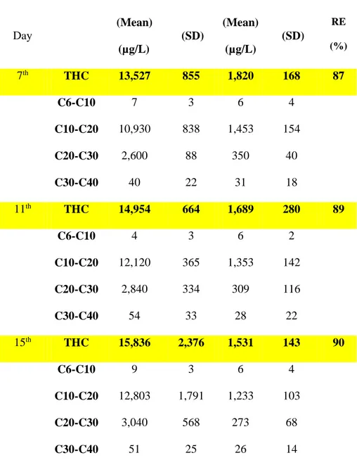
The values of the hydrocarbons for the three days of water sampling (average on the triplicate of the samples taken) are shown in the following table (Table 3.2). In the control, all the values are below the limits of detection. The level of PAHs in the grey mesocosm (1.53 ± 0.143 mg/L) after the wastewater treatment, was reduced of almost 10 times (90% efficiency) than black mesocosm (15.83 ± 2.37 mg/L). This efficiency is maintained during the various measurements carried out in the selected days as indicated above. These data point out the efficiency of BF-MBR to reduce the PAH concentration in oil wastewater discharge.
39
Table 3.2. Quantitative analysis of hydrocarbons mean (µg/L) and Standard Deviation (SD); RE (Removal Efficiency) in BLACK and GREY mesocosms
BLACK BLACK GREY GREY
Day (Mean) (µg/L) (SD) (Mean) (µg/L) (SD) RE (%) 7th THC 13,527 855 1,820 168 87 C6-C10 7 3 6 4 C10-C20 10,930 838 1,453 154 C20-C30 2,600 88 350 40 C30-C40 40 22 31 18 11th THC 14,954 664 1,689 280 89 C6-C10 4 3 6 2 C10-C20 12,120 365 1,353 142 C20-C30 2,840 334 309 116 C30-C40 54 33 28 22 15th THC 15,836 2,376 1,531 143 90 C6-C10 9 3 6 4 C10-C20 12,803 1,791 1,233 103 C20-C30 3,040 568 273 68 C30-C40 51 25 26 14
40
Histology
In the gills of M. galloprovincialis from the W mesocosm is possible to observe the regular morphology of gill tissue (Fig 3.2A.), consisting of parallel filaments whose whole surface is coated by various cilia. In the B mesocosm is showed a critical aberration of the morphology, with a relevant detachment of respiratory epithelium from the underlying connective tissue and loss of the cilia (3.2B.). In the G mesocosm instead, it is possible to observe a relevant reduction of cilia in respect to the control (3.2C.), but the tissue organization is maintained.
.
Fig.3.2. Haematoxylin and Eosin (H&E) staining in the gills of Mytilus galloprovincialis in Mesocosm W(A), Mesocosm B (B) and Mesocosm G (C). The arrow shows detachement of cells and * indicates loss of cilia. Scale bars, 20 μm.
A
B
C
41
Immunohistochemistry
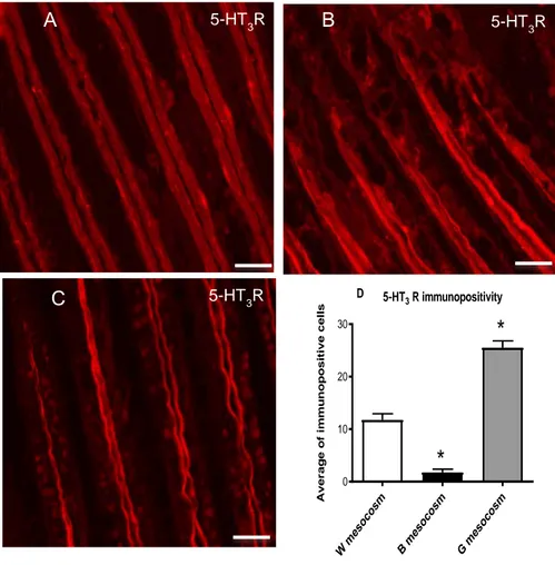
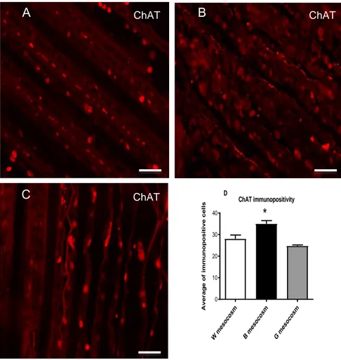
With regards the serotonergic system, in the mesocosm of control, it is possible to observe an intense immunopositivity for serotonin (5-HT) (3.3A.) within the cells, whilst there is a moderate immunopositivity of serotonin receptor (5-HT3R) (3.4A.). In the case of the B mesocosm, it is possible to recognize a drastic reduction of 5-HT immunopositivity (3.3B) and an intense positivity for 5-HT3R in fibers and a very low signal in the cells (3.4B.). In the G mesocosm (3.3C.; 3.4C.) the signal of 5-HT is high in fibers while the immunopositivity of 5-HT3R is high in the fiber and cells. Statistical analyses of immunohistochemical results for 5-HT and 5-HT3R are shown in 3.3D and 3.4D. About the cholinergic system, the immunopositivity of acetylcholinesterase (AChE) and acetyltransferase (ChAT) is low and present only in the cells (Fig. 3.5A.;3.6A.). In the B mesocosm (3.5B; 3.6B.), it is possible to evaluate an increase of the signal for the two enzymes in the cells. In the G mesocosm (3.5C.; 3.6C.), the intensity of the signal is also high, and in addition, it is possible to observe an immunopositivity also in fibers. Statistical analyses of immunohistochemical results for AChE and ChAT are shown in 3.5D. and 3.6D.
42
Fig 3.3. Immunohistochemical labeling for 5-HT in mussel gills in Mesocosm White (A), Mesocosm Black (B) and Mesocosm Grey (C).Mean and standard deviation (S.D.) of immunopositive cells (D).Significant
difference p < 0.05.between Control (White Mesocosm) and the Black and Grey Mesocosm is indicated by an asterisk. Scale bars 20 μm.
A 5-HT B 5-HT C 5-HT 5-HT immunopositivity W m esoc osm B m esoc osm G m esoc osm 0 5 10 15 20 25 A v e ra g e o f im m u n o p o s it iv e c e ll s
*
*
D43
Fig 3.4. Immunohistochemical labeling for 5-HT3 R in mussel gills in Mesocosm White (A), Mesocosm
Black (B) and Mesocosm Grey (C). Mean and standard deviation (S.D.) of immunopositive cells (D). Significant difference p < 0.05.between Control (White Mesocosm) and the Black and Grey Mesocosm is
indicated by an asterisk. Scale bars 20 μm.
A 5-HT3R B 5-HT3R C 5-HT3R 5-HT3 R immunopositivity W m eso cosm B m eso cosm G m eso cosm 0 10 20 30 A v e r a g e o f im m u n o p o s it iv e c e ll s
*
*
D44
Fig3.5 .Immunohistochemical labeling for AChE in mussel gills.in Mesocosm White (A),Mesocosm Black
(B) and Mesocosm Grey (C). ). Mean and standard deviation (S.D.) of immunopositive cells (D).Significant difference p < 0.05.between Control (White Mesocosm) and the Black and Grey Mesocosm is indicated by
an asterisk. Scale bars 20 μm.
A AChE B AChE C AChE AChE immunopositivity W m esoc osm B m esoc osm G m esoc osm 0 10 20 30 40 50 A v e ra g e o f im m u n o p o s it iv e c e ll s
*
D45
Fig. 3.6. Immunohistochemical labeling for ChAT in mussel gills in mesocosm White (a), mesocosm Black
(B) and mesocosm Grey (C). Mean and standard deviation (S.D.) of immunopositive cells (D).Significant difference p < 0.05.between Control (White Mesocosm) and the Black and Grey Mesocosm is indicated by
an asterisk. Scale bars 20 μm.
A ChAT B ChAT
C ChAT ChAT immunopositivity
W m eso co sm B m eso co sm G m eso co sm 0 10 20 30 40 A v e r a g e o f im m u n o p o s it iv e c e ll s
*
D46
Metabolomics
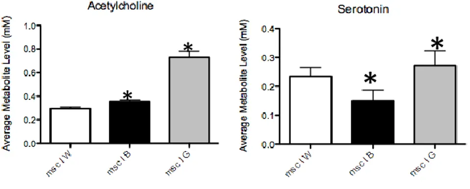
With regards to the amount of the two neurotransmitters, in the case of serotonin, in the B mesocosm it is possible to observe an almost half reduction of serotonin compared to the W mesocosm, while in the G mesocosm, the concentration is slight higher than the control. The level of acetylcholine is slight higher than the control in the B mesocosm, while there is a three time increase of acetylcholine in respect to the control in the G mesocosm (Fig 3.7.). All reported data are statistically significant data.
Fig 3.7. Average metabolite level of Serotonin and Acetylcholine in the three mesocosms.*:p value < 0.05
*
*
47
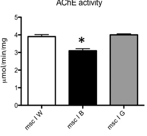
Enzymatics
From the comparison of the enzymatic activity in the three different mesocosms it is possible to observe a significant reduction of about 20% of AChE activity in the gills of mussels exposed to high concentrations of PAHs in the B mesocosm. In the G mesocosm, where wastewater was treated by BF-MBR, the enzymatic activity of AChE does not look inhibited hence the level of AChE activity appears equivalent to that recoreded in the W mesocosm (Fig. 3.8.).
Fig. 3.8. Evaluation of AChE activity in the three mesocosms. *:p value < 0.05
48
Molecular results
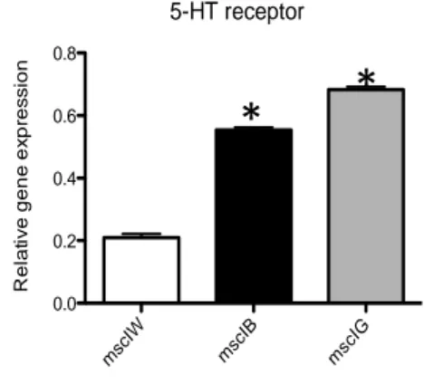
In regard to the gene expression of the serotonin receptor 5-HT3R, it is possible to observe variations in the different experimental conditions. In the black mesocosm, the gene expression of 5-HT3R is three times higher than that observed in the white mesocosm. In the grey mesocosm, where wastewater was treated by BF MBR, a three time increase in gene expression is observed compared to the W mesocosm, while the increase compared to the B mesocosm is slight (statistically significant data) (Fig 3.9.).
Fig 3.9. Evaluation of 5-HT receptor in the three mesocosms. * p. value< 0.05
5-HT receptor msc IW msc IB msc IG 0.0 0.2 0.4 0.6 0.8 R e la ti v e g e n e e x p re s s io n
*
*
49
3.3. Discussion
The pollution of water is the cause of severe alteration in the environment with serious damage to the ecosystem and living organisms. In particular, a great attention was focused over the pollution concerning the impact of the oil wastewater. Hence, despite events like oil spills look very impressing with regards to marine pollution, the higher oil load and consequent risk for marine pollution is mainly associated with routine operations deriving from slops, dirty ballast, sewage and bilge water management (Ciacci et al., 2012; Fasulo et al., 2015, Mancini et al., 2017). A common method used for oil wastewater treatment is a chemical de-emulsification followed by secondary clarifications. This kind of technique needs the use of a variety of chemical compounds such as sulphuric acid, iron and alumina sulphates, etc. (Al-Shamrani et al., 2002; Verma et al., 2010). The BioFilm Membrane Bio Reactor (BF-MBR), a growing biofilm system, became a very interesting method in the wastewater treatment sector (Barwal and Chaudhary, 2014; Gornati et al., 2018). The evaluation of BF-MBR efficiency is object of various studies. For instance, a pilot BF-MBR, inoculated with halophilic activated sludge and Alcanivorax
borkumensis, was able to remove 72 and 90% of COD from low
strength bilge water (1 g COD/L) in 30 days (Mancini et al., 2012). In Pirrone et al. (2018), the efficiency of a BF-MBR in reducing oily-wastewaters discharge impact to marine environment was investigated by small-scale (100 L) artificial systems (microcosms). In this work, the efficiency of BF-MBR is confirmed in a scale-up system mesocosom (3375 L). A 10-time reduction of PAH was observed.
50 Therefore, with regards to the chemical aspect, the BF-MBR looks useful to decrease the concentration of the petrochemical pollutants in the wastewater discharges. In this experiment, the efficiency of BF-MBR treatment was evaluated by a multi-biomarker approach in mussel’s gills in a mesocosm scale experiment. The use of mesocosm represents a very good tool to evaluate the effects of pollutants (Sanchis et al., 2018), and the microcosm and Mesocosm has just offered also good brilliant results in the evaluation of BF MBR (Pirrone et al., 2018; Gornati et al.,2018).
The mussel, in particular M. galloprovincialis, is a very common “sentinel organism” used in several biomonitoring programmes (Cappello et al., 2015; Fasulo et al. 2015; Maisano et al., 2017). The gills are important for various functions, i.e. respiratory gas exchange, blood haematosis, the capture of blood particles, uptake of nutrients and dissolved organic particles. All these functions are mainly performed by the ciliary and mucous cells of the gill epithelium, so the evaluation of their function is fundamental. The histomorphological analysis showed a severe aberration of the gill morphology in samples from the black mesocosm in respect to control, with a severe detachment of epithelium from the connective tissue. A better morphology, even if not regular such as in the control and with loss of cilia, was observed in the gills of samples from the G mesocosm. The observation of histomorphological alteration represents a signal of impairment of the functional integrity of gills (Cappello et al., 2013b; Maisano et al., 2017).
51 To better evaluate the alteration of cilia activity, which plays a key role in most of the gill functions (Sunila, 1998), the neurotransmission system, i.e. serotoninergic and cholinergic,was analysed by a multi-biomarker approach since the cilia activity in mussels is under control of the neurotransmission system (Gosselin, 1961; Carroll and Catapane, 2007; Matozzo et al., 2005). With regards to the serotoninergic system, the level of serotonin, evaluated by metabolomics and immunohistochemical approach, was lower in the B mesocosm, and higher in the G mesocosm in respect to the control. This data suggests that the BF-MBR treatment is able to create good condition for the synthesis of serotonin and partial improvement for functionality of the serotoninergic system in the G mesocosm than in B mesocosm. Instead, the evaluation of 5-HT receptor, performed by molecular and immunohistochemical analysis, showed an up-regulation in samples from both B and G mesocosms. The rise of 5-HT3R could be related to an adaptive response mediated by the paracrine signaling activities in order to recover a regular physiological activity in gills. Indeed, it has been reported that gill epithelial cells containing the lateral cilia present 5-HT receptors to increasing the ciliary beating rate (Cappello et al., 2015; Maisano et al., 2017).
The movement of cilia is also under control of the cholinergic system, responsible for the physiological functioning of the efferent nervous systems (Matozzo et al., 2005). The acetylcholinesterase (AChE) is a well known enzyme in ecotoxicology, since it possible to observe its inhibition by the presence of contaminants in complex mixtures (Matozzo et al., 2005; Cajaraville et al., 2000; Tsangaris et al., 2010;
52 Cravo et al., 2012; D'Agata et al., 2014), by pollutants such as organophosphate and carbamate pesticides (Lionetto et al., 2013), or heavy metals and hydrocarbons (Rank et al., 2007; Ciacci et al., 2012). With regards to AChE activity, it was evaluated by enzymatic assay combined with the metabolomic evaluation of acetylcholine. In addition, AChE level and distribution were observed by an immunohistochemistry assay. AChE enzymatic activity was reduced while the level of acetylcholine and AChE immunopositivity increased in the B mesocosm compared to the control. The increase of acetylcholine level is probably related to the inhibition of AChE. The enhancement of AChE level is a possible adaptive compensatory mechanism in order to heal a regular physiological function of gills. Except for the enzymatic activity, the observed results related to AChE appeared different from those reported by Cappello et al. (2015) and Maisano et al. (2017) in mussels caged in a petrochemical polluted site for 30 and 60 days. Choline acetyltransferase (ChAT), the key enzyme responsible for the synthesis of acetylcholine, was also evaluated. The ChAT immunopositivity increased in the B mesocosm. This enhancement, observed in Cappello et al. (2015), suggests the induction of adaptive compensatory responses mediated by the paracrine signalling activities in order to recover a regular physiological function of gills. In the G mesocosm, the AChE activity was not inhibited and the immunopostivity for AChE and ChAT was lower, while the acetylcholine level relevantly increased than in W and B mesocosms. Overall, this response pattern suggests that the BF MBR for wastewater
53 treatment is likely able to partly restore the condition observed in the W mesocosm.
3.4. Conclusion
The BF-MBR results a very good method to reduce the amount of the chemical concentration of pollutants in wastewater. The mesocosm scale experiment proved to be a very good tool to evaluate the effects of wastewater and BF MBR treated wastewater versus living organisms. Mussels, as seen in nature, were validated as very good biondicators also in this experimental approach. The multi-biomarker battery, applied in order to evaluate the condition of gills and its neurotransmission system, represents a good tool to understand better the health condition of samples in the three different experimental conditions. Hence, thanks to this approach, it is possible to report that the functionality of mussel gills in the G mesocosm was not so critical as that one observed in samples from the B mesocosm, but at the same time it was not possible to observe the same regular morphology and neurotransmission system activity found in the W mesocosm. Therefore, further studies are necessary for the improvement of this treatment.
54
CHAPTER IV
Biological evaluation of the Soil Washing remediation
versus natural mercury-polluted sediments in a
mesocosm scale experiment
4.1 Material and methods
Set-up of mesocosms
All tests were executed in the "Mesocosm Facility" of the IAMC-CNR of Messina (Italy). Animals were housed in fiberglass tanks (150 x 150 x 150 cm, volume 3375 L) filled in continuous (125 L/h) with seawater (salinity 37-38‰) directly collected, by a pipeline, from the station “Mare Sicilia” (38°12.23’N, 15°33.10’E; Messina, Italy), in order to guarantee periodic water turnover (Della Torre et al., 2012; Cappello et al., 2015). Natural seawater was filtered through a 300 μm nylon mesh to exclude large metazoans and detritus. To guarantee a constant level of water, each mesocosm was provided with a relief valve connected by a vertical conduct (PVC-u pn10, 200 mm Ø) located laterally of the tank to continuously remove the excess of seawater. Water within each mesocosm was lightly mixed in a continuous mode with a pump (35 L/h) placed close to the entrance of each tank to provide more homogenous conditions within each mesocosm. The measurement of pH and temperature, performed through a multi-parametric probe Waterproof CyberScan PCD 650 (Eutech Instruments, The Netherlands), revealed values of 19.5-20.5 °C (daily temperature